On the cell biology of pit cells, the liver-specific NK cells
←
→
Page content transcription
If your browser does not render page correctly, please read the page content below
PO Box 2345, Beijing 100023, China WJG, 2000 February; 6(1):1-11
Fax: +86-10-85381893 World Journal of Gastroenterology
E-mail: wjg@wjgnet.com www.wjgnet.com Copyright2000 by the WJG Press ISSN 1007-9327
Reviews
On the cell biology of pit cells, the liver-specific
NK cells
Luo DZ, Vermijlen D, Ahishali B, Triantis V, Plakoutsi G, Braet F, Vanderkerken K and
Wisse E
Subject headings pit cells; hepatic natural names, such as pit cells, hepatic NK cells and LGL
killer cells; large granular lymphocytes are used to describe pit cells, referring to different
aspects of their morphology or function[6]. We
INTRODUCTION prefer to use the first-given name of pit cells for
\Natural killer (NK) cells are functionally defined by these cells in the liver, because it is not related to
their ability to kill certain tumor cells and virus- the ever varying levels of function and morphology
infected cells without prior sensitization[1]. NK cells or a person (like Kupffer cells)[7]. In addition,
comprise about 10% to 15% of lymphocytes in the liver-associated lymphocytes (LAL), in some
peripheral blood and most of these cells in human instances, are used to describe the total population
and rat have the morphology of large granular of lymphoid cells in the liver[8,9]. However, LAL
lymphocytes (LGL)[2]. However, recent studies contain about 30% T lymphocytes, 3% B
have demonstrated that small agranular lymphocytes besides 43% pit cells in a human liver
lymphocytes, lacking CD3 expression, have washout[9]. Rat liver washouts contain 43% T
cytolytic activity comparable to NK cells[3]. These lymphocytes, 16% of B lymphocytes, 3.3% of
variations may be related to the stage of NK cell monocytes and 26% pit cells[10].
differentiation or heterogeneity[4]. Moreover, some The present review will discuss the biological
cytotoxic T lymphocytes (CTL) also display LGL relevance of pit cells with emphasis on rat liver.
characteristics[4]. Besides NK cells in peripheral
blood, NK cells are also found in tissue IDENTIFICATION, STRUCTURE AND TISSUE
compartments, such as the spleen, lung, intestine, DISTRIBUTION OF PIT CELLS
lymph nodes, bone marrow and liver[4]. NK cells in Pit cells were firstly described in 1976 by Wisse et
the liver, also called pit cells[5], constitute a al[5]. The name pit cell was introduced because of
unique, resident NK population in the liver the characteristic cytoplasmic granules, which in
sinusoids. Their immunophenotypical, Dutch language are called pit, resembling the pits in
morphological and functional characteristics are a grape[5]. The hypothesis that pit cells might
different from blood NK cells[6]. Presently, several possess NK activity was suggested by Kaneda et
al[11], because of their morphologic resemblance to
Dian Zhong Luo1,3, David Vermijlen 1 , Bülent Ahishali1, Vasilis LGL. The deve lopment of a method for the
Triantis1, Georgia Plakoutsi1, Filip Braet1*, Karin Vanderkerken2* and isolation and purification of pit cells from rat liver
Eddie Wisse1
1
Laboratory for Cell Biology and Histology, 2Department of Hematology and the evidence of pit cells possessing spontaneous
and Immunology, Free University of Brussels (VUB), Brussels-Jette, cytotoxicity against NK sensitive YAC-1 lymphoma
Belgium cells confirmed these cells to be hepatic NK
3
Department of Pathology, Guangxi Medical University, Nanning,
China cells[12,13].
*Filip Braet and Karin Vanderkerken are postdoctoral research fellows Pit cells exist in the liver sinusoids and often
of the Fund for Scientific Research Flanders. adhere to endothelial cells (Ec), although they
Dian Zhong Luo, male, born on 1955-07-07 in Guilin, Guangxi, incidentally contact Kupffer cells (Kc) (Figure 1).
graduated from Guangxi Medical University in 1982, got Master
degrees in 1987 in Guangxi Medical University and in 1994 in Free They face the blood directly. Pseudopodia of pit
University of Brussels, Professor of Pathology of Guangxi Medical cells can penetrate the fenestrae of the Ec, and
University, and now is following Ph.D. program in Medical Sciences enter the space of Disse and can directly contact the
in Free University of Brussels, Belgium, having more than 30 papers
published. microvilli of hepatocytes[5,14]. Their appearance in
Supported by grants 3.0053.92,3.0050.95, 9.0038.96,1.5.411.98 from the space of Disse is not a c ommon feature[15]. By
the National Foundation for Scientific Research (FWO) and grants morphological investigation, the frequency of pit
194.322.1740,195.332.1310,196.322.0140 and OZR.230 from the cells in liver tissue is about an average of 1 pit cell
Research Council of the Free Univ ersity of Brussels.
Correspondence to: Prof. Dr. Eddie Wisse, Laboratory for Cell per 10 Kupffer cells. The number of pit cells, in
Biology and Histology, Free University of Brussels (VUB), Laarbeeklaan untreated rats, is therefore estimated to be (1.4-2)
103, B-1090 Brussels, Belgium ×10 6 cells per gram liver weight [12]. By
Tel. (32)-2-477 4404, Fax. (32)-2-477 4405
Email. wisse@cyto.vub.ac.be immunohistoc hemistry, using mAb 3.2.3 against
Received 1999-09-22 Accepted 1999-11-15 NKR-P1A (a specific marker of NK cells), the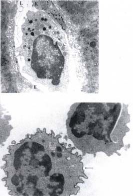
2                              ISSN 1007-9327     CN 14-1018/R     WJG February 2000      Volume 6   Number 1

number of pit cells in frozen sections of rat liver is     cells and considered as a characteristic of these
about 13.7 per mm2[ 16]. After intravenous injection       cells[6,23].
of biological response modifiers (BRM), the number
of pit cells increases 4-fold to 6-fold in rat liver       SURFACE PHENOTYPE OF PIT CELLS
treated with zymosan[17] and 43-fold with interleukin-     Extensive phenotypic analysis has shown that no
2 (IL-2)[18]. The surplus of pit cells is considered to    unique NK cell marker has been identified yet, but
originate from local proliferation and from the bone       expression of a set of differentiation antigens in the
marrow[17,18]. Pit cells were found to be more             absence of antigen-specific receptors of T and B
numerous in the periportal than in the pericentral         lymphocytes serves to identify NK cells. NKR-P1
region of the liver lobule[11,16].                         was first identified in the rat[25] and has now been
      Pit cells have essentially the same morphology       shown to be expressed also by mouse and human
as NK cells from blood and other organs, i.e. LGL          NK cells[26,27]. NKR-P1 is a type II membrane
(Figure 1). LGL morphology is characterized by a           glycoprotein of the C-type lectin superfamily[28]. The
relatively large size, the presence of granules in         N KR-P1 genes are located on mouse chromosome
the cytoplasm, a pronounced asymmetry of the cell          6[29], human chromosome 12 p 12-p13[27], and rat
and an indented or kidney shaped nucleus of high           chromosome 4 in a region designated as the ‘NK
density[10]. Pit cells in the rat are about 7µm in         gene complex’ (NKC)[28]. NKR-P1 antigen is present
diameter and vary in shape, while possessing well          on 94% of rat LGL and serves as a triggering
developed pseudopodia. They show a pronounced              structure on these cells[25]. NKR-P1 is consider ed
polarity with an eccentric nucleus and most organelles     to be a useful marker for NK cell identification[25].
lying at one side of the nucleus.
                                                           However, a subset of T lymphocytes and
      The most conspicuous organelles are the
                                                           polymorphonuclear leukocytes also express NKR-
electron-dense granules. These granules have several
                                                           P1 [25,27]. CD56 and CD16 are expressed, either
characteristics. They are azurophilic, therefore,
                                                           alone or in combination, on the majority of human
Giemsa staining of a cell smear or cytospin
                                                           NK cells and are most extensively used as human
preparation reveals the presence of the granules in
                                                           NK cell ‘markers’ for clinical and basic research
pit cells with light microscopic examination. As
                                                           purposes[2]. Other surface antigens expressed on
measured by electron microscopy, the granules differ
                                                           NK cells are: CD2, CD8, CD11a-c, CD18, CD45,
in size between different pit cell subpopulations (LD
                                                           CD54, CD56, CD58 and CD69[2,4,30,31].
an d HD pit cells, see next page), but within one
                                                                 Most surface antigens found on rat pit cells are
cell type the granules are very homogeneous with
                                                           similar to that found on spleen or peripheral blood
respect to size, shape and electronic density[19]. The
granules are membrane bound and range in size              NK cells (Table 1)[13,16,32]. All LGLs from a rat
between 0.2µm in LD pit cells an d 0.5µm in LAK            liver washout, in preparations for light and electron
cells. These granules contain a number of lysosomal        microscopy, were found to express NKR-P1[16]
enzymes, such as acid phosphatase[12,20]. Although         (Figure 2) as recognized by using the monoclonal
perforin and granzymes, which have been isolated           ant ibody ( mAb ) 3.2.3[25]. CD11a is present on
from NK cell granules[21,22], are not yet been             90% of rat pit cells, which is different from rat
identified in pit cell granules, it is believed by         peripheral blood NK cells ( 54% ) [32].
analogy that these molecules are present in the            Approximately 90% of rat pit cells express CD18,
granules of pit cells.                                     35% express CD54 and 80% express CD2[32].
      Rod-cored vesicles are small inclusions, ranging     Asialo-GM1, which is expressed by all rat blood NK
in diameter from 0.17 µm to 0.2 µm, and are                cells[19], is present on 36% of LD pit cells and 70%
exclusively found in LGL[11]. They contain a straight      of HD pit cells[13]. CD8, a marker of NK cells and
rod structure which is 30 nm-50 nm in length, that         cytotoxic T lymphocytes[2], is present on all rat pit
bridges the entire diameter of the vesicle[11,20]. Rod-    cells[13]. However, the composition of CD8 in NK
cored vesicles derive from and distribute preferentially   cells and T lymphocy tes is different. Most CD8+
around the Golgi apparatus. Possibly rod-cored             NK cells express CD8á/CD8á homodimers rather
vesicles may also contain cytotoxic factors functioning    than the CD8á/CD8â heterodimers prevalent on
in natural cytotoxicity[15].                               cytotoxic T cells[3 3]. In addition, rat pit cells do not
      Pit cells also exist in human and mouse liver,       express T cell receptor and CD5 antigen (a pan T
but the identification of pit cel ls in human and          cell marker)[13,15].
mouse liver is more difficult than in rat because                 LAL from human liver contain about 35%
they contain a lower number and smaller size of the        CD56+ cells, in which three subsets are found:
typical dense granules and very few rod-cored              CD3+/ CD16-,               CD3-/ CD16-,            CD3-/
                                                                   +[8]                            +
vesicles[9,23,24]. On the other hand, 5% to 25% of         CD16 . Moreover, all the CD56 LAL express
human pit cells contain ‘parallel tubular arrays’          CD11a and CD18, and partly express CD2, CD11b,
(PTA), that were also reported in human blood NK           CD11c, CD54 and CD58[8,34,35].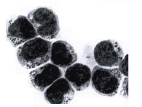
Luo DZ, et al. Biology of pit cells                                                                                    3

Table 1 Characteristics of LD, HD pit cells and peripheral                   ISOLATION AND PURIFICATION OF PIT CELLS
blood NK cells
                                                                             Our isolation method for rat pit cells is based on a
Item                            LD pit cell HD pit cell Blood NK cell        washout technique[12] followed by purification, based
Morphology*                                                                  on the magnetic negative-selection of cells using
Size(µm2)                           27.7          24.5         24.8
Rod-cored vesicles per cell          1.0           0.8           0.5
                                                                             mAbs against surface antigens and receptors found
Microvilli per cell                  5.2           5.9           7.1         on T and B cells[6,36]. Since pit cells are apparently
Size of granules (µm2)        Smaller(0.09) Intermediate(0.1) Larger(0.14)   not heavily anchored in the liver sinusoids, the cells
Number of granules per cell    Higher(50) Intermediate(20) Lower(1 0)
Surface antigens+                                                            could be washed out by this non- enzymatic, high-
CD2                                80            80            80            pressure (50cm water) perfusion of the liver via the
CD8                               100           100            40
CD11a                              90            90            54            portal vein with phosphate-buffered saline
CD18                               90            90            90            supplemented with 0.1% EDTA[12]. The washout
CD54                               35            35            35
Asialo-GM1                         36            70           100            was collected from the vena cava. The erythrocytes,
NKR-P1                             95            95            94            granulocytes and cell debris in the washout were
Functional features
NK activity                       High       Intermediate     Low            removed by Ficoll-Paque gradient centri fugation.
P815 cell killing                 Yes             No          No             The mononuclear cells recovered from the interface
*
 Data from reference 19.
                                                                             of Ficoll-Paque gr adient contained T cells, pit cells,
+
 Approximate % of cells that express antigen; data summarized                B cells, monocytes, and a few Ec and Kc. Ad
from referen ces 2, 13, 16, 19, 25, and 32.                                  herent monocytes and B cells in this population
                                                                             could be selectively removed in a nylon wool
                                                                             column[12]. Pit cells were further purified by magnetic
                                                                             cell sorting[36]. With this system, a highly purified
                                                                             population of pit cells was obtained by negative
                                                                             selection, i.e. by elimination of remaining monocytes,
                                                                             T and B cells using specific antibodies and
                                                                             immunomagnetic beads. By this method, we obtained
                                                                             pit cells with a purity of more than 90% and a
                                                                             viability of more than 95% (Figure 3). Moreover,
                                                                             this non-enzymatic method does not destroy cell
                                                                             surface molecules.
                                                                                   Alternatively, pit cells could be isolated by
                                                                             enzymatic methods[5]. Howe ver, this method is
                                                                             time consuming, labor-intensive and only provides
                                                                             pit cells with 30% purity and 90% viability[6,12].

                                                                             HETEROGENEITY AND ORIGIN OF PIT CELLS
                                                                             A considerable set of data indicates that rat pit cells
                                                                             constitute a heterogeneo us population. Based on
                                                                             the cell density, pit cells can be separated into a
                                                                             low density (LD) and high density (HD) fraction by
                                                                             45% iso-osmotic Percoll gradient centrifugation[19].
                                                                             These two cell populations have been shown to
                                                                             differ immunophenotypically, morphologically and
                                                                             functionally from each other and fro m blood LGL
                                                                             (Table 1)[19,37]. LD pit cells (Figure 4A) contain
Figure 1 Transmission electron micrograph of a pit cell in a
rat hepatic sinusoidal lumen (L). The pit cell shows polarity
                                                                             more rod-cored vesicles and more, but smaller
with an eccentric nucleus. The cytoplasm is abundant and                     granules than blood NK cells (Figure 4B)[19,37]. The
contains characteristic electr on-dense granules and other                   number of rod-cored vesicles and granule
organelles lying mainly on one side of the nucleus. The cell                 composition (number and size) of HD pit cells are
contacts an endothelial cell (E) and a portion of a Kupffer cell
                                                                             intermediate between LD pit cells and blood NK
(K) with a positive peroxidase reaction product in the rough
endoplasmic reticulum. Bar = 1 µm. (from Hepatology,1988;8:                  cells[19,37]. Immunophenotypically, almost all blood
46-52, with permission)                                                      NK cells are asia lo-GM1 positive, and 70% of HD
Figure 2 Immuno-transmission electron micrograph showing a                   pit cells are strongly positive, whereas only 36 % of
3.2.3 positive LGL (pit cell) (arrowhead) and a 3.2.3 negative               LD pit cells are weakly positive [19]. Furthermore,
agranular cell. The 3.2.3+ pit cell shows characteristic electron-
dense granules in the cyt oplasm and immunoperoxidase reaction
                                                                             functional differences have been observed among
product on the surface. Bar = 1 µm.(from Hepatology, 1995;21:                these three populations. The LD pit cells are five to
1690-1694, with permission)                                                  eight times more cytotoxic against YAC-1 cells and
4 ISSN 1007-9327 CN 14-1018/R WJG February 2000 Volume 6 Number 1
colon carcinoma cells than blood NK cells[37]. The diphosphonate[41]. The number of HD pit cells
HD pit cells have intermediate cytotoxic activity declined 3 days after the injection. Conversely, the
between LD pit cells and blood NK cells[37]. In LD pit cell population showed no change in number
addition, LD pit cells are able to lyse LAK-sensitive after 3 days, but a decline of about 80% was seen
P815 mastocytoma targets, which are resist ant to 7 days after the injection[41]. These data indicate
normal blood NK cells and hepatic HD pit cells[37]. that pit cells constitute a Kupffer cell-dependent
Pit cells are considered to originate from blood population and that Kc play an essential role in the
NK cells[38,39]. Several evidences support the differentiation of pit c ells in the liver. However, it
concept that blood NK cells immigrate into the remains unclear what factor(s) secreted by Kc is
hepatic sinusoids to become HD pit cells, which responsible for this differentiation. On the other
further differentiate into LD pit cells. Importantly, hand, other conditions present in the liver
the characteristics and functions of HD pit cells are microenvironment, i.e. Ec and their secreted
intermediate between blood NK cells and LD pit factors, may work synergically with Kc to
cells[19]. Kinetic experiments with sublethal total contribute to pit cell differentiation, since
body irradiation (700cGy) showed that blood NK coincuba tion of HD pit cells with Kc failed to
cells and HD pit cells were depleted in about one induce the full differentiation of HD into LD pit
week after irradiation, whereas LD pit cells totally cells[41].
disappeared at two weeks after irradiation[39].
Shielding of the liver gave similar results and FUNCTIONS OF PIT CELLS
splenectomy did not affect pit cell number[39]. By NK cells were initially defined as lymphoid cells
using intravenous anti-asialo-GM1 antiserum capable of mediating spontaneo us killing of target
injection, blood NK and HD pit cells totally cells, including tumor and virus- infected cells[1].
disappear ed within one week of treatment, whereas Such NK cytotoxic activity is mediated without prior
LD pit cells disappeared from the liver one week sensitization and any obvious stimulation or
later[39]. The direct evidence for LD pit cells activation[1]. In addition to this natural spontaneous
originating from asialo-GM1 positive precursors pathway of tumor killing, NK cells can mediate
(blood NK and HD pit cells) was given by the antibody-dependent cellular cytotoxicity ( ADCC )
adoptive transfer of fluorescent-labeled HD pit cells by a mechanism involving CD16, an IgG Fc
into syngeneic rats[39]. After three days, 5% of receptor[42]. Most human and mouse NK cells
labeled cells were recovered in the LD fraction and express CD16[2]. Rat NK cells contai n genes with
these cells displayed typical LD pit cell a high level of homology to human and murine Fc
morphology[39]. These observations also indicate receptors[43] and are able to display ADCC[44].
that the life span of pit cells in the liver is about two Unfortunately, no antibodies against rat CD16 are
weeks[6,39]. available yet.
The mechanism behind the migration of blood Although the cytotoxic function of NK cells is
NK cells to the liver sinusoids is not fully spontaneous, it can be significantly augmented by
understood. Several adhesion molecules were found several cytokines[2]. One of these, IL-2, has been
to be involved in the process[32]. Rat blood NK and shown to play a central role in the regulation of NK
pit cells express LFA-1 (CD11a/CD18) and CD2 cells, including augmenting NK cell cytotoxicity,
(LFA-2) adhesion molecules[32]. Their ligands, expanding NK cell antitumor spectrum and inducing
CD54(ICAM-1) and CD58 (LFA-3) were found to NK cell proliferation[4].
be present on liver Ec[40]. After intravenous NK cytotoxic (i.e. cytolytic) activity is usually
injection of antibodies against CD2, CD11a and determined by measuring the release of radiolabeled
CD18 into rats, the number of pit cells in the liver chromium from target cells after been exposed to
decreased significantly, indicating that the effector cells[2]. A new assay using flow cytometry
interactions of LFA-1/CD54 and CD2/CD58 are to assess NK cell activity, in which various dyes are
involved in the recruitment of pit cells in the used to differentiate viable from dead target cells,
liver[32]. has recently been described[45]. Initial studies have
Once marginated in the liver sinusoids, blood shown that this method is quick, reliable, and
NK precursors further differentiat e into HD pit correlates well with the standard 51Cr release
cells, then into LD pit cells. The microenvironment assay[45].
of the liver sinusoid is believed to be responsible for Besides the cytotoxic function, NK cells can
this differentiation process[41]. Vanderkerken et al produce various cytokines[46], regulate the growth
found that Kc were selectively eliminated 3 days of hemopoietic tissues and bone marrow
after intravenous injection of liposomes containing transplants[47], and participate in the resistance to
the cytotoxic drug dichloromethy lene microbial pathogens[2].Luo DZ, et al. Biology of pit cells 5
Most investigations on pit cell functions focus
on cytotoxic activity. Rat pit cells have high
spontaneous cytotoxic activity against various tumor
cell lines, such as YAC-1, P815, CC531s, DHD-
K12, L929, 3LL, and 3LL-R[10]. Comp ared with
blood NK cells, pit cells are four to eight times
more cytotoxic against YAC-1 and CC531s cells,
and are able to kill the NK-resistant but lymphokine
activated killer (LAK)-sensitive P815 cells (Figure
5)[19,48]. This evidence seems to support the idea
that pit cells become activated once they become
liver residents. However, it is not understood yet
what kind(s) of factor(s) is (are) responsible for
the activation of pit cells in the liver, although it
has been shown that pit cells are dependent on the
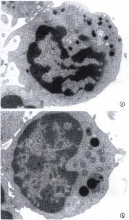
Figure 3 Light micrograph of an isolated and purified pit cell
presence of a healthy Kc population[41].
population in a May-Grünwald-Giemsa-stained cytospin. The cells
contain cytoplasmic granules, which can be used to recognize and Furthermore, NK activity in the liver could be
count the number of pit cells in freshly isolated liver-associated augmented by BRM, like Propionibacterium acnes
lymphocyte population. Bar = 5 µm. or maleic anhydride divinyl ether[24]. Interestingly,
an increase in function seems to coincide with a
large increase in the number of LGL[17,24]. IL-2
treatment results in a dramatic accumulation of pit
cells in the hepatic sinusoids in vivo[18], induces HD
cell proliferation and augments liver HD pit cell
cytotoxic activity in vitro[49]. In contrast, IL-2
treatment does not induce liver LD pit cell
proliferation[49].
MOLECULAR MECHANISMS IN NK CELL-MEDIATED
CYTOTOXICITY
It is believed that the cytotoxic function of NK cells
is mediated by multiple pathways, and each
pathway, in principle, encompasses a cascade of
events, including recognition of target cells,
binding of effector cells to target cells
(conjugation), activation of effector cells, delivery
of the lethal signal to tar get cells, and effector cell
detachment and recycling [2,4,50]. Although the
precise mechanisms of individual steps in this
process have not been fully elucidated, significant
progress has been made recently in identifying a
number of molecules participating in NK cell-
mediated cytotoxicity.
CONJUGATION
The prerequisite of NK cell killing is the binding of
one or more effector cells to a target cell, that is,
conjugation[6]. Several adhesion molecules on the
NK cell, such as CD2, CD28 and LFA-1, and on the
target cell, such as CD58, B7 and CD54, participate
Figure 4 Transmission electron micrographs of a typical LD pit in this process, and some of them may also possess
cell and a blood NK cell (B). co-stimulatory or even triggering capacity in the
(A) The main morphological characteristic of LD pit cells, cytotoxic cascade[28,50-52].
compared to HD cells and blood NK cells, is the presence of
numerous small cytoplasmic granules.
CD2 is an adhesion molecule of the
(B) Note the few, but large granules in blood NK cell. Bar = immunoglobulin (Ig) superfamily expressed on T
1 µm. (from Hepatology, 1990;12:70-75, with permission) cells and NK cells[53]. Approximately 80% of rat pit6 ISSN 1007-9327 CN 14-1018/R WJG February 2000 Volume 6 Number 1
cells express CD2[32]. Although CD2 is a well- lacking MHC class I. Expression of MHC class I
known activation structure on T cells[53], mAbs on a number of target cells is correlated with target
against CD2, depending on experimental cell resistance to natural killing[61-65]. Masking of
conditions, either induce[54,55] or inhibit NK MHC class I by a n mAb, enhances pit cell-
activity[31,56]. Anti-CD2 mAb had no effect on the mediated cytotoxicity against CC531s cells,
binding of pit cells to rat colon carcinoma cells indicating that MHC class I on CC531s cells
(CC531s), or on the cytotoxicity against protects these cells from being killed by rat pit
CC531s[57]. However, the anti-CD2 mAb enhanced cells[66].
the cytolytic function in rat pit cells against FcγR+ An explanation for these observations is, that
P815 target cells[57]. The ligand of CD2 is another the cytotoxic activity of NK cells is regulated by
adhesion molecule, CD58 (LFA-3) that is widely positive and negative signals from triggering and
expressed on various cell types[53]. Transfection of inhibitory membrane receptors. The final outcome,
CD58 into murine cell lines increased the lysis of i.e. triggering of cytotoxic activity or inhibition of
these targets by some human CD2+ NK cell cytotoxicity, appears to depend on the balance
clones[58]. However, expression of CD58 alone is between the positive and negative signals[51,67]. An
insufficient to confer cells sensitive to NK cell- increasing number of triggering and inhibitory
mediated lysis, indicating that CD2 may serve as a receptors have been described in recent
costimulatory receptor that augments, but not years[28,68,69]. In hibitory receptors on NK cells
initiates, the primary activation of NK cells[58]. recognize MHC class I, and these generally inhibit
The interaction between β2 integrins (CD11a- the lysis of MHC class I+ cells [28,67-71]. Three
c/CD18) and ICAMs (in tercellular adhesion receptor families , Ly49, CD94/NKG2 and Killer-
molecules) has been found to be important in the cell inhibitory receptors (KIRs), are reported to be
binding of NK cells to their targets[31,51,56]. β2 involved in the recognition of MHC class I
integrins are heterodimers containing a common β- molecules on target cells[28]. The Ly49 family is the
chain (CD18) and one of three different β-chains product of at least nine highly related genes
(CD11a,CD11b,CD11c). β2 Integrins are expressed (Ly 49A-Ly49I) present on mouse chromosome 6 in
only in leukocytes, including NK[56] and pit cells[32]. the NKC[72]. Ly-49 homologies have been identified
Besides the effect on the binding to target cells, on rat chromosome 4 in the ‘NKC’[73], but have not
LFA-1 (CD11a/CD18) also participates in signal been found in human. The Ly-49 molecules are type
transduction in NK cells required for NK cell II membrane glycoprotei ns and belong to the C-type
activation[59]. Cross-linking of LFA-1 on NK cells lectin superfamily[72]. Ly-49 receptors recognize a
with its antibody is known to induce a calcium trimeric MHC class I complex composed of the H-
influx, phosphoinositide turnover, tumor necrosis 2D or H-2K heavy chain, β2-microglobulin, and a
factor-α (TNF-α) production[59], and to inhibit the bound peptide. However, the composition of the
target cell killing by NK cells[60]. LFA-1 was also bound peptide does not appear to influence the
found to be involved in pit cell-mediated cytotoxicity. interaction to a large ex tent[68]. Most, but not all,
The antibody against LFA-1 inhibits not only the Ly-49 receptors contain an immunoreceptor
binding of pit cells to target cells, but also the killing tyrosine-based inhibitory motif (ITIM) in their
of target cells by pit cells[57]. Taken together, this cytoplasmic domains [28,67]. Ly-49 receptors
information suggests that LFA-1 on effector cells containing the ITIM sequence inhibit NK cell
may have a dual function of binding to target cells effector function[28,67], whereas Ly-49 lacking
and of triggering cytolysis. ITIM, such as Ly-49D and Ly-49H, can activate NK
Studies have shown that conjugation between
cell-mediated cytotoxicity when the receptor is
NK cells and target cells is essential but not
ligated by ant i-Ly49D mAb[74].
sufficient for NK activity[50]. After conjugation,
KIRs, the inhibitory receptors recognizing
further recognition events mediated by triggering
MHC class I in human NK cells, are monomeric
and inhibitory receptors on NK cells are required to
type I glycoproteins that contain Ig domains[67].
trigger NK cell cytotoxic activity[2,51].
They are enc oded by genes located on human
chromosome 19q13.4[28]. Two subfamilies of KIRs
NK CELL RECEPTORS INVOLVED IN MHC CLASS I can be identified by the number of Ig-like domains
RECOGNITION in the extracellular regions of the molecules[28]. The
NK cell-mediated cytotoxicity was originally KIR3D subfamily contains three Ig-like domains,
thought to be spontaneous and major whereas the KIR2D contains two Ig-like
histocompatibility complex ( MHC ) class I- domains[28]. A remar kable feature of both KIR2D
unrestricted. However, increasing evidence and KIR3D is the heterogeneity in the length of the
indicates that NK cells preferentially kill cells cytoplasmic domains. KIRs with long cytoplasmicLuo DZ, et al. Biology of pit cells 7
domains, i.e. KIR2DL (p58) and KIR3DL (p70), FcR - CC531s tar get killing[66]. However, the
contain two ITIM sequences that are responsible function of NKR-P1 on human NK cells appears
for the inhibitory function of these molecules[67,75]. more complex. Treatment of human NK cells with
KIRs containing short cytoplasmic domains, i.e. anti-NKR-P1 mAb gives cont roversial results, such
KIR2DS (p50) and KIR3DS, lack ITIM and as activation, inhibition or no effect, depending on
potentially activate NK activity[76,77]. Both KIR2D the NK cell population studied[27,87]. The conditions
and KIR3D molecules bind to HLA class I determining the outco me of the engagement of
trimers, composed of a class I heavy chain, β2 NKR-P1 in human NK cells are not known. When
microglobulin, and a bound peptide[28]. human NKR-P is compared with the corresponding
rat and mouse proteins, it was found that all rodent
In addition to KIRs, human NK cells also
NKR P1 has the C×CP motif that interacts with
express another type of receptor capable of
phosphorylated P561ck[88], whereas human NKR-P1
recognizing MHC class I, namely CD94/ lacks this motif[28].
NKG2[78-80]. This receptor is a heterodimer and is
composed of CD94 glycoprotein that is disulfide-
THE TWO MAJOR PATHWAYS OF NK CELL-MEDIATED
bonded to either a NKG2A or a NKG2C subunit[58].
CD94 and NKG2 genes are pre sent on human CYTOTOXICITY
chromosome 12p12.3 - p13.1 in the ‘NKC’[81]. CTL and NK cells, including rat pit cells, kill target
Both CD94 and NKG2 molecules belong to the C- cells by one of two disti nct mechanisms or both:
type lectin superfamily[81] . CD94 lacks a necrosis and apoptosis[19,48,50,89]. Necrosis or cytolysis
cytoplasmic domain, thus lacking intrinsic signal is characterized by swelling of the cell and
transduction capacity[81]. However, CD94 is organelles, and results in disruption and leakage of
required for the transport and membrane expression the cell membrane and in lysis[6]. Cell membrane
of the NKG2A or NKG2C glycoproteins[78,80]. Since damage is a key event in cytolysis and release of
NKG2A possesses an ITIM sequ ence in the the cytoplasmic contents possibly leads to an
cytoplasmic domain and NKG2C lacks an ITIM, the inflammatory response in vivo[6]. The 51Cr-release
CD94/ NKG2A complex acts as inhibitory receptor, assay is thought to reflect this type of damage[6].
whereas CD/NKG2C complex acts as a Apoptosis or programmed cell death is
morphologically recognizable by membrane
noninhi bitory ceceptor for MHC class I on NK
blebbing, chromatin condensation, nuclear
cells[28,68].
fragmentation, shrinking, condensation of cells and
their organelles, and fragmentation of the cells into
Triggering NK cell receptors
apoptotic bodies (Figure 6). The cellular remains
Several membrane molecules are described to serve
are phagocytosed by neighboring cells or
as triggering receptors on NK cells, including
macrophages. When phagocytosing cells are absent,
CD16, NKR-P1, NK-TR1, 2B4, P38 and apoptotic bodies progress to secondary
Lag3[28,51,69]. Only CD16 and NKR-P1 can be necrosis[6,90].
regarded as ‘established’ triggering receptors, while Recent studies have demonstrated that NK cell-
the role of the others is still undefined or mediated apoptosis can mainly be implemented by
controversial[51]. However, CD16 is responsible and two pathways, i.e. the perforin/granzyme (granule
necessary for ADCC and is not involved in natural exocytosis) pathway and the Fas/FasL (Fas ligand)
killing activity[28]. pathway[91,92]. NK cell-medi ated lysis is believed to
NKR-P1, a marker of NK cells[25], is expressed be mainly based on granule exocytosis[91], whereas
by rat [25], mous e[82] and human NK cells[27], Fas-mediated necrosis has been recently reported
including pit cells[16]. There are three homologous when caspases are blocked[93].
NKR-P1 genes, NKR-P1A, NKR-P1B and NKR- The Fas pathway of apoptosis is mediated by
P1C, in mice and rats[26,82,83], while only one the interaction of CD95 ligand (CD95 L, FasL) with
human NKR-P1 gene has been found[27]. MAbs the apoptosis-inducer CD95 (Fas/APO-1) molecule
against mouse and rat NKR-P1 were found to trigger expressed on target cells[91,94,95]. CD95 is a member
NK cell-mediated lysis of FcR+ target cells, termed of the tumor necrosis factor (TN F) and nerve
re-directed ADCC[25]. This action also involves a growth factor (NGF) receptor family[95,96]. CD95 is
rise in intracellular Ca2+ levels[84] and cytokine widely expressed on lymphoid and nonlymphoid
production[85]. Furthermore, mAbs to NKR-P1 tissues, and some tumor cells[89,95]. The expression
stimulate phosphoinositide turnover[84], arachidonic of CD95 can be up-regulated by interferon γ (IFN-
acid generation[86] and granule exocytosis[25]. NKR- γ) in various cell lines[96,97]. The cytoplasmic tail of
P1 on pit cells is involved in pit cell -mediated CD95 con tains a motif called ‘death domain’, that
cytotoxicity against FcR+ P815 target, but not in is essential for transmitting the apoptotic signal[98].8 ISSN 1007-9327 CN 14-1018/R WJG February 2000 Volume 6 Number 1
CD95L is a type II transmembrane protein of the
TNF family[95]. CD95L is expressed by activated T
cells, NK cells and pi t cells[89,95,99,100]. The binding
of CD95L to its receptor CD95 induces apoptosis of
CD95-bearing cells[94]. It is demonstrated that
CD95/ CD 95L plays an important role in the killing
of virus-infected cells and tumor ce lls by CTL and
NK cells[98]. Although CD95 is expressed on CC531s
cells and CD95L is expressed on rat pit cells, pit
cell-mediated CC531s apoptosis was found to be
exclusively implemented by the perforin/granzyme
exocytosis pathway[89].
The perforin/granzyme pathway is a Ca 2+ Figure 6 Transmission electron micrograph of an apoptotic CC531s
cell (T) coincubated with pit cells (E) for 3 hours. The apoptotic
dependent pathway and is mediat ed by the pore- CC531s cell (T) shows vacuolization (large arrowhead), blebbing
forming protein perforin and granzymes, especially of the cell surface (small arrowhead), chromatin condensation (thin
granzyme B, both of which are stored in NK cell arrow), and fragmentation of the nucleus (thick arrow). Bar:2µm.
granules[92]. After the contact between effector and (Hepatology,1999;29:51-56, with permission)
target cells, perforin and granzymes are released in
a directed manner into the intercellular space
between these cells. Perforin alone induces lysis
without inducing apoptosis, i.e. fragmentation of
target cell DNA. Gr anzymes play a critical role in
the rapid induction of DNA fragmentation by CTL,
NK cells and pit cells (Figure 7)[89,101]. Entrance of
granzymes into target cells is postulated to occur
through pores produced in the target cell me mbrane
by perforin. Recent studies have shown that
granzyme B is endocytosed by target cells
independently of perforin, possibly through
saturable high affinity cell surface binding sites. In
Figure 7 The involvement of the perforin/granzyme pathway in
the absence of perforin, granzyme B shows a pit cell-induced CC531s apoptosis. The ratio of freshly isolated pit
cytoplasmic localization. When perforin is added, cells to CC531s cells was 10:1. Apoptosis was measured in a 3
granzyme B relocalizes to a nuclea r position, rapidly hour DNA-fr agmentation assay. EGTA is a Ca2+ chelator that
inducing apoptosis[102,103]. These data indicate that blocks granule exocytosis and the action of perforin. DCI is a
the cooperation of the two molecules is necessary granzyme inhibitor. Z-DEVD-FMK is a inhibitor of caspase 3. These
treatments completely inhibit pit cell-induced CC531s apoptosis.
to induce apoptosis including DNA fragmentation.
Values were mean±SD of three independent experiments.
(Hepatology, 1999;29:51-56, with permission)
SUMMARY
There is s growing evidence that pit cells are highly
active, liver-specific NK cells. Pit cells are located
in the liver sinusoids and can be easily isolated and
purified by liver sinusoidal lavage and a magnetic
separation method. Furthermore, pit cells can be
separated into a LD and HD fraction by 45% iso-
osmotic Per coll gradient centrifugation. These two
populations are shown to differ morphol ogically,
phenotypically and functionally from each other and
Figure 5 Comparison of cytolysis between rat blood N K, HD from blood NK cells. LD pit cells contain more rod
and LD pit cells. The ratio of freshly isolated effector cells to -cored vesicles and more, but smaller granules than
target cells was 20:1.The cytolysis was measured in a 4 hour blood NK cells although both of them share LGL
51
Cr-rele ase assay for YAC-1and P815 cells and a 16 hour 51Cr- morphology. Phenotypically, LD cells have a
release assay for CC531s cells. The data show that LD pit cells higher expression of LFA-1 and a lower expression
are more cytotoxic against YA C-1, P815 and CC531s than HD
cells and blood NK cells. Values were means±SD of three to five
of asialo-GM 1 molecules compared to blood or
independent experiments. (Hepatology,1990;12:70-75, with spleen NK cells. Functionally, pit cells are more
permission) cytotoxic against several tumor cell lines asLuo DZ, et al. Biology of pit cells 9
compared to blood NK cells, and are able to kill- 14 Wisse E, De Zanger RB, Charels K, Van Der Smissen P, McCuskey
RS. The liver sieve: considerations concerning the structure and
NK- resistant but LAK-sensitive P815 cells. These function of endothelial fenestratae, the sinusoidal wall and the
data indicate that pit cells are a kind of naturally space of Disse.Hepatology,1985;5:683-692
15 Bouwens L, Wisse E. Pit cells in the liver.Liver,1992;12:3-9
activated NK cells and their cytotoxic function is 16 Luo D, Vanderkerken K, Bouwens L, Kuppen PJK, Crebbe E,
comparable to IL-2 in vitro activated NK cells. Wisse E. The number and distribution of hepatic natral killer cells
(pit cells) in normal rat liver: an immunohistochemical study.
The characteristics of HD cells are intermediate Hepatology,1995;21:1690-1694
between LD pit cells and blood NK cells. Pit cells 17 Bouwens L, Wisse E. Tissue localization and kinetics of pit cells
most probably originate from blood NK cells, although or large granular lymphocytes in the liver of rats treated with
biological response modifiers. Hepatology,1988;8:46-52
they show mitosis in the liver after certain stimuli. 18 Bouwens L, Marinelli A, Kuppen PJK, Eggermont AMM, Van De
The recruitment of pit cells in the liver is mediated Velde CJH, Wisse E. Electron microscopic observations on the
accumulation of large granular lymphocytes (pit cells) and Kupffer
by adhesion molecules. A major challenge is to cells in the liver of rats treated with continuous infusion of
achieve a better understanding of the mechanisms interleukin-2. Hepatology,1990;12:1365-1370
19 Vanderkerken K, Bouwens L, Wisse E. Characterization of a phe-
of pit cell cytotoxicity and the cooperation between notypically and functionally distinct subset of large granular lym-
pit cells and other cells in the liver, i.e. Kc, Ec and phocytes (pit cells) in rat liver sinusoids.Hepatology,1990;12:70-
75
LAL. Moreover, since pit cells are located in a 20 Kaneda K. Liver-associated large granular lymphocytes: morpho-
strategic position in the hepatic sinusoids, they logical and functional aspects. Arch Histol Cytol,1989;52:447-459
represent a first line of cellular defense against 21 Liu CC, Perussia B, Cohn ZA, Young JDE. Identification and
characterization of a pore-forming protein of human peripheral
metastasing colon cancer cells. The role of pit cells blood natural killer cells. J Exp Med,1986;164:2061-2076
in a number of liver pathologies deserves more 22 Kamada MM, Michon J, Ritz J, Holldack J, Serafin WE, Austen
KF, MacDermott RP, Stevens RL. Identification of carboxypep-
attention. tidase and tryptic esterase activities that are complexed to
proteoglycans in the secretory granules of human cloned natural
killer cells.J Immunol,1989;142:609-615
23 Bouwens L, Brouwer A, Wisse E. Ultrastructure of human hepatic
ACKNOWLEDGEMENTS We thank Carine Seynaeve and pit cells. In: Wisse E, Knook DL, Decker K, eds. Cells of the
Marijke Baekeland for their excellent technical support and hepatic sinusoid. The Kupper Cell Foundation, PO Box 5815,
Chris Derom for her photographic support. 2280 HV Rijwijk, The Netherlands,1989;2:471-476
24 Wiltrout RH, Mathieson BJ, Talmadge JE, Reynolds CW, Zhang
SR, Herberman RB, Ortaldo JR. Augmentation of organ-associ-
REFERENCES ated natural killer activity by biological response modifiers. Isola-
tion and characterization of large granular lymphocytes from the
1 Trinchieri G. Biology of natural killer cells. Adv Immunol, 1989; liver.J Exp Med,1984;160:1431-1449
47:187-376
25 Chambers WH, Vujanovic NL, DeLeo AB, Olszowy MW,
2 Robertson MJ, Ritz J. Biology and clinical relevance of human
Herberman RB, Hiserodt JC. Monoclonal antibody to a triggering
natural killer cells. Blood, 1990;76:2421-2438
structure expressed on rat natural killer cells and adherent lym-
3 Inveraldi L, Witson JC, Fuad SA, Winkler Pickett RT, Ortaldo JR,
Bach FH. CD3 negative “small agranular lymphocytes” are natu- phokine activated killer cells.J Exp Med, 1989;169:1373-1389
ral killer cells. J Immunol, 1991;146:4048-4052 26 Giorda R, Trucco M. Mouse NKR P1:a family of genes selectively
4 Lotzova E. Definition and functions of natural killer cells.Nat coexp ressed in adherent lymphokine activated killer cells. J
Immun,1993;12:169-176 Immunol,1991;147:1701-1708
5 Wisse E, van’t Noordende JM, van der Meulen J, Daems WTh. 27 Lanier LL, Chang C, Philips JH. Human NKR-P1A: a disulfide
The pit cell: description of a new type of cell occurring in rat liver linked homodimer of the C type lectin superfamily expressed by a
sinusoids and peripheral blood. Cell Tisses,1976;173:423-435 subset of NK and T lymphocytes. J Immunol,1994;153:2417-
6 Wisse E, Luo D, Vermijlen D, Kanellopoulou C, De Zanger R, 2428
Braet F. On the function of pit cells, the liver-specific natural 28 Lanier LL. NK cell receptors.Annu Rev Immunol,1998;16:359-
killer cells. Sem Liver Dis,1997;17:265-286 393
7 Wisse E, Braet F, Luo D, Vermoesen A, Jans D, Crabbé E, De 29 Yokoyama WM, Ryan JC, Hunter JJ, Smith HRC, Stark M, Sea-
Zanger R. On the tumoricide function of pit cells, the NK cells of man WE. cDNA cloning of mouse NKR-P1 and genetic linkage
the liver. In: Vidal-Vanaclocha F ed. Functional hererogeneity of with Ly 49: identification of a natural killer cell gene complex on
the liver tissue. New York: Springer Verlag, Medical Intelligence mouse chromosome 6. J Immunol, 1991;147:3229-3236
Unit, 1997:207-235 30 Siliciano RF, Pratt JC, Schmidt RE, Ritz J, Reinherz EL. Activa-
8 Winnock M, Lafon ME, Boulard A, Ferrer AM, Saric J, Dubuisson tion of cytolytic T lymphocyte and natural killer cell function
L, Bioulac-Sage P, Balabaud C. Characterization of liver-associ- through the T11 sheep erythrocyte binding protein. Nature, 1985;
ated natural killer cells in patients with liver tumors.Hepatology, 317:428-430
1991;13:676-682 31 Timonen T, Gahmberg CG, Patarroyo M. Participation of CD11a-
9 Hata K, Zhang XR, Iwatsuki S, Van Thiel DH, Herberman RB, c/CD18,CD2 and RGD binding receptors in endogenous and
Whiteside TL. Isolation, phenotyping, and functional analysis of interleukin-2-stimulated NK activity of CD3 negative large granu-
lymphocytes from human liver.Clin Immunol Immunopath,1990;
lar lymphocytes.Int J Cancer,1990;46:1035-1040
56:401-419
32 Luo D, Vanderkerken K, Bouwens L, Kuppen PJK, Baekeland M,
10 Bouwens L. Isolation and characteristics of hepatic NK cells. In:
Seynaeve C, Wisse E. The role of adhesion molecules in the re-
Bouwns L ed. NK cells in the liver. New York, Astin: Springer
Verlag, Medical Intelligence Unit, R.G. Landes Company, 1995: cruitment of hepatic natural killer cells (pit cells) in rat liver.
1-19 Hepatology,1996;4:1475-1480
11 Kaneda K, Wake K. Distribution and morphological characteris- 33 Baume DM, Caligiuri MA, Manley TJ, Daley JF, Ritz J. Differen-
tics of the pit cells in the liver of the rat. Cell Tissue Res,1983; tial expression of CD8á and CD8â associated with MHC-restricted
233:485-505 and non-MHC restricted cytolytic effector cells.Cell Immunol,
12 Bouwens L, Remels L, Baekeland M, Van Bossuyt H, Wisse E. 1990;131:352-365
Large granular lymphocytes or “pit cells” from rat liver: isolation, 34 Garcia-Barcina M, Winnock M, Huet S, Dubuisson L, Neaud V,
ultrastructural characterization and natural killer activity.Eur J Bidaurrazaga I, Bernard P, Bedin C, Saric J, Bioulac-Sage P, Balabaud
Immunol,1987;17:37-42 C. Expression of cell-adhesion molecules on liver-associated lym-
13 Bouwens L, Wisse E. Immuno-electron microscopic characteriza- phocytes and peripheral blood lymphocytes in patients with be-
tion of large granular lymphocytes (natural killer cells) from rat nign or malignant liver diseases. In: Knook DL, Wisse E, eds. Cells
liver.Eur J Immunol, 1987;17:1423-1428 of the hepatic sinusoid. The Kupffer Cell Foundation, Rijswijk,10 ISSN 1007-9327 CN 14-1018/R WJG February 2000 Volume 6 Number 1
The Netherlands, 1993;4:508-511 tein (CD2) and the Fc-receptor (CD16).J Immunol,1987;139:
35 Garcia-Barcina M, Winnock M, Bidaurrazaga I, Huet S, Bioulac- 1772-1779
Sage P,Balabaud C. Detection of cell-adhesion molecules on hu- 55 Van De Griend RJ, Bolhuis RLH, Stoter G, Roozemond RC. Regu-
man liver-associated ymphocytes.Immunology,1994;82:95-98 lation of cytolytic activity in CD3 and CD3+ killer cell clones by
36 Kanellopoulou C, Seynaeva C, Crabbé E, Baekeland M, Vermijlen monoclonal antibodies (anti-CD16, anti-CD2, anti CD3) depends
D, Vermoesen A, Braet F, De Zanger R, Wisse E. Isolation of pure on subclass specificity of target cell IgG FcR. J Immunol, 1987;
pit cells wit a magnetic cell sorter and effect of contaminating T 138:3137-3144
cells on their cytolytic capability against CC531. In: Wisse E, 56 Robertson MJ, Caligiuri MA, Manley TJ, Levine H, Ritz J. Human
Knook DL, Balabaud C, eds. Cells of the hepatic sinusoid. The natural killer cell adhesion molecules: differential expression after
Kupffer Cell Foundation, PO Box 2215,2301 CE Leiden, The activation and participation in cytolysis. J Immunol,1990;145:
Netherlands, 1997;6:471-473 3194-3201
37 Vanderkerken K, Bouwens L, Wisse E. Heterogeneity and differ- 57 Luo D, Vermijlen D, Vanderkerken K, Kuppen PJK, Seynaeve C,
entiation of pit cells or large granular lymphocytes of the rat. In: Eddouks M, Baekeland M, Wisse E. Involvement of LFA-1 in
Wisse E, Knook DL, Decker K, eds. Cells of the hepatic sinusoid. hepatic NK cell (pit cell)mediated cytolysis and apoptosis of co-
The Kupffer Cell Foundation, PO. Box 5815, 2280 HV Rijswijk, lon carcinoma cells.J Hepatol,1999;31:110-116
The Netherlands, 1989;2:456-461 58 Lanier LL, Corliss B, Phillips JH. Arousal and inhibition of human
38 Vanderkerken K, Bouwens L, Monden K, Van den Berg K, De NK cells.Immunol Rev,1997;155:145-154
Neve W, Wisse E. Kinetics of rat hepatic natural killer cells. In: 59 Melero I, Balboa M, Alonso JL, Yagüe E, Pivel J, Sanchez-Madrid
Wisse E, Knook DL, eds. Cells of the hepatic sinusoid. The Kupffer F, Lopez-Botet M. Signaling through the LFA-1 leucocyte integrin
Cell Foundation, PO Box 430,2300 Leiden, The Netherlands, actively regulates intercellular adhesion and tumor necrosis fac-
1993;4:483-486 tor-α production in natural killer cells. Eur J Immunol,1993;23:
39 Vanderkerken K, Bouwens L, De Neve W, Van den Berg K, 1859-1865
Baekeland M, Delens N, Wisse E. Origin and differentiation of 60 Smits KM, Kuppen PJK, Eggermont AMM, Tamatani T, Miyasaka
hepatic natural killer cells (pit cells).Hepatology,1993;18:919- M, Fleuren GJ. Rat interleukin-£²-activated natural killer (A-NK)
925 cell-mediated lysis is determined by the presence of CD18 on A-
40 Lukomska B, Garcia-Barcina M, Gawron W, Winnock M, Bioulac- NK cells and the absence of major histocompatibility complex
Sage P, Balabaud C, Olszewski WL. Adhesion molecules on liver class I on target cells. Eur J Immunol, 1994;24:171-175
associated lymphocytes and sinusoidal lining cells of human livers. 61 Giezeman-Smits KM, Kuppen PJK, Ensink NG, Eggermont AMM,
In: Wisse E, Knook DL, Wake K, eds. Cells of the hepatic sinusoid. Stals F, Wonigeit K, Fleuren GJ. The role of MHC classI expres-
The Kupffer Cell Fundation, PO Box 430, 2300 Leiden, The sion in rat NK cell-mediated lysis of syngeneic tumor cells and
Netherlands, 1995;5:99-102 virus-infected cells. Immunobiology, 1996;195:286-299
41 Vanderkerken K, Bouwens L, Van Rooijen N, Van den Berg K, 62 Carlow DA, Payne U, Hozumi N, Roder JC, Czitrom AA. Class I
Baekeland M, Wisse E. The role of Kupffer cells in the differen- (H-2K-b) gene transfection reduces susceptibility of YAC-1 lym-
tiation process of hepatic natural killer cells.Hepatology,1995; phoma targets natural killer cells. Eur J Immunol,1990;20:841-
22:283-290 846
42 Perussia B, Starr S, Abraham S, Fanning V, Trinchieri G. Human 63 Piontek GE, Taniguchi K, Ljunggren HG, Gr-nberg A, Kiessling R,
natural killer cells analyzed by B73.1, a monoclonal antibody Klein G, K-rre K. YAC-1 MHC class I variants reveal an associa-
blocking Fc receptor functions. I. Characterization of the lym- tion between decreased NK sensitivity and increased H-2 expres-
phocyte subset reactive with B73.1. J Immunol,1983;130:2133- sion after interferon treatment or in vivo passage. J Immunol,
2141 1985;135:4281-4288
43 Zeger DL, Hogarth PM, Sears DW. Characterization and expres- 64 Kraus E, Lambracht D, Wonigeit K, Hünig T. Negative regulation
sion of an Fc-gamma receptor cDNA cloned from rat natural of rat natural killer cell activity by major histocompatibility com-
killer cells.Proc Natl Acad Sci USA,1990;87:3425-3429 plex class I recognition.Eur J Immunol,1996;26:2582-2586
44 Song ES, Young K, Sears DW. Rat and human natural killers ex- 65 Storkus WJ, Howell DN, Salter RD, Dawson JR, Cresswell P. NK
hibit contrasting immunoglobulin G subclass specificities in anti- susceptibility varies inversely with target cell class I HLA antigen
body-dependent cellular cytotoxicity reflecting differences in their expression. J Immunol,1987;138:1657-1659
Fc receptors (Fc gammaR). J Leukoc Biol, 1990;48:524-530 66 Luo D, Vermijlen D, Vanderkerken K, Kuppen PJK, Seynaeve C,
45 Chang L, Gusewitch GA, Chritton DB, Folz JC, Lebeck LK, Nehlsen- Eddouks M, Wisse E. Participation of CD45 on pit cells and MHC
Cannarella SL. Rapid flow cytometric assay for the assessment of class I on target cells in rat hepatic NK cell (pit cell) mediated
natural killer cell activity. J Immunol Methods, 1993;166:45-54 cytotoxicity against colon carcinoma cells. In: Wisse E, Knook
46 Perussia B. Lymphokine-activated killer cells, natural killer cells DL, eds. Cells of the hepatic sinusoid. The Kupffer Cell Foundation.
and cytokines.Curr Opin Immunol,1991;3:49-55 PO Box 430,2300 Leiden, The Netherlands, 1999;7:in press
47 Horowitz MM, Gale RP, Sondel PM, Goldman JM, Kersey J, Kolb 67 Burshtyn DN, Long EO. Regulation through inhibitory receptors:
HJ, Rimm AA, Ringden O, Rozman C, Speck B, Truitt RL, Zwaan lessions from natural killer cells. Trends in Cell Biol,1997;7:473-
FE, Bortin MM. Graft-versus-leukemia reactions after bone mar- 479
row transplantation. Blood,1990;75:555-562 68 Lanier LL. Follow the leader: NK cell receptors for classical and
48 Bouwens L, Wisse E. Hepatic pit cells have natural cytotoxic nonclassical MHC class I.Cell,1998;92:705-707
(NC) activity against solid tumor-derived target cells. In: Wisse E, 69 Yokoyama WM. Natural killer cell receptors. Curr Opin Immunol,
Knook DL, Decker K, eds. Cells of the hepatic sinusoid. The 1995;7:110-120
Kupffer Cell Foundation, PO Box 5815, 2280 HV Rijwijk, The 70 Yokoyama WM, Daniels BF, Seaman WE, Hunziker R, Margulies
Netherlands, 1989;2:215-221 DH, Smith HRC. A family of murine NK cell receptors specific for
49 Vanderkerken K, Bouwens L, Baekeland M, Wisse E. Character- target cell MHC class I molecules.Sem Immunol,1995;7:89-101
ization of a liver specific population of large granular lympho- 71 Trinchieri G. Recognition of major histocompatibility complex
cytes (LGL) or pit cells.In:Wisse E, Knook DL, McCuskey RS, class I antigens by natural killer cells. J Exp Med,1994;180:417-
eds. Cells of the hepatic sinusoid. The Kupffer Cell Foundation. 421
PO Box 430,2300 Leiden, The Netherlands, 1991;3:291-294 72 Yokoyama WM. The Ly-49 and NKR-P1 gene families encoding
50 Berke G. The binding and lysis of target cells by cytotoxic lectin-like receptors on natural killer cells: the NK gene complex.
lymphocytes: molecular and cellular aspects. Ann Rev Immunol, Annu Rev Immunol,1993;11:613-635
1994;12:735-773 73 Dissen E, Ryan JC, Seaman WE, Fossum S. An autosomal domi-
51 Timonen T, Helander TS. Natural killer cell-target cell interactions. nant locus, NKa, mapping to the Ly-49 region of a rat natural
Curr Opin Cell Biol,1997;9:667-673 killer (NK) gene complex, controls NK cell lysis of allogeneic
52 Malorni W, Iosi F, Zarcone D, Grossi CE, Arancia G. Role of lymphocytes. J Exp Med, 1996;183:2197-2207
adhesion molecules in the mechanism of non-MHC (major histo- 74 Mason LH, Anderson SK, Yokoyama WM, Smith HRC, Winkler-
compatibility complex) restricted cell-mediated cytotoxicity.Scan- Pickett R, Ortaldo JR. The Ly-49D receptor activates murine
ning Microsc,1993;7:323-332 natural killer cells. J Exp Med,1996;184:2119-2128
53 Springer TA. Adhesion receptors of the immune system. Nature, 75 Burshtyn DN, Scharenberg AM, Wagtmann N, Rajagopalan S,
1990;346:425-434 Berrada K, Yi T, Kinet JP, Long EO. Recruitment of tyrosine
54 Anasetti C, Martin PJ, June CH, Hellstrom KE, Ledbeter JA, phosphatase HCP by the killer cell inhibitory receptor.Immunity,
Rabinovitch PS, Morishita Y, Hellstrom I, Hansen JA. Induction 1996;4:77-85
of calcium flux and enhancement of cytolytic activity in natural 76 Biassoni R, Cantoni C, Falco M, Verdiani S, Bottino C, Vitale M,
killer cells by cross-linking of the sheep erythrocyte binding pro- Conte R, Poggi A, Moretta A, Moretta L. The human leukocyteLuo DZ, et al. Biology of pit cells 11
antigen (HLA)-C-specific “activatory” or “inhibitory” natural CD8 is mediated by cysteine motifs. Cell, 1990;60:755-765
killer cell receptors display highly homologous extracellular do- 89 Vermijlen D, Luo D, Robaye B, Synaeve C, Baekeland M, Wisse E.
mains but differ in their transmembrane and intracytoplasmic Pit cells (hepatic natural killer cells) of the rat induce apoptosis in
portions.J Exp Med,1996;183:645-650 colon carcinoma cells by the perforin/granzyme pathway.Hepatology,
77 Moretta A, Sivori S, Vitale M, Pende D, Morelli L, Augugliaro R, 1999;29:51-56
Bottino C, Moretta L. Existence of both inhibitory (p58) and 90 Kerr JFR, Wyllie AH, Currie AR. Apoptosis: a basic biological
activatory (p50) receptor for HLA-C molecules in human natural phenomenon with wide ranging implications in tissue kinetics. Br
killer cells.J Exp Med,1995;182:875-884 J Cancer, 1972;26:239-257
78 Lazetic S, Chang C, Houchins JP, Lanier LL, Phillips JH. Human 91 Moretta A. Molecular mechanisms in cell-mediated cytotoxicity.
NK cell receptors involved in MHC class I recognition are disul- Cell, 1997;90:13-18
fide liked heterodimers of CD94 and NKG2 subunits.J Immunol, 92 Kagi D, Ledermann B, Burki K, Zinkernagel RM, Hengartner H.
1996;157:4741-4745 Molecular mechanisms of lymphocyte-mediated cytotoxicity and
79 Carretero M, Cantoni C, Bellon T, Bottino C, Biassoni R, Rodriguez their role in immunological protection and pathogenesis in vivo.
A, Perez-Villar JJ, Moretta L, Moretta A, Lopez-Botet M. The Annu Rev Immunol,1996;14:207-232
CD94 and NKG2A C-type lectins covalently assemble to form a 93 Vercammen D, Brouckaert G, Denecker G, Van de Craen M,
natural killer cell inhibitory receptor for HLA class I molecules. Declercq W, Fiers W. Dual signaling of the Fas receptor: initiation
Eur J Immunol,1997;27:563-575 of both apoptotic and necrotic cell death pathways. J Exp Med,
80 Brooks AG, Posch PE, Scorzelli CJ, Borrego F, Coligan JE. NKG2A 1998;188:919-930
complexed with CD94 declines a novel inhibitory NK cell receptor. 94 Berke G. The CTL’s kiss of death. Cell, 1995;81:9-12
J ExpMed,1997;185:795-800 95 Nagata S, Golstein P. The Fas death factor. Science,1995;267:
81 Chang C, Rodriguez A, Carretero M, Lopez-Botet M, Phillips JH, 1449-1456
Lanier LL. Molecular characterization of human CD94: a type II 96 Itoh N, Yonehara S, Ishii A, Yonehara M, Mizyshima SI, Sameshima
membrane glycoprotein related to the C-type lectin superfamily. M, Hase A, Seto Y, Nagata S. The polypeptide encoded by the
Eur J Immunol,1995;25:2433-2437 cDNA for human cell surface antigen Fas can mediate apoptosis.
82 Ryan JC, Turck J, Niemi EC, Yokoyama WM, Seaman WE. Mo- Cell,1991;66:233-243
lecular cloning of the NK1.1 antigen, a member of the NKR-P1 97 Watanabe-Fukunaga R, Brannan CI, Itoh N, Yonehara S, Copeland
family of natural killer cell activation molecules.J Immunol,1992; NG, Jen kins NA, Nagata S. The cDNA structure, expression and
149:1631-1635 chromosomal assignment of the mouse Fas antigen.J Immunol,
83 Giorda R, Rudert WA, Vavassori C, Chambers WH, Hiserodt JC, 1992;148:1274-1279
Trucco M. NKR-P1, a signal transduction molecule on natural 98 Ashkenazi A, Dixit VM. Death receptors: signaling and modulation.
killer cells. Science, 1990;249:1298-1300 Science,1998;281:1305-1308
84 Ryan JC, Niemi EC, Goldfien RD, Hiserodt JC, Seaman WE. NKR- 99 Oshimi Y, Oda S, Honda Y, Nagata S, Miyazaki S. Involvment of
P1, an activating molecule on rat natural killer cells, stimulates Fas ligand and Fas mediated pathway in the cytotoxicity of human
phosphoinositide turnover and a rise in intracellular calcium.J natural killer cells. J Immunol,1996;157:2909-2915
Immunol,1991;147:3244-3250 100 Lee RK, Spielman J, Zhao DY, Olsen KJ, Podack ER. Perforin,
85 Arase H, Arase N, Saito T. Interferon ã production by natural Fas ligand, and tumor necrosis factor are the major cytotoxic
killer (NK) cells and NK1-1+T cells upon NKR-P1 cross-linking. molecules used by lymphokine activated killer cells.J Immunol,
J Exp Med,1996;183:2391-2396 1996;157:1919-1925
86 Cifone MG, Roncaioli P, Cironi L, Festuccia C, Meccia A, D’Alo 101 Shresta S, MacIvor DM, Heusel JW, Russell JH, Ley T. Natural
S, Botti D, Santoni A. NKR-P1A stimulation of arachidonate- killer and lymphokine-activated killer cells require granzyme B
generating enzymes in rat NK cells is associated with granule for the rapid induction of apoptosis in susceptible target cells.
release and cytotoxic activity. J Immunol,1997;159:309-317 Proc Natl Acad SciUSA,1995;92:5679-5683
87 Poggi A, Costa P, Morelli L, Cantoni C, Pella N, Spada F, Biassoni 102 Jans DA, Jans P, Briggs LJ, Sutton V, Trapani JA. Nuclear trans-
R, Nanni L, Revello V, Tomasello E, Mingari MC, Moretta A, port of granzyme B (fragmentin 2). Dependence on perforin in
Moretta L. Expression of human NKR-P1A by CD34+ immature vivo and cytosolic factors in vitro. J Biol Chem,1996;271:30781-
thymocytes: NKR-P1A-mediated regulation of proliferation and 30789
cytolytic activity.Eur J Immunol,1996;26:1266-1272 103 Shi L, Mai S, Israels S, Browne K, Trapani JA, Greenberg AH.
88 Turner JM, Brodsky MH, Irving BA, Levin SD, Perlmutter RM, Granzyme B (GraB) autonomously crosses the cell membrane and
Littman DR. Interaction of the unique N-terminal region of ty- perforin initiates apoptosis and GraB nuclear localization.J Exp
rosine kinase P56-1ck with cytoplasmic domains of CD4 and Med,1997;185:855-866
Edited by Wu XN
Proofread by Miao QHYou can also read