An obligatory role for club cells in preventing obliterative bronchiolitis in lung transplants - JCI Insight
←
→
Page content transcription
If your browser does not render page correctly, please read the page content below
An obligatory role for club cells in preventing
obliterative bronchiolitis in lung transplants
Zhiyi Liu, … , Steven L. Brody, Andrew E. Gelman
JCI Insight. 2019;4(9):e124732. https://doi.org/10.1172/jci.insight.124732.

 Research Article   Immunology      Transplantation

Obliterative bronchiolitis (OB) is a poorly understood airway disease characterized by the
generation of fibrotic bronchiolar occlusions. In the lung transplant setting, OB is a
pathological manifestation of bronchiolitis obliterans syndrome (BOS), which is a major
impediment to long-term recipient survival. Club cells play a key role in bronchiolar
epithelial repair, but whether they promote lung transplant tolerance through preventing OB
remains unclear. We determined if OB occurs in mouse orthotopic lung transplants following
conditional transgene-targeted club cell depletion. In syngeneic lung transplants club cell
depletion leads to transient epithelial injury followed by rapid club cell–mediated repair. In
contrast, allogeneic lung transplants develop severe OB lesions that are largely devoid of
club cells despite immunosuppression treatment. Lung allograft club cell ablation also
triggers the recognition of alloantigens, and pulmonary restricted self-antigens reported
associated with BOS development. However, CD8+ T cell depletion restores club cell
reparative responses and prevents OB. In addition, ex vivo analysis reveals a specific role
for alloantigen-primed CD8+ T cells in inhibiting club cell proliferation and maintenance.
Taken together, our results demonstrate a vital role for club cells in maintaining lung
transplant tolerance and propose a model to identify the underlying mechanisms of OB.

Find the latest version:
http://jci.me/124732/pdf
RESEARCH ARTICLE

                                         An obligatory role for club cells in
                                         preventing obliterative bronchiolitis
                                         in lung transplants
                                         Zhiyi Liu,1,2 Fuyi Liao,1 Davide Scozzi,1 Yuka Furuya,3 Kaitlyn N. Pugh,1 Ramsey Hachem,3
                                         Delphine L. Chen,4 Marlene Cano,3 Jonathan M. Green,3,5 Alexander S. Krupnick,6 Daniel Kreisel,1,2
                                         Anne Karina T. Perl,7,8 Howard J. Huang,9 Steven L. Brody,3 and Andrew E. Gelman1,5
                                         Department of Surgery, Washington University School of Medicine, St. Louis, Missouri, USA. 2Department of Thoracic
                                         1

                                         Surgery, Shanghai Pulmonary Hospital, School of Medicine, Tongji University, Shanghai, China. 3Department of Medicine,
                                         4
                                             Department of Radiology, and 5Department of Pathology & Immunology, Washington University School of Medicine,
                                         St. Louis, Missouri, USA. 6Department of Surgery, University of Virginia School of Medicine, Charlottesville, Virginia, USA.
                                         7
                                             Department of Pediatrics, University of Cincinnati College of Medicine, Cincinnati, Ohio, USA. 8Division of Pulmonary
                                         Biology, Cincinnati Children’s Hospital Medical Center, Cincinnati, Ohio, USA. 9Houston Methodist J.C. Walter Jr. Transplant
                                         Center, Houston, Texas, USA.

                                                     Obliterative bronchiolitis (OB) is a poorly understood airway disease characterized by the generation
                                                     of fibrotic bronchiolar occlusions. In the lung transplant setting, OB is a pathological manifestation
                                                     of bronchiolitis obliterans syndrome (BOS), which is a major impediment to long-term recipient
                                                     survival. Club cells play a key role in bronchiolar epithelial repair, but whether they promote lung
                                                     transplant tolerance through preventing OB remains unclear. We determined if OB occurs in
                                                     mouse orthotopic lung transplants following conditional transgene-targeted club cell depletion. In
                                                     syngeneic lung transplants club cell depletion leads to transient epithelial injury followed by rapid
                                                     club cell–mediated repair. In contrast, allogeneic lung transplants develop severe OB lesions that are
                                                     largely devoid of club cells despite immunosuppression treatment. Lung allograft club cell ablation
                                                     also triggers the recognition of alloantigens, and pulmonary restricted self-antigens reported
                                                     associated with BOS development. However, CD8+ T cell depletion restores club cell reparative
                                                     responses and prevents OB. In addition, ex vivo analysis reveals a specific role for alloantigen-
                                                     primed CD8+ T cells in inhibiting club cell proliferation and maintenance. Taken together, our results
                                                     demonstrate a vital role for club cells in maintaining lung transplant tolerance and propose a model
                                                     to identify the underlying mechanisms of OB.

                                         Introduction
                                         For over 3 decades lung transplantation has remained the only viable option for many types of end-stage
                                         pulmonary diseases. Despite advances in surgical and postoperative management techniques the medium
                                         survival after lung transplant significantly lags behind other solid-organ recipients at approximately 5.8
                                         years (1). A major limitation to recipient survival is bronchiolitis obliterans syndrome (BOS), the most
                                         common form of chronic lung allograft dysfunction (CLAD) (2). BOS is diagnosed by the loss of forced
Conflict of interest: The authors have   expiratory volume to the exclusion of other causes of pulmonary dysfunction such as infection, acute rejec-
declared that no conflict of interest    tion, or anastomotic complications. BOS severity is driven by the progression of obliterans bronchiolitis
exists.
                                         (OB), inflammatory fibrotic eruptions that partially or completely obstruct the distal bronchioles (1).
Copyright: © 2019 American Society            Club cells are nonciliated epithelial secretory cells that are highly prevalent in the pulmonary distal bron-
for Clinical Investigation               chioles in both humans and mice (3). They are often identified by the expression of Scgb1a1, which encodes
Submitted: September 10, 2018            the club cell secretory protein (CCSP, CC10), a 16-kDa peptide with reported antiinflammatory properties
Accepted: March 27, 2019                 that coats airway surfaces (4). Club cells help maintain small airway homeostasis through detoxifying xeno-
Published: April 16, 2019.               biotic and oxidizing molecules, secretion of antimicrobial peptides, and promotion of mucociliary clearance.
Reference information: JCI Insight.      They also play a key role in bronchiolar epithelial repair through their ability to self-renew and differentiate
2019;4(9):e124732. https://doi.          into ciliated and goblet cells (5). In line with these observations, several studies have shown that targeted club
org/10.1172/jci.insight.124732.          cell ablation delays or prevents epithelial repair (6, 7). Interestingly, many nonalloimmune stressors of airway

insight.jci.org   https://doi.org/10.1172/jci.insight.124732                                                                                                            1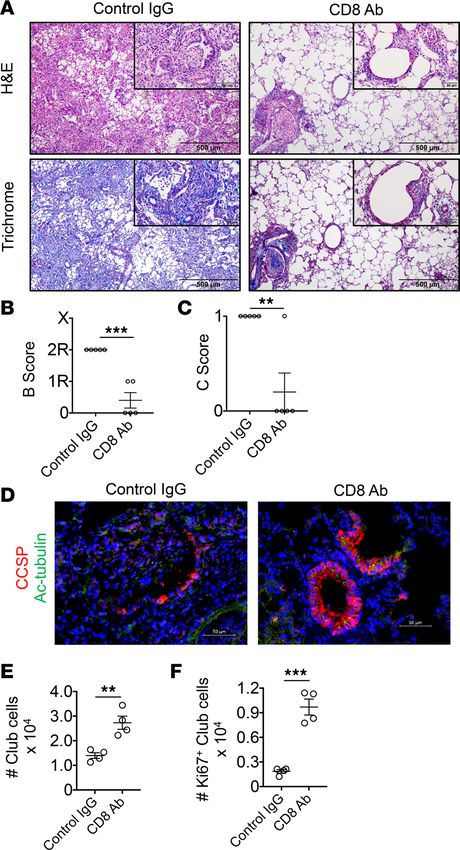
RESEARCH ARTICLE

                                  epithelium are risk factors for BOS development, which include primary graft dysfunction (8), Pseudomonas
                                  aeruginosa infection (9), community-acquired respiratory viral infections (10), and chronic aspiration of gas-
                                  tric acid (11, 12). There are also a few reports demonstrating club cell injury in lungs with BOS. For example,
                                  low CCSP levels in bronchiolar lavage fluid have been reported either as a risk factor for, or associated with,
                                  BOS development (13, 14). More recently, Palmer and colleagues demonstrated that patients with BOS have
                                  diminished CCSP expression in the airway epithelial cells of their terminal bronchioles (15). However, it
                                  remains to be investigated whether club cell loss is sufficient to trigger OB pathogenesis and promote immune
                                  responses known to be associated with BOS risk.
                                       Here, we describe a mouse orthotopic lung transplant (OLT) model that generates OB lesions in
                                  response to bronchiolar epithelial injury generated through the conditional activation of transgenes that
                                  direct club cell ablation. Club cell loss leads to the augmentation of adaptive immune responses that are
                                  coupled to BOS risk. Additionally, we find that CD8+ T cells play an important role in inhibiting club cell
                                  maintenance and proliferation.

                                  Results
                                  Club cell ablation triggers OB pathogenesis in lung transplant allografts. To determine if the loss of club cells
                                  promotes OB, we utilized triple-transgenic (3T) mice bearing the following genes: a reverse tetracycline
                                  activator gene driven by the CCSP promoter, Cre recombinase gene under control of the reverse tetracy-
                                  cline activator, and a lox-P–activated diphtheria toxin A gene (DT-A). Ingestion of doxycycline by 3T mice
                                  induces Cre-mediated recombination of the lox-P DT-A locus that promotes DT expression specifically in
                                  CSSP-expressing cells, resulting in their depletion and consequential injury to the bronchiolar epithelium
                                  (6). Because 3T mice were originally developed on a mixed histocompatibility antigen background, we
                                  extensively backcrossed these mice with FVB and C57BL/6 (B6) mice to generate fully defined minor and
                                  major histocompatibility 3T FVB and 3T B6 strains for syngeneic and allogeneic transplantation. To induce
                                  allograft acceptance in 3T FVB → B6 lung recipients, we administered CD40L-neuralizing (CD40L is also
                                  known as CD154) antibodies (Abs) and the CD80/86 antagonist CTLA4Ig (Figure 1A), which we have
                                  previously demonstrated induces established tolerance in the mouse OLT model 3 days after transplant
                                  (16). To implement club cell depletion, 3T B6 → B6 (syngeneic) and 3T FVB → B6 (allogeneic) recipient
                                  mice ingested doxycycline between postoperative day (POD) 7 and POD 9.5. Immunohistochemical anal-
                                  ysis of syngeneic and allogeneic transplants on POD 11 revealed small airways denuded of CCSP+ cells,
                                  but with the preservation of cells that expressed acetylated α-tubulin (Ac-tubulin), a marker for ciliated cells
                                  (Figure 1, B and C). By POD 16, however, syngeneic recipients had repaired their bronchiolar epithelium,
                                  as evident by reconstituted club and ciliated cell luminal monolayers that resembled preinjured bronchi-
                                  oles. In contrast, allografts contained club cells predominantly arranged in nonluminal monolayers without
                                  intervening ciliated cells. Additionally, many bronchioles had sharply reduced or completely absent lumi-
                                  nal expression of Ac-tubulin, although scattered ciliated cells could be detected throughout the interstitium
                                  (Supplemental Figure 1; supplemental material available online with this article; https://doi.org/10.1172/
                                  jci.insight.124732DS1). Histological assessment of POD 16, 3T FVB → B6 recipients that underwent club
                                  cell ablation revealed high-grade inflammatory bronchiolar injury and severe obliterative disease (Figure
                                  2, A–C). In contrast, analogously treated syngeneic lung transplants had at most mild graft inflammation,
                                  and lacked OB or peribronchial lesions up to POD 30 (Supplemental Figure 2). Total collagen deposition,
                                  which can be assessed by hydroxyproline assay or visualized by Masson’s trichrome stain, is promoted
                                  by chronic airway injury and accumulates to a higher degree in BOS patients when compared with stable
                                  lung recipients (17). Hydroxyproline accumulation was significantly greater in allografts with OB when
                                  compared with syngeneic transplants that underwent club cell ablation (Figure 2D). Masson’s trichrome
                                  staining of allograft OB lesions also revealed collagen deposition within transluminal fibrotic lesions (Fig-
                                  ure 2E). Finally, we did not observe the generation of OB lesions in allograft recipients following treatment
                                  with naphthalene (Supplemental Figure 3), an agent that has been used to ablate club cells (18) but may
                                  possibly inhibit lymphocyte activation (19).
                                       Previous work has demonstrated that club cells can be defined as pulmonary-restricted CD45–CD34–
                                  CD31–EpCAMhiCCSPhi cells (20). To further define the dynamics of bronchiolar epithelial repair in lung
                                  transplants we performed flow cytometric analysis to quantitate club cell abundance and proliferative
                                  responses following injury (Figure 3, A–H). In both syngeneic grafts and allografts there was an approx-
                                  imate 80% reduction in club cell abundance when measured 1.5 days after ablation. However, within the

insight.jci.org   https://doi.org/10.1172/jci.insight.124732                                                                                     2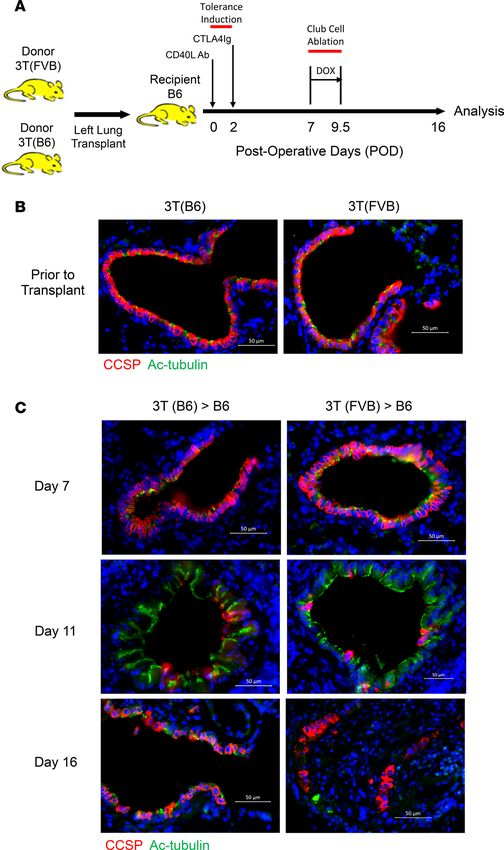
RESEARCH ARTICLE

                                  Figure 1. Lung allograft recipients are unable to repair their bronchiolar epithelium following club cell depletion.
                                  (A) A mouse OLT model that generates OB in 16 days. B6 recipients of 3T FVB left lungs treated with CD40L Abs
                                  (POD 0) and CTLA4Ig (POD 2) to establish tolerance were fed doxycycline by food and water (DOX) from POD 7 to 9.5
                                  and analyzed for graft inflammation on POD 16. 3T FVB and 3T B6 (B) donor lungs and (C) transplants stained with
                                  CCSP and Ac-tubulin Abs on POD 7, 11, and 16. Scale bars: 50 μm. Data shown are a representative result from at
                                  least 3 independent experiments.

insight.jci.org   https://doi.org/10.1172/jci.insight.124732                                                                                             3
RESEARCH ARTICLE

                                  remaining club cell compartment, the number undergoing replication was several-fold higher in synge-
                                  neic grafts when compared with allografts. Additionally, on POD 16, syngeneic grafts had nearly 3-fold
                                  more club cells relative to allografts. Therefore, club cell regenerative responses are sharply blunted in lung
                                  allografts that develop OB.
                                       Club cell ablation stimulates adaptive immune responses to lung transplants. We next determined if club cell
                                  depletion leads to a loss of immune tolerance. To this end, we compared effector T cell responses in 3T
                                  FVB allografts to double-transgene (2T FVB) allografts that lack the lox-P–activatable DT-A gene and
                                  therefore do not develop OB (Supplemental Figure 4). 3T and 2T FVB allograft recipients had comparable
                                  numbers of intragraft CD4+ T cells, Foxp3+CD4+, and IFN-γ+CD4+ T cells on POD 16 (Figure 4, A and B).
                                  However, club cell ablation led to greater accumulation of IL-17A+CD4+, total CD8+, and IFN-γ+CD8+ T
                                  cells within 3T FVB allografts. Additionally, specifically within airspaces of 3T FVB allografts, there was
                                  greater than a 3-fold increase of IFN-γ+CD8+ T cells (Figure 4C). The development de novo donor-anti-
                                  gen-specific Abs (DSAs) is a reported risk factor for BOS development (21). We therefore asked if club cell
                                  ablation promotes the accumulation of Abs that recognize FVB antigens (Figure 4D). When compared
                                  with recipients of 2T FVB allografts, there were significantly greater amounts of DSA against FVB anti-
                                  gens in the peripheral blood of 3T FVB allograft recipients.
                                       Because BOS progression is reportedly coupled to the loss of tolerance to alloantigens and the pulmo-
                                  nary self-restricted antigens, collagen V (ColV) and K-α1 tubulin (Kα1T) (22–24), we next assessed T cell
                                  antigen specificity through performing antigen recall assays on CD4+ and CD8+ T cells isolated from POD
                                  16 lung transplants (Figure 5A). Challenge of 3T FVB allograft CD4+ T cells with donor antigen-presenting
                                  cells (APCs) generated significantly higher amounts of IL-17A but not IFN-γ production relative to CD4+
                                  T cells from 2T FVB allografts. Syngeneic APCs bearing ColV or Kα1T peptides also stimulated higher
                                  amounts of IL-17A from 3T FVB allograft CD4+ T cells compared with those from 2T FVB allograft
                                  CD4+ T cells. Despite the lack of overall expansion of intragraft IFN-γ +CD4+ T cells there was a small but
                                  significant augmentation of IFN-γ production following ColV stimulation. In addition, club cell ablation
                                  led to more IFN-γ production from 3T FVB allograft CD8+ T cells following stimulation with donor anti-
                                  gens or self-antigens. In line with these observations, allografts with OB had accumulated higher intragraft
                                  ColV mRNA and protein along the epithelial basement membrane and within OB lesions (Figure 5, B–D).
                                  Together, these results demonstrate that club cell depletion induces the loss of tolerance to donor and pul-
                                  monary self-restricted antigens in lung transplants.
                                       Alloantigen-primed CD8+ T cells inhibit club cell regenerative responses. T cells are required for lung allograft
                                  rejection (25), but whether they regulate reparative responses by club cells remains unknown. Accordingly,
                                  we examined the role of T cells in our OB model by utilizing Ab-mediated T cell depletion (Figure 6, A–C,
                                  and Supplemental Figure 5). Pan–T cell depletion greatly reduced epithelial inflammation and eliminated
                                  OB lesions in 5 out of 7 allografts. Pan–T cell depletion also resulted in the repopulation of bronchiolar
                                  epithelium with club and ciliated cells (Figure 6D). Additionally, club cell proliferative responses were
                                  sharply elevated over recipients that received control Abs (Figure 6, E and F). Because we observed the high
                                  accumulation of airspace-resident effector CD8+ T cells in lung allografts with OB we next tested the effects
                                  of CD8-depleting Ab administration to recipients (Figure 7, A–F). Like pan–T cell depletion, CD8+ T cell
                                  depletion improved club cell regenerative responses and prevented OB lesion generation. In contrast, OB
                                  development was unaffected by Ab-mediated CD4+ T cell depletion (Supplemental Figure 6). To further
                                  examine the effects of CD8+ T cells on epithelial injury we cocultured club cells with alloantigen-primed,
                                  unprimed allogeneic, or syngeneic CD8+ T cells (Figure 8, A–C). Only alloantigen-primed CD8+ T cells
                                  inhibited club cell proliferation and maintenance. Noting our previous observations that apical surface
                                  MHC class I expression on airway epithelial cells is recognized by allogeneic CD8+ T cells (26), we asked if
                                  a neutralizing Ab that recognizes 1 of 3 MHC class I alleles (H-2Kq) expressed by FVB mice (27) prevents
                                  CD8+ T cell–mediated inhibition of club cell maintenance (Figure 8D). Blockade of H-2Kq significantly
                                  improved club cell maintenance, indicating that direct interactions between airway-infiltrating CD8+ T cells
                                  and club cells prevent bronchial epithelial repair in lung allografts.

                                  Discussion
                                  In the model of mouse orthotopic lung allograft transplantation reported in this study, we show that acute
                                  epithelial injury mediated by the ablation of club cells is sufficient to cause OB. The depletion and regener-
                                  ation of the club cell compartment was assessed by immunohistochemistry and flow cytometric analysis.

insight.jci.org   https://doi.org/10.1172/jci.insight.124732                                                                                          4
RESEARCH ARTICLE
Figure 2. Club cell ablation leads to severe OB lesions in lung allografts. (A) H&E histological staining of indicated
POD 16 transplants treated with or without DOX and blindly scored for (B) airway inflammation (B score) where 0
= none, 1R = low grade, 2R = high grade, and X = ungradable, and (C) the presence (1) or absence (0) of OB lesions
(C score). Dot plots represent mean score ± SEM of individual data obtained from 5 to 10 transplanted mice per
group. (D) Hydroxyproline content and (E) Masson’s trichrome staining of indicated transplant tissue at POD 16.
Data shown in D represent mean ± SEM (N ≥ 4/group). **P < 0.01, ***P < 0.001 by 2-tailed Mann-Whitney U test.
Images in A and E are representative histology from at least 5 transplants. Scale bars: 500 μm (upper images in
both) or 50 μm (lower images).
insight.jci.org https://doi.org/10.1172/jci.insight.124732 5RESEARCH ARTICLE
Figure 3. Club cell regenerative responses are suppressed in lung allografts. (A) FACS gating strategy for the identifi-
cation of club cells. Data shown are contour plots from a 3T FVB donor lung just prior to transplantation. 3T B6 and 3T
FVB transplants analyzed at indicated time points and assessed for club cell percentage abundance and proliferation
(Ki67). (B) Representative contour blots and histograms from at least 4 transplants. (C) Percentage reduction of club
cell abundance assessed on POD 11 calculated relative to levels prior to ablation on POD 7. (D) Percentage recovery of
the club cell compartment assessed on POD 16 calculated relative to levels prior to ablation on POD 7. (E) Total number
of intragraft club cells on POD 16. Club cell proliferation is shown as percentage abundance on (F) POD 11, (G) POD 16, or
as a total number on (H) POD 16. Dot plots represent mean ± SEM (N ≥ 4/group). n.s., not significant. *P < 0.05, **P <
0.01, ***P < 0.001 by 2-tailed Mann-Whitney U test.
Given CCSP expression within epithelial progenitor pools, the latter approach allowed us to more pre-
cisely monitor the regenerative dynamics of club cells through the use of lineage-specific markers and
intracellular CCSP expression (7, 20). Irrespective of whether the donor lung was on a B6 or FVB back-
ground we observed an average 80% reduction in club cells 1.5 days after doxycycline ingestion, indicating
there was not a strain-specific induction of bronchiolar epithelial injury. Another approach to ablate club
cells is through the systemic administration of naphthalene, which causes the production of cytotoxic
metabolites via oxidation by cytochrome P450 2F2, a monooxygenase that is expressed in club cells (28).
Naphthalene administration to our model was ineffective at producing OB lesions. Although the reasons
for this are not clear, it has been reported that toxic naphthalene metabolites generated by the liver inhibit
insight.jci.org https://doi.org/10.1172/jci.insight.124732 6RESEARCH ARTICLE
Figure 4. Club cell ablation promotes intragraft effector T cell accumulation and DSA. B6 recipients of 2T FVB and
3T FVB allografts that received DOX were analyzed by FACS on POD 16 for indicated intragraft T lymphocytes. (A)
Representative contour plots showing percentage abundance of graft-infiltrating effector T cells from B. (B) T lym-
phocyte numbers are shown for each transplant and as a mean per group ± SEM (N = 6 /group). *P < 0.05 by 2-tailed
Mann-Whitney U test. (C) Percentage abundance of airway-infiltrating effector T cells. Contour plots are a represen-
tative result from 3 transplants per group. (D) POD 16 recipient serum from 2T and 3T FVB recipients was coincubated
with FVB thymocytes at indicated dilution ratios and assessed for DSA with anti–mouse IgM antibodies. Reactivity is
shown as fold mean fluorescence intensity (MFI) relative to serum reactivity to syngeneic B6 thymocytes. Each data
point represents a mean from at least 4 recipients per group ± SEM. **P < 0.01 by 2-tailed Mann-Whitney U test.
Ab production by plasma cells (19). Of note, in a new mouse OLT model of lymphocytic bronchitis, B
cell deficiency protected allografts from club cell loss (29). Given that we observed DSA generation in our
model, these observations collectively raise the possibility that naphthalene prevents a B cell–dependent
response that promotes OB. Nevertheless, similar to reports of naphthalene-mediated club cell depletion,
we observed the transient spreading of ciliated cells around denuded bronchioles and the retention of a
population of injury-resistant club cells with high proliferative capacity (20, 30). This was most evident in
the high number of proliferating club cells within transplants approximately 2 days after injury induction.
Moreover, the relative abundance of proliferating club cells was nearly 3-fold lower in allografts when
compared with syngeneic grafts, indicating there is a critical threshold of regenerative capacity required to
adequately repair the bronchiolar epithelium.
insight.jci.org https://doi.org/10.1172/jci.insight.124732 7RESEARCH ARTICLE
Figure 5. Loss of allogeneic and pulmonary self-antigen tolerance following club cell ablation. (A) POD 16 intra-
graft CD4+ and CD8+ T cell recall assays for syngeneic (B6) APCs, allogeneic (FVB) APCs, and syngeneic APCs that bear
Kα1T or ColV peptides as measured by IFN-γ and IL-17A production. Data points shown are responses from individual
transplants and means per group ± SEM (N ≥ 5/group). (B) Semiquantitative RT-PCR measurements of intragraft ColV
(Col5a1) and Kα1T (Tuba1b) mRNA shown normalized to β-actin (Actb). Each data point represents a single transplant
along with a group mean ± SEM (N = 5/group). *PRESEARCH ARTICLE
Figure 6. Pan–T cell depletion inhibits OB and promotes club cell regenerative responses. 3T FVB → B6 recipients
that underwent club cell depletion received either control Ig or CD4- and CD8-depleting Abs on POD 6 and POD 11 were
analyzed on POD 16 for bronchiole injury by (A) H&E and Masson’s trichrome stain, (B) airway inflammation, and (C) OB
lesion score, or evaluated by immunohistochemistry for (D) bronchiolar epithelial repair with CCSP and Ac-tubulin Abs.
Transplant club cell (E) total numbers and (F) proliferating numbers (Ki67+) on POD 16. Results shown in A are represen-
tative stains used for grading in B and C, which show mean scores ± SEM (N = 7/group). The insets in A are ×400 (orig-
inal magnification). Scale bars: 500 μm (A) and 50 μm (D). Stains shown in D are representative of 7 transplants and E
and F show mean numbers ± SEM (N ≥ 4/group). *P < 0.05, **P < 0.01, ***P < 0.01 by 2-tailed Mann-Whitney U test.
Th17-mediated immunity (24), the enhanced recognition of Kα1T and ColV (23), the development of DSA
(21, 22), and high numbers of effector CD8+ T cells in the airway (32, 33). We also made insights into effector
T cell activation during OB development. To the best of our knowledge, this is the first report of the existence
of Kα1T- and ColV-specific effector CD8+ T cells in lung allografts. We also observed that increases in intra-
graft IL-17A+CD4+ T cells occur without significant changes in the overall number of graft-resident CD4+ T
cells. This finding suggests possible de novo generation of Th17 cells within graft tissue through the recruit-
ment of an uncommitted or naive CD4+ T cell pool. We have previously demonstrated that the targeted deple-
tion of lung allograft–resident Foxp3+CD4+ T cells abrogates established tolerance (34). Recent reports have
insight.jci.org https://doi.org/10.1172/jci.insight.124732 9RESEARCH ARTICLE
Figure 7. CD8+ T cell depletion inhibits OB and promotes club cell regenerative responses. 3T FVB → B6 recipients
that underwent club cell depletion received either control IgG or CD8-depleting Abs on POD 6 and POD 11 and were
analyzed on POD 16 for transplant bronchiole injury by (A) H&E and Masson’s trichrome stain, (B) airway inflamma-
tion, and (C) OB lesion score, or evaluated by immunohistochemistry for (D) bronchiolar epithelial repair with CCSP
and Ac-tubulin Abs. Transplant club cell (E) total numbers and (F) proliferating numbers (Ki67+) on POD 16. Results
shown in A are representative stains used for grading in B and C, which show mean scores ± SEM (N =5/group). The
insets in A are ×400 (original magnification). Scale bars: 500 μm (A) and 50 μm (D and insets in A). Stains shown in
D are representative of 5 transplants and E and F show mean numbers ± SEM (N = 4/group). **P < 0.01, ***P < 0.01
by 2-tailed Mann-Whitney U test.
also linked the loss of peripheral blood Foxp3+CD4+ T cells with increased risk for CLAD (35, 36). Although
we did not quantitate peripheral circulating Foxp3+CD4+ T cells, there was no significant change in absolute
numbers within graft tissues. One explanation for this discordant observation is that the ratio of Foxp3+CD4+
T cells to T cell effectors may be a more informative measure than is the assessment of absolute numbers (37).
Indeed, due to increased Th17 and effector CD8+ T cell numbers, this ratio was sharply lower in allografts
with OB. The development of Foxp3+CD4+ T cell regulatory dysfunction may also play an important role in
allograft rejection (38). Thus, future studies will be required to better understand the role of Foxp3+CD4+ T
cell abundance and function in OB pathogenesis.
Club cell ablation promoted elevated ColV mRNA and protein expression, which was especially
evident within the epithelial basement membranes and OB lesions. Of note, IL-17 has been previously
insight.jci.org https://doi.org/10.1172/jci.insight.124732 10RESEARCH ARTICLE
Figure 8. Alloantigen-primed effector CD8+ T cells inhibit club cell maintenance and proliferation. Club cells isolated
by lineage-negative enrichment from FVB lungs were cocultured without (Untreated) or with either unactivated
syngeneic FVB (Syn), unactivated allogeneic B6 (Unprimed Allo) or alloantigen-primed B6 (Primed Allo) CD8+ T cells, or
with supernatant from alloantigen-primed B6 CD8+ T cells (Primed Allo Supe). Thirty-six hours later, club cell abundance
was analyzed and shown as (A) representative contour plots from at least 4 independent experiments, or (B) total
numbers per experiment with a group mean ± SEM (N ≥ 4/group). Club cells analyzed for proliferation in the presence of
indicated CD8+ T cells by (C) CFSE dilution shown as a representative histogram from 5 independent experiments along
with a group mean ± SEM. **P < 0.01, ***P < 0.001 by 2-tailed Mann-Whitney U test (B and C). (D) Club cell numbers
remaining after coculture with indicated CD8+ T cells in the presence of control IgG or a neutralizing antibody directed
against the H-2Kq (FVB haplotype) MHC class I protein. Each data point represents a single determination from 4
independent experiments with a group mean ± SEM. *P < 0.05, **P < 0.01 by unpaired 2-tailed t test.
reported to promote ColV overexpression and the development of OB (39). In line with this previous
observation we demonstrated that CD4+ T cells isolated from allografts with OB made significantly more
IL-17A when challenged with ColV peptides when compared with CD4+ T cells isolated from tolerant
allografts. Despite increased T cell–mediated recognition of Kα1T we did not detect changes in its expres-
sion following club cell ablation. One possible reason for these differences comes from a recent finding
that Kα1T protein is highly enriched within exosomes released from human solid organ allografts, includ-
ing chronically rejected lung transplants (40). As exosome protein cargo can be endocytosed and present-
ed by dendritic cells (41), it remains plausible that T cells can be primed for recognition of Kα1T without
increased evidence of expression within parenchymal tissues.
In our model we used a well-established costimulatory blockade regimen (16, 42, 43) to induce lung
allograft tolerance, which interrupts both CD154/CD40 and B7/CD28 signaling in T cells. CD154 antag-
onism has recognized toxicity in humans due to CD154 expression on activated platelets (44). Also, in a
mouse heterotopic heart allograft model, the reported mechanism by which costimulatory blockade induc-
es acceptance is through the deletion of alloreactive T cells (45). In contrast, calcineurin inhibitors are
thought to promote tolerance by largely inhibiting T cell activation (46). These observations suggest that
the use of costimulatory blockade limits the clinical relevance of our model. However, OB lesions have
been reported to form spontaneously in about half of BALB/c → B6 orthotopic lung transplant recipi-
ents immunosuppressed with cyclosporine and steroids at between 1 and 3 months after engraftment (47).
insight.jci.org https://doi.org/10.1172/jci.insight.124732 11RESEARCH ARTICLE
Moreover, despite the use of costimulatory blockade, we could readily detect intragraft alloreactive T cells
in 2T FVB recipients, suggesting such clones are not efficiently deleted in lung transplants.
How T cells regulate epithelial cell repair in lung transplants remains an understudied area. This is likely
due to observations that several CD4+ and CD8+ T cell subsets are required for the induction of immuno-
suppression-mediated lung allograft tolerance (16, 42, 43). Accordingly, we chose to deplete T cells using
Abs after the establishment of tolerance (16), but just prior to the induction of epithelial injury to study club
cell responses. We observed that pan–T cell depletion largely reversed the loss of proliferating club cells,
restored the club cell compartment, and largely eliminated OB lesions. However, we still noted some epithe-
lial inflammation conceivably caused by the homeostatic expansion of residual intragraft alloreactive T cells
(48). We extended these studies to determine whether CD8+ or CD4+ T cells promote OB generation. CD8+
T cell depletion led to the promotion of club cell regenerative responses and protection from OB that was
comparable to pan–T cell depletion. In contrast, CD4+ T cell depletion had no effect on OB lesion develop-
ment, possibly due to the elimination of regulatory CD4+ T cells, which are reported to inhibit effector CD8+
T cell function in allografts (49, 50). We further explored the role of CD8+ T cells in OB by coculturing CD8+
T cells with club cells. Consistent with our observations of high numbers of airway-resident effector CD8+
T cells in lung allografts with OB, alloantigen-primed CD8+ T cells inhibited club cell maintenance and pro-
liferation. Additionally, in line with previous findings that airway-resident allogeneic CD8+ T cells recognize
apical MHC class I expressed on airway epithelium (26), we observed that neutralizing Abs against H-2Kq
improved club cell maintenance. Whether alloantigen-primed CD8+ T cells kill or suppress the proliferation
of club cells was not directly addressed by our studies, but is the subject of future investigation.
In summary, using a model of OLT we find that club cell ablation leads to development of OB and
the induction of adaptive immune responses associated with BOS risk. Given that severe and reliable OB
lesions are generated within 16 days after transplantation, this model should be useful for screening poten-
tial approaches to treating bronchiolar epithelial injury. To that end, our studies advocate for further defin-
ing T cell–dependent pathways that affect club cell regeneration and differentiation to identify new thera-
peutic targets for the prevention of BOS.
Methods
Mice and OLT. C57BL/6 (B6) and FVB/N (FVB) were purchased from Jackson Laboratories. CCSP rtTA/
TetOCre/DT-A (3T) mice were gifts from Jeffery Whitsett of The Children’s Hospital of Cincinnati. 3T
mice were backcrossed to greater than 99% FVB and C57BL/6 backgrounds, which was confirmed using
microsatellite-assisted accelerated backcrossing (MAX-BAX, Charles River). Mouse left OLT was conduct-
ed with 8- to 12-week-old donor and recipient mice as previously described by our group (42). To induce
allograft acceptance, recipients received i.p. 250 μg of CD40L Abs (MR1) on POD 0 and 200 μg of mouse
recombinant CTLA4Ig on POD 2 (16).
Club cell ablation and T cell depletion. Club cell injury was triggered by doxycycline ingestion via food (625
mg/kg chow, ENVIGO) and water (2 mg/ml, Sigma-Aldrich) from POD 7 to POD 9.5. T lymphocyte cell
depletion was accomplished by i.p. injection of anti-CD4 (500 μg GK1.5; Bio X Cell) or anti-CD8 (500 μg,
53-6.7, Bio-X-Cell) Abs or by using both aforementioned Abs for pan–T cell depletion. We used rat IgG2b
(clone LTF-2; Bio-X-Cell), rat IgG2a (clone 2A3; Bio-X-Cell), or both Abs for isotype controls for CD4+,
CD8+, or pan–T depletion, respectively. Abs were administered on POD 6 and 11.
Histological analysis. Harvested grafts were formaldehyde fixed, paraffin embedded, and stained with
H&E or Masson’s trichrome stain. Lung transplant histology was graded by a blinded pathologist using
the 2007 revision of the 1996 working formulation for the standardization of nomenclature in the diag-
nosis of lung rejection, where small-airway inflammation is scored Grade B0 (none), Grade B1R (low
grade), Grade B2R (high grade), and BX (ungradable). OB is graded without regard to inflammation as
C1 (present) or C0 (absent) (51).
Bronchiolar epithelium paraffin sections were first blocked with 5% goat serum and 2% fish gelatin
(both from Sigma-Aldrich) at 25°C for 45 minutes. Sections were then stained with 1:500 polyclonal rabbit
anti–mouse/rat CCSP (Seven Hills Bioreagents) and mouse anti–Ac-tubulin, 1:5000 (6-11B-1, Sigma-Al-
drich) overnight at 4°C. For immunofluorescent visualization, we used goat anti-mouse Alexa Fluor 488–
labeled secondary Ab, 1:1000 (Thermo Fisher Scientific) and goat anti-rabbit Alexa Fluor 555–labeled
secondary Ab, 1:1000 (Cell Signaling Technology). For ColV and Ka1T immunohistological analysis
1:100 polyclonal rabbit anti-ColV (Abcam), 1:200 polyclonal rabbit anti-Kα1T (Thermo Fisher Scientific),
insight.jci.org https://doi.org/10.1172/jci.insight.124732 12RESEARCH ARTICLE
or polyclonal rabbit IgG Isotype (Abcam) Ab dilutions were used in conjunction with a Vectastain Elite
System kit (LS Bio) for detection in accordance with the manufacturer’s recommendations. Quantitation
of stain was conducted with ImageJ bundled with Java version 1.8.0 (NIH) in which total pixels of stain
were assessed outside nuclear areas. For intragraft collagen measurements we used a Hydroxyproline
Assay Kit (Sigma-Aldrich) in accordance with manufacturer’s recommendations. Briefly, lung tissue was
extracted for protein with 6.0N HCl for 3 hours at 120°C and then dried at 60°C overnight. Then, 10 μg
of protein was assayed for reactivity for oxidized hydroxyproline with 4-(dimethylamino) benzaldehyde,
which in turn generates colorimetric product that was read by absorbance at 560 nm on a BioTek Synergy
HTX Microplate Analyzer.
Flow cytometric analysis and antigen recall assays. Lung tissue was minced and digested in RPMI 1640 with
type 2 collagenase (0.5 mg/ml) (Worthington Biochemical) and 5 units/ml DNAse (Sigma-Aldrich) for 90
minutes at 37°C and then filtered through a 70-μm cell strainer (Thermo Fisher Scientific) and treated with
ACK lysing buffer (Worthington Biochemical). Live cell discrimination was conducted with the Zombie
(Biolegend) fixable dye. Cell surface staining was conducted with the following Abs: CD45 (30-F11, eBio-
science), CD45.2 (104, Biolegend), CD90.2 (53-2.1, eBioscience), CD4 (clone RM4-5, eBioscience), CD8α
(53-6.7, eBioscience), CD31 (390, Biolegend), CD34 (HM34, Biolegend), and CD326 (G8.8, Biolegend).
Staining for Foxp3 (FJK-16s, eBioscience), Ki-67 (16A8, Biolegend), and polyclonal rabbit anti–mouse
CCSP (Seven Hills Bioreagents, catalog WRAB-3950) was conducted with an intranuclear Transcription
Factor Staining Buffer Kit (Invitrogen) in accordance with the manufacturer’s recommendations. For IFN-γ
and IL-17A expression, cells were first stimulated with 1 μM ionomycin (Sigma-Aldrich) and 20 ng/ml
PMA (Sigma-Aldrich) for 3.5 hours with 2 μM Golgi Plug (BD Biosciences) added for the last 3 hours of
stimulation and stained with anti–IFN-γ (XMG1.2, eBioscience) and anti–IL-17A (TC11-18H10.1, Bioleg-
end) using a Cytofix/Cytoperm kit (BD Biosciences) in accordance with the manufacturer’s recommenda-
tions. For antigen specificity measurements, T cells were fractionated by positive selection using CD4+ or
CD8+ immunomagnetic bead labeling kits (Miltenyi Biotec) from allograft cell suspensions and cocultured
at a 3:1 ratio with irradiated T cell–depleted FVB or B6 cell splenocytes for 96 hours pulsed with 0.5 μg/ml
Kα1T and ColV (obtained from T. Mohannakumar, St. Joseph’s Hospital, Phoenix, Arizona, USA). IFN-γ
and IL-17A were measured with uncoated ELISA kits from Invitrogen in accordance with the manufactur-
er’s recommendations.
Semiquantitative RT-PCR. Lung tissues were extracted for RNA with an RNAeasy kit (Qiagen) and
reverse transcribed with the High-Capacity cDNA Reverse Transcription Kit (Thermo Fisher Scien-
tific). Transcripts were semiquantified with PrimePCR Syber Green Assay (Bio-Rad) kits for Col5a1
(qMmuCED0046279), Tuba1b (qMmuCED0040564), and Atcb (qMmuCED0027505) in accordance with
the manufacturer’s recommendations.
Club cell enrichment and culture with CD8+ T cells. FVB lung tissue cell isolates were prepared as described
for FACS preparation and incubated with biotin-conjugated Abs (all from eBioscience) specific for CD45.1
(clone A20), CD34 (clone RAM34), CD31 (clone MEC13.3), CD90.1 (clone HIS51), and CD15 (clone
mc-480), washed, and then labeled with anti-biotin MicroBeads (Miltenyi Biotec) for negative selection on LS
columns (Miltenyi Biotec). Remaining cells were then incubated with biotin-conjugated CD326 Abs (clone
caa7-9G8, Miltenyi Biotec), washed, and then labeled anti-biotin MicroBeads for positive selection on an
MS column (Miltenyi Biotec). Enriched club cell fractions were resuspended in MTEC/Plus medium (52)
and seeded at 3.3 × 10⁴ cells per well in flat-bottom 96-well tissue culture plates (Thermo Fisher Scientific)
coated with 50 μg/ml type I rat tail collagen (Becton Dickinson). For proliferation analysis, club cells were
stained with 10 μM CFSE (Thermo Fisher Scientific) prior to seeding. CD8+ T cells were isolated from B6 or
FVB resting mouse spleens using CD8+ immunomagnetic bead labeling kits (Miltenyi Biotec) in accordance
with the manufacturer’s recommendations. To prime CD8+ T cells with alloantigen, T cell–depleted CD45.1+
FVB splenocytes were cocultured with B6 CD8+ T cells at a 3 to 1 respective ratio with 100 ng/ml LPS for 72
hours, which was used to augment alloantigen presentation. Alloantigen-primed CD8+ T cells were removed
from cultures by using immunomagnetic bead positive selection conducted with biotin-conjugated CD45.2
Abs (clone A20, eBioscience), anti-biotin MicroBeads, and LS columns. CD8+ T cells (1 × 10⁵) were added
to each club cell culture well for 24 hours. For proliferation analysis, CD8+ T cells were cocultured with
CFSE-labeled club cells for 36 hours. Four or 5 wells were used for each test condition. For MHC class I
blockade experiments 10 μg/ml of neutralizing Abs against H-2Kq (clone Y-3, Bio-X-Cell) or IgG2b isotype
control (clone MPC-11, Bio-X-Cell) was added to cultures 1 hour prior to the placement of CD8+ T cells.
insight.jci.org https://doi.org/10.1172/jci.insight.124732 13RESEARCH ARTICLE
Club cells were released from plates using 0.05% trypsin (Thermo Fisher Scientific), washed, and then ana-
lyzed on a Zombie–CD45–CD31–CD34–CD326+CCSP+ gate using Countbright beads (Thermo Fisher Scien-
tific) for quantitative FACS analysis.
Statistics. Unless indicated, a 2-tailed Mann–Whitney U test was performed to assess statistical rele-
vance using GraphPad Prism version 7.0. P < 0.05 was considered significant. n.s., not significant.
Study approval. Animal experiments were conducted in accordance with an approved Washington Uni-
versity Animal Care and Use Committee protocol (number 20160212).
Author contributions
AEG, ZL, HJH, AKTP, JMG, and SLB designed the experiments. AEG, HJH, and AKTP conceptualized
the model design. ZL, DS, MC, DLC, and HJH executed T cell studies. ZL performed immunohistochemical
staining, developed the club cell assays, and made figures. FL performed the mouse orthotopic lung trans-
plants. YF conducted the donor-specific antibody analysis. KNP validated and monitored the HLA haplotypes
of the triple-transgenic donor strains. AEG, ZL, RH, DK, ASK, and SLB wrote and edited the manuscript.
Acknowledgments
We thank Jihong Zhu for her excellent technical assistance. We also thank John H. Ritter for the analysis
of graft pathology. This work was supported by a Cystic Fibrosis Foundation research grant and by NIH
grants P01AI116501-01, R21AI119506, R01HL121218-01, and 2RHL094601.
Address correspondence to: Andrew E. Gelman, Maritz Professor of Immunology & Oncology, Campus
Box 8234, 660 South Euclid Avenue, Washington University in St. Louis, St. Louis, Missouri 63110-1013,
USA. Phone: 314.362.8382; Email: gelmana@wudosis.wustl.edu.
1. Yusen RD, et al. The Registry of the International Society for Heart and Lung Transplantation: Thirty-third Adult Lung and
Heart-Lung Transplant Report-2016; Focus theme: Primary diagnostic indications for transplant. J Heart Lung Transplant.
2016;35(10):1170–1184.
2. Verleden SE, Sacreas A, Vos R, Vanaudenaerde BM, Verleden GM. Advances in understanding bronchiolitis obliterans after
lung transplantation. Chest. 2016;150(1):219–225.
3. Rokicki W, Rokicki M, Wojtacha J, Dżeljijli A. The role and importance of club cells (Clara cells) in the pathogenesis of some
respiratory diseases. Kardiochir Torakochirurgia Pol. 2016;13(1):26–30.
4. Dierynck I, Bernard A, Roels H, De Ley M. Potent inhibition of both human interferon-gamma production and biologic activi-
ty by the Clara cell protein CC16. Am J Respir Cell Mol Biol. 1995;12(2):205–210.
5. Montoro DT, et al. A revised airway epithelial hierarchy includes CFTR-expressing ionocytes. Nature. 2018;560(7718):319–324.
6. Perl AK, Riethmacher D, Whitsett JA. Conditional depletion of airway progenitor cells induces peribronchiolar fibrosis. Am J
Respir Crit Care Med. 2011;183(4):511–521.
7. Teisanu RM, et al. Functional analysis of two distinct bronchiolar progenitors during lung injury and repair. Am J Respir Cell Mol
Biol. 2011;44(6):794–803.
8. Shah RJ, et al. Early plasma soluble receptor for advanced glycation end-product levels are associated with bronchiolitis obliter-
ans syndrome. Am J Transplant. 2013;13(3):754–759.
9. Borthwick LA, et al. Pseudomonas aeruginosa induced airway epithelial injury drives fibroblast activation: a mechanism in
chronic lung allograft dysfunction. Am J Transplant. 2016;16(6):1751–1765.
10. Khalifah AP, et al. Respiratory viral infections are a distinct risk for bronchiolitis obliterans syndrome and death. Am J Respir
Crit Care Med. 2004;170(2):181–187.
11. Mertens V, et al. Bile acids aspiration reduces survival in lung transplant recipients with BOS despite azithromycin. Am J Trans-
plant. 2011;11(2):329–335.
12. Neujahr DC, et al. Bile acid aspiration associated with lung chemical profile linked to other biomarkers of injury after lung
transplantation. Am J Transplant. 2014;14(4):841–848.
13. Nord M, Schubert K, Cassel TN, Andersson O, Riise GC. Decreased serum and bronchoalveolar lavage levels of Clara cell
secretory protein (CC16) is associated with bronchiolitis obliterans syndrome and airway neutrophilia in lung transplant recipi-
ents. Transplantation. 2002;73(8):1264–1269.
14. Bourdin A, et al. Donor clara cell secretory protein polymorphism is a risk factor for bronchiolitis obliterans syndrome after
lung transplantation. Transplantation. 2012;94(6):652–658.
15. Kelly FL, et al. Epithelial clara cell injury occurs in bronchiolitis obliterans syndrome after human lung transplantation. Am J
Transplant. 2012;12(11):3076–3084.
16. Li W, et al. Lung transplant acceptance is facilitated by early events in the graft and is associated with lymphoid neogenesis.
Mucosal Immunol. 2012;5(5):544–554.
17. Müller C, et al. Early extracellular matrix changes are associated with later development of bronchiolitis obliterans syndrome
after lung transplantation. BMJ Open Respir Res. 2017;4(1):e000177.
18. Reynolds SD, et al. Conditional clara cell ablation reveals a self-renewing progenitor function of pulmonary neuroendocrine
insight.jci.org https://doi.org/10.1172/jci.insight.124732 14RESEARCH ARTICLE
cells. Am J Physiol Lung Cell Mol Physiol. 2000;278(6):L1256–L1263.
19. Kawabata TT, White KL. Effects of naphthalene and naphthalene metabolites on the in vitro humoral immune response. J Toxi-
col Environ Health. 1990;30(1):53–67.
20. Guo L, et al. Generation of induced progenitor-like cells from mature epithelial cells using interrupted reprogramming. Stem
Cell Reports. 2017;9(6):1780–1795.
21. Safavi S, Robinson DR, Soresi S, Carby M, Smith JD. De novo donor HLA-specific antibodies predict development of bronchi-
olitis obliterans syndrome after lung transplantation. J Heart Lung Transplant. 2014;33(12):1273–1281.
22. Morrell MR, et al. De novo donor-specific HLA antibodies are associated with early and high-grade bronchiolitis obliterans syn-
drome and death after lung transplantation. J Heart Lung Transplant. 2014;33(12):1288–1294.
23. Hachem RR, Tiriveedhi V, Patterson GA, Aloush A, Trulock EP, Mohanakumar T. Antibodies to K-α 1 tubulin and collagen V
are associated with chronic rejection after lung transplantation. Am J Transplant. 2012;12(8):2164–2171.
24. Burlingham WJ, et al. IL-17-dependent cellular immunity to collagen type V predisposes to obliterative bronchiolitis in human
lung transplants. J Clin Invest. 2007;117(11):3498–3506.
25. Chen DL, et al. Increased T cell glucose uptake reflects acute rejection in lung grafts. Am J Transplant. 2013;13(10):2540–2549.
26. Kreisel D, et al. Polarized alloantigen presentation by airway epithelial cells contributes to direct CD8+ T cell activation in the
airway. Am J Respir Cell Mol Biol. 2011;44(6):749–754.
27. Sellers RS, Clifford CB, Treuting PM, Brayton C. Immunological variation between inbred laboratory mouse strains: points to
consider in phenotyping genetically immunomodified mice. Vet Pathol. 2012;49(1):32–43.
28. Stripp BR, Maxson K, Mera R, Singh G. Plasticity of airway cell proliferation and gene expression after acute naphthalene injury.
Am J Physiol. 1995;269(6 Pt 1):L791–L799.
29. Smirnova NF, et al. Inhibition of B cell-dependent lymphoid follicle formation prevents lymphocytic bronchiolitis after lung
transplantation. JCI Insight. 2019;4(3):123971.
30. Guha A, Vasconcelos M, Zhao R, Gower AC, Rajagopal J, Cardoso WV. Analysis of Notch signaling-dependent gene expres-
sion in developing airways reveals diversity of Clara cells. PLoS ONE. 2014;9(2):e88848.
31. Snyder JC, Reynolds SD, Hollingsworth JW, Li Z, Kaminski N, Stripp BR. Clara cells attenuate the inflammatory response
through regulation of macrophage behavior. Am J Respir Cell Mol Biol. 2010;42(2):161–171.
32. Zheng L, et al. Longitudinal comparisons of lymphocytes and subtypes between airway wall and bronchoalveolar lavage after
human lung transplantation. Transplantation. 2005;80(2):185–192.
33. Hodge S, et al. Increased levels of T cell granzyme b in bronchiolitis obliterans syndrome are not suppressed adequately by cur-
rent immunosuppressive regimens. Clin Exp Immunol. 2009;158(2):230–236.
34. Li W, et al. Bronchus-associated lymphoid tissue-resident Foxp3+ T lymphocytes prevent antibody-mediated lung rejection. J Clin
Invest. 2019;129(2):556–568.
35. Salman J, et al. Association of higher CD4+ CD25high CD127low, FoxP3+, and IL-2+ T cell frequencies early after lung transplan-
tation with less chronic lung allograft dysfunction at two years. Am J Transplant. 2017;17(6):1637–1648.
36. Piloni D, et al. Analysis of long term CD4+CD25highCD127- T-reg cells kinetics in peripheral blood of lung transplant recipients.
BMC Pulm Med. 2017;17(1):102.
37. Chung BH, et al. Clinical significance of the ratio between FOXP3 positive regulatory T cell and interleukin-17 secreting cell in
renal allograft biopsies with acute T-cell-mediated rejection. Immunology. 2012;136(3):344–351.
38. Bagley J, Yuan J, Chandrakar A, Iacomini J. Hyperlipidemia alters regulatory T cell function and promotes resistance to toler-
ance induction through costimulatory molecule blockade. Am J Transplant. 2015;15(9):2324–2335.
39. Vittal R, et al. IL-17 induces type V collagen overexpression and EMT via TGF-β-dependent pathways in obliterative bronchiol-
itis. Am J Physiol Lung Cell Mol Physiol. 2013;304(6):L401–L414.
40. Sharma M, Ravichandran R, Bansal S, Bremner RM, Smith MA, Mohanakumar T. Tissue-associated self-antigens containing
exosomes: Role in allograft rejection. Hum Immunol. 2018;79(9):653–658.
41. Morelli AE, et al. Endocytosis, intracellular sorting, and processing of exosomes by dendritic cells. Blood. 2004;104(10):3257–3266.
42. Krupnick AS, et al. Central memory CD8+ T lymphocytes mediate lung allograft acceptance. J Clin Invest. 2014;124(3):1130–1143.
43. Takahashi T, et al. PD-1 expression on CD8(+) T cells regulates their differentiation within lung allografts and is critical for tol-
erance induction. Am J Transplant. 2018;18(1):216–225.
44. Henn V, et al. CD40 ligand on activated platelets triggers an inflammatory reaction of endothelial cells. Nature.
1998;391(6667):591–594.
45. Wells AD, et al. Requirement for T-cell apoptosis in the induction of peripheral transplantation tolerance. Nat Med.
1999;5(11):1303–1307.
46. Hermann-Kleiter N, Baier G. NFAT pulls the strings during CD4+ T helper cell effector functions. Blood. 2010;115(15):2989–2997.
47. De Vleeschauwer S, et al. Chronic rejection pathology after orthotopic lung transplantation in mice: the development of a
murine BOS model and its drawbacks. PLoS ONE. 2012;7(1):e29802.
48. Moxham VF, et al. Homeostatic proliferation of lymphocytes results in augmented memory-like function and accelerated
allograft rejection. J Immunol. 2008;180(6):3910–3918.
49. van Maurik Av, Herber M, Wood KJ, Jones ND. Cutting edge: CD4+CD25+ alloantigen-specific immunoregulatory cells that can
prevent CD8+ T cell-mediated graft rejection: implications for anti-CD154 immunotherapy. J Immunol. 2002;169(10):5401–5404.
50. Lin CY, Graca L, Cobbold SP, Waldmann H. Dominant transplantation tolerance impairs CD8+ T cell function but not expan-
sion. Nat Immunol. 2002;3(12):1208–1213.
51. Stewart S, et al. Revision of the 1996 working formulation for the standardization of nomenclature in the diagnosis of lung
rejection. J Heart Lung Transplant. 2007;26(12):1229–1242.
52. You Y, Richer EJ, Huang T, Brody SL. Growth and differentiation of mouse tracheal epithelial cells: selection of a proliferative
population. Am J Physiol Lung Cell Mol Physiol. 2002;283(6):L1315–L1321.
insight.jci.org https://doi.org/10.1172/jci.insight.124732 15You can also read