Donor myeloid derived suppressor cells (MDScs) prolong allogeneic cardiac graft survival through programming of recipient myeloid cells in vivo
←
→
Page content transcription
If your browser does not render page correctly, please read the page content below
www.nature.com/scientificreports
OPEN Donor myeloid derived
suppressor cells (MDSCs) prolong
allogeneic cardiac graft survival
through programming of recipient
myeloid cells in vivo
Songjie Cai1,2,3, John Y. Choi1,3, Thiago J. Borges1,3, Hengcheng Zhang1, Ji Miao2,
Takaharu Ichimura1, Xiaofei Li1, Simiao Xu2, Philip Chu1, Siawosh K. Eskandari1,
Hazim Allos1, Juliano B. Alhaddad1, Saif A. Muhsin1, Karim Yatim1, Leonardo V. Riella1,
Peter T. Sage1, Anil K. Chandraker1* & Jamil R. Azzi1*
Solid organ transplantation is a lifesaving therapy for patients with end-organ disease. Current
immunosuppression protocols are not designed to target antigen-specific alloimmunity and are
uncapable of preventing chronic allograft injury. As myeloid-derived suppressor cells (MDSCs) are
potent immunoregulatory cells, we tested whether donor-derived MDSCs can protect heart transplant
allografts in an antigen-specific manner. C57BL/6 (H2Kb, I-Ab) recipients pre-treated with BALB/c
MDSCs were transplanted with either donor-type (BALB/c, H2Kd, I-Ad) or third-party (C3H, H2Kk,
I-Ak) cardiac grafts. Spleens and allografts from C57BL/6 recipients were harvested for immune
phenotyping, transcriptomic profiling and functional assays. Single injection of donor-derived MDSCs
significantly prolonged the fully MHC mismatched allogeneic cardiac graft survival in a donor-specific
fashion. Transcriptomic analysis of allografts harvested from donor-derived MDSCs treated recipients
showed down-regulated proinflammatory cytokines. Immune phenotyping showed that the donor
MDSCs administration suppressed effector T cells in recipients. Interestingly, significant increase in
recipient endogenous CD11b+Gr1+ MDSC population was observed in the group treated with donor-
derived MDSCs compared to the control groups. Depletion of this endogenous MDSCs with anti-Gr1
antibody reversed donor MDSCs-mediated allograft protection. Furthermore, we observed that the
allogeneic mixed lymphocytes reaction was suppressed in the presence of CD11b+Gr1+ MDSCs in a
donor-specific manner. Donor-derived MDSCs prolong cardiac allograft survival in a donor-specific
manner via induction of recipient’s endogenous MDSCs.
Solid organ transplantation is a lifesaving therapy for patients with end-organ disease. The Scientific Registry of
Transplant Recipients (SRTR) reported that 14,313 k idney1, 7,483 liver2, 2,465 lung3, 432 heart4, 80 p
ancreas5,
and 109 i ntestinal6 transplants were performed in 2017 in the US (Feb 2019 updated). Nevertheless, number of
challenges remain for organ recipients after the transplantation, as they suffer from acute and chronic rejection
as well as complications of i mmunosuppression7–9. One of the more critical issues pertains to the current state
of standard immunosuppressive protocols. Calcineurin inhibitor (CNI) based immunosuppressive protocols are
not designed to promote antigen-specific tolerance, and may exacerbate chronic allograft injury (i.e. calcineurin
toxicity)10. Therefore, there is an urgent need to establish safer anti-rejection strategies by targeting antigen-
specific alloimmunity.
1
Renal Division, Transplantation Research Center, Brigham and Women’s Hospital, Harvard Medical School,
221 Longwood Ave, Boston, MA 02115, USA. 2Division of Endocrinology, Boston Children’s Hospital, Harvard
Medical School, Boston, MA, USA. 3These authors contributed equally: Songjie Cai, John Y. Choi and Thiago
J. Borges. *email: achandraker@bwh.harvard.edu; jazzi@bwh.harvard.edu
Scientific Reports | (2020) 10:14249 | https://doi.org/10.1038/s41598-020-71289-z 1
Vol.:(0123456789)
www.nature.com/scientificreports/
Cell therapies, including regulatory dendritic cells (DCregs)11–14, regulatory T cells (Tregs)15 and regulatory
B cells (Bregs)16, have shown the potential application in solid organ transplantation. In particular, Thompson
and colleagues conducted a first-in-human study to demonstrate that adoptive transfer of DCreg allows complete
withdrawal of immunosuppression regimen in low risk p atients17–20. Myeloid derived suppressor cells (MDSCs)
comprise a heterogeneous cell population including immature myeloid cells, which thereafter differentiate into
monocytes, dendritic cells and neutrophils21. While sharing similar functional features, MDSCs are shown to
offer much powerful immunosuppression compared to DCregs. MDSCs inhibit T cell responses by inducing the
apoptosis of antigen-primed and activated T cells22,23 while inducing FoxP3+ Treg23,24. In vivo models have shown
MDSCs attenuate GvHD25 as well as autoimmunity26,27. Finally, higher frequency of MDSCs were detected in
blood samples from stable renal transplant patients24.
Based on the above clinical and preclinical studies, we hypothesized that donor-derived MDSCs may induce
donor-specific immune suppression in allogeneic organ transplantation. Of note, we modified standard in vitro
MDSCs generation p rotocol28 by culturing donor bone marrow cells within 6 days in the presence of GM-CSF29
, TGFβ30 and I L1031,32. In addition, we added IFNγ on day 5 to promote the suppressive function of M DSCs33.
Finally, we demonstrated that the administration of donor-derived MDSCs prolong allogeneic cardiac graft
survival in a donor-specific manner through induction of recipients’ endogenous MDSCs.
Results
Donor‑derived MDSCs suppress alloreactive T cell activation in vitro. Allogeneic mixed lympho-
cyte reaction (alloMLR) was performed to examine the regulatory function of donor-derived MDSCs. Co-cul-
turing naive T cells isolated from C57BL/6 mice (H2Kb, I-Ab) with BALB/c (H2Kd, I-Ad) derived conventional
DCs (cDCs) stimulates an alloreactive T cell proliferation. We observed that the addition of BALB/c MDSCs to
this alloMLR system significantly inhibited the proliferation of CD4+ and CD8+ T cells compared to conven-
tional myeloid derived cells (cMDCs) (Fig. 1A,B).
Donor‑derived MDSCs protect cardiac allografts from acute rejection. We then examined the
in vivo suppressive function of donor-derived MDSCs in the allogeneic cardiac transplantation model. C57BL/6
recipients received a single-dose of intravenous injection of 1 × 10⁶ BALB/c MDSCs or BALB/c cMDCs 7 days
prior to the cardiac transplantation with BALB/c donors (Fig. 1C). We found that BALB/c MDSCs significantly
prolonged allograft survival; in contrast, administration of cMDCs showed no difference to the group receiving
PBS (PBS control n = 9, median survival time (MST) 7 days; BALB/c cMDCs control n = 7, MST 7 days; BALB/c
MDSCs n = 8, MST 14 days, Fig. 1D).
Allografts were harvested on postoperative day (POD) 7 for hematoxylin and eosin (H&E) staining and
immune fluorescence staining. H&E staining revealed the attenuated myocardial lesion as well as the decreased
lymphocyte infiltration in the donor MDSCs treated group compared to the cMDC group (Fig. 2A). Immune
fluorescence staining showed that C D3+ T cells were significantly decreased in the MDSC group compared to
the cMDC group. Of note, there was no difference in C D11b+ cells infiltration between two groups (Fig. 2B).
Graft infiltration lymphocytes (GILs) were isolated from allografts (POD7) for flow cytometry analysis.
Gating on the CD4+FoxP3- helper T cells, the proportion of effector T cells (Teff) defined as CD44+CD62Llo
population, was significantly decreased in the MDSCs treated group compared to the cMDCs treated group
(Fig. 2C-upper). Within this Teff, the activation level measured by Ki67 was also significantly reduced in the
MDSCs treated group (Fig. 2C-lower).
Finally, qRT-PCR of allografts (POD3, whole tissue) showed that the treatment with donor-derived MDSCs
markedly downregulated the mRNA expression of proinflammatory cytokine: IL1α, IL2, IL4, IL13, NLRP3,
IFNγ, IRF1, and TNF (Fig. 2D and Table S4).
Allo‑immune response of effector T cells is reduced in recipients treated with donor MDSCs. T
cells were isolated from recipients’ splenocytes on POD7 for the ex vivo stimulation assay using BALB/c Anti-
gen Presenting Cells (APCs) at the ratio of 20:1 or 10:1 for 4 days (Fig. 3A). CD4+ and C D8+ Teff induction
was analyzed by flow cytometry. Cells were gated on CD4 Teff (CD4 FoxP3 CD44 CD62lo) and CD8+ Teff
+ + - +
(CD8+FoxP3-CD44+CD62lo). We found that the proportion of Ki67+ Teff (Ki67+CD44+CD62lo) in CD4+FoxP3-
as well as CD8+FoxP3- population were decreased in the MDSCs group in comparison to the cMDCs group
(Fig. 3B).
Donor‑derived MDSCs induce highly immune suppressive endogenous MDSCs. For mechanis-
tic studies, spleens from C57BL/6 recipients of BALB/c hearts treated with BALB/c MDSCs were isolated on
POD7 for flow cytometry analysis. We observed that a suppressive population CD11b+Gr1+34, defined broadly as
MDSCs34, was significantly increased in the donor-derived MDSCs treated group (Fig. 4A). Gating on this popu-
lation, we found that the PDL1 expression was markedly up-regulated in the MDSCs treated group compared to
the cMDCs treated group (Fig. 4B). These data indicate that the donor-derived MDSCs administration not only
increases the number of CD11b+Gr1+ population in recipients, but also program CD11b+Gr1+ cells to upregulate
PDL1 expression. PD-L1/PD-1 interactions were reported to deliver co-inhibitory signals leading to attenuation
of T cell responses both in vitro and in vivo35,36. Furthermore, PDL1 has been shown to be required for suppres-
sion of the autoimmune responses37. In consistent with this, we observed significantly increased CD11b+Gr1+
population in GILs from recipients treated with donor-derived MDSCs (Fig. 4C). We then hypothesized that
CD11b+Gr1+ cells in recipients detected upon administration of donor-derived MDSCs play a crucial role in
allogeneic immune suppression.
Scientific Reports | (2020) 10:14249 | https://doi.org/10.1038/s41598-020-71289-z 2
Vol:.(1234567890)
www.nature.com/scientificreports/
Figure 1. Donor-derived MDSCs suppress alloimmune reaction in vitro and in vivo. (A) Naïve C57BL/6T cells
were stimulated with BALB/c antigen presenting cells (APCs: BALB/c bone marrow derived cDCs). MDSCs
or cMDCs were added as modulator. C D4+ and CD8+ T cell proliferation in response to allogeneic cDCs was
analyzed by CellTrace violet dye dilution (n = 5 per group). (B) Graphs showing the proliferation of CD4+ and
CD8+ T cells in the presence of MDSCs compared to cMDCs. Mean ± SEM, *p < 0.05, **p < 0.01, two-tailed
unpaired t test. Data represents one of 4 separate experiments. (C) Schematic diagram of the experimental
design. C57BL/6 recipients received a single-dose intravenous injection of 1 × 10⁶ BALB/c MDSCs or cMDCs
7 days prior to the cardiac transplantation. (D) Kaplan–Meier cumulative survival of allograft shows the
prolonged survival in MDSCs treated group compared to control groups (n = 8–9 per group). **p < 0.01,
***p < 0.001, log-rank test.
To determine whether the increase of MDSCs ( CD11b+Gr1+) is from the clonal expansion of the transferred
donor-derived MDSCs or from the induction of recipients’ endogenous MDSCs, we traced the transferred
donor-derived MDSCs using H 2Kd antibody. 1 × 1 06 BALB/c MDSCs were intravenously injected to C57BL/6
recipients, and their splenocytes were examined at 3, 6 and 24 h post-injection. We found that the transferred
donor-derived MDSCs peaked 3 h post-injection (PIH3) and then disappeared at PIH24 (Fig. S7A). In parallel,
we analyzed the induction of endogenous MDSCs in recipients’ spleens by gating on the recipient type MHC-I,
H2Kb. We found significant increase of these endogenous MDSCs in recipients starting at PIH24 (Fig. S7B).
Next, we measured the MHC-II expression on the endogenous MDSCs as a marker of suppressive f unction38.
Indeed, the MHC-II expression was markedly elevated on the endogenous MDSCs (Fig. S7B). These data sug-
gest that while donor-derived MDSCs disappear within 24 h, the functional endogenous MDSCs are expanded
in the recipients’ lymphoid tissue.
We further characterized the composition of endogenous MDSCs in recipient’s spleen by using Ly6C and Ly6G
antibodies21. Gating on CD11b+ cells, flow cytometry analysis indicated that the induced endogenous MDSCs
were composed mostly of L y6C+Ly6G- monocytic MDSC (M-MDSC) and L y6C+Ly6G+ MDSC (Fig. S2C,D).
Previous studies have shown that systemic administration of donor cells undergoing apoptosis promote
donor-specific immunosuppression in vitro39 and in vivo40,41. To study whether our generated donor MDSCs
suppressed alloimmune reaction is related to the apoptotic donor cell mediated suppression, we used 7-AAD and
Annexin-V to measure the frequency of apoptotic cells in MDSCs and control cMDCs. Result showed approxi-
mately 3.3% early apoptotic and 6.1% late apoptotic cells in generated donor MDSCs, which are significantly
lower than in cMDCs (7.0% early apoptotic and 9.9% late apoptotic cells, p < 0.001) (Fig. S5). Taken together,
we concluded that the suppressive function of donor-derived MDSCs is independent of apoptotic-cell-induced
immune suppression.
Scientific Reports | (2020) 10:14249 | https://doi.org/10.1038/s41598-020-71289-z 3
Vol.:(0123456789)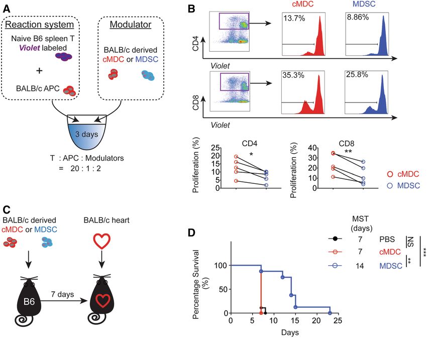
www.nature.com/scientificreports/
Figure 2. Allografts from donor-derived MDSCs treated recipients exhibit ameliorated T cell mediated
inflammation and increased recipient-derived endogenous MDSCs. (A) Representative example of the allograft
histology (H&E staining) from the MDSCs treated group and the cMDCs treated control on POD7. Scale bar
represent 500 μm (left) and 100 μm (right). Data represents the one of 3 separate experiments. (B) Allografts
were harvested on POD7 and stained with anti-CD3, anti-CD11b (green) and DAPI (blue). Scale bars represent
200 μm. Positive cells were qualitied by ImageJ and expressed as mean ± SEM. ****p < 0.0001, two-tailed
unpaired t test. (C) Flow cytometry analysis of BALB/c MDSCs treated C57BL/6 recipients’ graft infiltration
lymphocytes (GILs) on POD7. Graphs showing significant decrease of the proportion of C D44+CD62Llo and
+ + -
Ki67 in CD4 FoxP3 T cells in the MDSCs treated group. (D) qRT-PCR analysis of allografts (whole tissues) on
POD3 (n = 3 per group). Graph represented as RQ (relative quantification) = 2-ΔΔCt. Naïve BALB/c heart serve as
basic ΔCt (n = 3, not shown in graph). Mean ± SEM, *p < 0.05, **p < 0.01, ***p < 0.001, two-tailed unpaired t test.
Endogenous MDSCs inhibit effector T cells and prolong allograft survival. We then set up the
alloMLR to test the immunomodulatory function of these endogenous MDSCs in vitro. Naive T cells from
C57BL/6 mice were stimulated with BALB/c APCs. CD11b+Gr1+ cells were isolated by fluorescence activated
cell sorting (FACS) from recipients’ splenocytes on POD7 and were used as modulators. As the transferred
donor-derived MDSCs disappeared 24 h after injection, these C D11b+Gr1+ MDSCs are endogenous MDSCs
(Fig. S7A). Cells were incubated for 3 days and then stained with CD4, FoxP3 and Ki67 for flow cytometry
analysis (Fig. 4D). We observed that the proportion of Ki67+FoxP3- in CD4+ T cells was significantly decreased
in the MDSCs group (Fig. 4E).
Next, we depleted this population with a monoclonal antibody (mAb) anti-Gr1 (RB6-8C5) post-transplan-
tation and observed the allograft s urvival42–44. Recipients were treated with 1 × 10⁶ BALB/c MDSCs 7 days prior
to the transplantation, followed by the anti-Gr1 injection on days 0, 2, 4 and 6 post-transplantation (Fig. 4F).
Gr1+ depletion markedly shortened the allograft survival (anti-Gr1 n = 7, MST 10 days; Isotype IgG n = 7, MST
14 days, p = 0.0052; Fig. 4G) confirming that endogenous MDSCs that are characterized by CD11b+Gr1+ are
essential to the allograft protection.
Endogenous MDSCs protect cardiac allografts in a donor‑specific manner. We next tested
whether the prolongation of the allograft survival by the donor-derived MDSCs administration is donor-spe-
Scientific Reports | (2020) 10:14249 | https://doi.org/10.1038/s41598-020-71289-z 4
Vol:.(1234567890)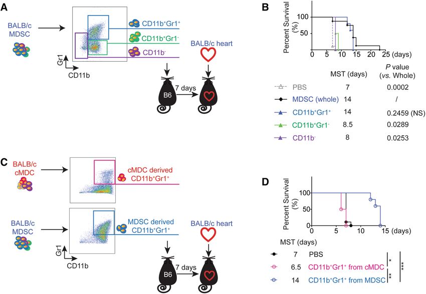
www.nature.com/scientificreports/
Figure 3. Donor-derived MDSCs suppress effector T cell activation. (A) Schematic diagram of the
experimental design. T cells were isolated from recipient splenocytes on POD7 and were stimulated
with BALB/c cDCs for 4 days (n = 4 per group). (B) CD4+ and CD8+ effector T cells induction was
analyzed by flow cytometry. Cells were gated on CD4+ Teff (CD4+FoxP3-CD44+CD62lo) and CD8+ Teff
(CD8+FoxP3-CD44+CD62lo). Graphs showing the percentage of activated Teff (Ki67+Teff). Mean ± SEM,
*p < 0.05, **p < 0.01, two-tailed unpaired t test. Data represents one of 4 separate experiments.
cific. C57BL/6 (H2Kb, I-Ab) mice received a single-dose intravenous injection of 1 × 10⁶ BALB/c (H2Kd, I-Ad)
or C3H ( H2Kk, I-Ak) MDSCs 7 days prior to the transplantation. C3H or BALB/c mice were used as cardiac
allograft donors (Fig. 5A). We observed that BALB/c MDSCs was able to prolong BALB/c allograft survival but
failed to prolong the C3H allografts survival. Likewise, C3H MDSCs was able to prolong C3H allograft survival
but failed to prolong the BALB/c allografts survival (Fig. 5B). Together, this suggests that donor-derived MDSCs
prolong the allogeneic cardiac graft survival in a donor-specific manner.
As we have identified that donor-derived MDSCs induced recipient’s endogenous MDSCs (CD11b+Gr1+)
cells in recipients play a key role in allogeneic immune suppression (Fig. 5C,D), we then tested the donor-specific
suppressive function of endogenous MDSCs by using the alloMLR. Naïve C57BL/6 spleen T cells were stimulated
with BALB/c cDCs or C3H cDCs as a third-party. CD11b+Gr1+ cells were isolated by FACS from BALB/c MDSCs
treated C57BL/6 recipient splenocytes at POD7 and were added as modulators (Fig. 5C). The proliferation of
CD4+ and CD8+ T cells were determined by using violet dye dilution by flow cytometry. As naïve C57BL/6 T cells
respond to BALB/c and C3H cDCs at a different rate, we calculated the relative suppression efficiency (SE) and
compared the SE between the primary donor type cDCs and the third-party cDCs stimulated group (Fig. 5D).
We observed doubled SE of CD4+ proliferation and a tripled SE of CD8+ proliferation in BALB/c cDCs stimu-
lated group compared to C3H cDCs stimulated group (Fig. 5D). This data supports that the immune suppressive
function of donor-derived MDSCs induce endogenous MDSCs is antigen-specific.
CD11b+Gr1+ cells among donor‑derived MDSCs prolong allograft survival. Further analysis
of in vitro generated MDSCs revealed the composition of donor-derived MDSCs is > 85% CD11b+Gr1+ cells
and < 15% non-CD11b+Gr1+ cells ( CD11b- and C D11b+Gr1-) (Fig. S1B). In above experiments pertain to Figs. 1,
2, 3, 4 and 5, we used whole MDSCs that were generated from donor bone marrow cells to achieve the donor-
specific alloimmune suppression. To test whether the population other than C D11b+Gr1+ cells contributes to
D11b Gr1 and CD11b- cells by FACS and per-
the biological changes, we isolated the other two populations, C + -
formed immune suppressive functional assay (Fig. 6A).
C57BL/6 recipients treated with CD11b+Gr1+ from donor-derived MDSCs achieved the similar allograft
survival with the whole MDSCs treatment group. However, C57BL/6 recipients treated with CD11b+Gr1- or
CD11b- cells from donor-derived MDSCs did not show significant graft survival benefit (Fig. 6B). Thus, we
concluded that the C D11b+Gr1+ cells, but not non-CD11b+Gr1+ cells, among donor-derived MDSCs play the
key role in the MDSCs mediated allograft survival.
MDSCs are a heterogeneous population of immature monocytes and granulocytes21. In addition to Gr-1,
CD11b combined with Ly6C and Ly6G has been utilized to distinguish the subpopulations of MDSCs45–47:
Ly6C+Ly6G- (monocytic MDSC, M-MDSC), Ly6G+Ly6C- (granulocytic MDSC, G-MDSC) and Ly6C+Ly6G+
MDSC. Our in vitro generated MDSCs are composed with approximately 47.14% of M-MDSC, 2.74% of
G-MDSC and 34.43% of Ly6C+Ly6G+ MDSC (Fig. S1B). To determine which population plays the key role in
the allograft survival prolongation, we flow-sorted the three subsets after MDSC differentiation in vitro and
transferred them to recipients 7 days before HTx. Because the dose of whole MDSC treatment group is 1 × 10⁶,
we adjusted the treatment dose to reflect the percentage of specific subset within the whole MDSC, that is: (1)
4.71 × 10⁵ of M-MDSC; (2) 3.44 × 10⁵ of Ly6C+Ly6G+ MDSC; (3) 2.74 × 10⁴ of G-MDSC. Surprisingly, none of
the single population treatment group (M-MDSC MST 7 days, n = 4; Ly6C+Ly6G+ MDSC MST 7 days, n = 4;
Scientific Reports | (2020) 10:14249 | https://doi.org/10.1038/s41598-020-71289-z 5
Vol.:(0123456789)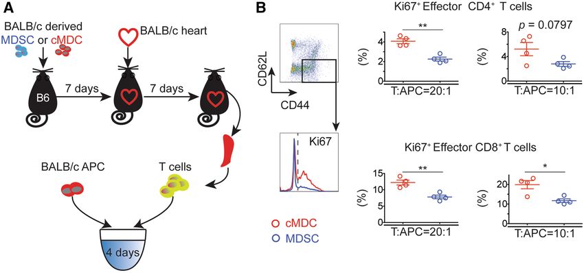
www.nature.com/scientificreports/
Figure 4. Endogenous MDSCs (CD11b+Gr1+) are essential to donor-derived MDSCs mediated alloimmune
suppression. (A,B) Flow cytometry analysis of BALB/c MDSCs treated C57BL/6 recipients’ splenocytes on
POD7 (n = 8–9 per group). (A) The proportion of CD11b+Gr1+ significantly increased in donor-derived the
MDSCs treated group. (B) Cells were gated on CD11b+Gr1+, PDL1 expression was up-regulated in the MDSCs
treated group. Mean ± SEM, * p < 0.05, ** p < 0.01, *** p < 0.001, one-way ANOVA and Tukey’s test. Data
represents one of 4 separate experiments. (C) Flow cytometry analysis of BALB/c MDSCs treated C57BL/6
recipients’ graft infiltration lymphocytes (GILs) on POD7. CD11b+Gr1+ population was significantly increased
in MDSCs treated group. n = 3–6 per group. Mean ± SEM. Data represents one of 3 separate experiments.
*p < 0.05, two-tailed unpaired t test. (D) Schematic illustration of the experimental design to test the ex vivo
immunosuppressive function of BALB/c MDSCs induced endogenous MDSCs ( CD11b+Gr1+) in recipients.
Naïve C57BL/6 T cells were stimulated with BALB/c APCs for 3 days. CD11b+Gr1+ cells were isolated by
fluorescence activated cell sorting (FACS) from C57BL/6 recipient splenocytes at POD7 (n = 3–4 per group)
and were added as modulator. (E) Cells were gated on CD4+FoxP3-, and the Ki67 expression was measured.
Graphs showing significant decrease of the proportion of K i67+FoxP3- in CD4+ T cells in the presence of
CD11b+Gr1+ cells induced with MDSCs compared to those induced with cMDCs. Mean ± SEM, * p < 0.05,
two-tailed unpaired t test. Data represents one of 3 separate experiments. (F) Schematic illustration of the
treatment protocol. C57BL/6 recipients received a single-dose intravenous injection of 1 × 10⁶ BALB/c 7 days
prior to the transplantation. Recipients were treated with anti-Gr1 mAb on POD 0, 2, 4, 6. (G) Anti-Gr1 mAb
administration attenuates MDSCs induced immune suppression. Kaplan–Meier cumulative survival of allograft
showing the shortened allograft survival in the anti-Gr1 Ab treated group compared to the IgG treated control
group (n = 7 per group). **p < 0.01, log-rank test.
Scientific Reports | (2020) 10:14249 | https://doi.org/10.1038/s41598-020-71289-z 6
Vol:.(1234567890)www.nature.com/scientificreports/
Figure 5. Donor-derived MDSCs prolong allogeneic cardiac graft survival in a donor-specific manner. (A)
Schematic diagram of the experimental design. C57BL/6 mice received a single-dose intravenous injection of
1 × 10⁶ BALB/c or C3H MDSCs. Cardiac grafts from C3H or BALB/c mice were transplanted into pre-sensitized
C57BL/6 recipients. (B) Kaplan–Meier cumulative survival of allograft showing that the BALB/c MDSCs
administration successfully prolong BALB/c allograft survival but fail to prolong C3H allograft survival, while
C3H MDSCs administration prolong C3H allograft survival but fail to prolong BALB/c allograft survival.
*p < 0.05, **p < 0.01, ***p < 0.001, log-rank test. (C) Schematic illustration of the experimental design to study
the ex vivo donor-specific immunosuppressive function of donor MDSCs induced endogenous MDSCs
(CD11b+Gr1+). Naïve C57BL/6 spleen T cells were labeled with CellTrace violet and were stimulated with
BALB/c cDCs or C3H cDCs (third-party). C D11b+Gr1+ cells were isolated by FACS from BALB/c MDSCs
treated C57BL/6 recipient splenocytes at POD7 (n = 3–4 per group) and were added as modulator. (D) CD4+
and CD8+ T cell proliferation in response to primary donor-type BALB/c cDCs or third-party C3H cDCs
was analyzed by CellTrace violet dye dilution. Graphs showed the attenuated suppression efficiency (SE) of
p(w/oMDSC)−p(MDSC)
CD11b+Gr1+ cells in third-party compared to donor-type allo MLR setting. SE = p(w/oMDSC) × 100%.
Mean ± SEM, *p < 0.05, **p < 0.01, two-tailed unpaired t test. Data represents one of 3 separate experiments.
G-MDSC MST 7 days n = 3) (Fig. S1C), but only the combination of the 3 populations significant improved
allograft survival (whole MDSC MST 14 days, n = 8). This result shows that the 3 different populations of MDSC
synergistically suppressed allo-immunity.
CD11b+Gr1+ cells from donor‑derived MDSCs, but not from donor‑derived cMDCs, suppress allo‑
immunity. In vitro generated donor-derived MDSCs contained > 85% CD11b+Gr1+ cells, while generated
donor-derived cMDCs (control) contained only 40% CD11b+Gr1+ cells (Fig. S1B). To study whether donor
D11b+Gr1+ cells rather than the dif-
MDSCs mediated alloimmune suppression is due to the different dose of C
Scientific Reports | (2020) 10:14249 | https://doi.org/10.1038/s41598-020-71289-z 7
Vol.:(0123456789)www.nature.com/scientificreports/
Figure 6. CD11b+Gr1+ population in donor-derived MDSCs is the effective suppressor subset. (A) Schematic
illustration of the experimental design to study which subset in donor-derived MDSCs impart the immune
suppressive function. C57BL/6 recipients received a single-dose intravenous injection of 1 × 1 06 CD11b+Gr1+
or CD11b+Gr1- or CD11b- cells FACS isolated from BALB/c derived MDSCs 7 days prior to BALB/c derived
cardiac graft transplantation. Whole MDSCs infusion and PBS administration groups served as positive and
negative control. (B) Kaplan–Meier cumulative survival of allograft showing only CD11b+Gr1+ subset rather
than CD11b+Gr1- and CD11b- subsets, reproduces similar allograft prolongation to the whole BALB/c MDSC
infusion (p = 0.2459, no significant difference). Log-rank test. (C) Schematic diagram of the experimental
design to study whether C D11b+Gr1+ population from MDSCs and cMDCs have the same immune suppressive
function. C57BL/6 recipients received a single-dose intravenous injection of 1 × 1 06 CD11b+Gr1+ cells
FACS isolated from BALB/c MDSC or cMDC 7 days prior to the cardiac transplantation. (D) Kaplan–Meier
cumulative survival of allograft showing CD11b+Gr1+ cells from MDSCs prolong allograft survival, while
C57BL/6 mice treated with CD11b+Gr1+ cells from cMDC slightly accelerate the rejection. *p < 0.05, **p < 0.01,
***p < 0.001, log-rank test.
ferent biological properties of cMDCs, we isolated CD11b+Gr1+ cells from MDSCs and cMDCs by FACS and
performed in vitro (Fig. S2A) and in vivo suppression assay (Fig. 6C).
The in vitro study showed that CD11b+Gr1+ cells from MDSCs suppressed the allo-reactive CD4+ as well
as CD8+ T cell proliferation. In contrast, CD11b+Gr1+ cells from cMDCs not only failed to suppress the allo-
reaction, but also showed enhanced CD8+ T cell proliferation (Fig. S2B). In accordance with this finding, C57BL/6
mice treated with CD11b+Gr1+ cells from cMDCs accelerated the allograft rejection instead of prolongation
(n = 4, MST = 6.5 days) (Fig. 6D).
We concluded that the CD11b+Gr1+ population in MDSCs versus cMDCs carry significantly distinct biologi-
cal property. Only MDSCs derived C D11b+Gr1+ cells were able to suppress the alloimmune reaction.
Discussion
Achieving donor-specific tolerance without compromising the overall immune response is the ultimate goal in
transplantation. Regulatory / tolerogenic DC (DCreg/ DCtol) based therapies have been shown to protect allo-
grafts and attenuate GvHD48–50. Previous study by others have demonstrated that in an NHP allogeneic kidney
transplant model, donor-derived D Creg17 administration prolonged the allografts survival, which indicated both
the safety and efficacy of a single donor-derived DCreg infusion. However, the suppressive function of DCreg
was limited due to the maturation and polarization under the stimulation of cytokines secreted by helper T cells
during early phase of allo-reaction. MDSCs are diverse collection of immature myeloid-lineage cells, which
show overlapping regulatory or suppressive properties with D Cregs51,52 . As MDSCs show more stable immature
Scientific Reports | (2020) 10:14249 | https://doi.org/10.1038/s41598-020-71289-z 8
Vol:.(1234567890)www.nature.com/scientificreports/
biological properties as well as enhanced immune suppressive capabilities53, we hypothesized that donor-type
MDSCs induce donor-specific immune suppression in solid organ transplantation.
We generated MDSCs from bone marrow cells within 6 days in the presence of GM-CSF29, TGFβ30 and
IL1031,32. As IFNγ is essential for the suppression capability of MDSCs via STAT1 signaling activation54,55 and
iNOS and NO p roduction56, we added IFNγ on day 5 to promote the suppressive function of M DSCs33 (Fig. S1A).
To optimize the MDSCs generation protocol, we compared the biological properties of GM-CSF/IL10/TGFβ1
generated cells, freshly isolated bone marrow cells and myeloid derived cells generated with GM-CSF only
(GM-MDCs). Results showed that only MDSCs generated under our protocol had the capability to suppress
alloimmune reaction in vitro and in vivo (Figs. S3, S4). As donor-derived MDSCs require 6 days of culture, the
clinical application of our approach is more suitable for living donor transplantation, such as lung, kidney and
liver. However, we employed in this study a very stringent, reproducible and well established mouse allogeneic
cardiac transplantation to test our h ypothesis57.
Several conclusions are drawn from the current study. First, the in vitro generated MDSCs showed immune
suppressive phenotype at the level of both protein and mRNA. Our generated MDSCs not only indicate the ≥ 85%
purity of CD11b+Gr1+ cells but also display a stable immune suppressive phenotype (Fig. S1B). In addition, the
low expression of costimulatory molecules, the generated MDSCs display a high level of PD1. Although PD1 is
known to be mainly expressed on T cells, evidence has emerged indicating that other non-lymphoid innate cells
also express PD136. Recent studies demonstrate that expression of PD1 on myeloid cells reduces proinflammatory
cytokine production58, diminishes innate immunity against bacterial i nfection59, and suppresses antigen-specific
CD8+ T cell proliferation via decreasing the production of IL-2 and IFNγ60. It is known that successful preven-
tion of acute allorejection using cellular approaches is dependent on migration of suppressor cells to secondary
lymphoid organs61. Our MDSCs show a high expression of CX3CR1, that not only directs trafficking62, but also
promotes migration63,64. CX3CR1 binds to CX3CL1, a membrane-bound chemokine that is highly expressed
in the spleen and lymph nodes, and provides a strong survival signal to MDSCs under both steady-state and
inflammatory conditions65. Transferred donor CX3CR1hi MDSCs may rapidly migrates into spleen and lymph
nodes, thus promoting an immune suppressive microenvironment. We also performed RNA-seq of MDSCs
and cMDCs to compare the transcriptomic signatures. When we compared the RNAseq of MDSCs and cMDCs
by principal component analysis (PCA), we found that MDSCs phenotypically separated from cMDCs, sug-
gesting distinct transcriptional programs (Fig. S1E). Differential gene expression analysis (Fig. S1D, Table S3)
showed the marked up-regulation of Tgfbi in MDSCs. Tgfbi is a secreted protein found in the ECM and it has
an N-terminal secretory signal, four FAS1 homologous internal domains, and a cell attachment site (RGD) at its
C terminus66. Tgfbi binds to the ECM through interaction of the YH motif in its FAS1 domains with collagens
I, II, IV, and VI67,68. Its FAS1 domain interacts with its α3β1, αVβ3, and αVβ5 integrins on the cell surface. Study
has shown that recombinant Tgfbi inhibited the proliferation and activation of CD4+ and C D8+ T cells stimulated
69
with anti-CD3 mAb via reducing the production of IFNγ and granzyme B in vitro . The mRNA expression of
S100A8 and S100A9 is also significantly up-regulated in MDSCs. S100A9 protein plays critical role in inhibition
of dendritic cell differentiation and accumulation of MDSCs via up-regulating reactive oxygen species (ROS)70.
Study of transplant patients has demonstrated that high expression of S100A9 predicts better graft o utcomes71.
In line with these positive clinical outcomes, DCs treated with S100A8 or S100A9 maintain their immature
phenotype, and show significant reduction in their capacity to induce T cell proliferation or to produce IFNγ71.
We also demonstrated that systemic administration of donor-derived MDSCs induced the endogenous
MDSCs, which showed the donor-specific alloimmune suppressive capability. A significant increased popula-
tion of C D11b+Gr1+ is observed in donor MDSCs treated recipients’ spleen as well as in allografts. Depletion
of this population in vivo abolished the donor MDSCs induced allograft protection. Furthermore, the addi-
tion of these recipients’ endogenous MDSCs showed a powerful immune suppression in primary donor type
APC (cDCs) stimulated alloMLR system in comparison to the third-party APC stimulated alloreaction, that
indicated the immune suppression function of the recipients’ endogenous MDSCs is donor-specific. While the
exact mechanism requires more investigation, this observation may be explained in a two-step process. First,
pretreatment with donor-derived MDSCs conditions recipients by inhibiting donor-reactive Teff. Pretreatment
with donor-type MDSCs acts as an immune suppressive vaccine, which leads to a primary host versus donor
antigen response. Recipient’s cognate T cells engage with donor MDSCs presenting allo-antigen with poor
co-stimulatory signal leading to the suboptimal activation72 which in turn results in the generation of donor-
specific Tregs73 and anergy of donor-specific T effs74. Our data supports this by showing increase in activated
Treg (Fig. S6C) as well as decrease in activated Teff (Fig. 2C). Another important immune reaction take place
simultaneously: recipient’s endogenous MDSCs are induced with high level of MHC-II (Fig. S1B) and PDL1
(Fig. 4B) expression. Inhibition of antigen-specific Teff depends on the sufficient level of MHC class II38 and
PDL135,36 expressed on MDSCs, which was consistent with our Teff analysis results in Figs. 2C and 3. Step 2: After
allografts are implanted, the passenger lymphocytes and the graft tissue itself serve as the permanent resource
of donor antigens. The microenvironment of the recipients promotes the donor-specific endogenous MDSCs
which leads to the donor-specific allograft protection.
In conclusion, this study suggests that systemic administration of donor-derived MDSCs leads to immune
regulation in a donor-specific manner via inducing endogenous MDSCs. Further research is needed to deter-
mine the detailed mechanism underlying in vivo MDSCs programming and to confirm these findings in human
transplant recipients.
Methods
Mice. Female mice 6–12 weeks of age were used for all experiments. Wide-type C57BL/6 J (C57BL/6, H
2Kb,
I-Ab), BALB/cByJ (BALB/c, H2Kd, I-Ad), C3H/HeJ (C3H, H2Kk, I-Ak) mice were from The Jackson Laboratory.
Scientific Reports | (2020) 10:14249 | https://doi.org/10.1038/s41598-020-71289-z 9
Vol.:(0123456789)www.nature.com/scientificreports/
All animal experiments and methods were performed in accordance with the relevant guidelines and regula-
tions approved by the Institutional Animal Care and Use Committee of Brigham and Women’s Hospital, Harvard
Medical School, Boston, MA (protocol number: 2016N000242/000,250 2018N000010).
Generation of MDSCs and cMDCs from bone marrow cells (BMCs). Femoral and tibial BMCs were
obtained from female BALB/c and C3H mice. To generate MDSCs, BMCs were cultured in RPMI-1640 medium
(Gibco, 61870036) supplemented with 10% FBS (Gibco, 10082147), 20 ng/mL GM-CSF, IL10 and TGFβ (Pep-
roTech, 315-03, 210-10, 100-21) and 50 mM 2-Mercaptoethanol (Sigma-Aldrich, M6250) for 6 days in 24-well
TC-treated plates (Costar, CLS3527). 10 ng/mL IFNγ (PeproTech, 315-05) was added on day 5.
To generate cMDCs, BMCs were cultured in RPMI-1640 supplemented with 10% FCS, 10 ng/mL GM-CSF,
IL4 (PeproTech, 214-14) and 50 mM 2-Mercaptoethanol for six days in 24-well cell culture plates. 10 ng/mL
IFNγ (PeproTech, 315-05) was added on day 5.
The graphic protocol is shown in Fig. S1A.
Flow cytometry. Cells were stained with antibodies from Biolegend (anti-CD4, RM4-5; anti-CD8, 53-5.8;
anti-CD11b, M1/70; anti-CD11c, N418; anti-Ly6G, 1A8; anti-Ly6C, HK1.4; anti-Gr1, RB6-8C5; anti-MHC-II,
M5/114.15.2; anti-CD40, 3/23; anti-CD80, 16-10A1; anti-CD86, GL-1; anti-CX3CR1, SA011F11; anti-PDL1,
MIH7; anti-IL4, 11B11; anti-CD44, IM7; anti-CD62L, MEL-14; anti-Ki67, 11F6; anti-CD25, PC61; anti-H2Kb,
AF6-88.5; anti-H2Kd, SF1-1.1), eBioscience (anti-FoxP3, FJK-16S). For intracellular staining, FoxP3/Transcrip-
tion Factor Staining Buffer Set was used (eBioscience, 00-5523-00). Flow cytometry was determined by Canto-II
instrument (BD) and analyzed by FlowJo v10.
For cell sorting, CD11b+ cells were pre-enriched with anti-CD11b microbeads (Miltenyi Biotec, 130-097-
142). Enriched C D11b+ cells were stained with anti-CD11b and anti-Gr-1 for FACS by using an MoFlo Astrios
system (Beckman Coulter).
Detailed reagents information is listed in Table S2.
RNA‑seq. RNAseq library preparations were performed as previously described75. Briefly, samples were
lysed with RLT Buffer (Qiagen) and RNA was isolated using MyOne Silane Dynabeads (Thermo Fisher Scien-
tific). RNA was fragmented and barcoded with 8 bp barcodes. Primers were removed using Agencourt AMPure
XP bead cleanup (Beckman Coulter/Agencourt). Samples were amplified with 14 PCR cycles. Libraries were
gel purified and quantified using a Qubit high sensitivity DNA kit (Invitrogen) and library quality was assessed
using Tapestation high sensitivity DNA tapes (Agilent Technologies). RNA was sequenced on an Illumina Next-
Seq sequencer (Illumina) according to manufacturer’s instructions, sequencing 50 bp single end reads. Analysis
was performed using the CLC Genomics Workbench version 8.0.1 RNAseq analysis software package (Qiagen).
Briefly, reads were aligned (mismatch cost = 2, insertion cost = 3, deletion cost = 3, length fraction = 0.8, similar-
ity fraction = 0.8) to the mouse genome and differential expression analysis was performed (total count filter
cutoff = 5.0). Results were normalized to reads per million.
Heterotopic cardiac transplantation. All transplant procedures were performed under anesthesia with
isoflurane. Fully vascularized heterotopic hearts from BALB/c or C3H were transplanted into C57BL/6 recipi-
ents using a microsurgical technique14,57. Graft survival was considered complete at the time of cessation of a
palpable heart beating and confirmed visually by laparotomy.
Isolation of lymphocytes from grafts. Tissues were disrupted mechanically in 10 ml digestion solu-
tion, which include 0.5 mg/ml collagenase IV (Sigma, 5138), 50 U/ml DNaseI (Invitrogen, 18047019) in RPMI-
1640 medium and incubated at 37 °C for 30 min. After that, 10 ml of iced RPMI 1640 with 5% FBS was added.
The suspension was filtered through a nylon mesh (70 μm) to remove aggregates. The resulted cell suspension
was centrifuged at 800g for 5 min to pellet the cells. The pellet was suspended in 5 ml PBS, loaded onto 5 ml
of Lympholyte-M (Cedarlane, CL5035) and centrifuged at 1500 g for 25 min at room temperature. Cells were
isolated from the Lympholyte-M interface and washed twice in PBS at 300 g for 5 min and prepared for flow
cytometry analysis.
Mixed lymphocyte reaction (MLR). Allogeneic MLR was performed in duplicate in 96-well, round-bot-
tom plates (Corning, 7007). Nylon wool-eluted spleen T cells (2 × 10⁵/well) were labeled with CellTrace violet
(Invitrogen, C34557) and used as responders. Cultures were maintained in the complete medium for 3–4 days at
37 °C in 5% C O2 in air. The reaction system and other details are shown in the associated figures.
Histopathology. Grafts harvested on POD7 were fixed in 10% formalin solution (Sigma, HT5011) and then
embedded in paraffin. Sections of 4 µm were made for hematoxylin and eosin (H&E) staining.
Immuno fluorescent staining. Grafts harvested on POD7 were embedded in OCT compound (Sakura,
4583) and preserved in − 80 °C freezer. Cryo sections of 4 µm were made for immuno fluorescent s taining76.
Antibodies used in this experiment is listed in Table S2.
RNA preparation and quantitative reverse transcriptase‑polymerase chain reaction
(qRT‑PCR). Cardiac grafts were harvested on POD3 and POD7 and submerged in RNAlater stabilization
for freezing (Sigma, R0901). Total RNA was extracted from frozen tissue samples using TRIzol method (Invit-
Scientific Reports | (2020) 10:14249 | https://doi.org/10.1038/s41598-020-71289-z 10
Vol:.(1234567890)www.nature.com/scientificreports/
rogen, 15596026). Then, RNA was reverse transcribed to cDNA using iScript cDNA Synthesis Kit (BIO-RAD,
1708891). Quantitative RT-PCR was performed using a SsoAdvance Universal SYBR Green system (BIO-RAD,
1725274) on the QuantStudio 6 Flex Real-Time PCR System (Applied Biosystems). The normalized threshold
cycle (Ct) value of each gene was obtained by subtracting the Ct value of 18S rRNA. The sequences used in our
study are shown in Table S1.
MDSCs depletion. Recipients received the intraperitoneal injection of 250 μg RB6-8C542–44 antibody
(BioXCell, BE0075) or a rat IgG2β isotype control (BioXCell, BE0090) at POD 0, 2, 4, 6.
Statistical analysis. The data were analyzed using GraphPad Prism, version 5.0 (GraphPad Software).
One-way ANOVA and Tukey’s test were used to compare the means of more than two groups. Student’s t test
was used to compare the means of two groups. A statistical evaluation of graft survival was performed using
Kaplan–Meier curves and compared using log-rank tests. RNA-seq data was analyzed by using extraction and
analysis of differential gene expression (EDGE) test. All in vitro experimental data were representative of at least
three independent experiments. p values of less than 0.05 were considered statistically significant.
Received: 23 December 2019; Accepted: 4 August 2020
References
1. Hart, A. et al. OPTN/SRTR 2017 annual data report: kidney. Am. J. Transplant. 19(Suppl 2), 19–123 (2019).
2. Kim, W. R. et al. OPTN/SRTR 2017 annual data report: liver. Am. J. Transplant. 19(Suppl 2), 184–283 (2019).
3. Valapour, M. et al. OPTN/SRTR 2017 annual data report: lung. Am. J. Transplant. 19(Suppl 2), 404–484 (2019).
4. Colvin, M. et al. OPTN/SRTR 2017 annual data report: heart. Am. J. Transplant. 19(Suppl 2), 323–403 (2019).
5. Kandaswamy, R. et al. OPTN/SRTR 2017 annual data report: pancreas. Am. J. Transplant. 19(Suppl 2), 124–183 (2019).
6. Smith, J. M. et al. OPTN/SRTR 2017 annual data report: intestine. Am. J. Transplant. 19(Suppl 2), 284–322 (2019).
7. Dandel, M. & Hetzer, R. Post-transplant surveillance for acute rejection and allograft vasculopathy by echocardiography: useful-
ness of myocardial velocity and deformation imaging. J. Heart Lung Transpl. 36, 117–131 (2017).
8. Costello, J. P., Mohanakumar, T. & Nath, D. S. Mechanisms of chronic cardiac allograft rejection. Tex Heart Inst. J. 40, 395–399
(2013).
9. Hornick, P. & Rose, M. Chronic rejection in the heart. Methods Mol. Biol. 333, 131–144 (2006).
10. Gueta, I. et al. High tacrolimus trough level variability is associated with rejections after heart transplant. Am. J. Transplant. 18,
2571–2578 (2018).
11. Morelli, A. E. & Thomson, A. W. Tolerogenic dendritic cells and the quest for transplant tolerance. Nat. Rev. Immunol. 7, 610–621
(2007).
12. Moreau, A. et al. Tolerogenic dendritic cells and negative vaccination in transplantation: from rodents to clinical trials. Front.
Immunol. 3, 218 (2012).
13. Raker, V. K., Domogalla, M. P. & Steinbrink, K. Tolerogenic dendritic cells for regulatory T cell induction in man. Front. Immunol.
6, 569 (2015).
14. Cai, S. et al. iPSC-derived regulatory dendritic cells inhibit allograft rejection by generating alloantigen-specific regulatory T cells.
Stem Cell Rep. 8, 1174–1189 (2017).
15. Noyan, F. et al. Prevention of allograft rejection by use of regulatory T cells with an MHC-specific chimeric antigen receptor. Am
J Transplant. 17, 917–930 (2017).
16. Durand, J. et al. Regulatory B cells with a partial defect in CD40 signaling and overexpressing granzyme B transfer allograft toler-
ance in rodents. J. Immunol. 195, 5035–5044 (2015).
17. Ezzelarab, M. B. et al. Regulatory dendritic cell infusion prolongs kidney allograft survival in nonhuman primates. Am. J. Trans-
plant. 13, 1989–2005 (2013).
18. Ezzelarab, M. B. et al. Renal allograft survival in nonhuman primates infused with donor antigen-pulsed autologous regulatory
dendritic cells. Am. J. Transplant. 17, 1476–1489 (2017).
19. Thomson, A. W., Metes, D. M., Ezzelarab, M. B. & Raich-Regue, D. Regulatory dendritic cells for human organ transplantation.
Transplant Rev. (Orlando). 33, 130–136 (2019).
20. Thomson, A. W., Humar, A., Lakkis, F. G. & Metes, D. M. Regulatory dendritic cells for promotion of liver transplant operational
tolerance: Rationale for a clinical trial and accompanying mechanistic studies. Hum. Immunol. 79, 314–321 (2018).
21. Nakamura, T., & Ushigome H. Myeloid-derived suppressor cells as a regulator of immunity in organ transplantation. Int. J. Mol.
Sci. 2018;19.
22. Mazzoni, A. et al. Myeloid suppressor lines inhibit T cell responses by an NO-dependent mechanism. J. Immunol. 168, 689–695
(2002).
23. Garcia, M. R. et al. Monocytic suppressive cells mediate cardiovascular transplantation tolerance in mice. J. Clin. Invest. 120,
2486–2496 (2010).
24. Luan, Y. et al. Monocytic myeloid-derived suppressor cells accumulate in renal transplant patients and mediate CD4(+) Foxp3(+)
Treg expansion. Am. J. Transplant. 13, 3123–3131 (2013).
25. Morecki, S. et al. CpG-induced myeloid CD11b+Gr-1+ cells efficiently suppress T cell-mediated immunoreactivity and graft-
versus-host disease in a murine model of allogeneic cell therapy. Biol. Blood Marrow Transplant. 14, 973–984 (2008).
26. Fujii, W. et al. Myeloid-derived suppressor cells play crucial roles in the regulation of mouse collagen-induced arthritis. J. Immunol.
191, 1073–1081 (2013).
27. Yin, B. et al. Myeloid-derived suppressor cells prevent type 1 diabetes in murine models. J. Immunol. 185, 5828–5834 (2010).
28. Joyce, D. et al. Induced pluripotent stem cells-derived myeloid-derived suppressor cells regulate the CD8(+) T cell response. Stem
Cell Res. 29, 32–41 (2018).
29. Abe, F. et al. Myeloid-derived suppressor cells in mammary tumor progression in FVB Neu transgenic mice. Cancer Immunol.
Immunother. 59, 47–62 (2010).
30. Bierie, B. & Moses, H. L. Transforming growth factor beta (TGF-beta) and inflammation in cancer. Cytokine Growth Factor Rev.
21, 49–59 (2010).
31. Bah, I., Kumbhare, A., Nguyen, L., McCall, C. E. & El Gazzar, M. IL-10 induces an immune repressor pathway in sepsis by promot-
ing S100A9 nuclear localization and MDSC development. Cell Immunol. 332, 32–38 (2018).
Scientific Reports | (2020) 10:14249 | https://doi.org/10.1038/s41598-020-71289-z 11
Vol.:(0123456789)www.nature.com/scientificreports/
32. Wu, L. et al. Ascites-derived IL-6 and IL-10 synergistically expand CD14(+)HLA-DR(-/low) myeloid-derived suppressor cells in
ovarian cancer patients. Oncotarget. 8, 76843–76856 (2017).
33. Okuma, A., Hanyu, A., Watanabe, S. & Hara, E. p16(Ink4a) and p21(Cip1/Waf1) promote tumour growth by enhancing myeloid-
derived suppressor cells chemotaxis. Nat. Commun. 8, 2050 (2017).
34. Wang, X. et al. The calcineurin-NFAT axis controls allograft immunity in myeloid-derived suppressor cells through reprogram-
ming T cell differentiation. Mol. Cell. Biol. 35, 598–609 (2015).
35. Latchman, Y. E. et al. PD-L1-deficient mice show that PD-L1 on T cells, antigen-presenting cells, and host tissues negatively
regulates T cells. Proc. Natl. Acad. Sci. U S A. 101, 10691–10696 (2004).
36. Keir, M. E., Butte, M. J., Freeman, G. J. & Sharpe, A. H. PD-1 and its ligands in tolerance and immunity. Annu. Rev. Immunol. 26,
677–704 (2008).
37. Ioannou, M. et al. Crucial role of granulocytic myeloid-derived suppressor cells in the regulation of central nervous system auto-
immune disease. J. Immunol. 188, 1136–1146 (2012).
38. Nagaraj, S. et al. Antigen-specific CD4(+) T cells regulate function of myeloid-derived suppressor cells in cancer via retrograde
MHC class II signaling. Cancer Res. 72, 928–938 (2012).
39. Kheradmand, T. et al. Ethylenecarbodiimide-fixed donor splenocyte infusions differentially target direct and indirect pathways
of allorecognition for induction of transplant tolerance. J. Immunol. 189, 804–812 (2012).
40. Morelli, A. E. & Larregina, A. T. Concise review: mechanisms behind apoptotic cell-based therapies against transplant rejection
and graft versus host disease. Stem Cells. 34, 1142–1150 (2016).
41. Wang, Z. et al. Use of the inhibitory effect of apoptotic cells on dendritic cells for graft survival via T-cell deletion and regulatory
T cells. Am. J. Transplant. 6, 1297–1311 (2006).
42. Wojtasiak, M. et al. Depletion of Gr-1+, but not Ly6G+, immune cells exacerbates virus replication and disease in an intranasal
model of herpes simplex virus type 1 infection. J. Gen. Virol. 91, 2158–2166 (2010).
43. Daley, J. M., Thomay, A. A., Connolly, M. D., Reichner, J. S. & Albina, J. E. Use of Ly6G-specific monoclonal antibody to deplete
neutrophils in mice. J. Leukoc. Biol. 83, 64–70 (2008).
44. Ma, C. et al. Anti-Gr-1 antibody depletion fails to eliminate hepatic myeloid-derived suppressor cells in tumor-bearing mice. J.
Leukoc. Biol. 92, 1199–1206 (2012).
45. Bronte, V. et al. Recommendations for myeloid-derived suppressor cell nomenclature and characterization standards. Nat Com-
mun. 7, 12150 (2016).
46. Youn, J. I., Nagaraj, S., Collazo, M. & Gabrilovich, D. I. Subsets of myeloid-derived suppressor cells in tumor-bearing mice. J.
Immunol. 181, 5791–5802 (2008).
47. Chabtini, L. et al. TIM-3 regulates innate immune cells to induce fetomaternal tolerance. J. Immunol. 190, 88–96 (2013).
48. Bonham, C. A. et al. Marked prolongation of cardiac allograft survival by dendritic cells genetically engineered with NF-kappa B
oligodeoxyribonucleotide decoys and adenoviral vectors encoding CTLA4-Ig. J. Immunol. 169, 3382–3391 (2002).
49. DePaz, H. A. et al. Immature rat myeloid dendritic cells generated in low-dose granulocyte macrophage-colony stimulating factor
prolong donor-specific rat cardiac allograft survival. Transplantation 75, 521–528 (2003).
50. Lutz, M. B. et al. Immature dendritic cells generated with low doses of GM-CSF in the absence of IL-4 are maturation resistant
and prolong allograft survival in vivo. Eur. J. Immunol. 30, 1813–1822 (2000).
51. Youn, J. I. & Gabrilovich, D. I. The biology of myeloid-derived suppressor cells: the blessing and the curse of morphological and
functional heterogeneity. Eur. J. Immunol. 40, 2969–2975 (2010).
52. Murdoch, C., Muthana, M., Coffelt, S. B. & Lewis, C. E. The role of myeloid cells in the promotion of tumour angiogenesis. Nat.
Rev. Cancer. 8, 618–631 (2008).
53. Zhang, W. et al. Myeloid-derived suppressor cells in transplantation: the dawn of cell therapy. J. Transl. Med. 16, 19 (2018).
54. Movahedi, K. et al. Identification of discrete tumor-induced myeloid-derived suppressor cell subpopulations with distinct T cell-
suppressive activity. Blood 111, 4233–4244 (2008).
55. Foks, A. C. et al. CD11b+Gr-1+ myeloid-derived suppressor cells reduce atherosclerotic lesion development in LDLr deficient
mice. Cardiovasc. Res. 111, 252–261 (2016).
56. Fortin, C., Yang, Y. & Huang, X. Monocytic myeloid-derived suppressor cells regulate T-cell responses against vaccinia virus. Eur.
J. Immunol. 47, 1022–1031 (2017).
57. Cai, S. et al. Prolonged mouse cardiac graft cold storage via attenuating ischemia-reperfusion injury using a new antioxidant-based
preservation solution. Transplantation 100, 1032–1040 (2016).
58. Krempski, J. et al. Tumor-infiltrating programmed death receptor-1+ dendritic cells mediate immune suppression in ovarian
cancer. J. Immunol. 186, 6905–6913 (2011).
59. Yao, S. et al. PD-1 on dendritic cells impedes innate immunity against bacterial infection. Blood 113, 5811–5818 (2009).
60. Lim, T. S. et al. PD-1 expression on dendritic cells suppresses CD8(+) T cell function and antitumor immunity. Oncoimmunology.
5, e1085146 (2016).
61. Zhang, X., Yu, S., Hoffmann, K., Yu, K. & Forster, R. Neonatal lymph node stromal cells drive myelodendritic lineage cells into a
distinct population of CX3CR1+CD11b+F4/80+ regulatory macrophages in mice. Blood 119, 3975–3986 (2012).
62. Nakano, H., Lyons-Cohen, M. R., Whitehead, G. S., Nakano, K. & Cook, D. N. Distinct functions of CXCR4, CCR2, and CX3CR1
direct dendritic cell precursors from the bone marrow to the lung. J. Leukoc. Biol. 101, 1143–1153 (2017).
63. Shi, C. & Pamer, E. G. Monocyte recruitment during infection and inflammation. Nat. Rev. Immunol. 11, 762–774 (2011).
64. Geissmann, F., Jung, S. & Littman, D. R. Blood monocytes consist of two principal subsets with distinct migratory properties.
Immunity 19, 71–82 (2003).
65. Ziegler-Heitbrock, L. et al. Nomenclature of monocytes and dendritic cells in blood. Blood 116, e74-80 (2010).
66. Skonier, J. et al. beta ig-h3: a transforming growth factor-beta-responsive gene encoding a secreted protein that inhibits cell attach-
ment in vitro and suppresses the growth of CHO cells in nude mice. DNA Cell Biol. 13, 571–584 (1994).
67. Hashimoto, K. et al. Characterization of a cartilage-derived 66-kDa protein (RGD-CAP/beta ig-h3) that binds to collagen. Biochim.
Biophys. Acta. 1355, 303–314 (1997).
68. Choi, Q., Hong, K. H., Kim, J. E. & Kim, H. K. Changes in plasma levels of natural anticoagulants in disseminated intravascular
coagulation: high prognostic value of antithrombin and protein C in patients with underlying sepsis or severe infection. Ann. Lab.
Med. 34, 85–91 (2014).
69. Patry, M. et al. betaig-h3 represses T-cell activation in type 1 diabetes. Diabetes 64, 4212–4219 (2015).
70. Cheng, P. et al. Inhibition of dendritic cell differentiation and accumulation of myeloid-derived suppressor cells in cancer is
regulated by S100A9 protein. J. Exp. Med. 205, 2235–2249 (2008).
71. Rekers, N. V. et al. Beneficial immune effects of myeloid-related proteins in kidney transplant rejection. Am. J. Transplant. 16,
1441–1455 (2016).
72. Mueller, D. L., Jenkins, M. K. & Schwartz, R. H. Clonal expansion versus functional clonal inactivation: a costimulatory signalling
pathway determines the outcome of T cell antigen receptor occupancy. Annu. Rev. Immunol. 7, 445–480 (1989).
73. Bakdash, G., Sittig, S. P., van Dijk, T., Figdor, C. G. & de Vries, I. J. The nature of activatory and tolerogenic dendritic cell-derived
signal II. Front. Immunol. 4, 53 (2013).
74. Sato, K., Yamashita, N., Baba, M. & Matsuyama, T. Modified myeloid dendritic cells act as regulatory dendritic cells to induce
anergic and regulatory T cells. Blood 101, 3581–3589 (2003).
Scientific Reports | (2020) 10:14249 | https://doi.org/10.1038/s41598-020-71289-z 12
Vol:.(1234567890)You can also read