Biocompatibility and Carcinogenicity of Carbon Nanotubes as Biomaterials - MDPI
←
→
Page content transcription
If your browser does not render page correctly, please read the page content below
nanomaterials

Review
Biocompatibility and Carcinogenicity of Carbon
Nanotubes as Biomaterials
Kaoru Aoki 1 and Naoto Saito 2, *
 1   Physical Therapy Division, School of Health Sciences, Shinshu University, 3-1-1 Asahi, Matsumoto,
     Nagano 390-8621, Japan; kin29men@shinshu-u.ac.jp
 2   Institute for Biomedical Sciences, Interdisciplinary Cluster for Cutting Edge Research, Shinshu University,
     3-1-1 Asahi, Matsumoto, Nagano 390-8621, Japan
 *   Correspondence: saitoko@shinshu-u.ac.jp; Tel.: +81-263-37-2409

 Received: 14 January 2020; Accepted: 31 January 2020; Published: 4 February 2020

 Abstract: With the development of nanotechnology in recent years, there have been concerns about the
 health effects of nanoparticles. Carbon nanotubes (CNTs) are fibrous nanoparticles with a micro-sized
 length and nano-sized diameter, which exhibit excellent physical properties and are widely studied
 for their potential application in medicine. However, asbestos has been historically shown to cause
 pleural malignant mesothelioma and lung cancer by inhalation exposure. Because carbon nanotubes
 are also fibrous nanotubes, some have raised concerns about its possible carcinogenicity. We have
 reported that there is no clear evidence of carcinogenicity by local and intravenous administration
 of multi-walled CNTs to cancer mice models. We firmly believe that CNTs can be a safe, new,
 and high-performance biomaterials by controlling its type, site of administration, and dosage.

 Keywords: carbon nanotubes; biocompatibility; carcinogenicity

1. Introduction
     In recent years, nano-sized materials, with particle sizes ranging from 1 to 100 nm, have been used
in various fields, and nanotechnology continues to make remarkable progress. These nanomaterials
include carbon nanotubes (CNTs) and carbonaceous materials, such as fullerenes; metal particles,
such as gold or silver nanoparticles and titanium oxide; ceramic particles, such as silicon dioxide
and; organic polymer particles. The diverse applications of nanomaterials, include zinc and titanium
oxides used in cosmetics [1,2]; nano Si cluster-SiOx -C composite material used in rechargeable lithium
batteries [3], and; CNTs used in aircrafts and space vehicles.
     Carbon is an indispensable element for organic compounds, which is abundant in the living body
and regarded for its excellent biocompatibility. In fact, carbon has already been used to improve the
function of biomaterials, such as prosthetic heart valves and coronary stents [4,5]. CNTs are made
of graphene sheets consisting of a series of carbon rings. These sheets are rolled into nano-sized
cylindrical fibers (Figure 1a) that are light-weight with higher tensile strength than diamond. CNTs
are widely researched for its potential industrial applications, due to their excellent electrical and
thermal conductivity. The material is used in lithium-ion batteries to extend battery life and contribute
to energy conservation. Furthermore, combining various materials with CNTs can reduce weight,
while preserving strength; therefore, the material is being developed for applications in automobiles,
aircrafts, ships, and space-crafts [6–9].
     On the other hand, asbestos is a natural nanomaterial that was widely used in buildings
from the 1950s to the 1970s for its exceptional heat insulation [10] (Figure 1b), fire resistance,
and sound insulation; however, inhalation exposure to asbestos nanoparticles has been shown
to cause malignant tumors, such as pleural mesothelioma, and their use was prohibited in various

Nanomaterials 2020, 10, 264; doi:10.3390/nano10020264                        www.mdpi.com/journal/nanomaterials
Nanomaterials 2020, 10, 264                                                                           2 of 16

countries [11–14]. Inhaled asbestos nanoparticles are believed to cause long-term inflammation in the
lungs, malignant transformation of mesothelial cells as a result of their reactivity, and formation of
malignant mesothelioma [15].

      Figure 1. (a) Scanning electron microscopy (SEM) image of MWCNTs. Scale bar, 2 µm. (b) SEM image
      of asbestos (chrysotile). Scale bar, 20 µm. Reproduced with permission from [10]. Elsevier, 2010.

      CNTs have excellent material properties and can reinforce the mechanical strength of composite
material, are used as a drug delivery system, and can potentially function as a high-performance
biomaterial. However, CNTs are fibrous nanoparticles and may give many researchers the impression
that the material is carcinogenic like asbestos. CNTs and asbestos often differ in their surface charge,
hydrophilicity/hydrophobicity, active metal properties, tensile strength, and bio-durability. Despite
these differences, the fibrous morphology of the nanoparticles has led some researchers to believe that
CNTs may potentially be carcinogenic like asbestos [16]. The pharmacokinetics of these nanoparticles,
as they are implanted in the body as biomaterials, remains unknown, and it is unclear how CNTs affect
the living body. In this review, we will summarize currently reported results from biocompatibility
and carcinogenicity tests on CNTs, in vitro and in vivo, and we will also discuss their effects on the
living body, methods of evaluation, and potential as biomaterials.

2. Biocompatibility Testing of CNTs

2.1. In Vitro Biocompatibility Testing of CNTs
     CNTs have been added to cultured cells in in vitro tests and evaluated for their reactions in
various cells.
     The greatest concern with exposure to nanoparticles in living bodies is its effects on respiratory
organs due to inhalation, which was also a problem associated with asbestos exposure. We added
multi-walled CNTs (MWCNTs) suspended in 0.1% polyvinyl alcohol (PVA) to V79 Chinese hamster lung
fibroblasts that are used in general cytotoxicity tests for the evaluation of cytotoxicity in colony-forming
assays. As a control, we used black tattoo ink, composed of carbon nanoparticles, which have been
proven to be safe and used throughout human history. Although, the number of colonies of V79 cells
decreased according to the concentration of MWCNTs, its cytotoxicity was comparable to tattoo ink.
As a result, we found that even if materials are considered safe for living bodies, they may be cytotoxic
at high concentrations. Moreover, the MWCNTs used in this study were only as cytotoxic as tattoo
ink for V79 cells, exhibiting excellent biocompatibility. Moreover, the evaluation of commercially
available tattoo inks showed that most were composed of carbon black, which are carbon nanoparticles
for industrial use. Thus, carbon black may be useful as a control for biological safety evaluations
of nanoparticles [17].
     In addition, we cultured human bronchial epithelial cell lines (BEAS-2B cells) with MWCNTs at
concentrations of 1, 10, and 50 µg/mL that were suspended in 0.1% gelatin solution. AlamarBlue assay
was used to evaluate cell viability, and the growth of BEAS-2B cells exhibited a concentration-dependent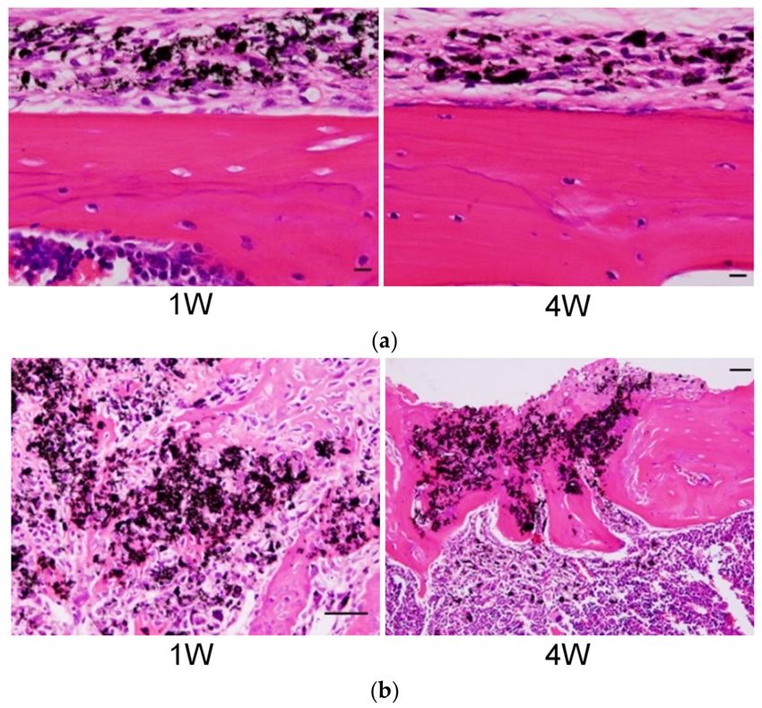
Nanomaterials 2020, 10, 264                                                                           3 of 16

suppression at 24 h after the addition of MWCNTs. Observation by transmission electron microscopy
(TEM) showed that MWCNTs were distributed in the lysosomes of the BEAS-2B cell. Flow cytometry
was performed using a total reactive oxygen species (ROS)/superoxide detection kit to measure total
ROS/superoxide production. As a result, some MWCNTs demonstrated higher oxidative stress and
increased activity of superoxide dismutase (SOD) that decomposes active oxygen, compared to the
positive control pyocianin, which induces ROS production [18]. Since most materials administered at
a high enough dose can cause toxicity, an examination of dosage is necessary.
      Nagai et al. [19] suspended MWCNTs with an approximate diameter of 115 nm at a concentration
of 5.0 µg/cm2 in 0.5% bovine serum albumin (BSA) solution with human peritoneal mesothelial cells
(HPMCs) [20]. Crocidolite, a type of asbestos, was used as a control. After 24 h of culturing, intracellular
fibers were observed by confocal microscopy. Although, crocidolite asbestos was observed within
cells, MWCNTs were only attached to the cellular surface. The side scatter (SSC) value [21,22] in
flow cytometry quantifies the uptake of fibers by cells. SSC values have been reported to increase for
crocidolite asbestos, but not for MWCNTs, and the diameter and rigidity of CNTs and crocidolite affect
their uptake into cells [19]. Ju et al. [23] conducted an experiment by which MWCNTs were added
to human pleural mesothelial cells (MeT-5A). MWCNTs were added in concentrations ranging from
1.25 to 40 µg/cm2 , and the cell viability showed concentration-dependent suppression at 24 h after
treatment. A different study by the same group [24] examined the genotoxicity of MWCNTs by using
a γH2AX foci formation technique, wherein MWCNTs were treated within 24 h at a concentration
of 10 µg/cm2 , which did not affect cell viability. Although few γH2AX foci formation was observed
by immunofluorescent microscopy within a short 24 h time frame after culturing, the foci formation
became more pronounced at 72 h, and the greatest γH2AX foci formation was demonstrated with the
longest exposure time at 90 days.
      The skin of living organisms in contact with the outside world may have more opportunities
to be exposed to nanoparticles. For skin cells, Murray et al. [25] added 1.5 nm diameter, 1–100 µm
long single-walled CNTs (SWCNTs) to murine epidermal cells and JB6 P+ cells, and investigated their
reaction. SWCNTs with 99.7% purity were added to JB6 P+ cells at 0.06 to 0.24 mg/mL concentration,
and cell viability was evaluated by alamarBlue assay at 24 h after treatment. Murray and other
research teams have also evaluated NF-κB [26–28], a redox-sensitive transcription factor involved in
the regulation of many inflammatory responses in the reaction between JB P+ cells and SWCNTs.
They also measured the activity of NF-κB with a luminometer which increased with the concentration
of SWCNTs, suggesting an association between SWCNTs and inflammatory responses. Furthermore,
Patlolla et al. [29] added MWCNTs to normal human dermal fibroblast (NHDF) cells and evaluated cell
viability by 3-(4,5-dimethylthiazol-2-yl)-2,5-diphenyl tetrazolium bromide (MTT) assay [30]. When the
concentration of MWCNTs was set to 40–400 µg/mL, the cell viability was suppressed, depending on
the concentration of MWCNTs. The DNA-damaging effect on NHDF cells was evaluated by comet
assay [31]. In this evaluation, there was an increase in the percent tail DNA of NHDF cells, exposed to
MWCNTs, demonstrating the genotoxicity of MWCNTs in NHDF cells.
      Due to their excellent mechanical strength, CNTs may potentially reinforce the mechanical
strength and durability of the original material when used as a composite material. Research has
been conducted to improve the performance of composite materials, such as artificial joints and
bone fixation materials [6,32]. We evaluated the cells related to the musculoskeletal system, such as
bones and joints. In a previous study, we cultured osteoblast-like stromal cells in an osteogenic
medium that were collected from the calvaria of 1-day old mice, in order to obtain osteoblasts to
be used for experiments. When MWCNTs or carbon black was added to osteoblasts and cultured
at a concentration of 0.5 to 50 µg/mL, neither MWCNTs nor carbon black affected the growth of
osteoblasts at a concentration of 5 µg/mL. In evaluating calcification using Alizarin Red S staining [33],
the group exposed to MWCNTs exhibited better staining compared to carbon black, in addition to
significantly higher levels of osteocalcin, a bone formation marker measured by real time PCR [34].
This suggests that MWCNTs, at lower concentrations, and do not exhibit cytotoxicity, may promote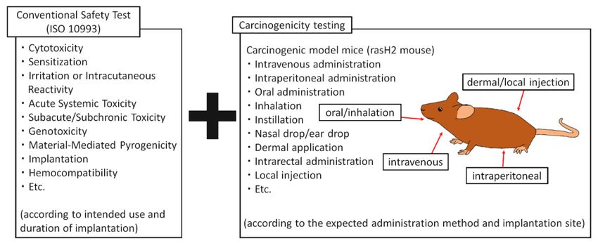
Nanomaterials 2020, 10, 264                                                                                                 4 of 16

bone formation. We also evaluated osteoclasts, which have a role in resorbing the bone matrix.
MWCNTs inhibited osteoclast growth, even at a concentration of 5 µg/mL, which did not affect cell
growth with osteoblasts. Further verification revealed that MWCNTs inhibited NFATc1 translocation
into the nucleus of osteoclast precursor cells in the IκBα/NF-κB pathway when osteoclast precursor
cells differentiate into osteoclasts [35]. Based on this result, osteoclasts are less likely to be activated
when MWCNTs are used as bone-related biomaterials, and biomaterials may be less likely to loosen or
fail. Macrophages in the bone marrow were also evaluated. Macrophage colony-stimulating factor
was added to tibial bone marrow cells of ddY mice and cultured to differentiate into macrophages.
In addition, MWCNTs were added into the culture, and inflammatory cytokines interleukin (IL)-6,
IL-1β, and tumor necrosis factor (TNF)-α in the culture solution were measured by enzyme-linked
immunosorbent assay (ELISA). Neither MWCNTs nor tattoo ink increased the inflammatory cytokines
in the culture [16]. CNTs have demonstrated favorable osteogenic responses in bone-related cells,
and we believe that CNTs can be applied to orthopedic and dental implants and bone regeneration
therapy. A summary of the literature discussed in this section is shown in Table 1.

                                       Table 1. In vitro biocompatibility testing of CNTs.

        Author, year             CNT           Cells           Origin of Cells        Control             Evaluations
                                                              Chinese hamster         tattoo ink
    Hara et al., 2011 [16]      MWCNT         V79 cell                                                colony-foaming assay
                                                               lung fibroblast      carbon black
    Hara et al., 2011 [16]      MWCNT             -        mouse bone macrophage     tattoo ink     IL-1β, IL-6, TNF-α, ELISA
                                                                                                        alamarBlue assay
                                                              human bronchial                         total ROS/superpxide
   Haniu et al., 2014 [18]      MWCNT       BEAS-2B cell                                  -
                                                               epithelial cell                             detection kit,
                                                                                                         flow cytometry
                                                              human peritoneal                        confoal microscopy
    Nagai et al., 2011 [35]     MWCNT             -                                  crocidolite
                                                               mesothelial cell                    SSC value, flow cytometry
                                                                                                          γH2AX foci
      Ju et al., 2014 [24]      MWCNT       MeT-5A cell    human mesothelial cell         -
                                                                                                      formation technique
                                                                                                       alamarBlue assay
   Murray et al., 2009 [25]     SWCNT        JB6 P+ cell    murine epidermal cell         -
                                                                                                   NF-κB actibity, luminometer
                                                                                                         MTT assay
                                                               normal human
   Patlolla et al., 2009 [29]   MWCNT             -                                       -          DNA damaging effect,
                                                              dermal fibroblast
                                                                                                        comet assay
                                                               mouse calvaria                         Alizarin Red S stain
  Shimizu et al., 2012 [34]     MWCNT             -            osteoblast-like      carbon black    mRNA of osteocalcin, real
                                                                stromal cell                               time PCR
   Narita et al., 2009 [35]     MWCNT             -           mouse osteoclast      carbon black    NFATc1, immunostaining
      IL: interleukin, TNF: tumor necrosis factor, ELISA: enzyme-linked immunosorbent assay, ROS: reactive oxygen
      species, SSC value: side scatter value, MTT assay: (3-(4,5-dimethylthiazol-2-yl)-2,5-diphenyl tetrazolium
      bromide) assay.

2.2. In Vivo Biocompatibility Testing of CNTs
      Here, we introduce the in vivo biocompatibility tests in which CNTs are administered to animals
in line with clinical medicine.
      Mercer et al. [36] administered MWCNTs to C57BL/6J mice by pharyngeal aspiration, and lung
tissues were removed 56 days later for histopathological evaluation. Most of the MWCNTs penetrated
into alveolar macrophages, but they reported increased thickness due to pulmonary fibrosis and
formation of granulomatous lesions. Pulmonary thickness increased over time as the duration of
MWCNTs administration increased to 7, 28, and 56 days. Dong et al. [37] also administered MWCNTs
to C57BL/6J mice by pharyngeal aspiration. They measured cells and cytokines in broncho-alveolar
lavage fluid (BALF). As a result, the BALF of mice exposed to MWCNTs contained a lot of neutrophils,
lymphocytes, and macrophages, while ELISA demonstrated many inflammatory cytokines such as
IL-6, IL-1β, and TNF-α.
      Deng et al. [38] measured the accumulation of intravenously injected 14 C-labeled MWCNTs in
the liver, spleen, and kidney. Many of the intravenously injected MWCNTs were accumulated in the
Nanomaterials 2020, 10, 264 5 of 16
liver and entrapped for 28 days after aspiration. At 90 days, the remaining MWCNTs in the liver
were reduced to approximately 20% of the dose. In order to evaluate the effects on the liver, serum
lactate dehydrogenase (LDH), total bilirubin (TBIL), total bile acid (TBA), alkaline phosphatase (ALP),
and alanine aminotransferase (ALT) were measured. Their bioactivity indices were equivalent to the
control and did not affect liver function. Tang et al. [39] also administered MWCNTs intravenously
to KunMing mice to assess their effects on the entire body. The blood at one day after MWCNTs
administration was evaluated. As a result, administration of MWCNTs did not affect the number of
blood cells such as white blood cells (WBC), red blood cells, platelets, bleeding time, and coagulation
time show different with the control. Histopathological evaluation of the heart, lung, liver, and kidney,
at 1 to 3 days after administration, showed no deposition of MWCNTs and no effect on the tissue.
In addition, there were no significant changes to creatine kinase (CK), urea nitrogen, and ALT levels in
serologic tests that indicate damage to the myocardium, kidney, and liver, respectively.
We have previously conducted in vivo biocompatibility tests by local administration of MWCNTs.
MWCNTs were locally injected into the dorsal subcutaneous tissue of ddY mice, and skin samples
were collected and histopathologically evaluated after 1, 4, 12, and 24 weeks of injection with an optical
microscope [16]. In both the group administered with MWCNTs and group administered with the tattoo
ink control, most of the particles penetrated the macrophages at one week after administration, and there
was a mild inflammatory reaction caused by fibroblasts, WBC, and lymphocytes in the surrounding
area. At 4 weeks after administration, the inflammatory response subsided and particles remained
within the macrophages. Our findings did not change at 12 and 24 weeks after administration (Figure 2),
and histological findings in the MWCNTs administration group and the tattoo ink administration
group were very similar to common histological findings found in tattooed skin [40].
Figure 2. A histopathological image of the skin of ddY mice, subcutaneously administered with
MWCNTs. (a) Group administered with MWCNT; (b) group administered with tattoo ink. In both
groups, carbon particles are observed in the macrophages at one week following administration with
slight inflammatory cell infiltration around them. At four weeks, carbon particles have remained in
the macrophages, and the inflammatory reaction has subsided. Scale bar, 20 µm. Reproduced with
permission from [16]. Elsevier, 2011.
In terms of the musculoskeletal system, we have previously administered MWCNTs into bone [41]
and within the joint [42]. When MWCNTs were administered to the subperiosteum of the skull in ddY
mice, MWCNTs did not cause osteolysis in the skull (Figure 3a). When MWCNTs were administered
into a tibial tunnel that was prepared with a drill, the tunnel was filled with new bone formation
at 4 weeks after implantation, and MWCNTs were incorporated into the bone matrix (Figure 3b).
In addition, there was significantly more bone mineral content in ectopic bone formed under theNanomaterials 2020, 10, 264 6 of 16
dorsal fascia of mice with an implant made by combining MWCNTs with recombinant human bone
morphogenetic protein-2 (rhBMP-2), compared to an implant that does not contain MWCNTs at
2 weeks after implantation. Similar to the results of in vitro studies, MWCNTs demonstrated a high
affinity for bone tissue and promoted bone formation. In addition, after administration of MWCNTs
into the knee joint of rats, mild inflammatory cell infiltration was observed in the synovial tissue within
one week of administration, and MWCNTs penetrated into macrophages and formed granulation
tissues. At 4 and 12 weeks after administration, the inflammatory reaction subsided and granulation
tissues remained. These CNTs can be expected to be relatively safe, even when added to biomaterials
used in bones and joints.
Although, the type and dosage of CNTs vary from report to report, CNTs generally produce
an inflammatory response when administered to the respiratory tract, but have been shown to be
relatively safe for intravenous and topical (subcutaneous, intraosseous) administration. A summary of
the literature discussed in this chapter is shown in Table 2.
Table 2. In vivo biocompatibility testing of CNTs.
Administration
Author, year CNT Animals Evaluations
Site/Route
light microscope
Mercer et al., 2011 [36] MWCNT C57BL/6J mouse pharyngeal aspiration
examination of lung
IL-6, IL-1β, TNF-α,
Dong et al., 2015 [37] MWCNT C57BL/6J mouse pharyngeal aspiration
ELISA of BALF
LDH, TBIL, TBA, ALP, ALT
Deng et al., 2007 [38] MWCNT KunMing mouse intravenously injection
in serum (effects to liver)
blood count (white blood
cells, red blood cells,
Tang et al., 2012 [39] MWCNT KunMing mouse intravenously injection
platelets), bleeding time,
coagulation time
Hara et al., 2011 [16] MWCNT ddY mouse subcutaneous tissue light microscopy
Usui et al., 2008 [41] MWCNT ddY mouse under dorsal fascia ectopic bone formation
Nomura et al., 2015 [42] MWCNT Wistar rat knee joint light microscopy
IL: interleukin, TNF: tumor necrosis factor, ELISA: enzyme-linked immunosorbent assay, BALF: bronchoalveolar
lavage fluid, LDH: lactate dehydrogenase, TBIL: total bilirubin, TBA: total bile acid, ALP: alkaline phosphatase,
ALT: alanine aminotransferase.
2.3. CNT Composites
In recent years, various CNT composites have been created and evaluated to increase the tissue
affinity and functional properties of CNTs. Prencipe et al. [43] produced SWCNTs loaded with
poly(ethylene glycol) (PEG) chains that were administered intravenously in balb/c mice to measure
their blood circulation half-life. PEGylated SWCNTs showed prolonged blood circulation half-life
and stability in serum. In addition, Meran et al. [44] evaluated the effects of PEGylated SWCNTs
on human umbilical vein endothelial cells (HUVECs). MTT assays were performed using longer
SWCNTs with a diameter of 7 Å and length of 120 Å and compared to shorter SWCNTs with a length
of 40 Å. Results confirmed that shorter PEGylated SWCNTs did not affect the cell growth of HUVECs,
while longer PEGylated SWCNTs suppressed cell growth. Although their mechanisms remain unclear,
the addition of substances may introduce new functions and effects to CNTs that are not found in each
individual substance.
In addition, a study to improve the function of CNTs by combining poly(ε-caprolactone) (PCL),
which is a high polymer evaluated as a bone scaffold. Huang et al. prepared PCL/MWCNT composite
scaffolds containing 0.35 wt%, 0.75 wt%, and 3 wt% of MWCNTs, and human adipose-derived stem
cells (hADSCs) were cultured on the scaffolds. Scaffolds containing 3 wt% MWCNTs showed the
highest cell growth in the alamarBlue assay [45]. Our aforementioned intraosseous implantation testNanomaterials 2020, 10, 264 7 of 16
on MWCNTs exhibited high biocompatibility with bone tissue and high osteogenic capacity, and we
believe that the MWCNTs promoted bone formation as a PCL scaffold.
Wu et al. [46] created a chitin/MWCNT composite obtained by blending 5 wt% MWCNTs with
chitin, a component found in the exoskeleton of crustaceans and insects, and examined the effect on
neuron cells (PC12 cells) isolated from the pheochromocytoma of rats. The evaluation of the MTT
assay showed that the chitin/MWCNT composite exhibited no cytotoxicity of PC12 cells, and the
MWCNT/chitin composite hydrogel was able to retain its morphological integrity longer in the
culture solution.
As described above, the biocompatibility of a CNT composite is affected by various factors such
as its blended substance, amount of CNTs, and length of CNTs. Even if each blended substance
demonstrates excellent biocompatibility, the composite itself should be tested separately as a new
material. A summary of the literature discussed in this chapter is shown in Table 3.
Table 3. Biocompatibility testing of CNT Composites.
Author, year CNT Composite Cells/Animals Administration Site/Route Evaluations
Prencipe et al.,
SWCNT PEG balb/c mouse intravenously injection circulation time
2009 [43]
Meran et al.,
SWCNT PEG HUVEC - MTT assay
2018 [44]
Huang et al.,
MWCNT PCL hADSC - alamarBlue assay
2019 [45]
Wu et al.,
MWCNT chitin rat neuron cell - MTT assay
2017 [46]
PEG: poly (ethylene glycol), HUVEC: human umbilical vein endothelial cell, MTT assay: (3-(4,5-dimethylthiazol-
2-yl)-2,5-diphenyl tetrazolium bromide) assay, PCL: poly (ε-caprolactone), hADSC: human adipose derived stem cell.
Figure 3. (a) Histopathology of ddY mice administered with MWCNTs in the subperiosteum of the
skull. MWCNTs were incorporated into macrophages, and no osteolysis was observed in the skull.
Scale bar, 20 µm; (b) Histopathology of ddY mice, in which MWCNTs were administered into a tibial
bone tunnel. One week after administration, woven bone is formed around MWCNTs, and after
4 weeks, bony cortex is regenerated with MWCNTs incorporated into the bone matrix. Scale bar, 20 µm.
Reproduced with permission from [41]. John Wiley and Sons, 2008.Nanomaterials 2020, 10, 264 8 of 16
2.4. Factors Affecting the Biocompatibility Testing of CNTs
Because there are various types of CNTs, we cannot discuss, in general terms, whether CNTs
are safe or dangerous for living organisms. The biocompatibility of CNTs is of great interest to many
scientists, and various biocompatibility tests have been performed on different types CNTs. This section
introduces the factors that affect the biocompatibility of CNTs according to their type.
CNTs are structurally classified into SWCNTs, double-walled CNTs, MWCNTs, and cup-stacked
carbon nanotubes (CSCNTs) [47,48]. Nahle et al. [49] evaluated the effect of MWCNTs and SWCNTs
on rat alveolar macrophage cell line (NR8383 cells). They added CNTs of various concentrations to
NR8383 cells and calculated its 50% inhibitory concentration (IC50 ) by alamarBlue assay. IC50 was
4.1 cm2 /cm2 for MWCNTs and 41.2 cm2 /cm2 for SWCNTs, and MWCNTs exhibited a higher cytotoxicity.
However, as claimed by these authors, MWCNTs and SWCNTs are different in thickness and length,
so the difference in structures may not be the only factor that affects CNTs. We added three different
MWCNTs and CSCNTs of different thicknesses and lengths to BEAS-2B cells and human malignant
pleural mesothelioma cell line (MESO-1 cells), and evaluated their effects using the alamarBlue assay.
In both types of cells, CSCNTs demonstrated higher cell viability than MWCNTs [18].
Zhao et al. [50] evaluated the effects of three different MWCNTs of comparable length (0.5–2 µm)
with different diameters on human umbilical vein endothelial cells (HUVECs). The respective diameters
of MWCNTs were 10.33 ± 3.50 nm, 21.55 ± 5.66 nm, 38.80 ± and 13.07 nm. IL-6 and ROS were measured
by ELISA. IL-6 and ROS showed the highest values in the thinnest MWCNTs, and narrower CNTs
showed stronger cytotoxicity. Wang et al. [51] evaluated the effect of CNTs length on biological responses.
They compared short and long MWCNTs, with an approximate diameter of 50 nm, and lengths of
0.5–2 µm and 20–50 µm, respectively. When these MWCNTs were added to the mouse leukemic monocyte
macrophage cell line (RAW264.7 cells), the production of transforming growth factor (TGF)-β1 that
induces extracellular matrix (ECM) protease inhibitors were increased by long MWCNTs. Furthermore,
with intra-tracheal administration to spontaneously hypertensive (SH) rats, TGF-β1 in BALF increased
in the group administered long MWCNTs, and pulmonary fibrosis was observed in the evaluation of
histopathological specimens of the lung.
The purity of CNTs is also known to be associated with biocompatibility, and Murray et al.
evaluated cell viability, using SWCNTs containing 0.23% iron impurity, and SWCNTs containing 30%
impurity using the aforementioned JB6 P+ cells [25]. In JB6 P+ cells with SWCNTs containing high iron
impurity, cell viability was decreased according to alamarBlue assay. The production of antioxidant
glutathione also decreased with impure SWCNTs.
Since CNTs present a fibrous morphology, the material tends to tangle. When administered to
a living body, it is necessary to disperse these entangled and solidified secondary particles. We dispersed
MWCNTs using three types of dispersants to evaluate BEAS-2B cells: Gelatin, carboxylmethyl cellulose
(CMC), and 1,2-dipalmitoylsn-glycero-3-phosphocholine (DPPC) [52]. After MWCNT dispersion to
the cells, the uptake of MWCNTs into the cells was measured by flow cytometry. At 24 h, the most
uptake of MWCNTs into cells was found in the order of gelatin, DPPC, and CMC. In addition,
when inflammatory cytokines in the culture medium were measured with a cytometric bead array flex
set system, the greatest amount of IL-6 and IL-8 were produced in the order of gelatin, DPPC, and CMC
in the same manner as the amount of MWCNTs uptake into the cells. This reveals that the uptake of
CNTs into cells is affected by the dispersant, and that cellular response is dependent on the amount of
CNT uptake into the cells. A summary of the literature discussed in this chapter is shown in Table 4.Nanomaterials 2020, 10, 264 9 of 16
Table 4. Factors affecting the biocompatibility testing of CNTs.
Administration
Author, year CNT Cells/Animals Factors Evaluations
Site/Route
Nahle et al., MWCNT NR8383 cell
- MWCNT or SWCNT alamarBlue assay
2019 [49] SWCNT (rat alveolar macrophage cell)
BEAS-2B cell
(human bronchial
Haniu et al., MWCNT epithelial cell)
- MWCNT or CSCNT alamarBlue assay
2014 [18] CSCNT MESO-1 cell
(human malignant pleural
mesothelioma cell)
Zhao et al.,
MWCNT HUVEC - diameter of fibers IL-6, ROS, ELISA
2019 [50]
RAW264.7 cell
TGF-β1, ELISA
(mouse leukemic
Wang et al., pharyngeal light microscope
MWCNT monocyte macrophage cell) length of fibers
2013 [51] aspiration examination of lung
spontaneously
TGF-β1, ELISA of BALF
hypertensive rat
alamarBlue assay
Murray et al., JB6 P+ cell purity of CNT
SWCNT - glutathione,
2009 [25] (murine epidermal cell) (containing iron)
fluorescence assay
flow cytometry
Haniu et al., dispersants IL-6, IL-8,
MWCNT BEAS-2B cell -
2011 [52] (gelatin, DPPC, CMC) cytometric bead array
flex set system
HUVEC: human umbilical vein endothelial cell, IL: interleukin, ROS: reactive oxygen species, ELISA: enzyme-linked
immunosorbent assay, TGF: transforming growth factor, BALF: bronchoalveolar lavage fluid, CMC: carboxylmethyl
cellulose, DPPC: 1,2-dipalmitoylsn-glycero-3-phosphocholine.
3. Carcinogenicity Testing of CNTs
As described above, CNTs are stable fibrous nanoparticles and have been studied for their potential
carcinogenicity, given their similarity with asbestos.
Takagi et al. [53] confirmed the formation of mesothelioma in the abdominal cavity following the
intraperitoneal administration of MWCNTs from carcinogenesis models consisting of p53+/− mice [54].
No tumor formation occurred in the group intraperitoneally administered with fullerene as a control.
In a separate study, they also reported that increasing the dose of MWCNTs increased the rate of
mortality for p53+/− mice due to mesothelioma [55].
Suzui et al. [56] evaluated tumor formation in intratracheally administering MWCNTs to F344/Crj
rats. None of the animals receiving the control vehicle showed tumor formation, but in the group
receiving MWCNTs, 6/38 animals presented with malignant mesothelioma and 14/38 animals exhibited
formation of lung tumors (bronchiolo-alveolar adenomas and carcinomas).
In order to evaluate carcinogenicity by local administration of CNTs, we subcutaneously
administered MWCNTs to rasH2 mice [57], a carcinogenic model mouse. RasH2 mice express
human-derived c-H-ras proto-oncogene and die as a result of multi-organ tumors throughout the body
after reaching approximately 35 weeks of age. When carcinogenic substances are administered to these
mice, tumor formation is accelerated and multi-organ tumors are formed within 26 weeks, which are
then used to evaluate the carcinogenicity of the substance. As a control, we used N-methyl-N-nitrosourea
(MNU), a known carcinogen. In the group in which MNU was subcutaneously injected in the dorsum,
papilloma and malignant lymphoma of the skin and stomach were observed, and the survival rate at
26 weeks after administration was 60%. However, in the group receiving MWCNTs, there was only one
case of inflammatory pseudotumor in the spleen, and the survival rate at 26 weeks after administration
was 100% (Figure 4) [58].Nanomaterials 2020, 10, 264 10 of 16
Figure 4. Survival curve of rasH2 mice with local subcutaneous administration of MWCNTs in the
dorsum. The survival rate at 26 weeks after administration was 60% in the group that received MNU,
a carcinogen, compared to 100% in the group that received MWCNTs. The survival rate of the carbon
black group is almost equivalent to tattoo ink at 90%, but the carcinogenic case is likely a coincidence.
Image is modified from a study by Takanashi et al. [58].
We have also evaluated the effects of systemic intravenously administration of MWCNTs into rasH2
mice [59]. Although, there were models that showed mesothelioma in the lung (Figure 5a) in the group
receiving MWCNTs, there was no significant difference in the incidence of tumor with the vehicle group.
In searching for MWCNTs using optical microscopy in organs throughout the body, 3.2% of mice showed
fiber deposition in pancreas (Figure 5b), but there was no tumor formation in the pancreas.
Figure 5. (a) Formation of Mesothelioma in the lung of rasH2 mice intravenously administered with
MWCNTs. The right is an enlarged image of the square in the left image; (b) histological image of
the pancreas in rasH2 mice administered intravenously with MWCNTs. Particles of MWCNTs are
deposited in the tissue. There is no tumor formation. The right is an enlarged image of the square in
the left image. Images are modified from a study by Sobajima et al. [59].Nanomaterials 2020, 10, 264 11 of 16
Donaldson et al. [60] hypothesized a carcinogenic mechanism by which macrophages elicit
a persistent inflammatory response when long and thin asbestos, and CNTs are not completely
enclosed by macrophage (frustrated phagocytosis), leading to cell tumorigenesis (Figure 6a). In fact,
in an experiment in which short and long MWCNTs were administered intraperitoneally to C57BL/6
mice. The formation of granuloma was observed around the MWCNT particles in the peritoneal
mesothelium for the long MWCNTs group. Donaldson et al. suggested that stable nanoparticles,
with long fibers that are not phagocytosed by macrophages, remain un-degraded in vivo, and that their
retention lead to inflammation and subsequent fibrosis and carcinogenesis [61] (Figure 6b). Short fibers
and tangled fibers are completely phagocytosed in macrophages, do not cause inflammation, and are
unlikely to be tumorigenic. A summary of the literature discussed in this chapter is shown in Table 5.
Table 5. Carcinogenicity testing of CNTs.
Author, year CNT Animals Administration Site/Route Control
Takagi et al., 2008 [53] MWCNT p53+/− mouse intraperitoneal injection fullerene
Suzui et al., 2016 [56] MWCNT F344/Crj rat pharyngeal aspiration vehicle
Takanashi et al., 2012 [58] MWCNT rasH2 mouse subcutaneous tissue MNU
Sobajima et al., 2019 [59] MWCNT rasH2 mouse intravenously injection MNU
MNU: N-methyl-N-nitrosourea.
Figure 6. (a) When nanofibers such as asbestos (left) and CNTs (right) are phagocytosed by macrophages,
inflammation may persist and tumors may be formed if long fibers trigger frustrated phagocytosis.
Image is modified from a study by Donaldson et al. [60]. (b) Long, biopersistent nanofibers may induce
inflammation, cancer, and fibrosis. Reproduced with permission from [61]. Elsevier, 2013.Nanomaterials 2020, 10, 264 12 of 16
4. Pharmacokinetics of CNTs
When administered into the body, the kinetics of nanoparticles, such as CNTs often remains
unclear. Since nanoparticles are small, it is difficult to grasp their distribution and amount in a small
quantity of particles. CNTs are also studied for their potential use as carriers in drug delivery systems
(DDS) for targeting organs. We believe the pharmacokinetics of CNT administration into the body is
an important research topic.
Singh et al. [62] intravenously administered Indium (111 In)-labeled SWCNTs to BALB/c mice and
evaluated the amount of CNTs that translocated to organs and tissues at 30 min, 3 h, and 24 h after
administration. At 30 min after administration, CNTs were observed in the blood, femur, liver, kidney,
muscle, and skin. However, there was almost no detection after 3 h, and fibers of CNTs were confirmed
in excreted urine.
Deng et al. [38] evaluated the pharmacokinetics of 14 C-tautine-labeled MWCNTs in KunMing
mice by intravenous injection, intra-tracheal administration, and oral administration. Many of the
MWCNTs were confirmed in the liver after intravenous injection and were entrapped for over 28 days.
Intra-tracheally administered MWCNTs were detected in the lung and decreased to approximately
25% after 28 days. Orally administered MWCNTs were gradually excreted in faeces at 12 h after
administration via the small and large intestines. In our experiment of intravenous injection of
MWCNTs into rasH2 mice, optical microscopy of organs at 26 weeks after administration showed that
MWCNTs were deposited only in the pancreas, but no MWCNTs were observed in other organs [59].
In the aforementioned experiment of S p53+/− mice by Takagi et al., MWCNTs were intraperitoneally
administered, and singular fibers were observed in the tissues of the choroid plexus at 168 days as well
as the lung and kidney at 197 days [53]. Regarding the pharmacokinetics of CNTs, there is no clear
agreement between each report, and further research should be conducted.
5. Conclusions
Various studies have been conducted on the biocompatibility and carcinogenicity of CNTs. Results
have varied widely, as some have reported that the material exhibits high biocompatibility while others
have reported that it is carcinogenic. In vivo studies, suggesting the carcinogenicity of CNTs in the
literature, were experiments by the inhalation or intraperitoneal administration [53,55,56], and no
previous studies have been conducted on intravenous or topical administration. We believe that the
carcinogenicity of CNTs is caused by its exposure to mesothelial tissues. Exposure to the abdominal
cavity occurs only by artificial administration, but exposure to the respiratory system caused by the
dispersion of CNTs into the air. When evaluating CNTs as a material in the industrial field, the aerial
dispersion of CNTs during the manufacturing process may risk exposure to the respiratory organs
and skin. However, this is an issue that can be overcome by improving the work environment and
minimizing the dispersion to undetectable levels.
When CNTs are used as biomaterials, the effect on the living body varies according to the type of
CNTs used, route of administration, and dose. If CNTs are intended to be used in the orthopedic field as
a locally implanted biomaterial such as artificial joints or fixation devices, nanoparticle pharmacokinetics
remains a problem that warrants further attention. However, there is no need to evaluate the response
from their intra-tracheal administration, intraperitoneal administration, and intravenous injection,
as safety evaluations for local administration/implantation should be sufficient.
When used as reagents for DDS or testing, evaluations should be conducted according to the
intended method of administration, such as intravenous administration, intraperitoneal administration,
oral administration, inhalation, instillation, nasal drop, dermal application, intra-rectal administration,
and local injection. Biological safety assessments of new biomaterials and medical devices are
internationally standardized by ISO 10993. Biologic effects, such as cytotoxicity, sensitization, irritation,
acute systemic toxicity, and implantation are evaluated according to their intended use and implantation
period [63,64]. It is necessary to evaluate the carcinogenicity of nanoparticles, such as CNTs (Figure 7).
rasH2 mice can be used to evaluate the carcinogenicity of test materials in a short period of time,Nanomaterials 2020, 10, 264 13 of 16
and are suitable for testing the carcinogenicity of new biomaterials. By evaluating an animal model,
according to the intended use, administration method, and administration site of the new biomaterial,
a realistic evaluation can be performed to simulate their clinical use.
Figure 7. Bio-safety testing of nano-sized biomaterials. Carcinogenicity tests are carried out by using
carcinogenic model mice, in addition to conventional safety tests according to the intended application
and implantation period of each biomaterial according to ISO 10993-1. The method of administration is
selected according to the expected administration method of nano-biomaterials to humans and the
implantation site.
CNTs need to be evaluated for their safety according to their type and intended use. In addition
to general biological safety tests, it is necessary to perform carcinogenicity tests. If the results of these
tests are good, we firmly believe that biological applications of CNTs may be feasible for their use as
new high-performance materials.
Author Contributions: Conceptualization, N.S.; methodology, K.A. and N.S.; validation, K.A. and S.N.; formal
analysis, K.A.; investigation, K.A.; resources, N.S.; data curation, K.A; writing—original draft preparation, K.A.;
writing—review and editing, N.S.; project administration, N.S. All authors have read and agreed to the published
version of the manuscript.
Funding: This research received no external funding.
Acknowledgments: We would like to thank Sho Steven Sugita (OrthoTranslations) for English language editing.
Conflicts of Interest: The authors declare no conflict of interest.
References
1. Lu, P.J.; Cheng, W.L.; Huang, S.C.; Chen, Y.P.; Chou, H.K.; Cheng, H.F. Characterizing titanium dioxide and
zinc oxide nanoparticles in sunscreen spray. Int. J. Cosmet. Sci. 2015, 37, 620–626. [CrossRef] [PubMed]
2. McSweeney, P.C. The safety of nanoparticles in sunscreens: An update for general practice. Aust. Fam. Phys.
2016, 45, 397–399.
3. Morita, T.; Takami, N. Nano Si cluster-SiOx -C composite material as high-capacity anode material for
rechargeable lithium batteries. J. Electrochem. Soc. 2006, 153, A425–A430. [CrossRef]
4. Aagaard, J. The Carbomedics aortic heart valve prosthesis: A review. J. Cardiovasc. Surg. (Torino) 2004, 45,
531–534.
5. Morice, M.C.; Bestehorn, H.P.; Carrie, D.; Macaya, C.; Aengevaeren, W.; Wijns, W.; Dubois, C.; de Winter, R.;
Verheye, S.; Hoffmann, S.; et al. Direct stenting of de novo coronary stenoses with tacrolimus-eluting versus
carbon-coated carbostents. The randomized JUPITER II trial. EuroIntervention 2006, 2, 45–52.
6. Saito, N.; Aoki, K.; Usui, Y.; Shimizu, M.; Hara, K.; Narita, N.; Ogihara, N.; Nakamura, K.; Ishigaki, N.;
Kato, H.; et al. Application of carbon fibers to biomaterials: A new era of nano-level control of carbon fibers
after 30-years of development. Chem. Soc. Rev. 2011, 40, 3824–3834. [CrossRef]Nanomaterials 2020, 10, 264 14 of 16
7. Saito, N.; Haniu, H.; Usui, Y.; Aoki, K.; Hara, K.; Takanashi, S.; Shimizu, M.; Narita, N.; Okamoto, M.;
Kobayashi, S.; et al. Safe clinical use of carbon nanotubes as innovative biomaterials. Chem. Rev. 2014, 114,
6040–6079. [CrossRef]
8. Jacobsen, N.R.; Moller, P.; Clausen, P.A.; Saber, A.T.; Micheletti, C.; Jensen, K.A.; Wallin, H.; Vogel, U.
Biodistribution of Carbon Nanotubes in Animal Models. Basic Clin. Pharmacol. Toxicol. 2017, 121, 30–43.
[CrossRef] [PubMed]
9. Choo, H.; Jung, Y.; Jeong, Y.; Kim, H.C.; Ku, B.C. Fabrication and Applications of Carbon Nanotube Fibers.
Carbon Lett. 2012, 13, 191–204. [CrossRef]
10. Kakooei, H.; Marioryad, H. Evaluation of exposure to the airborne asbestos in an automobile brake and
clutch manufacturing industry in Iran. Regul. Toxicol. Pharmacol. 2010, 56, 143–147. [CrossRef] [PubMed]
11. Luberto, F.; Ferrante, D.; Silvestri, S.; Angelini, A.; Cuccaro, F.; Nannavecchia, A.M.; Oddone, E.; Vicentini, M.;
Barone-Adesi, F.; Cena, T.; et al. Cumulative asbestos exposure and mortality from asbestos related diseases
in a pooled analysis of 21 asbestos cement cohorts in Italy. Environ. Health 2019, 18, 71. [CrossRef] [PubMed]
12. Gogou, E.; Hatzoglou, C.; Zarogiannis, S.G.; Malli, F.; Jagirdar, R.M.; Gourgoulianis, K.I. Mesothelioma
Mortality Rates in Greece for the Period 2005-2015 Is Increased Compared to Previous Decades. Medicina
2019, 55, 419. [CrossRef] [PubMed]
13. Krowczynska, M.; Wilk, E. Environmental and Occupational Exposure to Asbestos as a Result of Consumption
and Use in Poland. Int. J. Environ. Res. Public Health 2019, 16, 2611. [CrossRef] [PubMed]
14. Zha, L.; Kitamura, Y.; Kitamura, T.; Liu, R.; Shima, M.; Kurumatani, N.; Nakaya, T.; Goji, J.; Sobue, T.
Population-based cohort study on health effects of asbestos exposure in Japan. Cancer Sci. 2019, 110,
1076–1084. [CrossRef] [PubMed]
15. Carbone, M.; Yang, H. Mesothelioma: recent highlights. Ann. Transl. Med. 2017, 5, 238. [CrossRef] [PubMed]
16. Kane, A.B.; Hurt, R.H.; Gao, H. The asbestos-carbon nanotube analogy: An update. Toxicol. Appl. Pharmacol.
2018, 361, 68–80. [CrossRef] [PubMed]
17. Hara, K.; Aoki, K.; Usui, Y.; Shimizu, M.; Narita, N.; Ogihara, N.; Nakamura, K.; Ishigaki, N.; Sano, K.;
Haniu, H.; et al. Evaluation of CNT toxicity by comparison to tattoo ink. Mater. Today 2011, 14, 434–440.
[CrossRef]
18. Haniu, H.; Saito, N.; Matsuda, Y.; Tsukahara, T.; Usui, Y.; Maruyama, K.; Takanashi, S.; Aoki, K.; Kobayashi, S.;
Nomura, H.; et al. Biological responses according to the shape and size of carbon nanotubes in BEAS-2B and
MESO-1 cells. Int. J. Nanomed. 2014, 9, 1979–1990. [CrossRef]
19. Nagai, H.; Okazaki, Y.; Chew, S.H.; Misawa, N.; Yamashita, Y.; Akatsuka, S.; Ishihara, T.; Yamashita, K.;
Yoshikawa, Y.; Yasui, H.; et al. Diameter and rigidity of multiwalled carbon nanotubes are critical factors in
mesothelial injury and carcinogenesis. Proc. Natl. Acad. Sci. USA 2011, 108, E1330–E1338. [CrossRef]
20. Kajiyama, H.; Shibata, K.; Terauchi, M.; Ino, K.; Nawa, A.; Kikkawa, F. Involvement of SDF-1alpha/CXCR4
axis in the enhanced peritoneal metastasis of epithelial ovarian carcinoma. Int. J. Cancer 2008, 122, 91–99.
[CrossRef]
21. Stringer, B.; Imrich, A.; Kobzik, L. Flow cytometric assay of lung macrophage uptake of environmental
particulates. Cytometry 1995, 20, 23–32. [CrossRef]
22. Al-Jamal, K.T.; Kostarelos, K. Assessment of cellular uptake and cytotoxicity of carbon nanotubes using flow
cytometry. Methods Mol. Biol. 2010, 625, 123–134.
23. Ju, L.; Wu, W.; Yu, M.; Lou, J.; Wu, H.; Yin, X.; Jia, Z.; Xiao, Y.; Zhu, L.; Yang, J. Different Cellular Response of
Human Mesothelial Cell MeT-5A to Short-Term and Long-Term Multiwalled Carbon Nanotubes Exposure.
BioMed Res. Int. 2017, 2017, 2747215. [CrossRef]
24. Ju, L.; Zhang, G.; Zhang, X.; Jia, Z.; Gao, X.; Jiang, Y.; Yan, C.; Duerksen-Hughes, P.J.; Chen, F.F.; Li, H.; et al.
Proteomic analysis of cellular response induced by multi-walled carbon nanotubes exposure in A549 cells.
PLoS ONE 2014, 9, e84974. [CrossRef]
25. Murray, A.R.; Kisin, E.; Leonard, S.S.; Young, S.H.; Kommineni, C.; Kagan, V.E.; Castranova, V.; Shvedova, A.A.
Oxidative stress and inflammatory response in dermal toxicity of single-walled carbon nanotubes. Toxicology
2009, 257, 161–171. [CrossRef]
26. Driscoll, K.E.; Carter, J.M.; Hassenbein, D.G.; Howard, B. Cytokines and particle-induced inflammatory cell
recruitment. Environ. Health Perspect. 1997, 105, 1159–1164. [PubMed]
27. Mossman, B.T.; Churg, A. Mechanisms in the pathogenesis of asbestosis and silicosis. Am. J. Respir. Crit. Care Med.
1998, 157, 1666–1680. [CrossRef] [PubMed]Nanomaterials 2020, 10, 264 15 of 16
28. Schins, R.P.; Borm, P.J. Mechanisms and mediators in coal dust induced toxicity: A review. Ann. Occup. Hyg.
1999, 43, 7–33. [CrossRef]
29. Patlolla, A.; Knighten, B.; Tchounwou, P. Multi-Walled Carbon Nanotubes Induce Cytotoxicity, Genotoxicity
and Apoptosis in Normal Human Dermal Fibroblast Cells. Ethn. Dis. 2010, 20, S1-65–S1-72.
30. Mosmann, T. Rapid colorimetric assay for cellular growth and survival: application to proliferation and
cytotoxicity assays. J. Immunol. Methods 1983, 65, 55–63. [CrossRef]
31. Olive, P.L.; Banath, J.P.; Durand, R.E. Heterogeneity in radiation-induced DNA damage and repair in tumor
and normal cells measured using the “comet” assay. Radiat. Res. 1990, 178, 35–42. [CrossRef] [PubMed]
32. Ogihara, N.; Usui, Y.; Aoki, K.; Shimizu, M.; Narita, N.; Hara, K.; Nakamura, K.; Ishigaki, N.; Takanashi, S.;
Okamoto, M.; et al. Biocompatibility and bone tissue compatibility of alumina ceramics reinforced with
carbon nanotubes. Nanomedicine 2012, 7, 981–993. [CrossRef] [PubMed]
33. Gregory, C.A.; Gunn, W.G.; Peister, A.; Prockop, D.J. An Alizarin red-based assay of mineralization by
adherent cells in culture: comparison with cetylpyridinium chloride extraction. Anal. Biochem. 2004, 329,
77–84. [CrossRef] [PubMed]
34. Shimizu, M.; Kobayashi, Y.; Mizoguchi, T.; Nakamura, H.; Kawahara, I.; Narita, N.; Usui, Y.; Aoki, K.;
Hara, K.; Haniu, H.; et al. Carbon nanotubes induce bone calcification by bidirectional interaction with
osteoblasts. Adv Mater. 2012, 24, 2176–2185. [CrossRef]
35. Narita, N.; Kobayashi, Y.; Nakamura, H.; Maeda, K.; Ishihara, A.; Mizoguchi, T.; Usui, Y.; Aoki, K.;
Shimizu, M.; Kato, H.; et al. Multiwalled carbon nanotubes specifically inhibit osteoclast differentiation and
function. Nano Lett. 2009, 9, 1406–1413. [CrossRef]
36. Mercer, R.R.; Hubbs, A.F.; Scabilloni, J.F.; Wang, L.; Battelli, L.A.; Friend, S.; Castranova, V.; Porter, D.W.
Pulmonary fibrotic response to aspiration of multi-walled carbon nanotubes. Part Fibre Toxicol. 2011, 8, 21.
[CrossRef]
37. Dong, J.; Porter, D.W.; Batteli, L.A.; Wolfarth, M.G.; Richardson, D.L.; Ma, Q. Pathologic and molecular
profiling of rapid-onset fibrosis and inflammation induced by multi-walled carbon nanotubes. Arch. Toxicol.
2015, 89, 621–633. [CrossRef]
38. Deng, X.; Jia, G.; Wang, H.; Sun, H.; Wang, X.; Yang, S.; Wang, T.; Liu, Y. Translocation and fate of multi-walled
carbon nanotubes in vivo. Carbon 2007, 45, 1419–1424. [CrossRef]
39. Tang, S.; Tang, Y.; Zhong, L.; Murat, K.; Asan, G.; Yu, J.; Jian, R.; Wang, C.; Zhou, P. Short- and long-term
toxicities of multi-walled carbon nanotubes in vivo and in vitro. J. Appl. Toxicol. 2012, 32, 900–912. [CrossRef]
40. Ferguson, J.E.; Andrew, S.M.; Jones, C.J.; August, P.J. The Q-switched neodymium:YAG laser and tattoos:
a microscopic analysis of laser-tattoo interactions. Br. J. Dermatol. 1997, 137, 405–410. [CrossRef]
41. Usui, Y.; Aoki, N.; Narita, K.; Murakami, N.; Nakamura, I.; Nakamura, K.; Ishigaki, N.; Yamazaki, H.;
Horiuchi, H.; Kato, H.; et al. Carbon nanotubes with high bone-tissue compatibility and bone-formation
acceleration effects. Small 2008, 4, 240–246. [CrossRef] [PubMed]
42. Nomura, H.; Takanashi, S.; Tanaka, M.; Haniu, H.; Aoki, K.; Okamoto, M.; Kobayashi, S.; Takizawa, T.;
Usui, Y.; Oishi, A.; et al. Specific biological responses of the synovial membrane to carbon nanotubes. Sci. Rep.
2015, 5, 14314. [CrossRef] [PubMed]
43. Prencipe, G.; Tabakman, S.M.; Welsher, K.; Liu, Z.; Goodwin, A.P.; Zhang, L.; Henry, J.; Dai, H. PEG branched
polymer for functionalization of nanomaterials with ultralong blood circulation. J. Am. Chem. Soc. 2009, 131,
4783–4787. [CrossRef] [PubMed]
44. Meran, M.; Akkus, P.D.; Kurkcuoglu, O.; Baysak, E.; Hizal, G.; Haciosmanoglu, E.; Unlu, A.; Karatepe, N.;
Guner, F.S. Noncovalent Pyrene-Polyethylene Glycol Coatings of Carbon Nanotubes Achieve in Vitro
Biocompatibility. Langmuir 2018, 34, 12071–12082. [CrossRef]
45. Huang, B.; Vyas, C.; Roberts, I.; Poutrel, Q.A.; Chiang, W.H.; Blaker, J.J.; Huang, Z.; Bartolo, P.
Fabrication and characterisation of 3D printed MWCNT composite porous scaffolds for bone regeneration.
Mater. Sci. Eng. C Mater. Biol. Appl. 2019, 98, 266–278. [CrossRef]
46. Wu, S.; Duan, B.; Lu, A.; Wang, Y.; Ye, Q.; Zhang, L. Biocompatible chitin/carbon nanotubes composite
hydrogels as neuronal growth substrates. Carbohydr. Polym. 2017, 174, 830–840. [CrossRef]
47. Tilmaciu, C.M.; Morris, M.C. Carbon nanotube biosensors. Front. Chem. 2015, 3, 59. [CrossRef]
48. Manawi, Y.M.; Samara, A.; Al-Ansari, T.; Atieh, M.A. A Review of Carbon Nanomaterials’ Synthesis via the
Chemical Vapor Deposition (CVD) Method. Materials 2018, 11, 822. [CrossRef]Nanomaterials 2020, 10, 264 16 of 16
49. Nahle, S.; Safar, R.; Grandemange, S.; Foliguet, B.; Lovera-Leroux, M.; Doumandji, Z.; Le Faou, A.; Joubert, O.;
Rihn, B.; Ferrari, L. Single wall and multiwall carbon nanotubes induce different toxicological responses in
rat alveolar macrophages. J. Appl. Toxicol. 2019, 39, 764–772. [CrossRef]
50. Zhao, X.; Chang, S.; Long, J.; Li, J.; Li, X.; Cao, Y. The toxicity of multi-walled carbon nanotubes (MWCNTs)
to human endothelial cells: The influence of diameters of MWCNTs. Food Chem. Toxicol. 2019, 126, 169–177.
[CrossRef]
51. Wang, P.; Nie, X.; Wang, Y.; Li, Y.; Ge, C.; Zhang, L.; Wang, L.; Bai, R.; Chen, Z.; Zhao, Y.; et al. Multiwall
carbon nanotubes mediate macrophage activation and promote pulmonary fibrosis through TGF-beta/Smad
signaling pathway. Small 2013, 9, 3799–3811. [CrossRef]
52. Haniu, H.; Saito, N.; Matsuda, Y.; Kim, Y.A.; Park, K.C.; Tsukahara, T.; Usui, Y.; Aoki, K.; Shimizu, M.;
Ogihara, N.; et al. Effect of dispersants of multi-walled carbon nanotubes on cellular uptake and biological
responses. Int. J. Nanomed. 2011, 6, 3295–3307. [CrossRef]
53. Takagi, A.; Hirose, A.; Nishimura, T.; Fukumori, N.; Ogata, A.; Ohashi, N.; Kitajima, S.; Kanno, J. Induction of
mesothelioma in p53+/− mouse by intraperitoneal application of multi-wall carbon nanotube. J. Toxicol. Sci.
2008, 33, 105–116. [CrossRef]
54. Blanchard, K.T.; Barthel, C.; French, J.E.; Holden, H.E.; Moretz, R.; Pack, F.D.; Tennant, R.W.; Stoll, R.E.
Transponder-induced sarcoma in the heterozygous p53+/− mouse. Toxicol. Pathol. 1999, 27, 519–527.
[CrossRef]
55. Takagi, A.; Hirose, A.; Futakuchi, M.; Tsuda, H.; Kanno, J. Dose-dependent mesothelioma induction by
intraperitoneal administration of multi-wall carbon nanotubes in p53 heterozygous mice. Cancer Sci. 2012,
103, 1440–1444. [CrossRef] [PubMed]
56. Suzui, M.; Futakuchi, M.; Fukamachi, K.; Numano, T.; Abdelgied, M.; Takahashi, S.; Ohnishi, M.; Omori, T.;
Tsuruoka, S.; Hirose, A.; et al. Multiwalled carbon nanotubes intratracheally instilled into the rat lung induce
development of pleural malignant mesothelioma and lung tumors. Cancer Sci. 2016, 107, 924–935. [CrossRef]
[PubMed]
57. Mitsumori, K.; Koizumi, H.; Nomura, T.; Yamamoto, S. Pathological features of spontaneous and induced
tumors in transgenic mice carrying a human prototype c-Ha-ras gene used for six-month carcinogenicity
studies. Toxicol. Pathol. 1998, 26, 520–531. [CrossRef] [PubMed]
58. Takanashi, S.; Hara, K.; Aoki, K.; Usui, Y.; Shimizu, M.; Haniu, H.; Ogihara, N.; Ishigaki, N.; Nakamura, K.;
Okamoto, M.; et al. Carcinogenicity evaluation for the application of carbon nanotubes as biomaterials in
rasH2 mice. Sci. Rep. 2012, 2, 498. [CrossRef] [PubMed]
59. Sobajima, A.; Haniu, H.; Nomura, H.; Tanaka, M.; Takizawa, T.; Kamanaka, T.; Aoki, K.; Okamoto, M.;
Yoshida, K.; Sasaki, J.; et al. Organ accumulation and carcinogenicity of highly dispersed multi-walled
carbon nanotubes administered intravenously in transgenic rasH2 mice. Int. J. Nanomed. 2019, 14, 6465–6480.
[CrossRef] [PubMed]
60. Donaldson, K.; Murphy, F.A.; Duffin, R.; Poland, C.A. Asbestos, carbon nanotubes and the pleural
mesothelium: A review of the hypothesis regarding the role of long fibre retention in the parietal pleura,
inflammation and mesothelioma. Part Fibre Toxicol. 2010, 7, 5. [CrossRef]
61. Donaldson, K.; Poland, C.A.; Murphy, F.A.; MacFarlane, M.; Chernova, T.; Schinwald, A. Pulmonary toxicity
of carbon nanotubes and asbestos—Similarities and differences. Adv. Drug Deliv. Rev. 2013, 65, 2078–2086.
[CrossRef] [PubMed]
62. Singh, R.; Pantarotto, D.; Lacerda, L.; Pastorin, G.; Klumpp, C.; Prato, M.; Bianco, A.; Kostarelos, K. Tissue
biodistribution and blood clearance rates of intravenously administered carbon nanotube radiotracers.
Proc. Natl. Acad. Sci. USA 2006, 103, 3357–3362. [CrossRef] [PubMed]
63. ISO 10993-1:2018. Biological Evaluation of Medical Devices—Part 1: Evaluation and Testing within a Risk
Management Process. Available online: https://www.iso.org/standard/68936.html (accessed on 1 February 2020).
64. ISO 10993-3:2014. Biological Evaluation of Medical Devices—Part 3: Tests for Genotoxicity, Carcinogenicity and
Reproductive Toxicity. Available online: https://www.iso.org/standard/55614.html (accessed on 1 February 2020).
© 2020 by the authors. Licensee MDPI, Basel, Switzerland. This article is an open access
article distributed under the terms and conditions of the Creative Commons Attribution
(CC BY) license (http://creativecommons.org/licenses/by/4.0/).You can also read