Live imaging of bidirectional traffic from the ERGIC
←
→
Page content transcription
If your browser does not render page correctly, please read the page content below
Research Article                                                                                                                               357

                          Live imaging of bidirectional traffic from the ERGIC
                          Houchaima Ben-Tekaya1, Kota Miura2, Rainer Pepperkok2 and Hans-Peter Hauri1,*
                          1
                           Department of Pharmacology and Neurobiology, Biozentrum, University of Basel, Klingelbergstrasse 70, 4056 Basel, Switzerland
                          2
                           Department of Cell Biology and Biophysics, European Molecular Biology Laboratory Heidelberg, 69117 Heidelberg, Germany
                          *Author for correspondence (e-mail: hans-peter.hauri@unibas.ch)

                          Accepted 27 October 2004
                          Journal of Cell Science 118, 357-367 Published by The Company of Biologists 2005
                          doi:10.1242/jcs.01615

                          Summary
                          The     endoplasmic      reticulum-Golgi    intermediate                           that the stationary elements are sites of repeated sorting of
                          compartment (ERGIC) defined by the cycling lectin                                  retrograde and anterograde cargo, and are interconnected
                          ERGIC-53 consists of tubulovesicular clusters, but it is                           by highly mobile elements. These results suggest that the
                          unknown if these membranes are transport vehicles or                               ERGIC is stationary and not simply a collection of mobile
                          stationary entities. Here, we show by live imaging that                            carriers that mediate protein traffic from endoplasmic
                          GFP-ERGIC-53 mainly localizes to long-lived stationary                             reticulum to Golgi.
                          and some short-lived highly mobile elements. Unlike the
                          anterograde marker VSV-G-GFP, GFP-ERGIC-53 does                                    Supplementary material available online at
                          not vectorially move to the Golgi upon exit from the                               http://jcs.biologists.org/cgi/content/full/118/2/357/DC1
                          ERGIC, as assessed by a novel quantitative vector field
Journal of Cell Science

                          method. Dual-color imaging of GFP-ERGIC-53 and a                                   Key words: ERGIC-53, Live cell imaging, Membrane traffic,
                          secretory protein (signal-sequence-tagged dsRed) reveals                           Secretory pathway, Vesicular stomatitis virus G protein

                          Introduction                                                                       al., 1997) by a route that appears to largely bypass the Golgi
                          Newly synthesized secretory proteins, also termed cargo, leave                     apparatus (Klumperman et al., 1998).
                          the endoplasmic reticulum (ER) in COPII-coated vesicles at                            The view that ERGIC clusters are transport vehicles rather
                          the part rough-part smooth transitional elements of the ER                         than stable entities is largely based on studies where transport
                          (Palade, 1975; Bannykh et al., 1996; Schekman and Orci,                            of GFP-labeled vesicular stomatitis virus G protein (VSV-G-
                          1996). These ER-exit sites (ERES) are localized both in the                        GFP) through the secretory pathway was visualized in living
                          proximity of the Golgi apparatus and more peripherally in the                      cells (Presley et al., 1997; Scales et al., 1997; Lippincott-
                          cell. Two major hypotheses explain how cargo is transported                        Schwartz et al., 2000). Upon ER exit, VSV-G-GFP becomes
                          from ER to Golgi in higher eukaryotic cells (Farquhar, 1985;                       concentrated into bright fluorescent dots, localized adjacent to
                          Pelham, 1989; Bannykh and Balch, 1997; Glick and Malhotra,                         ERES that rapidly move to the Golgi area in a microtubule-
                          1998; Lippincott-Schwartz et al., 2000; Stephens and                               dependent manner. Nevertheless, nothing in this data set
                          Pepperkok, 2001; Beznoussenko and Mironov, 2002; Storrie                           precludes the possibility that the VSV-G-containing transport
                          and Nilsson, 2002). According to the stable compartment                            complexes, despite their considerable size and complexity,
                          model, cargo is packaged into COP II vesicles that fuse with                       originate from a stationary ERGIC compartment by a
                          pre-existing tubulovesicular membrane clusters of the ER-                          dissociative process. By recording anterograde cargo only, a
                          Golgi intermediate compartment (ERGIC). Transport vesicles                         stationary ERGIC may not become apparent. What is needed,
                          subsequently bud from the ERGIC and fuse with the cis-Golgi.                       therefore, is the visualization of anterograde and retrograde
                          According to the now prevailing maturation model, COP II                           traffic from the ERGIC. If the ERGIC clusters are just maturing
                          vesicles fuse with one another to form the ERGIC. The ERGIC                        transport carriers, anterograde and retrograde sorting would be
                          clusters then move as mobile transport complexes to the Golgi                      expected to consume them. In the opposite scenario, the sorting
                          and form a new cis-cisterna by homotypic fusion.                                   event would leave behind ERGIC structures that persist, at least
                             The ERGIC (Hauri et al., 2000) consists of a constant                           for a certain period, and are capable of multiple rounds of
                          average number of tubulovesicular clusters that stain positive                     sorting.
                          for the type I membrane lectin ERGIC-53 and the COPI                                  In this report, we have visualized and quantified in living
                          subunit β-COP (Schweizer et al., 1988; Bannykh et al., 1996;                       cells the trafficking of the recycling marker ERGIC-53 tagged
                          Klumperman et al., 1998). It is equivalent to the site at which                    with green fluorescent protein (GFP-ERGIC-53) and compared
                          ER-Golgi transport of the anterograde marker membrane                              it with that of the well established anterograde reporter protein
                          protein VSV-G and some other secretory proteins is blocked at                      VSV-G-GFP and of a soluble secretory version of DsRed. We
                          15°C (Schweizer et al., 1990; Lotti et al., 1992; Blum et al.,                     found GFP-ERGIC-53 in two distinct structures, long-lived
                          2000). Morphological and biochemical data indicate that                            and short-lived. The long-lived structures, corresponding to the
                          ERGIC-53 recycles from ERGIC to ER (Lippincott-Schwartz                            previously described tubulovesicular clusters, are localized
                          et al., 1990; Aridor et al., 1995; Tang et al., 1995; Kappeler et                  close to ERES, move little, and can undergo multiple rounds
358       Journal of Cell Science 118 (2)
                          of anterograde and retrograde sorting as well as cleavage and           or a 100× 1.4 NA lens, a pinhole diameter of 1 Airy unit, and 488 nm
                          fusion. The short-lived structures are highly dynamic, move in          laser excitation for GFP and 568 nm for Alexa® 565.
                          all directions without preference for the Golgi area and are
                          proposed to connect the stationary elements. These                      Live cell imaging
                          observations support the notion of a stationary ERGIC that
                                                                                                  HeLa cells were cultured on 18 mm round glass coverslips and treated
                          is highly active in anterograde/retrograde sorting and can              with sodium butyrate overnight followed by incubation without
                          exchange material laterally.                                            sodium butyrate for a few hours. They were then transferred to
                                                                                                  imaging medium 1 (Ham’s F12 supplemented with 20 mM HEPES,
                                                                                                  pH 7.4) in a Ludin chamber (Life Imaging Services GmbH,
                          Materials and Methods                                                   Switzerland, www.lis.ch) and imaged with a ×63 1.4 NA Plan-
                          Recombinant DNAs                                                        Apochromat oil objective on a Zeiss Axiovert 135M microscope at
                          Standard molecular biology protocols including PCR-based splicing       37°C. Images were taken with a CCD camera (SensiCam; PCO
                          and mutagenesis were used. Oligonucleotides were from Microsynth        Computer Optics) using a filter wheel to switch between excitation
                          (Switzerland) and enzymes from New England BioLabs. GFP-                and emission wavelengths. The excitation/emission combinations
                          ERGIC-53 was constructed in three steps: (1) The first AUG in           used were at 480/525 for GFP and 565/620 for DsRed (Chroma
                          the GFP coding sequence of the pEGFP-C1 vector (Clontech                Technology). ImagePro® Plus software (Media Cybernetics®) was
                          Laboratories) was removed using the gene splicing by overlap            used for both recording and image processing, which essentially
                          extension procedure; (2) The prolactin signal sequence (PRL) was        consisted of narrowing the look-up table range and using a High
                          amplified from a pCB6 vector construct in which it is upstream of a     Gauss and sharpening filters. Image J from NIH Image was also used
                          HA tag, and NheI restriction sites were introduced. The resulting PCR   for image processing (http://rsb.info.nih.gov/nih-image/). Speeds and
                          product was inserted into either pDsRedT1-N1 vector (Bevis and          displacements were measured using a macro written in NIH Image by
                          Glick, 2002) or the modified pEGFP-C1 vector (from step 1); (3)         Jens Rietdorf (ALMF, EMBL, Heidelberg). Fast dual recording and
                          GFP-ERGIC-53 was engineered from pBluescript SK-ERGIC-53                4D imaging was done at the Advanced Light Microscopy Facility
                          construct (Schindler et al., 1993). The original signal sequence of     (ALMF) EMBL, Heidelberg. The imaging medium was MEM
                          ERGIC-53 was replaced by an XmaI restriction site and the generated     without phenol red, supplemented with 30 mM HEPES, pH 7.4 and
                          PCR product was inserted into the pEGFPC1 vector containing the         0.5 g/l sodium bicarbonate. For the dual color fast time-lapse
Journal of Cell Science

                          pre-prolactin signal sequence. Throughout this manuscript, GFP          recording, a temperature-controlled Olympus TILL/Photonics® time-
                          refers to the enhanced version of GFP. VSV-G tsO45 C-terminally         lapse microscope, equipped with an emission beam splitter
                          tagged with GFP (here termed VSV-G-GFP) was as described (Scales        (DualView, OpticalInsights) that splits the emitted light into two
                          et al., 1997). All constructs were verified by sequencing.              spectrally distinct channels was used. The sample was excited at 488
                                                                                                  nm and the fluorescence signal was split into two channels using a
                                                                                                  dichroid mirror (centered around 560 nm) and two emission filters
                          Cell culture, pulse-chase and immunoprecipitation                       (BP530/30 and LP590) introduced into the emission beam splitter. A
                          HeLa cells (ATCC) were grown in DMEM supplemented with 10%              Perkin Elmer spinning disc confocal microscope (UltraVIEWRS)
                          fetal bovine serum, non-essential amino acids, fungizone, penicillin    mounted on a Zeiss Axiovert 200 microscope was used for 4D image
                          and streptomycin. Calcium phosphate precipitation or Fugene6            acquisition. Individual z-stacks at distinct time-points were acquired
                          (Roche) was used for transient transfections unless stated otherwise.   using a ×60 Plan Neofluar objective and subsequently projected for
                          24 hours later, the cells were processed for imaging or for stable      each time-point separately using a macro written in IDL by Timo
                          transfection. HeLa stable cell lines were produced by selection for     Zimmerman (ALMF, EMBL, Heidelberg).
                          G418 resistance (0.6 mg/ml; Sigma-Aldrich). Single clones were
                          screened for expression with or without sodium butyrate induction
                          (10 mM). Results are from at least two independent clones. HeLa cells   Transport blocks
                          grown in 3.5 cm dishes were subjected to pulse-chase using              Cells were incubated in HEPES-buffered medium (20 mM HEPES,
                          [35S]methionine, followed by immunoprecipitation with mAb G1/93         pH 7.4). To reversibly block traffic in the ERGIC, cells were incubated
                          (Schweizer et al., 1988) or anti-GFP (Boehringer Mannheim,              in DMEM at 15/16°C for 2-3 hours then rewarmed to 37/32°C and
                          Germany).                                                               imaged. To block ERGIC-53 in the ER, cells were incubated on the
                                                                                                  temperature-controlled microscope stage for 20 minutes in imaging
                                                                                                  medium 1 supplemented with 50-90 µM H89 (Calbiochem). The drug
                          Immunofluorescence microscopy                                           was removed by flushing with fresh medium 1 warmed to 37°C. Data
                          Cells were cultured in eight-well Lab-Tek glass chamber slides (Nalge   for quantification of sorting in stationary GFP-ERGIC-53 structures
                          Nunc International). Cells were fixed with 3% paraformaldehyde and      was collected from six different cells (each corresponding to a
                          permeabilized with 0.1% saponin or 0.2% Triton X-100, 10 mM             separate experiment) imaged every 10 seconds during rewarming.
                          glycine and 0.1% sodium azide in PBS (solution 1). Non-specific         Yellow structures were counted and the first event to take place was
                          binding was blocked by a 10-minute incubation in solution 1             scored. To quantify sorting in fast-moving structures, cells were
                          containing 0.3% BSA. Cells were then incubated with primary             imaged every 0.2 second for 1 minute at different rewarming times.
                          antibodies diluted in solution 1 followed by appropriate secondary      The RGB movies collected were extracted into green and red, and all
                          antibodies for 45-90 minutes. After several washings, the cells were    fast-moving structures were scored in both channels separately. Their
                          embedded in Mowiol 4-88 (Calbiochem) supplemented with 1.3              overlap was then assessed over time.
                          mg/ml DABCO (Sigma-Aldrich). The following antibodies were
                          used: mAb G1/296 anti-CLIMP63 (Schweizer et al., 1993), mAb
                          G1/221 anti-transferrin receptor (Vollenweider et al., 1998), mAb       Quantification of directionality
                          maD anti-β-COP (Pepperkok et al., 1993), anti-Sec31 (Shugrue et al.,    HeLa stable cell lines were pretreated for 16 hours with sodium
                          1999) and anti-Sec13 (Tang et al., 1997). Primary antibodies were       butyrate, which was removed 6 hours prior to imaging. The cells were
                          detected with affinity-purified Alexa® 565 either goat-anti-mouse or    infected with adenovirus carrying the ts045-VSV-G-GFP DNA as
                          goat anti-rabbit (Molecular Probes). Images were obtained using a       described (Scales et al., 1997) and recorded during recovery from a
                          Leica TCS NT confocal laser-scanning microscope, a 63× 1.32 NA          15/16°C block using a temperature-controlled spinning disk confocal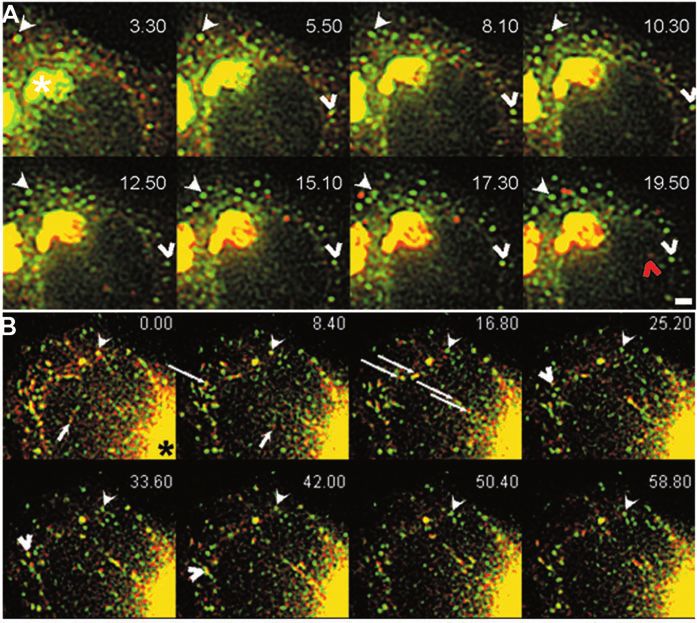
ERGIC clusters are stationary sorting sites                    359
                          microscope (ALMF, EMBL, Heidelberg, Germany). Protein transport          measured for no longer than 10 seconds because during this time
                          was measured at different rewarming times in six cells expressing        interval movement of spots and tubules could be tracked pixel by pixel
                          GFP-ERGIC-53 or VSV-G-GFP using a program written in IGOR Pro            and the precise direction quantified. Increasing the analysis time
                          (Wavemetrics, Lake Oswego, OR). The program is based on gradient-        resulted in a more complex movement with several spots and tubules
                          based optical flow estimation (Nomura et al., 1991) that measures the    intersecting making it impossible to track them. Protein mass flow
                          speed and direction of moving objects in an image sequence. We           rates in each direction were calculated by multiplying fluorescence
                          scored directions towards a reference point in the Golgi apparatus.      intensity and speed. The flow rate is thus a measure of transported
                          Velocity and intensity filters were used to eliminate the apparent       protein per unit of time. Statistical significance (P≤0.05) of the
                          vectors generated by noise in the image sequence. The direction          preference in a certain direction among these three categories was
                          corresponds to the angle between the moving direction and the            probed by Student’s t-test.
                          reference point. The direction is 0° when the spot moves straight to
                          the reference point and 180° or –180° when moving straight away
                          from the reference point. Directions were categorized by angle           Results
                          intervals. Particles moving in the interval of –60° to 60° were
                          designated ‘towards’, those moving in the interval –120° to 120° were
                                                                                                   Features of GFP-ERGIC-53 and endogenous ERGIC-53
                          designated ‘away’, and the remaining angle values (–60° to –120° and     are indistinguishable
                          60° to 120°) were designated ‘non-directional’. The directionality was   To visualize the ERGIC and retrograde traffic from the ERGIC
Journal of Cell Science

                          Fig. 1. GFP-ERGIC-53 localizes and
                          oligomerizes like endogenous ERGIC-
                          53. (A) HeLa cells stably expressing
                          GFP-ERGIC-53 were treated with
                          sodium butyrate overnight, fixed with
                          paraformaldehyde, and stained for β-
                          COP, Sec31 or transferrin receptor.
                          Cells were observed with a confocal
                          fluorescence microscope. Insets show
                          higher magnifications. (B) GFP-
                          ERGIC-53 cells treated with sodium
                          butyrate or non-transfected cells were
                          pulsed with [35S]methionine and
                          chased for the indicated times. Proteins
                          were immunoprecipitated either with
                          anti-ERGIC-53 for the control cells or
                          with anti-GFP for the stable clone.
                          Immunoprecipitates were analyzed by
                          non-reducing SDS-PAGE and
                          visualized by fluorography. During the
                          time course, dimers (lower bands)
                          disappear as hexamers (higher bands)
                          form until a steady state is reached
                          when each species is represented as
                          50%. GFP-ERGIC-53 dimers appear as
                          two bands corresponding to
                          homodimers and heterodimers (lower
                          arrowhead) with endogenous ERGIC-
                          53. These dimers are converted to
                          hetero-hexamers (upper arrowheads)
                          and homo-hexamers.
                          (C) Quantification of dimer and
                          hexamer formation during the chase
                          period. 100% is the sum of homo-
                          dimers and homo-hexamers at a given
                          chase time. A representative
                          experiment is shown. Bar, 5 µm.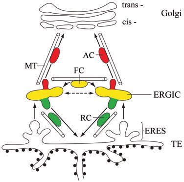
360 Journal of Cell Science 118 (2)
in living cells we tagged ERGIC-53 with green fluorescent different expression levels of up to fourfold that of endogenous
protein (GFP-ERGIC-53) and stably expressed it in HeLa cells. ERGIC-53. All the tested clones, however, gave similar results.
GFP was attached to the N-terminus of ERGIC-53 in order not By confocal microscopy, GFP-ERGIC-53 localized to the
to interfere with its trafficking, which is controlled by multiple Golgi area, peripheral dots and, less prominently, to the
position-dependent, C-terminal transport determinants ER (Fig. 1A). This distribution is very similar to that
(Kappeler et al., 1997; Nufer et al., 2002; Nufer et al., 2003). of endogenous ERGIC-53 in non-transfected cells
In some of the obtained clones GFP-ERGIC-53 was inducible (supplementary material Fig. S1A). In the present work, we
by sodium butyrate, providing a means to study the effect of mainly focused on peripheral structures, as labeling in the
Golgi area is too dense to distinguish individual ERGIC spots.
Like endogenous ERGIC-53, GFP-ERGIC-53 colocalized with
the COP I subunit β-COP and there was partial overlap with
the distribution of the COP II subunit Sec31 (Fig. 1A)
(Klumperman et al., 1998; Shima et al., 1999; Hammond and
Glick, 2000). In transiently transfected COS cells highly
overexpressing ERGIC-53, a fraction of the protein escapes to
the plasma membrane and is subsequently endocytosed by a
signal-mediated process (Hauri et al., 2000). This is not the
case, however, in our HeLa clones expressing GFP-ERGIC-53,
as no co-labeling with the endosomal marker transferrin
receptor (Fig. 1A) or the lysosomal marker Lamp1 (data not
shown) was observed.
To test if GFP-ERGIC-53 oligomerizes correctly into
disulfide-linked dimers and hexamers (Schweizer et al.,
1988), cells were metabolically labeled with [35S]methionine
Journal of Cell Science
in a pulse-chase experiment and ERGIC-53 was
immunoprecipitated and analyzed by SDS-PAGE under
non-reducing conditions. Fig. 1B shows that GFP-ERGIC-53
initially appears as a 160 kDa band corresponding to
homodimers which is in part converted to a 480 kDa species
corresponding to homohexamers. These high molecular weight
species break down to an 80-kDa monomeric form under
reducing conditions (data not shown). Importantly, the kinetics
of conversion of dimeric to hexameric forms is identical to that
of endogenous ERGIC-53 (Fig. 1C). Additional bands on the
gel of GFP-ERGIC-53-expressing cells are hetero-oligomers
formed between GFP-ERGIC-53 and the endogenous protein
as these bands immunoprecipitated with both anti-GFP and
anti-ERGIC-53 (supplementary material Fig. S1B). This
hetero-oligomerization is an additional indication for correct
folding of GFP-ERGIC-53.
Fig. 2. Live imaging of GFP-ERGIC-53 reveals two types of
structures with different dynamics. (A) A whole cell and its
schematic representation where paths of stationary structures imaged
every 10 seconds have been traced (see supplementary material
Movie 2). Note that these structures undergo very short range to no
movement. Lower panel shows image series of the outlined portion
in the upper panel. Arrowheads point two peripheral ERGIC
structures fusing with each other. Arrows indicate elongated ERGIC
structures. (B) Representative images from supplementary material
Movie 3. GFP-ERGIC-53-expressing HeLa cells were imaged in the
xyz directions every 10 seconds. Arrows indicate long-lasting
structures that can persist for up to 31 minutes. (C) Data taken from
supplementary material Movie 4 imaged with an interval of 0.2
second. Representative frames show three peripheral ERGIC
structures (arrowheads) that hardly move during the entire imaging
interval. The arrow points to a peripheral stationary spot shooting out
an elongated structure. Lower panel shows the whole cell and a
schematic representation of paths that GFP-ERGIC-53 fast-moving
structures followed during the imaging time. Note that the movement
does not follow a defined direction, and often seems to connect two
peripheral stationary structures. Bar, 5 µm. Time is shown as
minutes.seconds.ERGIC clusters are stationary sorting sites 361
Fluorescence recovery after photobleaching was then used move to the Golgi area but rather hovered about in place.
to test if GFP-ERGIC-53 recycles. When the peripheral We call them ‘stationary’ structures. They most probably
cytoplasm was bleached throughout the z-axis, fluorescent dots correspond to the COP I-positive tubulovesicular clusters of the
reappeared within 2 minutes (supplementary material Movie ERGIC previously identified by immunoelectron microscopy
1). We did not see non-bleached GFP-ERGIC-53 spots moving (Klumperman et al., 1998). Some stationary structures
to refill the bleached area, suggesting that the reappearing dots persisted throughout the entire recording time and occasionally
largely originate from a recycling process involving the ER fused with one another (Fig. 2A, arrowheads) or split. Others
rather than a massive lateral transfer. The reappearance is disappeared or appeared de novo in non-labeled areas. The
not due to new protein synthesis as it also occurred in fluorescence intensity of the spots did not remain constant but
cycloheximide-treated cells. A similar recovery was observed fluctuated with time indicating the continuous recycling of
when the Golgi area was bleached. Collectively, these data GFP-ERGIC-53. Recycling was confirmed by the recovery of
suggest that the GFP tag does not interfere with the folding and fluorescence observed after partial bleaching of these spots (see
recycling of ERGIC-53. arrows in supplementary material Movie 1). In rare cases,
some dynamic tubular structures were observed that rapidly
disappeared (Fig. 2A, arrows). They probably correspond to
GFP-ERGIC-53 imaging reveals two populations with the fast-moving structures seen with fast imaging (see below).
different dynamics To test if the appearance and disappearance of the stationary
To explore the dynamics of GFP-ERGIC-53 in living HeLa structures correspond to de novo formation and consumption
cells we used bright field and confocal time-lapse imaging with events or to movement into or out of the focal plane, we
recording intervals of 10 seconds (‘slow imaging’) or 0.2 performed 4D imaging (3D over time) using an imaging
second (‘fast imaging’) to follow long- and short-time events, interval of 10 seconds and a step size of 0.8 µm. This approach
respectively. showed that stationary GFP-ERGIC-53 spots can indeed form
Slow imaging of GFP-ERGIC-53 for 15 minutes (Fig. 2A) de novo or be consumed. Moreover, imaging for more than
and longer (data not shown) revealed fluorescent puncta with 30 minutes confirmed that many structures are long-lived,
a diameter of 0.9-1.2 µm exhibiting short-range movement of
Journal of Cell Science
can undergo several fusion and splitting events, and do not
1.2 µm on average with a maximal velocity of 0.2 µm/second exhibit a preferential movement to the Golgi area (Fig. 2B;
(see supplementary material Movie 2). These structures did not supplementary material Movie 3). These features were
Fig. 3. ERGIC stationary
structures in the periphery and in
the Golgi area are equivalent.
(A) Time points from
supplementary material Movie 5
showing the relocalization of
GFP-ERGIC-53 to the ER when
cells are treated with H89. The
arrow points to a peripheral
ERGIC structure that loses
fluorescence and disappears
without moving to the Golgi
area. (B) Time series from
supplementary material Movie 6
showing the recovery of the
same cell after H89 washout.
The arrow points to a peripheral
ERGIC structure that gets
brighter during recovery but
does not move to the Golgi area
(arrowhead) which is refilled at
the same time as peripheral
ERGIC structures appear.
(C) NEM blocks H89-induced
recycling of GFP-ERGIC-53 to
the ER. HeLa cells stably
expressing GFP-ERGIC-53 were
treated with H89 for 20 minutes
in the presence or absence of
NEM or left untreated (control),
fixed with paraformaldehyde,
stained for Sec13 (red), and
analyzed by confocal
microscopy. Insets show higher
magnifications. Bar, 5 µm. Time
is indicated as minutes.seconds.362 Journal of Cell Science 118 (2)
Fig. 4. GFP-ERGIC-53 tubule formation
during rewarming from 16°C. GFP-ERGIC-
53-expressing HeLa cells were incubated for
3 hours at 16°C, shifted to 37°C for 1 minute
53 seconds and imaged every 0.6 second (see
supplementary material Movie 8). 0 second
indicates the time at which imaging started.
Extensive tubular structures appear from
peripheral ERGIC spots and move with no
preference in different directions. A single
ERGIC structure can simultaneously extend
tubules in opposite directions without being
consumed (arrow). Several rounds of tubule
emission from the same ERGIC spot do not
consume it (arrowhead). Bar, 5 µm. Time,
minutes.seconds.
colocalized with the ER marker CLIMP-
63, whereas the structure of the Golgi
defined by giantin remained unchanged
(data not shown). Addition of H89 rapidly
stopped the movement as well as the
unchanged when cells were imaged in the presence of splitting and fusion activities of the peripheral stationary spots,
cycloheximide to block protein synthesis. and, synchronously with the Golgi area, the spots lost
Fast imaging (0.2-second intervals) of GFP-ERGIC-53 fluorescence within 20 minutes at the expense of an
Journal of Cell Science
revealed, in addition to the stationary structures, short-lived increasingly fluorescent ER (arrow in Fig. 3A; supplementary
fast-moving structures that were difficult to track under slow- material Movie 5). H89 also stopped the fast moving structures
imaging conditions. The short-lived elements moved in all (data not shown). Upon removal of H89, GFP-ERGIC-53
directions and could occasionally be seen originating from reappeared simultaneously in peripheral spots and the Golgi
stationary structures (supplementary material Movie 4; Fig. area (Fig. 3B; supplementary material Movies 6 and 7) whereas
2C). They had an apparent average diameter of 0.5 µm and the ER fluorescence decreased concomitantly (compare Fig.
moved in a stop-and-go fashion along curvilinear trajectories 3B, recovery 2.0 and 26.1). Of note, the peripheral ERGIC
with speeds ranging from 1 to 7 µm/s. Often these structures structures did not directionally move to refill the Golgi area
seemed to cross each other, to move from one stationary (arrow in Fig. 3B). This suggests that ERGIC structures in the
structure to another or to cross several structures during their periphery and in the Golgi area independently received GFP-
long-range movement of up to 6 µm. They frequently changed ERGIC-53 from the ER. Fast-moving ERGIC-53 structures
shape by becoming slightly elongated (supplementary material only appeared after almost full recovery of the stationary
Movie 4). Tracking several of these structures for up to 70 structures and often derived from them moving in all directions
seconds showed that they moved only rarely in the direction of (data not shown).
the Golgi area (tracks in Fig. 2C). At 37°C, the fast-moving To evaluate if the H89-induced redistribution of GFP-
structures represented ~20% of all GFP-ERGIC-53 spots. Long ERGIC-53 to the ER involves vesicular traffic we additionally
tubules were infrequently seen. Their number was increased up included n-ethylmaleimide (NEM), which is known to block
to eightfold, however, when GFP-ERGIC-53 expression was COP I vesicle fusion but not budding both in vitro and in vivo
induced by sodium butyrate treatment for 40 hours (data not (Rothman and Orci, 1990). NEM prevented the retrograde
shown). Fast movement was absent in cells preincubated on ice traffic of GFP-ERGIC-53 to the ER although ER exit was
for 15 minutes and imaged at 37°C in the presence of blocked, as indicated by the diffuse appearance of the COPII
nocodazole, suggesting microtubule dependence (data not component Sec13 (Fig. 3C). In the presence of NEM peripheral
shown). Collectively, our live-imaging approach revealed that GFP-ERGIC-53 spots were slightly less abundant than in
most ERGIC-53 spots in the periphery are stationary and a control cells presumably owing to uninhibited vesicular
minor population is highly dynamic. budding from ERGIC membranes. NEM did not alter the high
mobility of GFP-ERGIC-53 trapped in the ER by H89 as
monitored by FRAP (data not shown). These results are
Trafficking routes of GFP-ERGIC-53 consistent with vesicular ERGIC to ER transport.
To explore the dynamics of GFP-ERGIC-53 in more detail we To study trafficking from the ERGIC, low temperature/
used conditions that reversibly block protein recycling in the rewarming experiments were performed. Incubation at 15-
ER or ERGIC. Incubating the cells with the kinase inhibitor 16°C is known to reversibly accumulate ERGIC-53 in the
H89 known to block protein export from the ER (Aridor and ERGIC (Lippincott-Schwartz et al., 1990; Schweizer et al.,
Balch, 2000), relocalized GFP-ERGIC-53 to the ER (Fig. 3A; 1990; Klumperman et al., 1998). Upon rewarming from 16°C
supplementary material Movie 5) as reported previously for to 37°C, the stationary peripheral GFP-ERGIC-53 spots
endogenous ERGIC-53 (Lee and Linstedt, 2000). Antibody rapidly emitted tubules moving with an average velocity of
staining of fixed cells confirmed that during the H89 block, around 1 µm/second (Fig. 4; supplementary material Movie 8),
GFP-ERGIC-53 and endogenous ERGIC-53 increasingly whereas the fast-moving structures had essentially the sameERGIC clusters are stationary sorting sites 363
based on the optical flow estimation (see Materials and
Methods). HeLa cells either stably expressing GFP-ERGIC-53
or infected with adenovirus carrying the ts045-VSV-G-GFP
DNA were subjected to the low temperature/rewarming
procedure and imaged at different rewarming times for
approximately 100 seconds. We analyzed moving entities with
speed ranges between 200 nm/second and 400 nm/second from
several 10 seconds sub-sequences of each movie. There was
enough displacement during this short period to measure
directionality relative to the Golgi optimally. During
rewarming from 16°C to 37°C, the movement of GFP-ERGIC-
53 had a slight preference away from the Golgi (Fig. 5A). This
Fig. 5. GFP-ERGIC-53 and VSV G-GFP take different routes from is consistent with and quantitatively supports our visual
the ERGIC. Quantification of the flow rate direction of GFP-ERGIC- impression that GFP-ERGIC-53 does not preferentially move
53 (A) and VSV-G-GFP (B) in cells rewarmed from 16°C to 37°C or to the Golgi. For VSV-G-GFP, rewarming from 15°C to 32°C
15°C to 32°C, respectively. The protein flow rate is plotted in
generated a preferential movement toward the Golgi (Fig. 5B).
arbitrary units (normalized to 1) against the direction of movement.
Note that VSV-G-GFP is directed toward the Golgi whereas GFP- This finding is in accord with previous reports on the dynamics
ERGIC-53 is directed away from the Golgi. These differences are of VSV-G-GFP during rewarming from 15°C (Presley et al.,
statistically significant (Student’s t-test, P≤0.05). Bars show the 1997; Scales et al., 1997). The seemingly small differences
mean±s.d. (GFP-ERGIC-53, n=67; VSV-G-GFP, n=73); No, non- between towards and away unravel the particularities of VSV-
directional movement. G-GFP dynamics during exit from the ERGIC in a 15°C
rewarming experiment. VSV-G-GFP exit from the ERGIC is
movement as observed at 37°C (data not shown). Single gradual rather than a rapid switch. Simulations show that such
stationary structures could repeatedly extend multiple tubules small differences between towards and away produce a net
Journal of Cell Science
in different directions. The tubules formed and detached within directional transport of VSV-G to the Golgi as a result of
seconds and stayed at peripheral sites for only brief periods cumulative effects. Collectively, these results show
before vanishing in the proximity of a stationary structure quantitatively that GFP-ERGIC-53 and VSV-G-GFP leave the
or disappearing in the cell periphery towards no defined ERGIC in different directions, consistent with their opposed
structures. Formation and translocation of GFP-ERGIC-53- transport to the ER and to the Golgi, respectively.
enriched tubules decreased with time until the activity entirely
stopped 20 minutes after rewarming (data not shown). Tubule
formation in general (81% of cases) did not consume the Sorting of anterograde and retrograde cargo in the
stationary structure. Moreover, the tubules had no apparent ERGIC
preference for moving to the Golgi area (Fig. 5). To obtain sorting information also on a soluble secretory
To investigate whether these tubular processes were directed protein and to visualize the sorting of anterograde and
to the Golgi area, we photobleached this region in cells retrograde cargo directly in the same cell we constructed a
rewarmed from 16°C. At early recovery and rewarming times, secretory form of pDsRedT1 (Bevis and Glick, 2002), termed
tubules forming from peripheral spots extended randomly into ssDsRed, by attaching an N-terminal signal sequence. ssDsRed
different directions whereas the Golgi area was refilled was transfected into HeLa cells stably expressing GFP-
homogeneously and independently of the tubules (data not ERGIC-53 and transport was studied by dual color imaging.
shown). Obviously, the refilling of the Golgi area reflects direct Using immunoprecipitation experiments we verified that
transport of GFP-ERGIC-53 from the ER into ERGIC clusters ssDsRed was indeed secreted into the culture medium (data not
that are concentrated near the Golgi apparatus and cannot be shown). When transport was blocked at 16°C, ssDsRed
resolved by light microscopy (Klumperman et al., 1998). After displayed an enhanced ER pattern, and some ERGIC-53 spots
10 minutes of recovery, the profile of GFP-ERGIC-53- also co-labeled with ssDsRed. Rewarming to 37°C led to a
containing structures, including the Golgi area, was similar to gradual decrease of ER fluorescence followed by an increase
that of the pre-bleached state. Collectively, these experiments in peripheral ERGIC structures and subsequently the Golgi
suggest that the dynamic tubules indicate the ERGIC to ER area (Fig. 6A; supplementary material Movie 9). Large
recycling route and that the recycling of GFP-ERGIC-53 ssDsRed spots were clearly seen to leave the peripheral ERGIC
largely bypasses the Golgi. spots. The number of exiting ssDsRed spots correlated with the
expression level. We chose to analyze moderately expressing
cells as they are closer to physiological conditions. To quantify
GFP-ERGIC-53 and VSV-G-GFP take different routes ssDsRed segregation from peripheral ERGIC clusters we
from the ERGIC tracked structures colocalizing with GFP-ERGIC-53 in six
Our conclusion regarding the recycling pathway of GFP- cells and classified the different initial sorting events during
ERGIC-53 is based on visual analysis. To test this visual rewarming from 16°C.
impression further, we sought to compare the traffic route of Table 1 shows that 55% of the stationary ERGIC structures
GFP-ERGIC-53 from the ERGIC quantitatively with that of efficiently sorted ssDsRed and ERGIC-53 in a single step,
the well-studied anterograde marker membrane protein VSV- whereby ssDsRed structures of considerable size left the
G-GFP. To this end, we measured the directionality of protein ERGIC and moved to the Golgi area while the GFP-ERGIC-
flow rate after a low temperature block by a vector field method 53 spot remained stationary. In 12% of cases, the sorting was364 Journal of Cell Science 118 (2)
incomplete. In 31% of the cases, no sorting was observed, but Table 1. Quantification of GFP-ERGIC-53 and ssDsRed
many of these elements underwent fission and fusion. In only initial sorting in the ERGIC of live cells following
2% of the cases did the sorting result in complete consumption rewarming from 15 to 37°C*
of the yellow ERGIC spot. Thus, in a majority of sorting Consistent with stationary
events, the ERGIC remained stationary and was not consumed. Observation % compartment model?
Many of the ERGIC structures segregated ssDsRed repeatedly Complete sorting
(Fig. 6A, empty arrowheads; supplementary material Movie 9) Export of ssDsRed spot, 55 Yes
indicating anterograde flow through a persisting ERGIC GFP-ERGIC-53 spot remains
structure. Importantly, the export of a ssDsRed dot often stationary
Consumption by sorting 2 No
changed the color of the initial ERGIC cluster from yellow to
more greenish in colour, suggesting that individual clusters act Incomplete sorting
as entities in cargo sorting. Export of ssDsRed spot 7 Yes
Export of GFP-ERGIC-53 spot 5 Yes
To make sure we did not miss fast sorting events we studied
ssDsRed and GFP-ERGIC-53 segregation by the fast recording No sorting 31 –
procedure. As with the slow recording, ssDsRed segregated *Recordings were made at intervals of 10 seconds for 20 minutes (see
from GFP-ERGIC-53 stationary structures (Fig. 6B, filled Materials and Methods). % refers to total number of analyzed stationary
arrowheads). Of the fast moving structures, 29% were positive ERGIC structures.
for both markers and therefore appeared yellow in merged
images. When observed for up to 1 minute, 77% of these
yellow structures showed no sign of sorting and often the dynamics of sorting of the retrograde marker protein
eventually fused with stationary structures indicating intra- ERGIC-53 and the anterograde markers VSV-G and ssDsRed.
ERGIC transport; 23% of the fast-moving yellow structures Our findings shed new light on the nature of the ERGIC and
separated into a red and a green vehicle, which moved in on protein trafficking early in the secretory pathway. They
opposite directions. Hence, some of the fast-moving structures support the notion of a stationary ERGIC consisting of
Journal of Cell Science
exhibited bidirectional sorting. numerous discontinuous elements that operate in bidirectional
sorting to the ER and Golgi. This conclusion is based on three
major observations. First, GFP-ERGIC-53 is localized in
Discussion stationary spots displaying short-range non-directional
Although no stable marker for the ERGIC is known, the movement. Unlike VSV-G-GFP spots, GFP-ERGIC-53 spots
continuous recycling of ERGIC-53 has allowed us to visualize do not show a preferential movement toward the Golgi region
the ERGIC for prolonged times in living cells and to compare and hence do not exhibit typical features of anterograde
Fig. 6. Sorting of GFP-ERGIC-53 and ssDsRed
in peripheral ERGIC structures. Data from
cells co-expressing GFP-ERGIC-53 and
ssDsRed rewarmed after a 3-hour 16°C block.
(A) Time series (taken every 10 seconds) from
supplementary material Movie 9. 3.30 indicates
the time after rewarming to 37°C. The filled
arrowhead indicates a stationary GFP-ERGIC-
53 structure from which ssDsRed segregates.
Note that even in the proximity of the Golgi,
the ERGIC stationary structure does not move
toward this area. The empty arrowhead
indicates another stationary structure from
which ssDsRed segregates. 11 minutes 50
seconds later, this same structure receives new
ssDsRed material that is shot toward the Golgi
at time point 19.50 (red arrowhead). Note that
the Golgi (asterisk) becomes redder as the
rewarming proceeds. Time, minutes.seconds.
(B) Cell imaged with an interval of 0.2 second,
12 minutes after shifting the temperature from
15°C to 37°C. 0.00 indicates the time at which
the imaging started. The filled arrowhead
points to a stationary structure that segregates
ssDsRed as in A. Some GFP-ERGIC-53
structures are positive for ssDsRed and move
together towards (arrow) the Golgi (asterisk) or toward other ERGIC structures (empty arrowhead). Often, moving structures carrying both
GFP-ERGIC-53 and ssDsRed cross several GFP-ERGIC-53 stationary structures (long arrows). Time, seconds.milliseconds. Bar, 5 µm.ERGIC clusters are stationary sorting sites 365
Quantification of the directionality of movement by a
numerical processing procedure supports our visual impression
that GFP-ERGIC-53 largely escapes packaging into ACs.
Unlike VSV-G-GFP, GFP-ERGIC-53 shows no preferential
movement to the Golgi upon exit from the ERGIC: it moves
in the opposite direction. Considering the apparently random
distribution of ERGIC clusters in the peripheral cytoplasm one
would expect no preferred directionality for GFP-ERGIC-53
cycling back to the ER. However, low temperature blocks tend
to concentrate the ERGIC clusters closer to the Golgi apparatus
(Klumperman et al., 1998). Therefore, the measured net flow
for ERGIC-53 results from a combined effect: repositioning of
ERGIC clusters and recycling of ERGIC-53. At first glance,
the measured difference of 13.5% between anterograde and
retrograde movements of VSV-G-GFP appears to be small. It
should be noted, however, that the quantification time was short
(10 seconds) and that extrapolation to a longer time results in
Fig. 7. Model of the organization of the early secretory pathway and a net flow to the Golgi within a few minutes. Thus, directed
sorting of anterograde and retrograde traffic in the ERGIC. The transport is the consequence of a slight preference in movement
cycling protein GFP-ERGIC-53 (green) and anterograde cargo, such towards one direction. Overall, this novel vector field method
as VSV-G-GFP or ssDsRed (red), leave the ER at ER-exit sites based on the optical flow estimation validates previous
(ERES) of the transitional elements of the rough ER (TE) and are conclusions derived from fixed cells (Klumperman et al.,
transported to the ER-Golgi intermediate compartment (ERGIC) by a 1998), proposing that ERGIC to ER retrograde transport
microtubule-independent process. The ERGIC is a collection of largely bypasses the Golgi. It is also consistent with our
stationary tubulovesicular clusters exhibiting short range, non-
Journal of Cell Science
ssDsRed/GFP-ERGIC-53 dual-imaging data showing
directional, but microtubule-dependent movement. The clusters can preferential sorting of anterograde and retrograde traffic in the
split or fuse with one another (dashed arrow). Sorting in the ERGIC
leads to anterograde carriers (AC), mediating transport of ssDsRed to
ERGIC.
the cis-Golgi and to retrograde carriers (RC) mediating transport of ERGIC clusters defined by ERGIC-53 and COP I are
GFP-ERGIC-53 back to the ER. Both pathways are dependent on localized in close proximity to ERES (Bannykh et al., 1996;
intact microtubules. Individual ERGIC-clusters are connected by Klumperman et al., 1998; Martinez-Menarguez et al., 1999;
fast-moving carriers (FC) that carry both markers and are transported Stephens et al., 2000), which precludes the visualization of
along microtubules. MT, microtubules. protein transport from ERES to ERGIC in live cells owing to
the insufficient resolution of light microscopy. Therefore, a
simple alternative interpretation of our data would be that GFP-
carriers (ACs). Their short-range movement is dependent on ERGIC-53, unlike endogenous ERGIC-53, is trapped in ERES
intact microtubules as it is lost in nocodazole-treated cells. and has no access to the ERGIC. Accordingly, the dispersal of
Second, many of the stationary GFP-ERGIC-53 structures are GFP-ERGIC-53 induced by H89 could reflect diffusion within
long-lived and persist for more than 30 minutes, another the plane of the ER membrane. Several observations argue
feature that is inconsistent with an exclusive AC function. ER against this interpretation. Like endogenous ERGIC-53, GFP-
to Golgi transport is a rapid event. The ERGIC spots can ERGIC-53 co-localizes with the COP I subunit β-COP but
undergo splitting and occasionally fuse with one another; some shows only partial overlap with COPII subunits, Sec13 and
appear de novo, others disappear. Interestingly, similar features Sec31, in fixed cells analyzed by confocal microscopy.
have been observed for ERES (Stephens, 2003; Bevis and Likewise, our biochemical analysis indicates normal folding of
Glick, 2002) suggesting that the dynamics of ERES and GFP-ERGIC-53. Furthermore, NEM blocks the H89-induced
ERGIC clusters may be regulated in concert. Third, the ERGIC dispersal of ERGIC-53 from the stationary structures
spots are not consumed by the sorting of GFP-ERGIC-53 and indicating discontinuity with the ERES. The combined data
ssDsRed. They can undergo multiple rounds of sorting of suggest that the GFP-ERGIC-53 stationary structures
anterograde and retrograde cargo. correspond to the tubulovesicular ERGIC clusters defined by
Our conclusion regarding the nature of the ERGIC is ERGIC-53 and COP I at the ultrastructural level (Schweizer et
different from previous live-imaging studies on VSV-G-GFP al., 1988; Klumperman et al., 1998).
transport (Presley et al., 1997; Scales et al., 1997). These By imaging at high temporal resolution of 5 frames per
authors concluded that the ERGIC clusters are transport second (fast imaging) we have uncovered a third pathway not
vehicles for protein delivery to the Golgi, rather than a stable previously described. This pathway is mediated by fast-moving
compartment (Lippincott-Schwartz et al., 2000). In accord with carriers (FCs) a fraction of which contains both GFP-ERGIC-
these studies, we observed that the ACs moving to the Golgi, 53 and ssDsRed. The pathway is highly sensitive to
containing ssDsRed (or VSV-G-GFP), are rather large and microtubule-disrupting drugs as well as H89, and requires the
cannot be small transport vesicles. However, they derive from existence of stationary ERGIC structures as unveiled by H89
the stationary ERGIC defined by ERGIC-53. Only by wash-out experiments. As FCs do not exhibit preferential
visualization of the sorting of anterograde and retrograde movement to the Golgi area and can occasionally be seen to
traffic from the ERGIC in living cells did the stationary nature originate from and fuse with stationary ERGIC structures, we
of this compartment become apparent. propose that they functionally connect ERGIC clusters by366 Journal of Cell Science 118 (2)
lateral exchange. Although a majority of the FCs remains the ERGIC is stationary, it is likely to have additional functions
unsorted and appears to eventually fuse with a stationary that remain to be uncovered.
structure, some can separate into a GFP-ERGIC-53-containing
and an ssDsRed-containing dot, which move in opposite We thank Beat Ludin, Jens Rietdorf and Timo Zimmermann for
directions. The GFP-ERGIC-53 dot tends to rapidly disappear, imaging assistance; Benjamin Glick for providing pDsRedT1; Fred
Gorelick and Wanjin Hong for providing antibodies to Sec31 and
whereas the ssDsRed dot moves to the Golgi. This suggests
Sec13; Käthy Bucher, Maria Susanna Balda and Karl Matter for
that some FCs are involved in anterograde/retrograde sorting. continuous support; and the members of the Hauri and Pepperkok
Integrating our new data with previously published findings, groups for suggestions. The study was supported by the Swiss
the following picture regarding the organization and traffic National Science Foundation (H.-P.H.), the University of Basel (H.-
routes in the early secretory pathway emerges (Fig. 7). Newly P.H.) and a Quality of Life EU NetworkGrant QCRI-CT-2002-01272
synthesized secretory proteins and ERGIC-53 are transported (R.P.)
from the ER to stationary ERGIC clusters that are lying close
to, but separate from ERES (Bannykh et al., 1996; Mezzacasa
and Helenius, 2002). Although there is agreement that ER exit References
is COP II-dependent (Horstmann et al., 2002; Mironov et al., Aridor, M. and Balch, W. E. (2000). Kinase signaling initiates coat complex
II (COPII) recruitment and export from the mammalian endoplasmic
2003), the precise mechanism of membrane traffic from ERES reticulum. J. Biol. Chem. 275, 35673-35676.
to ERGIC remains to be elucidated. Once in the ERGIC, Aridor, M., Bannykh, S. I., Rowe, T. and Balch, W. E. (1995). Sequential
anterograde cargo is sorted from ERGIC-53 into rather large coupling between COPII and COPI vesicle coats in endoplasmic reticulum
ACs by a dissociative process. The size of ACs may vary to Golgi transport. J. Cell Biol. 131, 875-893.
Bannykh, S. I. and Balch, W. E. (1997). Membrane dynamics at the
according to cargo flux and can be considerable under endoplasmic reticulum-Golgi interface. J. Cell Biol. 138, 1-4.
conditions of massive synchronized release of VSV-G from the Bannykh, S. I., Rowe, T. and Balch, W. E. (1996). The organization of
ER (Horstmann et al., 2002). ACs then rapidly move to the endoplasmic reticulum export complexes. J. Cell Biol. 135, 19-35.
Golgi in a microtubule-dependent way. In contrast, ERGIC-53 Bevis, B. J. and Glick, B. S. (2002). Rapidly maturing variants of the
Discosoma red fluorescent protein (DsRed). Nat. Biotechnol. 20, 83-87.
is packaged into retrograde carriers (RCs), the size and shape
Journal of Cell Science
Beznoussenko, G. V. and Mironov, A. A. (2002). Models of intracellular
of which can also vary. RCs must also be of a considerable size transport and evolution of the Golgi complex. Anat. Rec. 268, 226-238.
to be visible. When traffic is inhibited at 15°C followed by Blum, R., Stephens, D. J. and Schulz, I. (2000). Lumenal targeted GFP, used
rewarming to 37°C, RCs emanating from the ERGIC clusters as a marker of soluble cargo, visualises rapid ERGIC to Golgi traffic by a
are often tubular. It appears that conditions of massive cargo tubulo-vesicular network. J. Cell Sci. 113, 3151-3159.
Farquhar, M. G. (1985). Progress in unraveling pathways of Golgi traffic.
transport favor the formation of tubules regardless of the Annu. Rev. Cell Biol. 1, 447-488.
pathway. Like ERGIC to Golgi anterograde transport, efficient Glick, B. S. and Malhotra, V. (1998). The curious status of the Golgi
ERGIC to ER retrograde transport requires intact microtubules. apparatus. Cell 95, 883-889.
What is the role of the newly discovered FCs that connect Hammond, A. T. and Glick, B. S. (2000). Dynamics of transitional
endoplasmic reticulum sites in vertebrate cells. Mol. Biol. Cell 11, 3013-
individual stationary ERGIC elements and are only visible at 3030.
high temporal resolution? FCs may functionally link the Hauri, H. P., Kappeler, F., Andersson, H. and Appenzeller, C. (2000).
ERGIC clusters allowing the exchange of critical components ERGIC-53 and traffic in the secretory pathway. J. Cell Sci. 113, 587-596.
within the discontinuous ERGIC system. Alternatively, FCs Horstmann, H., Ng, C. P., Tang, B. L. and Hong, W. (2002). Ultrastructural
characterization of endoplasmic reticulum – Golgi transport containers
may be incompletely sorted ACs that, because of their (EGTC). J. Cell Sci. 115, 4263-4273.
incomplete sorting, are unable to travel to the Golgi. Thereby, Kappeler, F., Klopfenstein, D. R., Foguet, M., Paccaud, J. P. and Hauri,
the FC pathway would operate as a backup system in order to H. P. (1997). The recycling of ERGIC-53 in the early secretory pathway.
prevent anterograde transport of incompletely sorted ERGIC-53 carries a cytosolic endoplasmic reticulum-exit determinant
interacting with COPII. J. Biol. Chem. 272, 31801-31808.
membranes. Klumperman, J., Schweizer, A., Clausen, H., Tang, B. L., Hong, W.,
Our study does not lend support to the notion of an ERGIC- Oorschot, V. and Hauri, H. P. (1998). The recycling pathway of protein
53-positive late subdomain of ERGIC close to the Golgi as ERGIC-53 and dynamics of the ER-Golgi intermediate compartment. J. Cell
proposed by Marra and colleagues (Marra et al., 2001). Sci. 111, 3411-3425.
Inconsistent with such a view, ERGIC-53 appears with Lee, T. H. and Linstedt, A. D. (2000). Potential role for protein kinases in
regulation of bidirectional endoplasmic reticulum-to-Golgi transport
indistinguishable kinetics in clusters close to the Golgi and in revealed by protein kinase inhibitor H89. Mol. Biol. Cell 11, 2577-2590.
the periphery after H89 wash-out, and ACs moving from Lippincott-Schwartz, J., Donaldson, J. G., Schweitzer, A., Berger, E. G.,
stationary ERGIC spots to the Golgi do not comprise Hauri, H.-P., Yuan, L. C. and Klausner, R. D. (1990). Microtubule-
detectable levels of ERGIC-53. Moreover, GFP-ERGIC-53 dependent retrograde transport of proteins into the ER in the presence of
brefeldin A suggests an ER recycling pathway. Cell 60, 821-836.
spots in the periphery and in the Golgi region are GM-130- Lippincott-Schwartz, J., Roberts, T. H. and Hirschberg, K. (2000).
negative in these cells (data not shown). Our data rather suggest Secretory protein trafficking and organelle dynamics in living cells. Annu.
that all ERGIC clusters are qualitatively very similar or Rev. Cell Dev. Biol. 16, 557-589.
identical. Lotti, L. V., Torrisi, M. R., Pascale, M. C. and Bonatti, S. (1992).
Immunocytochemical analysis of the transfer of vesicular stomatitis virus G
In conclusion, we find that in living cells the ERGIC defined glycoprotein from the intermediate compartment to the Golgi complex. J.
by ERGIC-53 is composed of stationary long-lived structures Cell Biol. 118, 43-50.
close to ERES. The ERGIC structures are sites of active sorting Marra, P., Maffucci, T., Daniele, T., Tullio, G. D., Ikehara, Y., Chan, E.
of anterograde and retrograde cargo. Both anterograde and K., Luini, A., Beznoussenko, G., Mironov, A. and de Matteis, M. A.
(2001). The GM130 and GRASP65 Golgi proteins cycle through and define
retrograde transport from the ERGIC must involve a a subdomain of the intermediate compartment. Nat. Cell Biol. 3, 1101-1113.
dissociative process, the precise molecular mechanism of Martinez-Menarguez, J. A., Geuze, H. J., Slot, J. W. and Klumperman, J.
which remains to be uncovered. In view of the new finding that (1999). Vesicular tubular clusters between the ER and Golgi mediateERGIC clusters are stationary sorting sites 367
concentration of soluble secretory proteins by exclusion from COPI-coated Schweizer, A., Fransen, J. A. M., Baechi, T., Ginsel, L. and Hauri, H.-P.
vesicles. Cell 98, 81-90. (1988). Identification, by a monoclonal antibody, of a 53-kD protein
Mezzacasa, A. and Helenius, A. (2002). The transitional ER defines a associated with a tubular-vesicular compartment at the cis-side of the Golgi
boundary for quality control in the secretion of tsO45 VSV glycoprotein. apparatus. J. Cell Biol. 107, 1643-1653.
Traffic 3, 833-849. Schweizer, A., Fransen, J. A. M., Matter, K., Kreis, T. E., Ginsel, L. and
Mironov, A. A., Mironov, A. A., Jr, Beznoussenko, G. V., Trucco, A., Hauri, H.-P. (1990). Identification of an intermediate compartment involved
Lupetti, P., Smith, J. D., Geerts, W. J., Koster, A. J., Burger, K. N., in protein transport from ER to Golgi apparatus. Eur. J. Cell Biol. 53, 185-
Martone, M. E. et al. (2003). ER-to-Golgi carriers arise through direct en 196.
bloc protrusion and multistage maturation of specialized ER exit domains. Schweizer, A., Ericsson, M., Bachi, T., Griffiths, G. and Hauri, H. P.
Dev. Cell 5, 583-594. (1993). Characterization of a novel 63 kDa membrane protein. Implications
Nomura, A., Miike, H. and Koga, K. (1991). Field theory approach for for the organization of the ER-to-Golgi pathway. J. Cell Sci. 104, 671-683.
determining optical flow. Pattern Recog. Lett. 12, 183-190. Shima, D. T., Scales, S. J., Kreis, T. E. and Pepperkok, R. (1999).
Nufer, O., Guldbrandsen, S., Degen, M., Kappeler, F., Paccaud, J. P., Tani, Segregation of COPI-rich and anterograde-cargo-rich domains in
K. and Hauri, H. P. (2002). Role of cytoplasmic C-terminal amino acids endoplasmic-reticulum-to-Golgi transport complexes. Curr. Biol. 9, 821-
of membrane proteins in ER export. J. Cell Sci. 115, 619-628. 824.
Nufer, O., Kappeler, F., Guldbrandsen, S. and Hauri, H. P. (2003). ER Shugrue, C. A., Kolen, E. R., Peters, H., Czernik, A., Kaiser, C., Matovcik,
export of ERGIC-53 is controlled by cooperation of targeting determinants L., Hubbard, A. L. and Gorelick, F. (1999). Identification of the putative
in all three of its domains. J. Cell Sci. 116, 4429-4440. mammalian orthologue of Sec31P, a component of the COPII coat. J. Cell
Palade, G. (1975). Intracellular aspects of the process of protein synthesis. Sci. 112, 4547-4556.
Science 189, 347-358. Stephens, D. J. (2003). De novo formation, fusion and fission of mammalian
Pelham, H. R. (1989). Control of protein exit from the endoplasmic reticulum. COPII-coated endoplasmic reticulum exit sites. EMBO Rep. 4, 210-217.
Annu. Rev. Cell Biol. 5, 1-23. Stephens, D. J. and Pepperkok, R. (2001). Illuminating the secretory
Pepperkok, R., Scheel, J., Horstmann, H., Hauri, H. P., Griffiths, G. and pathway: when do we need vesicles? J. Cell Sci. 114, 1053-1059.
Kreis, T. E. (1993). Beta-COP is essential for biosynthetic membrane Stephens, D. J., Lin-Marq, N., Pagano, A., Pepperkok, R. and Paccaud, J.
transport from the endoplasmic reticulum to the Golgi complex in vivo. Cell P. (2000). COPI-coated ER-to-Golgi transport complexes segregate from
74, 71-82. COPII in close proximity to ER exit sites. J. Cell Sci. 113, 2177-2185.
Presley, J. F., Cole, N. B., Schroer, T. A., Hirschberg, K., Zaal, K. J. and Storrie, B. and Nilsson, T. (2002). The Golgi apparatus: balancing new with
Lippincott-Schwartz, J. (1997). ER-to-Golgi transport visualized in living old. Traffic 3, 521-529.
cells. Nature 389, 81-85. Tang, B. L., Low, S. H., Hauri, H. P. and Hong, W. (1995). Segregation of
Rothman, J. E. and Orci, L. (1990). Movement of proteins through the Golgi ERGIC53 and the mammalian KDEL receptor upon exit from the 15 degrees
Journal of Cell Science
stack: a molecular dissection of vesicular transport. FASEB. J. 4, 1460-1468. C compartment. Eur. J. Cell Biol. 68, 398-410.
Scales, S. J., Pepperkok, R. and Kreis, T. E. (1997). Visualization of ER-to- Tang, B. L., Peter, F., Krijnse-Locker, J., Low, S. H., Griffiths, G. and
Golgi transport in living cells reveals a sequential mode of action for COPII Hong, W. (1997). The mammalian homolog of yeast Sec13p is enriched in
and COPI. Cell 90, 1137-1148. the intermediate compartment and is essential for protein transport from
Schekman, R. and Orci, L. (1996). Coat proteins and vesicle budding. the endoplasmic reticulum to the Golgi apparatus. Mol. Cell. Biol. 17, 256-
Science 271, 1526-1533. 266.
Schindler, R., Itin, C., Zerial, M., Lottspeich, F. and Hauri, H. P. (1993). Vollenweider, F., Kappeler, F., Itin, C. and Hauri, H. P. (1998). Mistargeting
ERGIC-53, a membrane protein of the ER-Golgi intermediate compartment, of the lectin ERGIC-53 to the endoplasmic reticulum of HeLa cells impairs
carries an ER retention motif. Eur. J. Cell Biol. 61, 1-9. the secretion of a lysosomal enzyme. J. Cell Biol. 142, 377-389.You can also read