Regulation of c-Raf Stability through the CTLH Complex - MDPI
←
→
Page content transcription
If your browser does not render page correctly, please read the page content below
International Journal of
           Molecular Sciences

Article
Regulation of c-Raf Stability through the
CTLH Complex
Christina J. McTavish 1,2,† , Wesley Bérubé-Janzen 1,2,† , Xu Wang 1 , Matthew E. R. Maitland 1,2 ,
Louisa M. Salemi 1,2 , David A. Hess 1,3 and Caroline Schild-Poulter 1,2, *
 1    Robarts Research Institute, Schulich School of Medicine & Dentistry, The University of Western Ontario,
      London, ON N6A 5B7, Canada; christina.mctavish@gmail.com (C.J.M.);
      wesleyjanzen@hotmail.com (W.B.-J.); xwang287@uwo.ca (X.W.); mmaitla2@uwo.ca (M.E.R.M.);
      louisa.salemi@gmail.com (L.M.S.); dhess@robarts.ca (D.A.H.)
 2    Department of Biochemistry, Schulich School of Medicine & Dentistry, The University of Western Ontario,
      London, ON N6A 5C1, Canada
 3    Department of Physiology and Pharmacology, Schulich School of Medicine & Dentistry,
      The University of Western Ontario, London, ON N6A 5C1, Canada
 *    Correspondence: cschild-poulter@robarts.ca; Tel.: +1-519-931-5777
 †    These authors contributed equally to this work.

 Received: 1 February 2019; Accepted: 14 February 2019; Published: 21 February 2019

 Abstract: c-Raf is a central component of the extracellular signal-regulated kinase (ERK) pathway
 which is implicated in the development of many cancer types. RanBPM (Ran-Binding Protein
 M) was previously shown to inhibit c-Raf expression, but how this is achieved remains unclear.
 RanBPM is part of a recently identified E3 ubiquitin ligase complex, the CTLH (C-terminal to LisH)
 complex. Here, we show that the CTLH complex regulates c-Raf expression through a control of its
 degradation. Several domains of RanBPM were found necessary to regulate c-Raf levels, but only the
 C-terminal CRA (CT11-RanBPM) domain showed direct interaction with c-Raf. c-Raf ubiquitination
 and degradation is promoted by the CTLH complex. Furthermore, A-Raf and B-Raf protein levels are
 also regulated by the CTLH complex, indicating a common regulation of Raf family members. Finally,
 depletion of CTLH subunits RMND5A (required for meiotic nuclear division 5A) and RanBPM
 resulted in enhanced proliferation and loss of RanBPM promoted tumour growth in a mouse model.
 This study uncovers a new mode of control of c-Raf expression through regulation of its degradation
 by the CTLH complex. These findings also uncover a novel target of the CTLH complex, and suggest
 that the CTLH complex has activities that suppress cell transformation and tumour formation.

 Keywords: c-Raf; CTLH complex; RanBPM; RMND5A; ubiquitination; cancer; ERK pathway

1. Introduction
     Hyperactivation of the extracellular signal-regulated kinase (ERK) signaling pathway occurs
in up to one third of human cancers of various origins and promotes cell proliferation, survival
and transformation through activation of signaling pathways targeting various cytoplasmic and
nuclear targets [1–3]. Central to these pathways are the Raf kinases, of which three isoforms exist
in mammals, A-Raf, B-Raf and c-Raf which share a common structure [3]. Each isoform consists of
three conserved regions (CR), each possessing their own distinct function necessary to the activity and
regulation of the Raf kinases. CR1 contains the Ras binding domain necessary for Ras binding and
subsequent attachment to the plasma membrane for activation. CR2 contains activating and inhibitory
phosphorylation sites regulating Ras binding and Raf activation, while CR3 contains the kinase
domain, which is activated upon phosphorylation of the activating segment contained in the CR3 [4–6],
reviewed in [2]. While B-Raf has the highest occurrence of mutations in human cancers and therefore

Int. J. Mol. Sci. 2019, 20, 934; doi:10.3390/ijms20040934                          www.mdpi.com/journal/ijms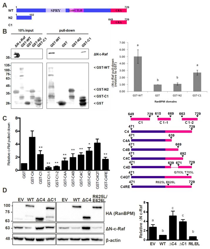
Int. J. Mol. Sci. 2019, 20, 934                                                                      2 of 20

appears to have a dominant role in the ERK signaling pathway, c-Raf, also known as Raf-1, has been
the most extensively studied and thus is the best characterized Raf kinase [2]. c-Raf activation is
tightly regulated through a complex regulatory process involving phosphorylation/dephosphorylation
events, translocation to the membrane, and subsequent homo- or heterodimerization [2]. During
the inactive state, c-Raf is held in a closed conformation by the N-terminal regulatory region folding
over the C-terminal catalytic domain, with the 14-3-3 dimer stabilizing the conformation by binding
phosphorylated S259 (pS259) of the N-terminal, and pS621 of the C-terminal [2]. Activation is initiated
by pS259 dephosphorylation, releasing 14-3-3 from the N-terminal and revealing the Ras binding sites
allowing for Ras binding and recruitment to the plasma membrane [5,7]. The activation segment of
CR3 is then phosphorylated, specifically at S338, to achieve full kinase activation with Raf homo- or
heterodimerization, leading to its subsequent interaction with MEK [8,9]. Dimerization is required for
Raf activation. Heterodimerization of c-Raf and B-Raf was demonstrated to occur following mitogen
activation, while A-Raf weakly dimerizes with B-Raf [8,10]. In addition, the interaction between Heat
Shock Protein 90 (Hsp90) and c-Raf is essential to c-Raf stability and its activity as a signal transducer
within the ERK signaling pathway [11,12]. c-Raf stability is also regulated through ubiquitination by
CHIP (C-terminus of constitutive heat shock protein (Hsc) 70-interacting protein), a highly conserved
E3 ubiquitin ligase, which associates with the molecular chaperone proteins Hsc70–Hsp70 and Hsp90
causing client proteins to be ubiquitinated and subsequently degraded via the proteasome [13,14].
X-linked inhibitor of apoptosis proteins (XIAP), another E3 ubiquitin ligase, has also been found to
interfere with c-Raf stability, promoting ubiquitination through Hsp90-mediated CHIP, independent
of its own E3 ligase activity [15].
      Interestingly, incidences of CHIP-independent modes of ubiquitination of c-Raf have been
documented. For successful activation of C-Raf, S621 must be autophosphorylated in order to allow
for correct folding and stability, as pS621 is necessary to bind 14-3-3 to its C-terminal [16]. Without
the phosphorylation of S621, c-Raf is effectively kinase-dead and is degraded by the proteasome [17].
However, degradation is not exclusively regulated by CHIP, as siRNA knockdown of CHIP did
not yield altered levels of kinase-dead c-Raf [17]. Treatment with the oxidative glucose metabolite
methylglyoxal and abolishing extracellular adhesion has been shown to cause degradation of c-Raf
through the ubiquitin-proteasome system [18,19]. However, the E3 ubiquitin ligase was not identified
in either case.
      A lesser-known regulator of c-Raf stability is the protein RanBPM (Ran-Binding Protein M), which
was initially identified to bind the c-Raf kinase domain in a yeast two-hybrid analysis [20]. Subsequently,
our studies showed that RanBPM and c-Raf form a complex and that RanBPM downregulates c-Raf at
the protein level [21]. RanBPM also had a repressive effect on ERK phosphorylation, suggesting that,
through its effect on c-Raf, RanBPM is an inhibitor of the ERK pathway [21]. However, the mechanism
by which RanBPM downregulates c-Raf remains unknown. RanBPM has been previously been
implicated in the regulation of several cancer pathways and has been suggested to have tumour
suppressive functions [22,23]. While initially studied in isolation, it has now become evident that
RanBPM is part of a large complex called the C-terminal to LisH (CTLH) complex that has E3 ligase
activity [23,24]. However, the targets and activities of the CTLH complex are still largely unknown.
In this study, we show that downregulation or knockout of CTLH complex subunits RanBPM and
RMND5A (Required for Meiotic Nuclear Division 5A) lead to increased cell proliferation and that
RanBPM downregulation promotes tumour formation in a mouse tumour model. We show that
RanBPM binds c-Raf directly and that this interaction is dependent on the RanBPM C-terminal CRA
(CT11-RanBPM) domain. The stability of all three Raf kinases (A-Raf, B-Raf and c-Raf) was found to
be dependent on CTLH complex member RMND5A expression and c-Raf ubiquitination is regulated
in a CTLH complex-dependent manner. Overall, this study uncovers a novel regulation of c-Raf by the
CTLH complex that may contribute to the tumour-suppressive activity of this novel E3 ligase complex.
Int. J. Mol. Sci. 2019, 20, 934                                                                                                    3 of 20

2. Results

2.1. CTLH Complex Members Regulate c-Raf Levels and Cell Growth
         Int. J. Mol. Sci. 2019, 19, x                                                                                   3 of 20
     We previously found that RanBPM forms a complex with c-Raf and that RanBPM downregulation
results 2.1.  CTLH Complex
         in increased        Members
                         c-Raf         Regulate[21].
                                 expression     c-Raf Levels and Cell Growth
                                                      To determine      whether this effect was mediated through
the CTLH complex,        as opposed
               We previously    found to   a regulation
                                         that  RanBPM formsby RanBPM       alone,
                                                                   a complex      we tested
                                                                                 with          c-Rafthat
                                                                                       c-Raf and      expression
                                                                                                           RanBPM in cells
        downregulation     results in increased   c-Raf expression   [21]. To
lacking RMND5A, a CTLH complex member containing a conserved RING domain [25].determine   whether  this  effect wasWestern
        mediated through the CTLH complex, as opposed to a regulation by RanBPM alone, we tested c-Raf
blot analysis of RMND5A CRISPR knockout HEK293 cell extracts showed over sixfold increase of
        expression in cells lacking RMND5A, a CTLH complex member containing a conserved RING
endogenous c-Raf protein levels compared to wild-type HEK-293 (Figure 1A), which were decreased
        domain [25]. Western blot analysis of RMND5A CRISPR knockout HEK293 cell extracts showed over
by nearly    fivefold
        sixfold        upon
                 increase     re-expression
                          of endogenous        of protein
                                            c-Raf  RMND5A. levelsThese  results
                                                                  compared       demonstrate
                                                                              to wild-type       that endogenous
                                                                                            HEK-293   (Figure 1A), c-Raf
proteinwhich
         levelswere
                  are decreased
                      affected by   RMND5A       expression,    and   therefore  imply   that the CTLH
                                 by nearly fivefold upon re-expression of RMND5A. These results demonstratecomplex plays
a role in the regulation of endogenous c-Raf protein levels.
        that  endogenous   c-Raf protein  levels are affected by  RMND5A     expression,  and therefore  imply  that
         the CTLH complex plays a role in the regulation of endogenous c-Raf protein levels.

              1. CTLH
      Figure Figure      complex
                    1. CTLH         members
                              complex  members   RMND5A
                                                   RMND5A and      RanBPM
                                                              and RanBPM       regulate
                                                                             regulate     c-Raf
                                                                                      c-Raf levelslevels  and
                                                                                                    and cell    cell proliferation.
                                                                                                             proliferation.
             (A) RMND5A
      (A) RMND5A             regulates
                        regulates      endogenous c-Raf
                                     endogenous        c-Rafprotein  levels.
                                                               protein       WholeWhole
                                                                         levels.    cell extracts
                                                                                              cell from   wild-type
                                                                                                     extracts    from(WT)
                                                                                                                        wild-type
             HEK293 cells
      (WT) HEK293      cellsand
                             and CRISPR
                                   CRISPR KO KO RMND5A
                                                    RMND5A HEK293   cells untransfected
                                                                 HEK293                  (−) or transfected
                                                                             cells untransfected       (−) orwith   pCGN- with
                                                                                                                transfected
             HA-RMND5A (+) were prepared and analyzed by Western blot. The top shows a representative
      pCGN-HA-RMND5A           (+) were prepared and analyzed by Western blot. The top shows a representative
             analysis using c-Raf, HA (hemagglutinin), RMND5A, and β-actin antibodies to detect endogenous c-
      analysis using c-Raf, HA (hemagglutinin), RMND5A, and β-actin antibodies to detect endogenous
             Raf, exogenous HA-RMND5A, endogenous RMND5A, and β-actin, respectively. Below, relative
      c-Raf, exogenous
             endogenous HA-RMND5A,
                           c-Raf protein levels endogenous      RMND5A,
                                                  were quantified              and β-actin,
                                                                    by normalizing    c-Raf torespectively.       Below, relative
                                                                                                β-actin, and comparing
      endogenous
             valuesc-Raf   proteinHEK293
                     to wild-type   levels were
                                              whenquantified    by normalizing
                                                     set to a value                c-Raf toare
                                                                     of 1. Quantifications       shownand
                                                                                              β-actin,    withcomparing
                                                                                                                 error bars values
      to wild-type  HEK293
             indicating  SD. pwhen
                                < 0.05set
                                       (*).to(B)
                                               a value
                                                 RMND5A of 1.regulates
                                                              Quantifications    are shown
                                                                        ERK signaling.   Whole withcellerror barsfrom
                                                                                                        extracts    indicating
                                                                                                                       WT      SD.
                HEK293 cells and CRISPR KO RMND5A HEK293 cells were analyzed by Western blot for ERK and
                MEK phosphorylation. The same extracts were run on two different gels and equal loading was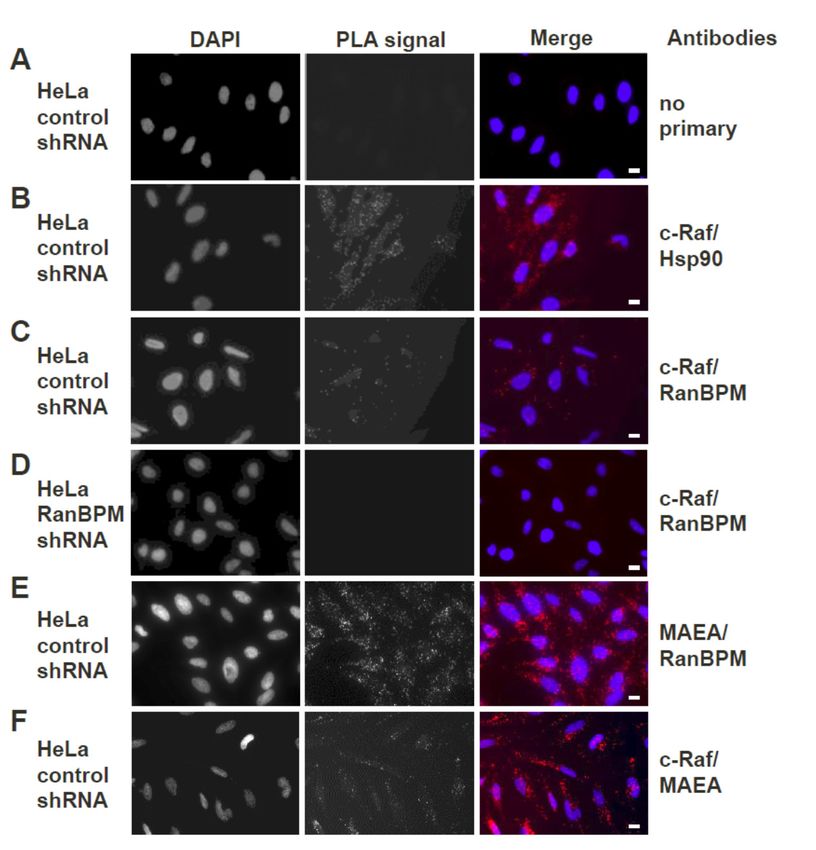
Int. J. Mol. Sci. 2019, 20, 934                                                                                4 of 20

      p < 0.05 (*). (B) RMND5A regulates ERK signaling. Whole cell extracts from WT HEK293 cells and
      CRISPR KO RMND5A HEK293 cells were analyzed by Western blot for ERK and MEK phosphorylation.
      The same extracts were run on two different gels and equal loading was assessed for both analyses
      using total ERK and total MEK and a tubulin antibody. (C) RMND5A knockout HEK293 cells show
      increased proliferation. Growth rates for HEK293 control (WT, blue) and three different RMND5A
      CRISPR KO cell lines (clones #1, red, 3, green and 14, purple) were assessed for six days. Data represents
      average cell number from at least three experiments with error bars indicating SEM. p < 0.05 (*), p < 0.01
      (**), p < 0.001 (***); (D) c-Raf expression is increased in primary RanBPM knockout mouse embryonic
      fibroblasts (MEFs). MEFs were isolated from RanBPM WT, and knockout (KO) embryos at D13.5. In the
      top, whole cell extracts were analyzed by Western blot with antibodies to RanBPM, c-Raf and β-actin.
      Below, quantification of relative amounts of c-Raf normalized to β-actin. Results are averaged from 13
      paired MEFs samples from five different sets of embryos with error bars indicating SEM. p < 0.05 (*);
      (E) RanBPM knockout MEFs proliferate faster than WT MEFs. Growth rates for primary wildtype (WT,
      blue) and RanBPM knockout (KO, red) MEFs were assessed for five days. Data represents average
      cell number from three independent experiments performed in triplicate. Error bars represent SEM.
      p < 0.01 (**), p < 0.001 (***).

     As RanBPM downregulation was previously reported to result in increased cellular
proliferation [21,26], we evaluated whether the loss of RMND5A could also confer similar properties.
Comparison of growth curves of wild-type (WT) and three different RMND5A CRISPR knockout
HEK293 clonal derivatives showed that control cells slowed down after four days, whereas cells lacking
RMND5A proliferated markedly faster starting at day 3 (Figure 1C). We also determined that, similar
to the loss of RanBPM that we previously showed induced MEK and ERK phosphorylation [21],
the knockout of RMND5A resulted in increased MEK and ERK phosphorylation (Figure 1B).
Interestingly, we found that primary mouse embryonic fibroblasts (MEFs) isolated from RanBPM KO
mice also displayed increased c-Raf expression and increased proliferation (Figure 1D,E), suggesting
that the consequences of the loss of RanBPM/CTLH complex are not restricted to immortalized cells.

2.2. RanBPM Expression Prevents Tumour Formation in Mouse Models
      Our observations that RanBPM downregulation promotes c-Raf expression and ERK activation [21]
suggested that loss of RanBPM function could promote tumour formation in vivo. Moreover,
downregulation of RanBPM in Hela and HCT116 cells causes extensive changes in the expression of
several genes implicated in oncogenesis [27]. In particular, overexpression of RON (Recepteur d’origine
nantais) kinase, L1 cell adhesion molecule (L1CAM), ELF3 (E74-like factor 3), transglutaminase 2 (TG2)
(all increased in RanBPM shRNA cells [27]) have all been reported in various tumour types and were
shown to be directly implicated in cancer development [28–31]. Thus, loss of RanBPM affects several
pathways which collectively promote many aspects of tumorigenesis.
      We tested whether RanBPM downregulation could promote tumour formation in a xenograft
model. For this assay, we generated a pool of early passage HEK293 cells stably expressing
RanBPM shRNA or control shRNA (Figure 2A). HEK293 cells are immortalized with Adenovirus
5 E1A expression but exhibit weak tumorigenicity [32]. RanBPM shRNA HEK293 injected into
NOD/SCID/gamma (NSG) mice caused a marked and statistically significant increase in tumour
volume over control cells (Figure 2C). Tumours also appeared earlier (day 17, versus day 31) and were
on average twentyfold larger (220 mm3 versus 11 mm3 ) than those observed with control cells at day 35.
These results suggest that RanBPM downregulation can enhance tumorigenic properties of HEK293
cells in this model. To demonstrate that these effects were not due to off-target effects of the RanBPM
shRNA, we generated HEK293 RanBPM shRNA cells with stably integrated Tetracyclin (Tet)-off
vector pBIG-RanBPM in which RanBPM can be re-expressed upon removal of Tet/doxycycline (Dox)
(Figure 2A, lanes 3,4). We verified that re-expression of RanBPM reduced ERK phosphorylation to
wild-type levels (Figure 2B). Interestingly, RanBPM shRNA cells also showed an upregulation of A-Raf
and B-Raf protein levels, which specifically decreased upon re-expression of RanBPM, suggesting that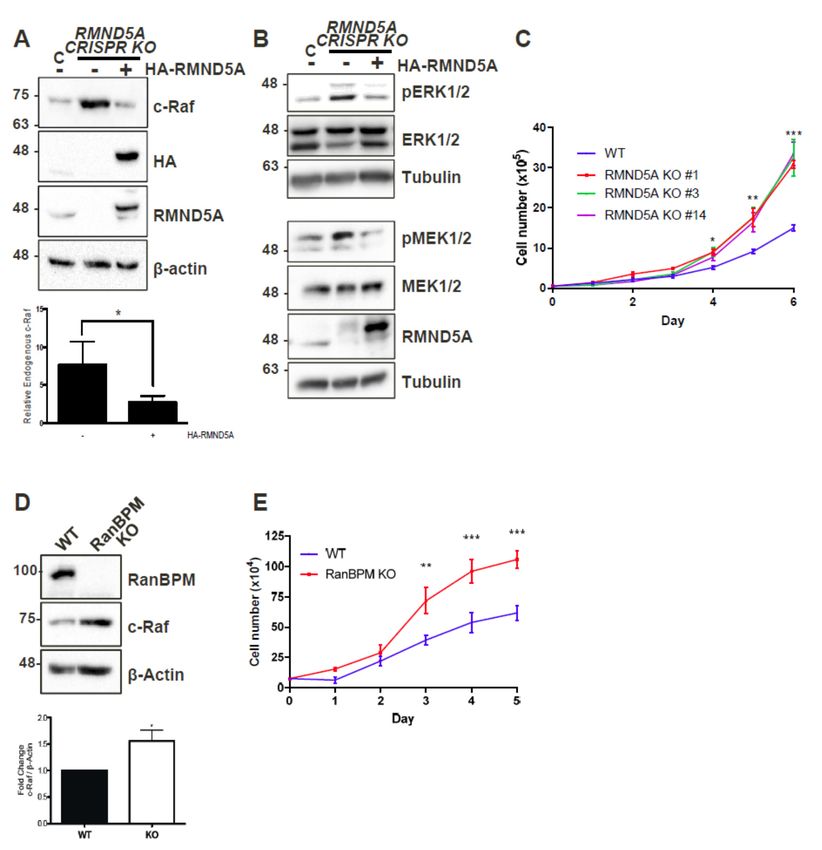
Int. J. Mol. Sci. 2019, 20, 934 5 of 20
ERK pathway activation could result from all three Raf kinases activation in these cells. pBIG RanBPM
shRNA cells were injected into NSG mice. Mice fed with Dox-containing chow developed tumours,
whereas those fed with regular chow did not, demonstrating that RanBPM re-expression effectively
prevents
Int.tumour formation
J. Mol. Sci. 2019, 19, x (Figure 2D). 5 of 20
2. Downregulation
Figure Figure 2. Downregulation of ofRanBPM
RanBPM promotes tumour
promotes tumour formation
formation in NOD/SCID/gamma
in NOD/SCID/gamma mice. (A) mice.
(A) re-expression
re-expression ofofRanBPM
RanBPM in in HEK293 cellsvia
HEK293 cells viaTet-off
Tet-off pBIG
pBIG expression
expression vector.
vector. HEK293 HEK293
pool ofpool
cells of cells
stably expressing
stably expressing RanBPM RanBPM
shRNA shRNA were
were transfectedwith
transfected with pBIG-RanBPM
pBIG-RanBPM WT WT (si-mt) andand
(si-mt) maintained
maintained in
in media
media with with 2 μg/mL
2 µg/mL Tetracycline
Tetracycline andand250250 μg/mLhygromycin
µg/mL hygromycin toto select for for
select integration of the of
integration pBIGthe pBIG
vector. Following selection, cells were either maintained (+) in Tetracyclin-containing media, or
vector. Following selection, cells were either maintained (+) in Tetracyclin-containing media, or cultured
cultured in absence of Tetracyclin (−) for 24 h to allow induction of RanBPM. Tetracyclin removal
in absence of Tetracyclin (−) for 24 h to allow induction of RanBPM. Tetracyclin removal leads to
leads to re-expression of RanBPM (lane 4); (B) ERK pathway activation in RanBPM shRNA cells.
re-expression
Samplesof RanBPM
shown (laneanalyzed
in (A) were 4); (B) ERK
for ERKpathway activation
phosphorylation and in RanBPM
A-Raf and B-RafshRNA cells.The
expression. Samples
shown Western
in (A) were analyzed for ERK phosphorylation and A-Raf and B-Raf expression.
blot was analyzed with the indicated antibodies; (C) injections with HEK293 control and The Western
blot was analyzed
RanBPM with the
shRNA poolsindicated antibodies;
were injected (C) injections
subcutaneously withflank
in the HEK293 control
of 6–8 weeksandold RanBPM
shRNANOD/SCID/gamma.
pools were injected Tumour measurements
subcutaneously wereflank
in the takenoftwice
6–8per weekold
weeks andNOD/SCID/gamma.
a digital caliper was usedTumour
to measure
measurements Length
were taken× Width
twice× per
Depth of the
week tumour
and uponcaliper
a digital excisionwas
in order
usedtotocalculate
measure volume.
Lengthn =× 7, Width
error bars represent SEM; (D) injections with HEK293 RanBPM shRNA pool of cells stably re-
× Depth of the tumour upon excision in order to calculate volume. n = 7, error bars represent SEM;
expressing RanBPM via pBIG Tet-off expression system (see C, lanes 3,4). Mice were fed chow
(D) injections with HEK293 RanBPM shRNA pool of cells stably re-expressing RanBPM via pBIG Tet-off
containing Dox (purple line) or regular chow (green line). n = 6, error bars represent SEM. p < 0.05 (*),
expression system
p < 0.01 (see bars
(**). Error C, lanes 3,4). Mice
are included were
for all datafed chow
points butcontaining
may not be Dox (purple
visible line) orthan
when smaller regular
line chow
size. n = 6, error bars represent SEM. p < 0.05 (*), p < 0.01 (**). Error bars are included for all
(green line).
data points but may not be visible when smaller than line size.
2.3. Both Endogenous RanBPM and MAEA Form a Complex with c-Raf in Situ
2.3. Both Endogenous RanBPM and MAEA Form a Complex with c-Raf In Situ
Previous co-immunoprecipitation and pull-down experiments in our laboratory have
demonstrated
Previous that RanBPM and c-Raf
co-immunoprecipitation andexist togetherexperiments
pull-down in a complex in
[21]. Since
our this was have
laboratory shown using
demonstrated
that RanBPM and c-Raf exist together in a complex [21]. Since this was shown using ectopically the
ectopically expressed protein constructs, we sought to confirm that the complex occurs with expressed
endogenous proteins in vivo. The in situ Proximity Ligation Assay (PLA), which allows visualization
protein constructs, we sought to confirm that the complex occurs with the endogenous proteins in vivo.
of protein–protein interactions in cells using fluorescence microscopy [33], was thus used to visualize
The in situ Proximity between
the interaction Ligationendogenous
Assay (PLA), CTLHwhich allows
complex visualization
members MAEA of protein–protein
(Macrophage interactions
erythroblast
in cellsattacher)
using fluorescence
and RanBPM and microscopy
c-Raf in HeLa[33],
cells.was thus used to visualize the interaction between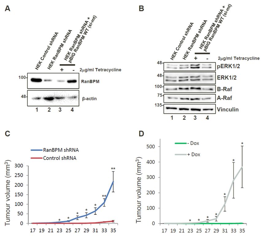
Int. J. Mol. Sci. 2019, 20, 934 6 of 20
endogenous CTLH complex members MAEA (Macrophage erythroblast attacher) and RanBPM and
c-Raf inInt.
HeLa cells.
J. Mol. Sci. 2019, 19, x 6 of 20
Stable HeLa control or RanBPM shRNA (clone 2–7), previously generated in our laboratory [34],
were used inStablethis HeLa
experiment.
control orAs a control,
RanBPM shRNA HeLa cells
(clone were
2–7), incubated
previously without
generated primary
in our laboratoryantibodies
[34],
(Figure were
3A). used
Given in that
this experiment.
c-Raf is known As a control, HeLawith
to interact cellsHsp90
were incubated without control
[35], a positive primary was
antibodies
included
(Figure
where HeLa 3A). Given
control that c-Raf
cells were is known
incubated to interact
with antibodieswithagainst
Hsp90 [35],
c-Rafa and
positive control
Hsp90. Thiswas
didincluded
expectedly
where HeLa control cells were incubated with antibodies against c-Raf and Hsp90. This did
produce fluorescent dots representing interactions (Figure 3B). In HeLa control cells in which antibodies
expectedly produce fluorescent dots representing interactions (Figure 3B). In HeLa control cells in
against c-Raf and RanBPM were included for the assay, fluorescent dots were observed, confirming
which antibodies against c-Raf and RanBPM were included for the assay, fluorescent dots were
that the observed,
two proteins are found
confirming that together in a complex
the two proteins in vivo
are found (Figure
together 3C). To in
in a complex confirm the specificity
vivo (Figure 3C). To of
the RanBPM
confirm the specificity of the RanBPM primary antibody, another control was included wherecells
primary antibody, another control was included where HeLa RanBPM shRNA HeLawere
incubated with primary
RanBPM shRNA cells antibodies againstwith
were incubated RanBPM
primaryand c-Raf (Figure
antibodies against3D). As expected,
RanBPM and c-Raf fluorescent
(Figure
3D). As expected,
dots representing fluorescent
interactions weredotsnot representing
observed ininteractions
absence ofwere not observed
RanBPM. To confirmin absence
that c-Rafof is
RanBPM.
interacting with the To confirm that c-Raf we
CTLH complex, is interacting
repeated with the CTLH complex,
the experiment with CTLHwe repeated
complex themember
experimentMAEA.
with CTLH complex member MAEA. A positive control using primary antibodies against MAEA
A positive control using primary antibodies against MAEA and RanBPM presented numerous dots,
and RanBPM presented numerous dots, consistent with these two proteins being present in a
consistent with these two proteins being present in a complex (Figure 3E). MAEA and c-Raf antibodies
complex (Figure 3E). MAEA and c-Raf antibodies also yielded fluorescent dots (although in
also yielded fluorescent
noticeably dots (although
fewer numbers than the MAEAin noticeably
and RanBPM)fewer numbers
indicative of than theproximity
the close MAEA and of theRanBPM)
two
indicative
proteins (Figure 3F), suggesting that endogenous c-Raf associates with the CTLH complex as a wholec-Raf
of the close proximity of the two proteins (Figure 3F), suggesting that endogenous
associates with the CTLH complex as a whole in vivo.
in vivo.
Endogenous
Figure 3.Figure RanBPM
3. Endogenous andand
RanBPM c-Raf interaction
c-Raf interactionininHeLa
HeLa cells usingPLA.
cells using PLA. Duolink
Duolink II proximity
II proximity
ligation ligation
assay (PLA)
assaywas performed
(PLA) in: (A)in:
was performed control shRNA
(A) control HeLa HeLa
shRNA cells, without the addition
cells, without of primary
the addition of
primary
antibodies antibodies
(negative (negative
control); control);
(B) control (B) control
shRNA HeLashRNA
cells,HeLa
with cells,
Hsp90 with
andHsp90
c-Rafand c-Raf primary
primary antibodies
(positiveantibodies
control); (positive
(C) controlcontrol);
shRNA (C) HeLa
controlcells,
shRNA HeLa
using cells,
c-Raf and using
RanBPMc-Raf and RanBPM
primary primary (D).
antibodies;
antibodies; (D). HeLa RanBPM shRNA cells, with c-Raf and RanBPM primary antibodies (negative
HeLa RanBPM shRNA cells, with c-Raf and RanBPM primary antibodies (negative control); (E) control
control); (E) control shRNA HeLa cells, using MAEA (Macrophage erythroblast attacher) and
shRNA HeLa cells, using MAEA (Macrophage erythroblast attacher) and RanBPM primary antibodies
RanBPM primary antibodies (positive control). (F) Control shRNA HeLa cells, using c-Raf and MAEA
(positiveprimary
control). (F) Control shRNA HeLa cells, using c-Raf and MAEA primary antibodies. The DAPI
antibodies. The DAPI filter was used to visualize the nuclei, while the Cyanine 3 (Cy3) filter
filter was
was used tovisualize
used to thePLA
visualize the nuclei,
dotswhile the Cyanine
representing 3 (Cy3) filter
protein–protein was used
interactions. to visualize
Representative the PLA
images
dots representing
from one ofprotein–protein
three independentinteractions.
experiments areRepresentative images
shown. Scale bars, from one of three independent
10 μm.
experiments are shown. Scale bars, 10 µm.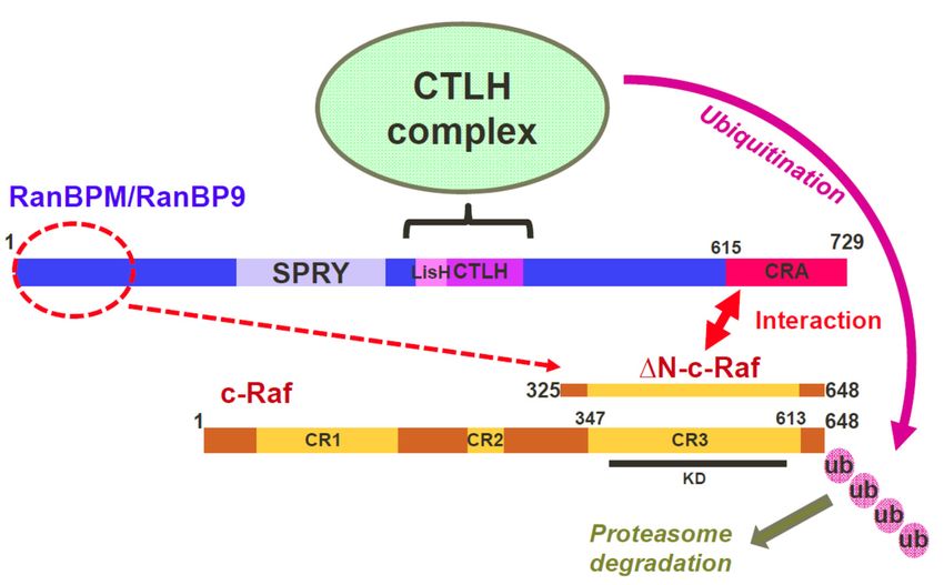
Int. J. Mol. Sci. 2019, 20, 934 7 of 20
2.4. The N-Terminus, CRA and LisH/CTLH Domains of RanBPM Are Required for c-Raf Downregulation
Given that RanBPM contains a number of conserved domains, we sought to determine which
regions of RanBPM were necessary for downregulation of c-Raf. We used a series of RanBPM deletion
mutant constructs (Figure 4A) sub-cloned into the mammalian expression vector pCMV-HA [34,36],
to test their effects on levels of c-Raf. ∆N-c-Raf, a constitutively active construct of c-Raf containing
only amino acids 325–648 [9], was used instead of full-length c-Raf because RanBPM has been shown
to have a greater effect on activated c-Raf [21]. Since RanBPM is able to dimerize [37], we performed
the experiments in HeLa RanBPM shRNA cells to avoid dimerization between mutant RanBPM
and endogenous WT RanBPM monomers. As previously reported [21], WT RanBPM was able to
significantly downregulate ∆N-c-Raf compared to the levels of ∆N-c-Raf seen in the pCMV-HA control
(Figure 4B). The RanBPM deletion mutant ∆212, in which the SPRY domain is deleted, was also able to
downregulate ∆N-c-Raf, indicating that the SPRY domain is not required for c-Raf destabilization.
Next, we tested the effect of N-terminal deletions of RanBPM. We previously reported that ∆N2
RanBPM is expressed at lower levels than WT RanBPM [36], which made the results difficult to
evaluate since lower expression levels may have accounted for the inefficiency of this mutant to
regulate ∆N-c-Raf. However, even in experiments where higher levels of ∆N2 RanBPM were obtained,
∆N2 RanBPM was unable to effectively downregulate ∆N-c-Raf (Figure 4B). Therefore, ∆N2 RanBPM
appeared to have lost its ability to downregulate ∆N-c-Raf, implying that the N-terminus of RanBPM
is required for its effect on c-Raf.
The ∆360 and ∆C4 RanBPM mutants also did not effectively downregulate ∆N-c-Raf, as ∆N-c-Raf
expression levels were significantly higher than those seen in response to WT RanBPM, and not
significantly different than those seen in the pCMV-HA control (Figure 4B). This occurred despite
the fact that expression of these mutants was noticeably higher than that of WT RanBPM. These
results suggest that the LisH/CTLH domains and the C-terminus of RanBPM play a role in c-Raf
downregulation. However, considering that the ∆C4 deletion removes a very large portion of RanBPM,
we repeated the experiment using a construct harboring only a deletion of the CRA domain, namely
the ∆C1 RanBPM construct. Like ∆C4, ∆C1 was unable to downregulate ∆N-c-Raf (Figure 4C).
This implies that, within the C-terminus of RanBPM, it is specifically the CRA domain that is needed
for c-Raf downregulation.
Altogether, these results demonstrate that the N-terminus, LisH/CTLH and CRA domains are
required for c-Raf destabilization, since loss of any of these regions render RanBPM unable to effectively
downregulate c-Raf.
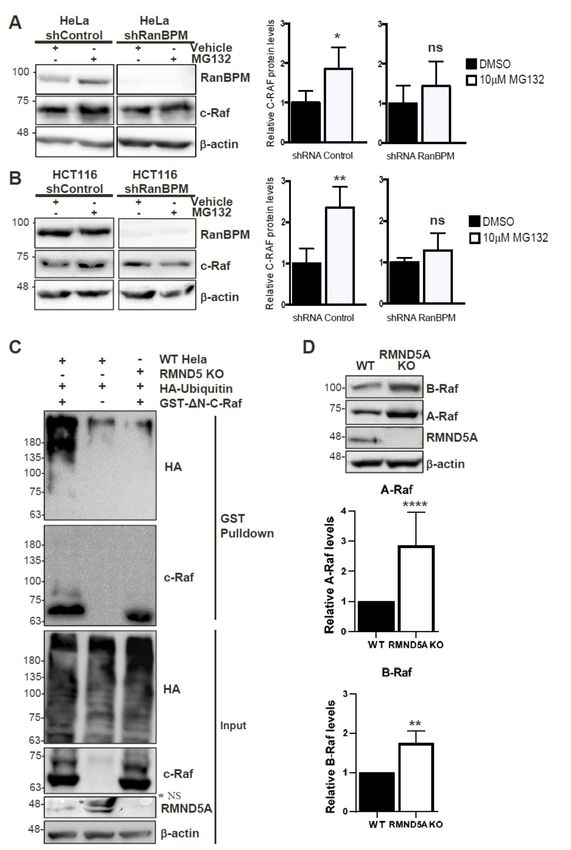
2.5. The CRA Domain of RanBPM Is Required for Interaction with c-Raf
Next, we aimed to determine the RanBPM domain(s) required for interaction with c-Raf.
To accomplish this, we co-transfected either GST or GST-∆N-c-Raf with RanBPM deletion mutants
in RanBPM shRNA HeLa cells and performed GST pull-down assays. Due to its poor stability,
∆N2 RanBPM was not tested as we were unable to obtain sufficient levels of the protein to detect
it in this type of assay. As anticipated based on previous studies [21], GST-∆N-c-Raf was able to
successfully pull-down WT RanBPM (Figure 4D). Both ∆212 RanBPM and ∆360 RanBPM associated
with ∆N-c-Raf similar to WT, indicating that that the SPRY and LisH/CTLH domains are not required
for the interaction between RanBPM and c-Raf (Figure 4D,E). However, ∆C1 RanBPM was not able to
effectively interact with ∆N-c-Raf, showing a twofold decrease compared to WT RanBPM (Figure 4D).
Thus, the data suggest that, of the domains tested here, the CRA domain is the only one required for
the interaction between RanBPM and c-Raf.Int. J. Mol. Sci. 2019, 20, 934 8 of 20
Int. J. Mol. Sci. 2019, 19, x 8 of 20
Figure 4. Analysis of RanBPM
Figure 4. Analysis of RanBPMdomains
domains that
thatcontrol C-Rafstability.
control C-Raf stability. (A) schematic
(A) schematic representation
representation of of
RanBPM mutants.
RanBPM mutants. (B) and (B)(C)
andWestern
(C) Western blot
blot analyses of
analyses ofHeLa
HeLa RanBPM
RanBPMshRNA cells transfected
shRNA with
cells transfected with
pEBG-GST-ΔN-c-Raf and either pCMV-HA (empty vector), pCMV-HA-WT-RanBPM or pCMV-HA
pEBG-GST-∆N-c-Raf and either pCMV-HA (empty vector), pCMV-HA-WT-RanBPM or pCMV-HA
RanBPM mutant constructs as indicated. c-Raf and HA antibodies were used to detect the levels of
RanBPM mutant constructs as indicated. c-Raf and HA antibodies were used to detect the levels of
ΔN-c-Raf and RanBPM, respectively. β-actin was used as a loading control. A representative Western
∆N-c-Raf and RanBPM, respectively. β-actinofwas
blot is shown (top) and quantifications c-Rafused
levelsasarea shown
loading control.
(bottom A representative
graph) with error bars Western
blot is shown (top) and quantifications of c-Raf levels are shown (bottom graph)
indicating SEM (n = 5). Deletion of RanBPM C-terminal domain (ΔC1) impairs RanBPM with error bars
interaction
with GST-ΔN-c-Raf. (D) and (E) GST-Pull-down assays. HeLa RanBPM shRNA
indicating SEM (n = 5). Deletion of RanBPM C-terminal domain (∆C1) impairs RanBPM interaction cells were transfected
with pEBG-ΔN-c-Raf and either pCMV-HA (empty vector), pCMV-HA-WT-RanBPM or pCMV-HA
with GST-∆N-c-Raf. (D) and (E) GST-Pull-down assays. HeLa RanBPM shRNA cells were transfected
RanBPM mutant constructs. ΔN-c-Raf was pulled down through binding to glutathione-sepharose
with pEBG-∆N-c-Raf and either
beads and interaction pCMV-HA
of RanBPM (empty
WT and mutants vector),
with pCMV-HA-WT-RanBPM
GST-ΔN-c-Raf assessed by Western blot or withpCMV-HA
RanBPM mutant
an HA antibody. Below:∆N-c-Raf
constructs. waswere
Quantifications pulled downbythrough
performed binding
normalizing RanBPM to mutant
glutathione-sepharose
levels to
beads andpulled-down
interactionGST or GST-ΔN-c-Raf
of RanBPM WT and andmutants
statistical with
analyses were performedassessed
GST-∆N-c-Raf (n = 4–7, SEM shown). blot with
by Western
Different letters are statistically different (p < 0.05).
an HA antibody. Below: Quantifications were performed by normalizing RanBPM mutant levels to
pulled-down GST or GST-∆N-c-Raf and statistical analyses were performed (n = 4–7, SEM shown).
Different letters are statistically different (p < 0.05).
2.6. RanBPM Interacts Directly with c-Raf through the CRA Domain
To confirm the interaction between the CRA domain of RanBPM and c-Raf and determine whether
the interaction is direct, we performed pull-down experiments using bacterially expressed mammalian
c-Raf and RanBPM. In addition to full-length RanBPM, we tested RanBPM C1 (encoding only the CRAInt. J. Mol. Sci. 2019, 19, x 9 of 20
Int. J. Mol. Sci. 2019,
2.6. 20, 934 Interacts Directly with c-Raf through the CRA Domain
RanBPM 9 of 20
To confirm the interaction between the CRA domain of RanBPM and c-Raf and determine
whether the interaction is direct, we performed pull-down experiments using bacterially expressed
domain) and mammalian
the N2 region (Figure
c-Raf and RanBPM. 5A), whichtowas
In addition unexamined
full-length RanBPM, wein theRanBPM
tested mammalian cell-based
C1 (encoding GST
pull-down assays.
only the CRA domain) and the N2 region (Figure 5A), which was unexamined in the mammalian
cell-based GST pull-down assays.
Figure 5. RanBPM Figure C-terminal
5. RanBPM C-terminal CRA domain
CRA domain directly
directly interacts with
interacts ∆N-c-Raf
with ΔN-c-Raf and and
is necessary for C- for C-Raf
is necessary
Raf regulation. (A) diagram of WT RanBPM, N2 domain and C1 domain cloned into the bacterial
regulation. (A) diagram of WT RanBPM, N2 domain and C1 domain cloned into the bacterial expression
expression vector pGEX-4T-1; (B) Left, Western blot analysis of GST pull-down assays for N c-Raf
vector pGEX-4T-1; (B) Left,
performed using Western blot analysis
GST, GST-WT-RanBPM, of GST pull-down
GST-N2-domain assays for E.Ncoli
and GST-C1-domain c-Raf performed
extracts. A using
GST, GST-WT-RanBPM, GST-N2-domain and GST-C1-domain E. coli extracts. A representative image
representative image is shown. Right, pull down assays experiments were quantified by normalizing
ΔN-c-Raf levels to pulled-down GST, GST-WT-RanBPM, GST-N2 or GST-C1 and statistical analyses
is shown. Right, pull down assays experiments were quantified by normalizing ∆N-c-Raf levels to
were performed (n = 6, error bar indicates SEM) with different letters indicating statistical difference
pulled-down GST, GST-WT-RanBPM,
(p < 0.05); GST-N2
(C) analysis of RanBPM CRAor mutants
GST-C1usingand instatistical analyses
vitro pull-down. were
Right, performed (n = 6,
schematic
error bar indicates SEM) with different letters indicating statistical difference (p < 0.05); (C) analysis
of RanBPM CRA mutants using in vitro pull-down. Right, schematic representation of the mutants
analyzed. Left, Quantifications of pull down experiments performed with the CRA mutants as
described in (B). Relative ∆N-C-Raf protein levels were quantified by normalizing ∆N-C-Raf to the
GST-fusion protein product, and comparing values to GST when set to a value of 1. Quantifications are
shown with error bars indicating SD (n = 3–5). Statistical difference with respect to GST-C4 is indicated,
* p < 0.05; ** p < 0.01; (D) analysis of RanBPM CRA domain mutants in mammalian cells. HeLa RanBPM
shRNA cells were transfected with pEBG-GST-∆N-c-Raf and either pCMV-HA empty vector, RanBPM
WT, RanBPM-∆C4, RanBPM-∆C1, or RanBPM-R625L E626L, and whole cell extracts were prepared
24 h post-transfection and analyzed by Western blot. HA, c-Raf and β-actin antibodies were used to
detect HA-RanBPM constructs, ∆N-c-Raf and β-actin proteins, respectively. Right, relative ∆N-c-Raf
protein levels were quantified by normalizing ∆N-c-Raf to β-actin, and comparing values to HA-WT
when set to a value of 1. Different letters are statistically different (p < 0.05). Error bars indicate SD
(n = 4).Int. J. Mol. Sci. 2019, 20, 934 10 of 20
GST-RanBPM constructs were purified on glutathione beads and incubated with a crude cell
lysate from E. coli expressing ∆N-c-Raf. Both GST-WT-RanBPM and GST-C1 were able to pull-down
∆N-c-Raf (Figure 5B), indicating that the interaction between RanBPM and c-Raf is direct and that the
CRA domain is sufficient for the interaction. GST-N2 was unable to pull-down ∆N-c-Raf (Figure 5B),
suggesting that the N-terminus of RanBPM is unable to directly interact with c-Raf.
2.7. Analysis of the RanBPM CRA Domain Interaction with c-Raf
To identify sub-domains that might mediate interaction with c-Raf, we examined structural
features of the CRA domain to design partial deletions. As there is no crystal structure of RanBPM or
the CRA domain currently available, a predicted tertiary structure was elucidated by the RAPTORX
online server [38]. The predicted tertiary structure corroborated the previous assumption that
the CRA domain has a high propensity for α-helices, with 6 α-helices spanning the entirety of
the domain [23,39] (Figure S1A). Deletion mutants of the RanBPM CRA domain were created to
identify sub-domains that might mediate interaction with c-Raf (Figure 5C). Deletions were guided
by thepredicted tertiary structure (Figure S1B) and data shown above demonstrating that amino
acids 649–729 of RanBPM, denoted C1, bound directly to ∆N-c-Raf (Figure 5B). Only a portion of
the CRA domain is present in the C1 construct, namely helices IV, V and VI, and a small portion of
helix III. We therefore derived two GST-fusion CRA constructs which contained amino acids 615–669
spanning helices I, II, and III, denoted C1-1, and amino acids 663–729 containing helices IV, V, and VI,
denoted C1-2. GST pull down assays were normalized to the negative control, GST, and compared
to the positive ∆N-c-Raf binding control, GST-C4, which contains amino acids 471–729 of RanBPM.
(Figure 5C, representative experiments shown in Figure S2A). GST-C1 displayed significant interaction
with ∆N-c-Raf, although quantifications showed reduced binding compared to GST-C4, by 2.5-fold.
Both GST-C1-1 and GST-C1-2 appeared to lack protein stability as they bound less ∆N-c-Raf than that
of the GST negative control.
To increase protein stability, CRA domain constructs were expanded to include sequences
N-terminal to the CRA domain (Figure 5C). Similar to the strategy used for the two previous constructs,
CRA deletion mutations were guided by the predicted tertiary structure. We derived an additional
four GST-fusion CRA constructs: C4A which spans amino acids 471–639 and helices I and II; C4B
contains amino acids 471–669 and helices I–III; C4C is comprised of amino acids 471–692 and helices
I–IV; and finally, C4D which includes amino acids 471–639 and 671–729, and helices I–II and IV–VI.
∆N-c-Raf binding was again tested by GST pull down assay and the mutants binding to ∆N-c-Raf
were quantified and compared to that of GST-C4 (Figure 5C and Figure S2B). All mutants showed
decreased binding compared to C4, suggesting that each helix of the CRA domain plays a role in
binding ∆N-c-Raf. As more helices were deleted from the CRA domain, less binding of ∆N-c-Raf was
observed. However, two portions of the CRA domain seemed worth investigating, helices I and II,
and helices V and VI, since deletion of helices I and II (C1), and helices V and VI (C4C) resulted in a
sharp decrease in ∆N-c-Raf binding with the least amount of the CRA domain deleted.
Point mutations were designed to target the center of helices I and II, or helix V, in efforts to
disrupt the helical structures within the CRA domain and the binding interface between CRA and
∆N-c-Raf. Site-directed mutagenesis was used to create C4 with point mutations R625L and E626L
within helix I and II, denoted C4-R625L E626L, or with point mutations Q703L and T705L within helix
V, denoted C4-Q703L T705L. GST-C4-R625L E626L showed a significant loss in ∆N-c-Raf binding with
over threefold decrease compared to C4, whereas GST-C4-Q703L T705L did not disrupt the interaction,
showing only a slight decrease in ∆N-c-Raf binding (Figure 5C and Figure S2C). These results suggest
that the mutation R625L E626L made to helix I and II of the CRA domain, is sufficient to disrupt the
interaction between the CRA domain and ∆N-c-Raf in vitro.
To validate the in vitro binding data, mutant RanBPM CRA domains were cloned into the
mammalian expression vector pCMV, and co-transfected with pEBG-GST ∆N-c-Raf in HeLa RanBPM
shRNA cells. RanBPM- ∆C1 and RanBPM- R625L E626L were chosen to investigate their effect onInt. J. Mol. Sci. 2019, 20, 934 11 of 20
∆N-c-Raf regulation. RanBPM-∆C1 showed significant loss of ∆N-c-Raf regulation, compared to WT
RanBPM, confirming our in vitro binding data (Figure 5D). However, the RanBPM R625L E626L point
mutant maintained the regulation of ∆N-c-Raf protein levels to a level comparable to WT RanBPM,
suggesting that the mutant could still interact with c-Raf in the cellular context. Similarly, the C4C
deletion (693–729) did not affect ∆N-c-Raf regulation when transfected in Hela RanBPM shRNA cells
(Figure S2D). This suggests that the interaction in vivo may be enhanced or stabilized by other domains
of RanBPM or by other members of the CTLH complex.
2.8. c-Raf Stability Is Regulated through the Proteasome and Its Ubiquitination Is Dependent on the
CTLH Complex
Since the CTLH complex has E3 ubiquitin ligase activity [24], we wanted to determine whether
it regulates c-Raf protein stability through the proteasomal degradation pathway. RanBPM shRNA
and control shRNA HeLa, and HCT116 cells were used to assess c-Raf protein levels in conditions of
proteasomal impairment in order to determine whether RanBPM loss of function would affect c-Raf
degradation. All cell lines were treated with either 10 µM of the proteasome inhibitor, MG132,
or with the vehicle, dimethyl sulfoxide (DMSO). HeLa and HCT116 cells expressing RanBPM
shRNA showed minimal accumulation of c-Raf protein levels during proteasomal inhibition, whereas
control shRNA cells exhibited a significant increase of c-Raf expression when compared to their
respective vehicle-treated controls (Figure 6A,B). This suggests that the downregulation of RanBPM
impairs proteasome-dependent degradation of c-Raf. To determine whether this could occur through
ubiquitination of c-Raf by the CTLH complex, we co-transfected control and RMND5A KO HeLa
cells with GST-∆N-c-Raf and HA-Ubiquitin constructs to investigate the involvement of RMND5A in
c-Raf ubiquitination. GST-∆N-c-Raf ubiquitination was observed in control Hela cells but was strongly
reduced in RMND5A KO cells confirming that the CTLH complex regulates c-Raf ubiquitination
(Figure 6C). Taken together, our data suggests that c-Raf ubiquitination and proteasomal degradation
is regulated by the CTLH complex.
2.9. The CTLH Complex Regulation Is Conserved for A- and B-Raf
The c-Raf region that interacts with the RanBPM/CTLH complex comprises the CR3 (Conserved
Region 3) which is the most conserved region between the three Raf isoforms and includes the kinase
domain [2]. Our observation that RanBPM shRNA cells display increased expression of A-Raf and
B-Raf (Figure 2B) prompted us to evaluate whether RMND5A could also regulate the levels of the other
two Raf kinases, A-Raf and B-Raf, which would confirm the involvement of the CTLH complex in the
regulation of all three Raf kinases. Indeed, levels of both A- and B-Raf were increased in RMND5A
KO cells (Figure 6D), with the most pronounced effect on A-Raf, which showed an almost threefold
increase compared to nearly twofold for B-Raf. This effect was not due to an isolated clonal effect,
as all RMND5A CRISPR KO clones obtained showed increased expression of the three Raf kinases
compared to wild-type cells (Figure S3). This suggests that the CTLH complex regulates all three Raf
kinases likely through a similar mechanism.Int. J. Mol. Sci. 2019, 20, 934 12 of 20
Int. J. Mol. Sci. 2019, 19, x 12 of 20
FigureFigure 6. C-Raf
6. C-Raf is regulated
is regulated by theby the proteasome
proteasome throughthe
through theCTLH
CTLH complex.
complex. Non-targeting
Non-targeting shRNA
shRNA
control and shRNA RanBPM cells were treated with 10 μM MG132 or DMSO, as vehicle, for 24 h.
control and shRNA RanBPM cells were treated with 10 µM MG132 or DMSO, as vehicle, for 24 h. RIPA
RIPA buffered whole cell extracts of HeLa (A), and HCT116 (B) were analyzed by Western blot with
buffered whole cell extracts of HeLa (A), and HCT116 (B) were analyzed by Western blot with RanBPM,
RanBPM, c-Raf and β-actin antibodies to detect RanBPM, c-Raf and β-actin proteins, respectively. c-
c-Raf and β-actin antibodies to detect RanBPM, c-Raf and β-actin proteins, respectively. c-Raf protein
Raf protein levels were normalized to β-actin levels. Quantifications of relative c-Raf protein levels
levels were normalized to β-actin levels. Quantifications of relative c-Raf protein levels are shown with
are shown with error bars indicating SD (n = 4). * p < 0.05; ** p < 0.01; ns, no significance. (C) c-Raf is
error bars indicatinginSD
ubiquitinated (n = 4).
a CTLH * p < 0.05; ** p < manner.
complex-dependent 0.01; ns,Hela
no significance. (C) c-Raf
control (WT Hela) is ubiquitinated
and RMND5A KO cellsin a
CTLH were
complex-dependent manner. Helaand/or
transfected with GST-ΔN-c-Raf control (WT Hela) and
HA-Ubiquitin RMND5A
as indicated. GSTKO cells were
pull-downs transfected
performed
with GST-∆N-c-Raf
using whole cell and/or HA-Ubiquitin
extracts were analyzed as indicated.
by Western GSTblotpull-downs performed
with the indicated using whole
antibodies. Inputcell
extractsrepresent
were analyzed
5% of thebyextracts
Westernusedblot
forwith the indicated
pull-down. antibodies.
The asterix Input
(*) indicates represent 5%
a non-specific of (NS).
band the extracts
(D)
used forA-Raf and B-Raf
pull-down. Theexpression is indicates
asterix (*) increased ainnon-specific
RMND5A KO cells.(NS).
band Extracts from HEK293
(D) A-Raf control
and B-Raf and
expression
RMND5A
is increased CRISPR KO
in RMND5A KO cells.
cells Extracts
were analyzed
from HEK293by Western
controlblotand with
RMND5Athe indicated
CRISPR KO antibodies.
cells were
analyzed by Western blot with the indicated antibodies. Quantifications are shown below with error
bar indicating SD, n = 8 for A-Raf, **** p < 0.0001 and n = 3 for B-Raf, ** p < 0.01.Int. J. Mol. Sci. 2019, 19, x 13 of 20
Quantifications are shown below with error bar indicating SD, n = 8 for A-Raf, **** p < 0.0001 and n =
Int. J. Mol. Sci. 2019, 20, 934 13 of 20
3 for B-Raf, ** p < 0.01.
3. Discussion
3. Discussion
The regulation
The regulation ofof Raf
Raf kinases
kinases isiscentral
centraltotothe
thesignaling
signalingactivities ofof
activities thethe
ERKERKpathway
pathway[1–3]. In
[1–3].
this study, we have identified a novel regulatory mechanism of c-Raf protein expression.
In this study, we have identified a novel regulatory mechanism of c-Raf protein expression. The data The data
presentedhere
presented hereshow
show that
that thethe CTLH
CTLH complex,
complex, a yetauncharacterized
yet uncharacterized E3 ubiquitin
E3 ubiquitin ligase complex,
ligase complex, affects
affects
c-Raf c-Raf stability
stability and promotes
and promotes its ubiquitination.
its ubiquitination. One of theOne of the
CTLH CTLH complex
complex subunits,subunits,
RanBPM,RanBPM,
interacts
interacts directly with the C-terminal domain of c-Raf through its CRA domain. Preventing this
directly with the C-terminal domain of c-Raf through its CRA domain. Preventing this interaction
interaction or interfering with CTLH subunit expression results in increased c-Raf expression and
or interfering with CTLH subunit expression results in increased c-Raf expression and loss of its
loss of its ubiquitination. Our data suggest a model whereby RanBPM is the targeting subunit
ubiquitination. Our data suggest a model whereby RanBPM is the targeting subunit necessary for
necessary for c-Raf recruitment to the CTLH complex for ubiquitination (Figure 7).
c-Raf recruitment to the CTLH complex for ubiquitination (Figure 7).
Figure
Figure 7. Model of
7. Model of regulation
regulation ofofc-Raf
c-Rafbybythe
theRanBPM/CTLH
RanBPM/CTLH complex.
complex. Three
Three RanBPM
RanBPM regions,
regions, N-
N-terminal (1–102), LisH/CTLH (360–460) and C-terminal CRA (615–729) are
terminal (1–102), LisH/CTLH (360–460) and C-terminal CRA (615–729) are necessary to regulate c-Raf necessary to regulate
c-Raf expression/stability,
expression/stability, butthe
but only only
CRA thedomain
CRA domain
is able toisdirectly
able to interact
directly with
interact
c-Rafwith c-RafOur
in vitro. in vitro.
data
Our data suggest that RanBPM interacts with c-Raf through the CRA domain
suggest that RanBPM interacts with c-Raf through the CRA domain and recruits c-Raf to the CTLH and recruits c-Raf to the
CTLH
complex complex to which
to which RanBPM
RanBPM is associated
is associated through
through itsitsLisH/CTLH
LisH/CTLHdomain.
domain.The The CTLH
CTLH complex
complex
promotes
promotes c-Raf ubiquitination and degradation. The role of RanBPM N-terminal domain is
c-Raf ubiquitination and degradation. The role of RanBPM N-terminal domain is unclear,
unclear,
but
but itit may
may bebe involved
involved inin RanBPM
RanBPM stability
stability and
and folding
folding andand potentially
potentially stabilizes
stabilizes c-Raf
c-Raf interaction
interaction
(dashed
(dashedline).
line).The
Theminimum
minimumregion
regionofofc-Raf
c-Rafdefined
definedsosofarfarasasnecessary
necessary forfor
interaction
interactionwith RanBPM
with RanBPM is
∆N-c-Raf, which is comprised of conserved region CR3 and short flanking sequences.
is ∆N-c-Raf, which is comprised of conserved region CR3 and short flanking sequences. The position The position of
the CR1,
of the CR2CR2
CR1, andandCR3CR3conserved
conserved regions are shown.
regions The location
are shown. of theofc-Raf
The location catalytic
the c-Raf domain
catalytic (KD,
domain
kinase domain) is indicated. The thick double-head arrow indicates interaction. The dashed arrow
(KD, kinase domain) is indicated. The thick double-head arrow indicates interaction. The dashed
indicates a regulation of c-Raf by the RanBPM N-terminal domain. Ubiquitination of c-Raf by the
arrow indicates a regulation of c-Raf by the RanBPM N-terminal domain. Ubiquitination of c-Raf by
CTLH complex is indicated by the pink arrow. The bracket indicates that the LiSH/CTLH domain
the CTLH complex is indicated by the pink arrow. The bracket indicates that the LiSH/CTLH domain
mediates interaction with CTLH complex members.
mediates interaction with CTLH complex members.
Based on the results obtained from this study, we propose a mechanism by which RanBPM
Based on the results obtained from this study, we propose a mechanism by which RanBPM
downregulates c-Raf through tethering c-Raf to the CTLH complex for ubiquitination. We showed
downregulates c-Raf through tethering c-Raf to the CTLH complex for ubiquitination. We showed
that both MAEA and RanBPM co-localize with c-Raf using PLA in HeLa cells. In addition, c-Raf
that both MAEA and RanBPM co-localize with c-Raf using PLA in HeLa cells. In addition, c-Raf
protein levels were affected by loss of CTLH complex members RMND5A and RanBPM. Deletion of
protein levels were affected by loss of CTLH complex members RMND5A and RanBPM. Deletion of
the CRA domain of RanBPM prevented c-Raf downregulation, suggesting the RanBPM CRA domain
the CRA domain of RanBPM prevented c-Raf downregulation, suggesting the RanBPM CRA domain
is essential for the targeting of c-Raf by the CTLH complex. In addition, deletion of RanBPM CRA
is essential for the targeting of c-Raf by the CTLH complex. In addition, deletion of RanBPM CRA
domain prevented the interaction between RanBPM and c-Raf and the RanBPM CRA domain alone
domain prevented the interaction between RanBPM and c-Raf and the RanBPM CRA domain alone
was capable of interacting directly with c-Raf. This suggests that RanBPM recruits c-Raf to the CTLH
was capable of interacting directly with c-Raf. This suggests that RanBPM recruits c-Raf to the CTLH
complex through a direct interaction with the CRA domain. Deletion of the LisH/CTLH domains
complex through a direct interaction with the CRA domain. Deletion of the LisH/CTLH domains also
also inhibited c-Raf downregulation; however, RanBPM still retained its ability to interact with c-Raf.
While the CTLH complex topology remains to be confirmed, studies of its yeast counterpart, the GIDInt. J. Mol. Sci. 2019, 20, 934 14 of 20
complex, have shown that RanBPM is connected to the other CTLH complex members through its
LisH and CTLH domains [40]. Therefore, the RanBPM LisH/CTLH deletion mutant would still retain
the ability to interact with c-Raf, but would no longer be able to connect c-Raf to the CTLH complex for
ubiquitination. Deletion of the N-terminus of RanBPM also resulted in a loss of c-Raf downregulation
while not interacting directly with c-Raf. RanBPM N-terminus is proline-rich and predicted to be an
unstructured, flexible region which could fold over to stabilize the protein or promote interactions
with other proteins [41]. The N-terminus could therefore be involved in stabilizing c-Raf interaction
with the CRA domain.
We were unable to identify a distinct motif in the CRA domain that mediates c-Raf interaction.
Our data suggest that each helix of the CRA domain has a role to play in c-Raf binding, with helices I,
II, V and VI being most important for the interaction. Site-directed mutagenesis was used to alter the
secondary structure of the CRA domain helices I and II or helix V in hopes of disrupting the interaction
between the CRA domain and c-Raf. In vitro binding data suggested that the amino acid substitutions
in helices I and II (R625L/E626L) were effective in disrupting this interaction. However, ectopically
expressed RanBPM-R625L/E626L was unable to downregulate ∆N-c-Raf, suggesting that the point
mutations did not disrupt the interaction between the CRA domain when tested in mammalian
cells. Our in vitro experiments employed CRA domain constructs lacking the central and N-terminal
portions of RanBPM. Therefore, the direct interaction of c-Raf with the RanBPM CRA domain may
be stabilized by additional interactions with the RanBPM N-terminal domain and/or other CTLH
complex members. It is worth noting that three other CTLH subunits, TWA1 and the two RING
domain subunits MAEA and RMND5A, also contain CRA domains [25], thus it is possible that these
subunits also help stabilize the c-Raf interaction with RanBPM.
We propose that c-Raf is targeted for degradation by the CTLH complex in a RanBPM-dependent
manner, with c-Raf being tethered to the complex by RanBPM through its CRA domain. RanBPM
is a promiscuous protein, reported to interact with over 75 proteins [23]. The ability of RanBPM to
associate with many proteins has given rise to the notion that it functions as an adaptor or scaffolding
protein and that it may function to bring substrate proteins into proximity of the complex [23,25,40].
Our data show that, in addition to c-Raf, both A- and B-Raf protein levels are regulated by the
CTLH complex. The c-Raf region interacting with RanBPM (∆N-c-Raf, aa 325–648) consists of little
more than the conserved region 3 (CR3) which is extremely well conserved between the three Raf
kinases and contains the kinase domain [2]. RanBPM could therefore interact with either of the Raf
kinases to target them for degradation, although an interaction between RanBPM and A- and B-Raf
remains to be demonstrated.
Four other E3 ubiquitin ligases have previously been shown to be involved in c-Raf ubiquitination/
degradation. The CHIP E3 ligase was shown to regulate c-Raf ubiquitination, however, CHIP
downregulation only partially affected c-Raf degradation suggesting that alternative mechanisms
exist involving (an) unidentified E3 ligase(s) [17]. The E3 ligase XIAP was shown to promote c-Raf
ubiquitination, but this occurred in a RING-independent manner, suggesting that XIAP does not target
c-Raf directly [15]. A recent study identified the E3 ubiquitin ligase HERC1 as a regulator of the ERK
pathway through a regulation of c-Raf stability [42]. HERC1 was shown to interact with c-Raf and
mediate c-Raf ubiquitination. Finally, the E3 ubiquitin ligase HUWE1, was also shown to regulate
c-Raf degradation, functioning indirectly through ubiquitination of the Raf scaffold protein Shoc2 [43].
Whether the CTLH complex directly targets c-Raf or targets other components of the c-Raf regulatory
complex remains to be investigated.
The functions and targets of the CTLH complex are still elusive, however several subunits of the
complex, studied in isolation, have been implicated in various cellular processes and regulations [23].
RanBPM in particular has been implicated in the regulation of several signaling pathways involved
in cancer development [22,23]. The basis for this study was our previous observation that RanBPM
downregulation correlated with ERK phosphorylation and pathway activation [21]. Our results
presented here suggest that RanBPM is the CTLH complex subunit that recruits c-Raf for degradation,You can also read