LRP2 controls sonic hedgehog-dependent differentiation of cardiac progenitor cells during outflow tract formation
←
→
Page content transcription
If your browser does not render page correctly, please read the page content below
Human Molecular Genetics, 2020, Vol. 29, No. 19           3183–3196

                                                                               doi: 10.1093/hmg/ddaa200
                                                                               Advance Access Publication Date: 8 September 2020
                                                                               General Article

GENERAL ARTICLE

LRP2 controls sonic hedgehog-dependent

                                                                                                                                                         Downloaded from https://academic.oup.com/hmg/article/29/19/3183/5902918 by guest on 11 December 2020
differentiation of cardiac progenitor cells
during outflow tract formation
Annabel Christ*,† , Maike Marczenke and Thomas E. Willnow
Max-Delbrueck-Center for Molecular Medicine, 13125 Berlin, Germany
*To whom correspondence should be addressed at: Max-Delbrueck-Center for Molecular Medicine, Robert-Roessle-Str. 10, D-13125 Berlin, Germany.
Tel: +49-30-9406-3747; E-mail: annabel.christ@mdc-berlin.de

Abstract
Conotruncal malformations are a major cause of congenital heart defects in newborn infants. Recently, genetic screens in
humans and in mouse models have identified mutations in LRP2, a multi-ligand receptor, as a novel cause of a common
arterial trunk, a severe form of outf low tract (OFT) defect. Yet, the underlying mechanism why the morphogen receptor LRP2
is essential for OFT development remained unexplained. Studying LRP2-deficient mouse models, we now show that LRP2 is
expressed in the cardiac progenitor niche of the anterior second heart field (SHF) that contributes to the elongation of the
OFT during separation into aorta and pulmonary trunk. Loss of LRP2 in mutant mice results in the depletion of a pool of
sonic hedgehog-dependent progenitor cells in the anterior SHF due to premature differentiation into cardiomyocytes as
they migrate into the OFT myocardium. Depletion of this cardiac progenitor cell pool results in aberrant shortening of the
OFT, the likely cause of CAT formation in affected mice. Our findings identified the molecular mechanism whereby LRP2
controls the maintenance of progenitor cell fate in the anterior SHF essential for OFT separation, and why receptor
dysfunction is a novel cause of conotruncal malformation.

† Annabel   Christ, http://orcid.org/0000-0002-2019-176X

Received: June 30, 2020. Revised: August 21, 2020. Accepted: August 24, 2020
© The Author(s) 2020. Published by Oxford University Press. All rights reserved. For Permissions, please email: journals.permissions@oup.com
This is an Open Access article distributed under the terms of the Creative Commons Attribution Non-Commercial License (http://creativecommons.org/
licenses/by-nc/4.0/), which permits non-commercial re-use, distribution, and reproduction in any medium, provided the original work is properly cited.
For commercial re-use, please contact journals.permissions@oup.com

                                                                                                                                                3183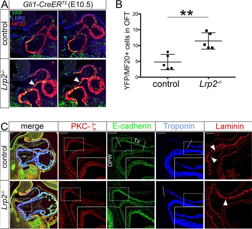
3184       Human Molecular Genetics, 2020, Vol. 29, No. 19

Graphical Abstract

                                                                                                                                                Downloaded from https://academic.oup.com/hmg/article/29/19/3183/5902918 by guest on 11 December 2020
Introduction                                                             left and right ventricles, respectively. Defects in OFT formation
                                                                         produce conotruncal malformations characterized by incom-
LRP2 is a member of the LDL receptor gene family, a class
                                                                         plete or absent separation of the aorta and pulmonary trunk,
of multifunctional endocytic receptors that play key roles in
                                                                         resulting in low oxygen supply due to the provision of mixed
embryonic development and that result in severe developmental
                                                                         blood to the circulation. OFT malformations account for almost
malformations in humans and animal models when dysfunc-
                                                                         30% of all congenital heart defects in humans (15). Recently, a
tional (1). During neurulation, LRP2 is prominently expressed
                                                                         range of cardiac anomalies including CAT but also ventricular
in the neuroepithelium that gives rise to the various parts of
                                                                         septal defects and atrioventricular canal malformations were
the developing central nervous system. Loss of receptor expres-
                                                                         reported in mice with targeted Lrp2 gene disruption (16). Still, the
sion in this tissue in gene-targeted mice results in the fusion
                                                                         molecular mechanism of receptor action in heart development
of the forebrain hemispheres (holoprosencephaly) (2,3) and in
                                                                         remained unexplained.
the overgrowth of the eye globe (buphthalmia) (4,5). Similar
                                                                             In this study, we focused on CAT formation, the most
defects are seen in patients with Donnai-Barrow/Facio-oculo-
                                                                         consistent feature of heart malformations observed in Lrp2
acoustico-renal (DB/FOAR) syndrome, an autosomal recessive
                                                                         mutant mice. We demonstrate that LRP2 is specifically expressed
disorder caused by inheritable LRP2 mutations (6–9).
                                                                         in SHH-responsive progenitor cells in the dorsal pericardial
    Concerning its mode of action, LRP2 has been shown to act as
                                                                         wall (DPW) that contribute to the formation of the OFT. Loss
an auxiliary receptor for sonic hedgehog (SHH) and to activate
                                                                         of receptor activity in Lrp2 mutant mice impairs SHH signaling
or inhibit this morphogen pathway dependent on the cellular
                                                                         in the DPW, resulting in decreased numbers and disturbed
context. In the neuroepithelium, LRP2 acts as a co-receptor to
                                                                         myocardial differentiation of progenitors that migrate into
Patched1 to promote SHH signaling and to pattern the ventral
                                                                         the OFT myocardium. Ultimately, these morphogenetic defects
midline of the forebrain (10). In contrast, in the developing eye,
                                                                         cause insufficient elongation of the OFT, which may cause
it operates as a clearance receptor for SHH to antagonize growth
                                                                         the conotruncal malformations seen in mice, and possible in
promoting signals by this morphogen in the retina (11). Concep-
                                                                         patients lacking LRP2.
tually, these functions of LRP2 in control of SHH signaling explain
the forebrain and eye phenotypes observed in patients with
DB/FOAR syndrome. However, LRP2 has also been shown to bind              Results
fibroblast growth factor (FGF) 8 (12) and bone morphogenetic
                                                                         Loss of LRP2 results in formation of a common arterial
protein (BMP) 4 (3) and may thus have the potential to regulate
                                                                         trunk including defective endocardial cushion
multiple morphogen pathways during organogenesis.
    Surprisingly, unbiased screens using exome sequencing now            formation
implicated mutations in LRP2 in congenital heart disease in              To elucidate the molecular mechanism(s) of LRP2 action in
humans, a receptor function not considered thus far (13). Addi-          mammalian heart development, we studied mice homozygous
tional evidence for a role in heart development came with ENU-           for a targeted Lrp2 gene disruption (Lrp2−/− ) generated by us
induced mutagenesis studies that revealed Lrp2 mutations as a            previously (2) and compared them to somite-matched control
prominent cause of cardiac outflow tract (OFT) defects in mice           embryos. Lrp2 mutant embryos were compared with their
(14). The cardiac OFT is a transient structure at the arterial pole of   respective wild-type or heterozygous littermates, as no defects
the embryonic heart. During development, it separates into the           were observed in Lrp2+/− animals. The latter two genotypes are
ascending aorta and pulmonary trunk, outlets of the definitive           jointly referred to as controls herein.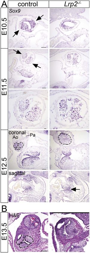
Human Molecular Genetics, 2020, Vol. 29, No. 19 3185
Downloaded from https://academic.oup.com/hmg/article/29/19/3183/5902918 by guest on 11 December 2020
Figure 1. Loss of LRP2 causes the formation of a common arterial trunk. (A)
Whole mounts (left) and coronal sections (right) of Lrp2−/− and control mouse
hearts at embryonic (E) day 18.5 and 15.5. In control embryos, separation of the
OFT into aorta (Ao) and pulmonary trunk (Pa) is evident in both embryonic stages.
In Lrp2−/− embryos, aorta and pulmonary trunk fail to separate resulting in a
common arterial trunk (CAT, indicated by asterisks). This CAT phenotype showed
full penetrance and was seen in 23 out of 23 Lrp2−/− embryos analyzed. (B) Coro-
nal sections of control and Lrp2−/− hearts at E13.5 and E12.5. In control embryos,
aorta (Ao) and pulmonary trunk (Pa) are present. Stippled circles indicate two
distinct swellings forming the endocardial cushions in the aortic valve in control
hearts. In Lrp2−/− hearts, a common outflow vessel (indicated by asterisks)
above the right ventricle is formed. Instead of two endocardial swellings, only
one cell cluster in endocardial cushion formation (stippled circle) is apparent. At
E13.5, Lrp2−/− embryos display ventricular septal defects (arrowhead). At E12.5,
arrowheads indicate blebbing of the epicardium and hemorrhages in Lrp2−/−
embryos. Scale bars: A, 750 μm; B, 250 μm.
Loss of LRP2 activity in Lrp2−/− mice leads to defects in OFT
formation characterized by incomplete or absent separation of
the aorta (Ao) and pulmonary trunk (Pa), a defect referred to
as a common arterial trunk (CAT; persistent truncus arteriosus).
CAT formation was evident at embryonic (E) day 18.5 and 15.5
when control embryos showed properly separated Ao and Pa
(Fig. 1A), while Lrp2−/− embryos exhibited a single vessel exiting
the heart from the right ventricle (Fig. 1A, asterisks). Histological
alterations indicative of an OFT defect manifested around E12.5
to E13.5 when OFT septation normally occurs. At this time point,
septation of Ao and Pa through the formation of the major septal
cushions was seen in control but not in LRP2-deficient embryos
(Figs 1B and Supplementary Material, S1A). In addition to CAT,
other malformations of the heart were also detected in mutant
mice, albeit at more variable levels. Thus, some Lrp2−/− embryos
failed to form distinct endocardial valve cushions but exhibited
only one cell cluster in the common outlet valve (Fig. 1B, stippled
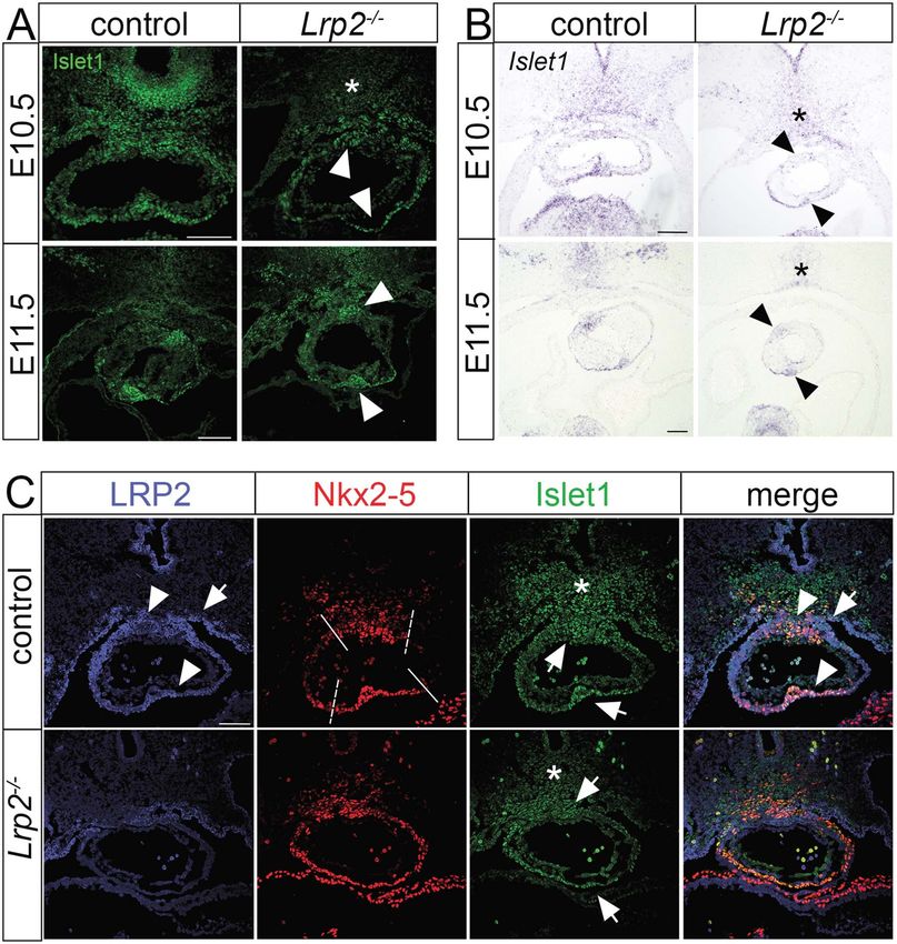
Figure 2. Altered endocardial cushion formation in Lrp2−/− hearts. (A) ISH for
circle), the likely cause for the hypoplastic semilunar valves of Sox9 on coronal and sagittal sections of Lrp2−/− and control embryos depicts
the common trunk seen in E15.5 mutants (Supplementary Mate- endocardial cushion formation in the OFT. At E10.5, two endocardial cushions
rial, Fig. S1B, arrows). Deeper intertrabecular spaces (Supplemen- are formed by two distinct tissue swellings in the control OFT tissue (arrows).
tary Material, Fig. S1A, arrows), defective formation of ventricular In Lrp2−/− OFT tissue, these two tissue swellings are not visible. At E11.5, Sox9
septum (Fig. 1B, arrowhead; Supplementary Material, Fig. S1A, positive cells cluster in the endocardial cushions (arrows) in control OFT tissue.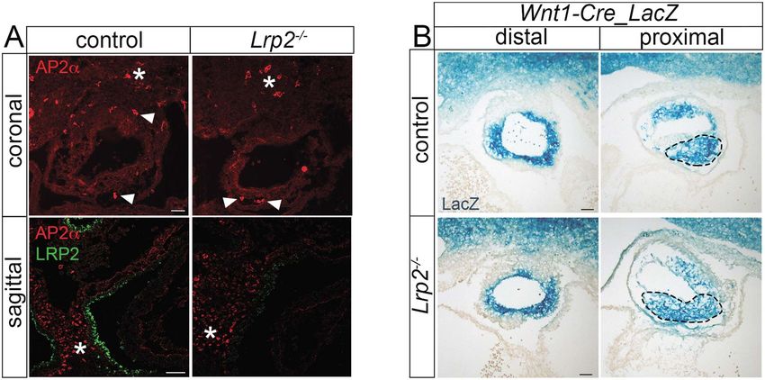
3186         Human Molecular Genetics, 2020, Vol. 29, No. 19

arrowhead in E13.5) as well as blebbing of the epicardial cell layer                        enveloping the distal OFT (Fig. 3A, E9.5 and E10.5). Upon closer
and hemorrhages (Fig. 1B, arrowheads; Supplementary Material,                               inspection of the distal OFT, LRP2 expression regionalized mainly
Fig. S1A, arrowheads in E12.5) were other variable pathological                             to the superior and inferior OFT regions (Fig. 3B, coronal view).
features of mutant hearts.                                                                      From the DPW, progenitor cells move along a path of
    Because the formation of a CAT was the most prominent                                   differentiation via an intermediate state in the transition zone
and most consistent phenotype seen in Lrp2 mutant mice (100%                                (Tz) to being fully differentiated cardiomyocytes when reaching
penetrance), we focused on elucidating its molecular cause in                               the OFT myocard (20). To better visualize LRP2 expression along
our mouse model of (DB/FOAR) syndrome. Given the impor-                                     this migratory path, we used higher magnification of a sagittal
tance of the major endocardial septal cushion formation for OFT                             view of E10.5 control hearts (Fig. 3B, sagittal). In this plane of
septation, we compared this process in wild-type and Lrp2−/−                                section, LRP2 levels were high in cells of the DPW but reduced in
embryos using in situ hybridization (ISH) for Sox9 (Fig. 2A). Sox9                          the Tz and completely absent from the OFT myocardium. This
is expressed in the cardiac cushion mesenchyme and promotes                                 observation argued for a function of LRP2 in SHF progenitors
cardiac valve progenitor proliferation (17). At E10.5, two Sox9                             that was lost as differentiation to cardiomyocytes proceeded.
positive major septal endocardial cushions were visible in con-                             At E12.5, when the OFT was already septated, robust LRP2

                                                                                                                                                                                     Downloaded from https://academic.oup.com/hmg/article/29/19/3183/5902918 by guest on 11 December 2020
trol embryos (arrows) while these distinct tissue swellings were                            expression persisted in the DPW and in the epithelial sheet
not detectable in Lrp2−/− embryos, despite the presence of Sox9                             enclosing the most distal regions of the Ao and Pa, while only
positive cells. By E11.5, these spiraling clusters of Sox9 positive                         minor receptor levels were visible in tissue surrounding the atria
cells were less compacted in Lrp2−/− as compared with control                               and ventricles (Fig. 3A, arrowheads in E12.5). LRP2 was absent
embryos (Fig. 2A, E11.5 lower panel). Of note, ISH for Sox9 also                            from cardiomyocytes and endothelial cells as documented
identified defects in endocardial swellings of the aortic valve                             by lack of co-expression with cardiomyocyte marker alpha-
that formed an unorganized Sox9 positive cell cluster in the                                smooth muscle actin (SMA) and endothelial cell marker platelet
mutant embryo (Fig. 2, stippled circle in E12.5 Lrp2−/− embryo).                            endothelial cell adhesion molecule (PECAM) (Fig. 3A, lower panel
This defect likely causes the hypoplastic semilunar valves seen                             in E12.5).
in the common trunk in mutants (Supplementary Material, Fig.
S1B). In some Lrp2−/− embryos, signs of mild hypoplasia of the
                                                                                            LRP2 deficiency specifically impacts SHF
atrioventricular cushions were observed (Fig. 2A, arrow in E12.5
                                                                                            progenitor cells
sagittal). Investigating the formation of aortic valve leaflets at
E13.5 in more detail, we observed clearly defined left coro-                                While LRP2 expression in neural crest cells had been reported
nary, right coronary and noncoronary leaflets in the control                                before (19,20), we failed to detect the receptor in the CNCC
embryos (Fig. 2B, stippled circles). In contrast, Lrp2−/− embryos                           population using immunohistology (Fig. 4A). Still, the major sep-
displayed deformed common valve leaflets at this embryonic                                  tal cushion defect observed in Lrp2−/− embryos was consistent
stage (Fig. 2B), defects in line with the observation of malformed                          with a potential defect in CNCCs. To query an indirect effect
endocardial cushions at E11.5 (Fig. 2A).                                                    of receptor deficiency on this cell population, we crossed the
                                                                                            Lrp2 deficient mouse strain with the Wnt1-Cre_LacZ reporter line
                                                                                            to specifically mark CNCCs (23). Staining lacZ activity at E10.5
LRP2 is expressed in the progenitor domain                                                  revealed a comparable pattern of CNCCs in the OFT of Lrp2−/−
of the second heart field                                                                   and control embryos (Fig. 4B). Subtle differences were seen in
The formation, elongation and septation of the cardiac OFT                                  more proximal regions of the spiraling OFT cushions, where
depends on the interaction of two distinct cell populations,                                CNCCs appeared less compacted in Lrp2−/− embryos compared
namely cardiac neural crest cells (CNCCs) and second heart field                            with controls (Fig. 4B, stippled circle). Based on these data, we
(SHF) cells. CNCCs are a subpopulation of neural crest cells that                           concluded that LRP2 deficiency did not profoundly impact the
originate from the dorsal neural tube and that migrate into                                 migration of CNCCs into the OFT or their integration in the major
pharyngeal arches 3, 4 and 6. Starting from E9.5, CNCCs populate                            septal cushions. Rather subtle alterations in CNCC organization
the cardiac OFT and move into the OFT cushions (18). There, they                            seen in the proximal OFT of mutant mice were considered
give rise to the condensed mesenchyme in the major endocardial                              secondary to defects in cushion formation.
septal cushions forming the aorticopulmonary septation com-                                     Since Lrp2 deficiency did not affect the migration of CNCC, we
plex that divides the distal OFT into Ao and Pa (19). In contrast,                          turned our attention to SHF cells that express LRP2 and that are
SHF cells are cardiac progenitor cells located in the pharyngeal                            necessary for OFT septation. Among other markers, these cells
mesoderm. During heart development, they are added to the                                   are characterized by the expression of insulin gene enhancer
arterial pole of the OFT driving OFT elongation.                                            protein (Islet1) (24). Islet1 protein as well as Islet1 transcript
   To interrogate the role of LRP2 during OFT formation, we                                 showed decreased expression levels in the pharyngeal meso-
investigated the expression pattern of the receptor during                                  derm and in the OFT of Lrp2−/− embryos compared with controls
OFT septation using immunohistology. At E9.5 and E10.5,                                     (Fig. 5A and B) possibly explaining defective OFT development.
when CNCCs invaded the OFT cushions, LRP2 showed strong                                     Islet1 protein and transcript were especially reduced in the
expression in the surface ectoderm as well as in the epithelium                             superior and inferior intercalated cushions, which were visible
of the pericardial cavity, including the DPW harboring SHF                                  at E11.5 in both genotypes (Fig. 5A and B, arrowheads). Reduced
progenitors. In addition, LRP2 was detected in the epithelium                               levels of Islet1 in the intercalated cushions likely contribute to

In mutants, Sox9 positive cells are less compacted and poorly organized in the endocardial cushions as depicted in the higher magnification view at E11.5. At E12.5 in
control OFT tissue, the OFT septum has formed and divides aorta (Ao) and pulmonary trunk (Pa). Sox9 positive cells are localized in the endocardial swellings of the
aortic valve (stippled circles), while in Lrp2−/− OFT vessel, only a disorganized cluster of Sox9 positive cells is visible (stippled circle). On E12.5 sagittal sections (arrow),
mild hypoplasia of the atrioventricular cushions with a loss of fusion is observable in Lrp2−/− embryos compared with controls. (B) Hematoxylin and eosin-stained
coronal sections of Lrp2−/− and control embryos at E13.5 documenting the left coronary, right coronary and noncoronary leaflets in the aortic valve of control embryos
(stippled circles) but malformed common valve leaflets in Lrp2−/− embryos. Scale bar: 100 μm.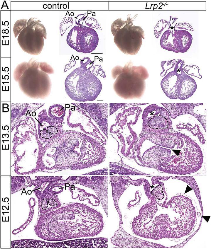
Human Molecular Genetics, 2020, Vol. 29, No. 19        3187

                                                                                 the defective development of the common non-coronary valve
                                                                                 leaflet observed in Lrp2−/− hearts (Fig. 2B).
                                                                                     To investigate which cells in the SHF express LRP2, we
                                                                                 studied its localization with respect to SHF marker Islet1 and
                                                                                 cardiac mesoderm marker NK2 transcription factor-related
                                                                                 locus 5 (Nkx2–5), a myocardial transcription factor regulated by
                                                                                 Islet1 and required for SHF development. LRP2 was expressed
                                                                                 in the DPW (Fig. 5C, arrow) but also in the epithelial sheet
                                                                                 surrounding the intercalated cushions in the OFT (Fig. 5C,
                                                                                 arrowheads), partially overlapping in these regions with Nkx2–
                                                                                 5 and Islet1 in controls. In Lrp2−/− mutants, Nkx2–5 protein
                                                                                 in the distal OFT was less restricted to the superior and
                                                                                 inferior regions (Fig. 5C, stippled lines in control embryos) but
                                                                                 showed a more widespread localization throughout the distal

                                                                                                                                                        Downloaded from https://academic.oup.com/hmg/article/29/19/3183/5902918 by guest on 11 December 2020
                                                                                 OFT. Islet1 demonstrated decreased levels in the pharyngeal
                                                                                 mesoderm (Fig. 5A and C, asterisks) as well as in the superior
                                                                                 and inferior intercalated cushions (Fig. 5A arrowheads; Fig. 5C
                                                                                 arrows).
                                                                                     We also tested the expression of two additional SHF mark-
                                                                                 ers, namely neurovascular guiding factor Sema3c, important to
                                                                                 correctly navigate CNCCs towards the OFT (25), as well as T-box
                                                                                 transcription factor 1 (Tbx1), stimulating the proliferation of mul-
                                                                                 tipotent heart progenitors (26). Overall, the expression pattern
                                                                                 of Sema3c was largely unchanged in Lrp2−/− embryos compared
                                                                                 with controls, although slightly reduced levels were noted in
                                                                                 the intercalated cushions of the OFT (Supplementary Material,
                                                                                 Fig. S2A, arrowheads). Also, expression levels of Tbx1 were com-
                                                                                 parable between genotypes (Supplementary Material, Fig. S2B),
                                                                                 suggesting mis-patterning rather than complete absence of SHF
                                                                                 progenitors in LRP2 mutant mice.

                                                                                 LRP2 promotes SHH signaling during OFT formation
                                                                                 So far, our data documented a decrease in Islet1-positive SHF
                                                                                 progenitor cells and a shifted localization of Nkx2–5 positive
                                                                                 cells in LRP2 mutant embryos, arguing for a role of this recep-
                                                                                 tor in controlling cardiac progenitor maintenance during OFT
                                                                                 septation. Several signaling pathways have been implicated in
                                                                                 the formation, proliferation and differentiation of multipotent
                                                                                 cardiac progenitor cells in the SHF, including signaling through
                                                                                 Wnts, BMPs and SHH (27). Since LRP2 is known to act in mul-
                                                                                 tiple morphogen pathways, we investigated these pathways in
                                                                                 Lrp2−/− embryos during OFT development.
                                                                                     Canonical Wnt signaling has been shown to act upstream of
                                                                                 other signaling pathways in control of Islet1-positive progenitor
                                                                                 cell proliferation (28). Non-canonical Wnt signaling is implicated
                                                                                 in OFT development (26,27) and important for planar cell polarity
                                                                                 during myocardialization of the OFT cushions. It regulates polar-
                                                                                 ity and intracellular cytoskeletal rearrangements, important to
                                                                                 prevent premature differentiation of SHF progenitor cells into
                                                                                 cardiomyocytes (31–33). To explore potential defects in canonical
                                                                                 Wnt signaling during OFT formation, we crossed the Lrp2 mutant
                                                                                 strain with the Tcf/Lef_LacZ reporter line. Detecting lacZ activity
                                                                                 as a read-out of Wnt signaling, we failed to observe any changes
Figure 3. LRP2 expression in the developing OFT. Immunhistological detection
of LRP2, SMA, and PECAM on the coronal sections of the embryonic mouse
                                                                                 in Wnt activity in the SHF of embryos lacking LRP2 (Supplemen-
heart at the indicated embryonic days. (A) At E9.5 and E10.5, LRP2 expression    tary Material, Fig. S3A). Also, the non-canonical Wnt pathway
is seen along the ectoderm as well as in the epithelium enclosing the            was unchanged as deduced from investigating the expression
pericardial cavity, including the DPW and in the distal OFT myocardium           pattern of Wnt11 in the OFT using ISH (Supplementary Material,
of controls. No immunoreactivity for LRP2 is detected in receptor mutant         Fig. S3B). BMP signaling downregulates proliferation when SHF
hearts. At E12.5, LRP2 expression is visible in the DPW and in myocardial
                                                                                 cells enter the OFT and drives myocardial differentiation (34).
cells surrounding the pulmonary artery (Pa) as well as the aorta (Ao) at the
side facing the Pa. Minor LRP2 levels are visible in the epithelium enveloping
                                                                                 Analyzing the BMP pathway by studying Bmp4 expression in the
atria and ventricles (arrowheads). Cardiomyocytes are stained with SMA           distal OFT, we failed to detect obvious changes in LRP2-deficient
and endocardial cells are stained with PECAM. (B) Higher magnification of        as compared with control embryos (Supplementary Material,
the region corresponding to the boxed area in control embryos in panel           Fig. S3B).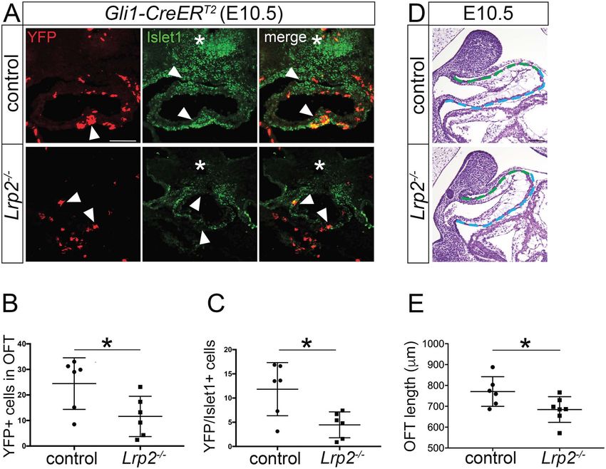
3188        Human Molecular Genetics, 2020, Vol. 29, No. 19

                                                                                                                                                                             Downloaded from https://academic.oup.com/hmg/article/29/19/3183/5902918 by guest on 11 December 2020
Figure 4. LRP2 deficiency does not affect the CNCC population migrating into the OFT. (A) Immunohistological detection of AP2α on coronal and of AP2α and LRP2 on
sagittal sections of the hearts of control and Lrp2−/− embryos at E10.5. Similar numbers of AP2α positive cells are detected in the SHF region (asterisks) and in the OFT
(arrowheads) of Lrp2−/− and control embryos. AP2α and LRP2 show non-overlapping expression domains, suggesting the absence of LRP2 from CNCCs. (B) Staining for
lacZ activity in CNCCs on coronal sections of E10.5 mouse OFT tissues from the Wnt1-Cre_LacZ reporter line. Mice express (control) or lack LRP2 (Lrp2−/− ). In sections of
the distal OFT region (left panels), the presence of CNCCs in the outflow vessel is detected in control and Lrp2−/− embryos. In more proximal regions of the OFT (right
panel), CNCCs fail to accumulate in the endocardial cushions in Lrp2−/− as compared with the control OFT vessel (stippled circle). Scale bars: 50 μm.

    Another essential morphogenetic signal in SHF patterning                                Next, we performed co-immunostaining for YFP and Islet1
is provided by SHH secreted from the pharyngeal endoderm.                               to test if SHF progenitor cells lacking LRP2 were less respon-
SHH regulates both CNCC survival and OFT development (35).                              sive to SHH signals. YFP positive cells in the inferior OFT of
Using the Gli1-LacZ reporter allele introduced into the Lrp2                            control embryos co-immunostained with Islet1. Lrp2−/− embryos
mutant strain, we detected reduced SHH activity in the distal                           showed a significant reduction in Islet1 immunoreactivity in the
OFT of Lrp2−/− embryos as compared with controls (Fig. 6).                              pharyngeal mesoderm (Fig. 7A, asterisk) as well as in the supe-
At 28 somites even before intercalated cushion, developed                               rior and inferior OFT (Fig. 7A, arrowheads). Quantification of car-
SHH activity is already reduced. Still, we cannot exclude that                          diac progenitor cells double positive for Islet1 and YFP substan-
impaired formation of the intercalated cushions may also                                tiated a significant reduction of SHH-responsive Islet1-positive
secondarily contribute to the reduction in Gli1_lacZ activity                           progenitor cells in the distal OFT of LRP2-deficient embryos
seen in the mutant tissue. Nevertheless at 30 and 32 somites,                           (Fig. 7C).
reduced SHH activity was especially evident in the inferior                                 During OFT formation, SHF progenitor cells located in the
OFT region where SHH signaling was significantly decreased                              DPW move along a path of differentiation to the Tz, where
in LRP2-deficient embryos (Fig. 6). To test if reduced SHH                              they initiate a myocardial gene expression program. They finally
signaling in Lrp2−/− embryos was connected to the SHF defects                           reach the OFT myocardium to fully differentiate into cardiomy-
as characterized by reduced Islet1 expression, we crossed the                           ocytes. By this movement, cardiac SHF progenitor cells con-
LRP2-deficient line with the Gli1-CreERT2 reporter strain. In this                      tribute to the elongation of the OFT, necessary for correct align-
line, expression of a tamoxifen-inducible Cre transgene under                           ment. Disturbances in this differentiation program result in a
control of the Gli1 promoter enables labeling of SHH-responsive                         shortened OFT and can give rise to a CAT (37). A significant
cells by Cre-induced expression of YFP (36). To induce YFP                              decrease in OFT length was seen in the hearts of Lrp2−/− embryos
expression, pregnant mice were injected with tamoxifen at E7.5                          compared with controls (Fig. 7D and E), arguing for a defect in
and embryos were analyzed at E10.5. Immunodetection of YFP                              progenitor movement in Lrp2−/− embryos.
demonstrated SHH-responsive cells accumulating in the inferior                              To investigate the consequences of reduced SHH signaling in
OFT region in control embryos (Fig. 7A). In Lrp2−/− embryos, YFP-                       DPW progenitors for their differentiation potential, we studied
positive cells were reduced about 50% in numbers and failed                             sagittal sections of Gli1-CreERT2 control and LRP2-deficient
to accumulate in the inferior OFT region (Fig. 7A, arrowheads;                          embryos at E10.5. In controls, SHH-responsive cells (as deduced
Fig. 7B).                                                                               by YFP expression) concentrated in the DPW and distal Tz, a

A. On a coronal view (left panel), robust LRP2 expression is seen in cells of the SHF in the DPW and in distal myocardial cells of the OFT. In the OFT, LRP2
immunoreactivity is most prominent in the superior and inferior OFT regions. On a sagittal view (right panel), prominent levels of LRP2 are seen in epithelial cells of
the DPW. Receptor expression is significantly reduced in the Tz and completely absent from the proximal OFT myocardium (OFT). Boundaries between DPW, TZ and
OFT myocardium are marked by stippled white lines. Scale bar: 100 μm.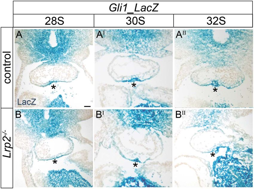
Human Molecular Genetics, 2020, Vol. 29, No. 19                   3189

                                                                                                                                                                           Downloaded from https://academic.oup.com/hmg/article/29/19/3183/5902918 by guest on 11 December 2020
Figure 5. a reduction in SHF cardiac progenitor cells. (A) Immunodetection of Islet1 and (B) ISH for Islet1 on coronal OFT sections of control and Lrp2−/− embryos at
E10.5 and E11.5. In Lrp2−/− embryos, immunosignals for Islet1 are decreased in the pharyngeal mesoderm (A, asterisk) and in the superior and inferior intercalated
cushions of the OFT (A, arrowheads) when compared with controls. The reduction in Islet1 protein levels in mutants is paralleled by decreased Islet1 transcript levels
at both embryonic stages in the pharyngeal mesoderm (B, asterisk) and in the intercalated cushions (B, arrowheads) as compared with controls. Scale bar: 100 μm. (C)
Immunohistological detection of LRP2, of SHF marker Nkx2–5 and of SHF cardiac progenitor marker Islet1 on coronal sections of E10.5 control and Lrp2−/− embryos.
Single channels as well as merged images are shown. Cardiac progenitor cells of the SHF in the DPW (arrow in LRP2 and merged channels) and in the myocardium
lining the intercalated cushions (arrowheads in LRP2 and merged channels) stain positive for LRP2. Lack of LRP2 in Lrp2−/− embryos results in the presence of Nkx2–5
positive cells throughout the OFT. This contrasts with the more restricted pattern in the superior and inferior OFT region (stippled white lines) in the control embryo.
Furthermore, Lrp2−/− embryos show reduced numbers of Islet1 positive cells in the pharyngeal mesoderm (asterisk in single channel) as well as in the intercalated
cushions (arrows in single channel) when compared with controls. Scale bar: 75 μm.

region overlapping with the LRP2 expression domain in the                              myocardium in Lrp2−/− embryos (Fig. 8B). Jointly, the reduction
DPW (Fig. 8A). Very few SHH-responsive cells were detected                             of YFP/Islet1 double positive cells in the distal OFT and the
in the OFT myocardium of control embryos as evidenced                                  increased amount of YFP/MF20 double positive cells in the OFT
by almost complete absence of co-staining of YFP with the                              myocardium in Lrp2−/− embryos argued for a role of LRP2 in
cardiomyocyte marker MF20 (Fig. 8A). In contrast, in Lrp2−/−                           facilitating the response of SHF progenitors in the DPW to SHH.
embryos, an increased number of SHH-responsive cells were                              Such a response is required to control migratory movement
co-stained by YFP and MF20 in the OFT myocardium (Fig. 8A,                             and differentiation along their path to the OFT myocardium.
arrowheads). Quantification of YFP and MF20 double positive                            Reduced responsiveness to SHH in the LRP2-deficient DPW was
cells corroborated this observation, demonstrating a signifi-                          not due to the impaired formation of primary cilia, essential
cant increase in YFP-MF20 double positive cells in the OFT                             for SHH signal reception, as documented by staining for the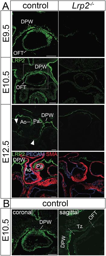
3190        Human Molecular Genetics, 2020, Vol. 29, No. 19

                                                                                                                                                                          Downloaded from https://academic.oup.com/hmg/article/29/19/3183/5902918 by guest on 11 December 2020
Figure 6. Reduced SHH signaling activity in the Lrp2−/− OFT. Staining for lacZ activity on coronal sections from 28 somites, 30 somites and 32 somites old Gli1_LacZ
reporter embryos to visualize SHH signaling. Animals express (control) or lack LRP2 (Lrp2−/− ). In all three examples, the activity of the GLI1-dependent LacZ reporter
is significantly reduced in Lrp2−/− embryos compared with their somite-matched control (compare asterisks in B to A, BI to AI , in BII to AII ). Scale bar: 50 μm.

ciliary marker Arl13b in Islet1-positive progenitor cells in both                     posterior regions in the DPW (Fig. 8C, compare localization of the
genotype groups (Supplementary Material, Fig. S4).                                    stippled lines). This shifted cell identity was also confirmed by
                                                                                      a shift in localization of cardiac troponin I, a marker for mature
LRP2 defines the boundary of the cardiac SHF                                          cardiomyocytes (Fig. 8C). Cardiac troponin I was exclusively
progenitor zone                                                                       expressed in cells of the OFT myocardium in controls. In Lrp2−/−
                                                                                      OFT tissue, also cells in the TZ stained positive for this protein.
To further substantiate a role for LRP2 in control of SHF progeni-
                                                                                      Furthermore, laminin, a marker for the basal lamina, also
tor cell differentiation along a path from the DPW to the Tz and
                                                                                      showed an altered localization in Lrp2−/− embryos. While in
finally to the OFT myocardium, we performed immunodetection
                                                                                      the control DPW and Tz, laminin showed a punctuated pattern
of markers representative of these three tissues in control-
                                                                                      in apical cell regions; this punctuated pattern was lost in the
and receptor-deficient embryos. Previous studies demonstrated
                                                                                      Lrp2−/− DPW and Tz (Fig. 8C, arrowheads). Finally, in control
an atypical apicobasal polarization of SHF progenitor cells
                                                                                      embryos, the basal lamina formed a continuous pattern in
and their basal filopodia in the DPW, and their increasing
                                                                                      the OFT region but not in the Tz, whereas in Lrp2−/− hearts,
epithelial characteristics in the Tz before differentiation in the
                                                                                      this continuous pattern of the basal lamina reached inside the
OFT myocardium (20,26,32,38–40). Therefore, we focused on the
                                                                                      Tz (Fig. 8C). Collectively, these findings argued for a reduction
immunodetection of proteins having a role in apical cell polarity
                                                                                      in numbers and for premature differentiation of Lrp2−/− SHF
comparing patterns in DPW and Tz of mutant and control
                                                                                      progenitor cells as they transit from the DWP via the Tz into OFT
embryos. Immunodetection of the apical tight junction marker
                                                                                      myocardium. These defects confound the contribution of SHF
aPKC-ζ (41) demonstrated robust expression at the apical cell
                                                                                      progenitors to OFT elongation and result in OFT shortening, the
membrane in the DPW of controls (Fig. 8C). No changes in signal
                                                                                      likely cause of CAT formation in Lrp2−/− mice.
intensity or localization in aPKC-ζ were detected in the Lrp2−/−
DPW, arguing against an overall effect of LRP2 deficiency on
polarization of cells. However, the result was different for the
adherens junction marker E-cadherin. Although localization
                                                                                      Discussion
of E-cadherin to the apical cell membrane was comparable                              Previous studies implicated LRP2 in OFT formation (16). Still,
between the two genotypes, the distribution of E-cadherin-                            the exact mechanism whereby this multifunctional endocytic
positive cells was clearly shifted in the mutants. As shown in                        receptor may control the formation of the cardiac OFT and why
Fig. 8C, in controls, E-cadherin was expressed in the anterior                        receptor dysfunction results in conotruncal malformations in
DPW and further increased in expression in the Tz, consistent                         patients and in mouse models remained unexplained. Here, we
with an increasing epithelial cohesion before cells differentiate                     uncovered a crucial role for LRP2 in SHH signaling in progenitor
into OFT myocardium. Vanishing E-cadherin protein levels                              cells of the DPW, signals required to ensure their timely differ-
were detected in the posterior region of the DPW closer to the                        entiation into mature cardiomyocytes as they migrate into the
venous pole. In contrast, in Lrp2−/− embryos, the localization                        OFT tissue. Loss of LRP2 results in a decrease in SHH-responsive
of E-cadherin-positive cells in the DPW was shifted to more                           progenitor cells in the DPW and in a concomitant appearance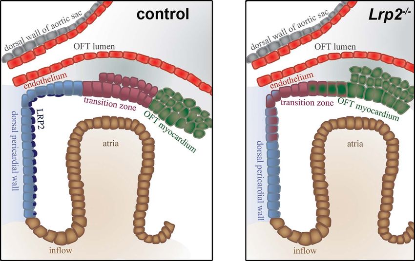
Human Molecular Genetics, 2020, Vol. 29, No. 19                    3191

                                                                                                                                                                              Downloaded from https://academic.oup.com/hmg/article/29/19/3183/5902918 by guest on 11 December 2020
Figure 7. Reduced SHH responsiveness of cardiac progenitor cells in the Lrp2−/− OFT. (A) Immunohistological detection of YFP and Islet1 on coronal sections of E10.5
mouse hearts from the tamoxifen-inducible Gli1-CreERT2 reporter strain. The animals express (control) or lack LRP2 (Lrp2−/− ). Single channels as well as merged images
are shown. In control embryos, YFP-labeled cells are visible throughout the OFT but accumulate in the inferior intercalated cushion (arrowhead). Islet1-positive cardiac
progenitor cells are seen in the pharyngeal mesoderm (asterisk) as well as in the superior and inferior OFT (arrowheads). In Lrp2−/− embryos, YFP positive cells fail to
accumulate in the inferior OFT but are dispersed throughout the OFT vessel (arrowheads). Also, Islet1 positive cells are reduced in the pharyngeal mesoderm (asterisk)
and in the OFT vessel (arrowheads). Scale bar: 100 μm. (B) YFP positive cells (as exemplified in panel A) were quantified on 5–9 coronal sections through the distal OFT
vessel of 6 control and 6 Lrp2−/− embryos. YFP positive cells are reduced about 50% in Lrp2−/− embryos. (C) Cardiac progenitor cells double positive for Islet1 and YFP
(as exemplified in panel A) were quantified on 5–9 coronal sections through the distal OFT vessel of 6 control and 6 Lrp2−/− embryos. The numbers of YFP/Islet1 double
positive cells in the OFT are significantly reduced in mutant as compared with control embryos (∗ P ≤ 0.05; unpaired Student’s t test). (D) Exemplary hematoxylin and
eosin-stained sagittal sections of Lrp2−/− and control embryos at E10.5. The OFT length was determined by taking the average of the measured upper (green line) and
lower (blue line) OFT wall lengths for each section. Scale bar: 100 μm. (E) OFT length in control and Lrp2−/− OFT tissues as quantified on 5 sagittal sections each through
the OFT vessel of 6 control and 7 Lrp2−/− embryos. OFT length is significantly reduced in mutant as compared with control embryos (∗ P ≤ 0.05; unpaired Student’s t
test).

of cardiomyocytes in the Tz, documenting depletion of this                              a significantly shortened OFT (Fig. 7E), most likely due to a
progenitor niche due to premature differentiation of cardiac                            reduced number of incorporated myocardial cells derived from a
progenitors along their path to an OFT myocardial fate.                                 diminished progenitor pool in the anterior SHF. In addition to
                                                                                        OFT elongation, SHF progenitors also contributed to the for-
Loss of anterior SHF progenitors is the likely cause                                    mation of the intercalated cushions and, to a minor extent,
of CAT formation in Lrp2−/− embryos                                                     to the OFT cushion mesenchyme (42). Conceptually, impaired
Formation and subsequent septation of the OFT is dependent                              OFT cushion formation in Lrp2−/− mice may be a secondary
on two cell populations: the SHF and the CNCCs. We traced                               consequence of OFT shortening in these animals (Fig. 7 D, E).
the primary defect in LRP2 deficiency to the progenitor cells in                        More excitingly, defects in intercalated cushions as well as in
the anterior SHF that express this receptor (Fig. 5C). While the                        the OFT cushions may be explained by a role for LRP2 in also
CNCC population normally migrated into the OFT to reach the                             controlling migration of SHF progenitor cells into these tissues.
distal OFT cushions (Fig. 4), the number of Islet1-positive SHF                         This hypothesis is supported by the malformed formation of OFT
progenitor cells was significantly reduced in receptor-deficient                        cushions (Fig. 2) and by the reduced number of Islet1-positive
hearts (Fig. 5). Cardiac progenitors in the DPW contribute to                           cells seen in the intercalated cushions of Lrp2−/− mice (Fig. 5)
the growth and elongation of the OFT, a process essential for                           caused by reduced cell migration from the SHF inside these
proper septation into Ao and Pa (37). Lrp2−/− mice exhibited                            tissues.
3192 Human Molecular Genetics, 2020, Vol. 29, No. 19
Downloaded from https://academic.oup.com/hmg/article/29/19/3183/5902918 by guest on 11 December 2020
Figure 8. Loss of LRP2 decreases the SHF progenitor niche. (A) Immunodetection of YFP (green), LRP2 (blue) and MF20 (red) on two exemplary sagittal sections for E10.5
control and for Lrp2−/− hearts of the Gli1-CreERT2 reporter line. In control embryos, SHH responsive (YFP positive) cells cluster at the border between DPW and Tz
(arrow). Very few double positive cells for YFP-MF20 are detected in the OFT of control embryos. In the two Lrp2−/− heart tissue sections, MF20-positive cardiomyocytes
in the OFT myocardium are positive for YFP (arrowhead), indicating aberrant differentiation of SHH responsive cells. Scale bar: 100 μm. (B) Cardiomyocytes double
positive for MF20 and YFP (as exemplified in panel A) were quantified on 5–8 sagittal sections through the OFT vessel of 5 control and 5 Lrp2−/− embryos. The
numbers of YFP/MF20 double positive cells in the OTF are significantly reduced in mutant as compared with control embryos (∗∗ P ≤ 0.01; unpaired Student’s t test).
(C) Immunohistological detection of PKC-ζ , E-cadherin, troponin and laminin on sagittal sections of E10.5 control and Lrp2−/− heart tissues. To visualize the overall
morphology of the heart, merged overview pictures are shown for both genotypes. Detailed comparison of PKC-ζ , E-cadherin, troponin and laminin immunosignals are
given in single channel configuration in higher magnification images (boxed areas in overview pictures). The insets depict cellular localization of PKC-ζ , E-cadherin
and troponin corresponding to the boxed area in the higher magnification images. Signal intensity and distribution for adherens junction component PKC-ζ (red) are
unchanged comparing control and Lrp2−/− hearts. In control hearts, E-cadherin (green) exhibits strong expression in the Tz but reduced levels in the DPW. In Lrp2−/−
hearts, the E-cadherin expression domain extends into the DPW (to the stippled line). Troponin (blue) marks differentiated cardiomyocytes in the OFT myocardium. In
contrast to control hearts, troponin expression extends from the OFT myocardium into the transition zone (stippled line) in Lrp2−/− hearts. Laminin, a marker for the
basal lamina, shows a punctuated pattern with apical vesicles (arrowheads) in the DPW and distal Tz in controls. In Lrp2−/− hearts, laminin-positive apical vesicles
are reduced in numbers (arrowhead) and the regular pattern of the basal lamina from the OFT continues into the Tz and DPW. Scale bar: 50 μm.
LRP2 deficiency impairs SHH-dependent maintenance differentiation fate decisions in the SHF (24,39). These pathways
of the DPW progenitor zone include canonical and non-canonical Wnt signaling, as well as
To facilitate OFT elongation, SHF progenitor cells exhibit an signals downstream of FGF, BMP and SHH (27,40–46). Our studies
increased proliferative capacity and a delayed propensity to failed to provide evidence for defects in Wnt and BMP signaling
differentiate into myocardium (43). Several signaling path- as a primary cause of CAT formation in the LRP2-deficient
ways have been implicated in balancing proliferative versus heart (Supplementary Material, Fig. S3). Rather, our findingsHuman Molecular Genetics, 2020, Vol. 29, No. 19 3193
from Gli1_LacZ (Fig. 6) and Gli1_CreERT2 (Figs 7A–C and 8A and B) LRP2 being one of these essential SHH co-receptors. Dependent
reporter strains argue for a defect in SHH signaling in the on the biological context, LRP2 may activate (by facilitating cell
progenitor niche of the DPW as the underlying molecular surface binding of the morphogen) (10) or inhibit (by directing
mechanism. Our model is consistent with the established role SHH to lysosomal degradation) the SHH pathway (11). Data in
of LRP2 in promoting SHH signal reception in other embryonic here argue that during OFT formation, LRP2 acts as an agonist to
tissues, such as the neuroepithelium (10). Hypothetically, we promote the responsiveness of SHF progenitor cells to SHH cues.
cannot exclude that LRP2 deficiency may also impact signaling This hypothesis is supported by a recent study documenting a
pathways other than through SHH, although the nature of such crucial role for SHH in activating progenitor gene expression and
pathways remains unknown. in inhibiting premature differentiation, thereby maintaining the
The manifold cardiac phenotypes observed upon intercep- progenitor cell population in the SHF (57). We also show that
tion with the SHH pathway uncovered multiple roles for this LRP2 ensures the proper migration pattern of SHH-responsive
morphogen in heart morphogenesis (51). Among other func- SHF progenitors during OFT elongation. These results are in line
tions, SHH is necessary for arterial pole formation as loss of SHH with findings that SHH-responsive progenitors from the anterior
signaling in Shh−/− mice or in chick embryos treated with the SHF migrate into the pulmonary vessel (58). Finally, a recent
Downloaded from https://academic.oup.com/hmg/article/29/19/3183/5902918 by guest on 11 December 2020
SHH antagonist cyclopamine results in CAT formation (48,49). study on GATA4-regulated SHH signaling in cardiac progenitor
Conditional disruption of the SHH pathway mediator Smoothened cells documented the importance of SHH signals for SHF cell
(Smo) in Islet1-positive progenitor cells or a Shh conditional migration in mice (59). In this study, the migratory defect of Gata4
mutant using the Nkx2.5cre deleter strain substantiated a role for haploinsufficient SHF progenitor cells was rescued by the over-
SHH in OFT elongation and identified SHF progenitors as cell activation of the hedgehog pathway using the constitutively
population receptive to SHH signals (35,54). Now, our findings activate Smo mutant SmoM2, reconstituting cell migration from
identify LRP2 as an essential component of the SHH signaling the SHF into the OFT and ensuring proper OFT formation.
machinery during OFT formation and implicate defects in SHH In conclusion, we uncovered a crucial role for LRP2 in main-
signaling in SHF progenitors of the DPW in CAT formation in tenance of a pool of SHH-dependent progenitors in the anterior
receptor mutant mice. Using the Gli1-CreERT2 reporter line, we SHF, ensuring their differentiative fate as they migrate into the
showed an overall reduction in the number of SHH-responsive OFT tissue. These findings have identified a novel component
Islet1-positive progenitor cells in the Lrp2−/− distal OFT tissue of the SHH signaling machinery essential for OFT development
(Fig. 7A and C). Also, while these cells were mainly restricted and uncovered the molecular cause of conotruncal malforma-
to the DPW and the Tz in control hearts, they showed a more tions in humans and mouse models lacking this receptor. Addi-
dispersed localization in the OFT tissue in mutants, overlapping tional defects seen in LRP2-deficient hearts suggest that receptor
with MF20 positive cardiac cells in Tz and OFT myocardium activity may not be restricted to SHH-dependent progenitor cell
(Fig. 8A). In line with an impaired demarcation of DPW, Tz and fate specification during OFT formation but encompass other
OFT myocardium in Lrp2−/− hearts, the expression domain for aspects of heart development, such as OFT cushion formation.
cardiac troponin I in mutants reached into the Tz, while E- The molecular mechanisms of LRP2 function in the later pro-
cadherin expression, restricted to the Tz in controls, aberrantly cesses still require further elucidation.
extended more posteriorly into the DPW (Fig. 8C). Finally, the
pattern of laminin, indicative of a more mature development
of the basal lamina was restricted to the proximal regions of Materials and Methods
the Tz and the OFT myocardium in controls but extended into
Mouse models
the distal Tz and anterior DPW in Lrp2−/− heart tissue. Collec-
tively, these findings support a role for LRP2 in establishing an Generation of mice with targeted Lrp2 gene disruption has
SHH-dependent niche for cardiac progenitor cells in the DPW been described before (2). The Lrp2 gene defect was analyzed
and in controlling their differentiation along a path to the OFT in receptor-deficient and somite-matched littermates either
myocardium. wild-type (Lrp2+/+ ) or heterozygous for the mutant Lrp2 allele
(Lrp2+/− ). Since heterozygous animals demonstrate no LRP2
haploinsufficiency phenotype, Lrp2+/+ and Lrp2+/− embryos
LRP2 likely acts as agonist for SHH signaling during were combined and referred to as control group in this study.
OFT formation Where indicated, the Lrp2 deficient line was crossed with the
In the LRP2 mutant OFT, both the number of Islet1-positive cells Gli1-CreERT2 (JAX; Stock 007913), the Gli1_LacZ (JAX; Stock 008211)
receptive to SHH signals but also the overall expression domain or the Tcf/Lef_LacZ (Mohamed et al., 2004) reporter strains. Also,
of Islet1-positive progenitor cells were reduced (Fig. 5). Thus, where applicable, the Lrp2 deficient line was crossed with the
we cannot distinguish with certainty whether LRP2 deficiency Wnt1-Cre line (obtained from C. Birchmeier, Max-Delbrueck-
primarily impacts SHH signaling in progenitor cells, thereby Center, Berlin) and subsequently with the R26R (JAX; Stock
decreasing their numbers, or whether LRP2 is required for other 003474) reporter line to generate the Wnt1-Cre_LacZ reporter
aspects of cell maintenance and loss of SHH activity in this niche strain. All experiments involving animals were performed
is a secondary consequence of progenitor cell loss. However, according to institutional guidelines following approval by local
based on evidence provided in this study and in published work authorities (X9007/17).
(10,11), an instructive role for LRP2 in facilitation SHH signaling
in SHF progenitors seems likely.
The SHH signaling machinery is well characterized at the
Immunohistology
molecular level. In addition to the primary SHH receptor PTCH1, For hematoxylin and eosin (H + E) staining, embryos were fixed
auxiliary SHH binding proteins are necessary to activate or overnight in 4% paraformaldehyde in PBS at 4◦ C, followed by
inhibit the pathway in a context-dependent manner. This routine paraffin embedding and sectioning at 10 μm thickness.
modulatory activity of SHH binding proteins has mainly been For lacZ or immunofluorescence stainings, embryos were fixed
documented in the embryonic neuroepithelium (55,56), with for 1–4 h in 4% paraformaldehyde in PBS at 4◦ C. Fixed embryos3194 Human Molecular Genetics, 2020, Vol. 29, No. 19
were infiltrated with 30% sucrose in PBS overnight at 4◦ C, fol- Student’s t test (Figs 7B, C, E and 8B). P values are indicated in the
lowed by 2 h incubation in 50% cryoprotectant Tissue-Tek® OCT respective figure legends.
in 30% sucrose, followed by 2 h incubation in 75% Tissue-Tek®
OCT in 30% sucrose. Finally, the embryos were embedded in
Tissue-Tek® OCT and subjected to 10 μm cryo-sectioning. For Supplementary Material
immunodetection of E-cadherin, PKC-ζ , cardiac troponin I or Supplementary Material is available at HMG online.
laminin, embryos were dehydrated and embedded in paraffin
and sectioned at 10 μm.
Immunohistochemical analysis was carried out by incuba- Acknowledgement
tion of tissue sections with primary antibodies at the following
We are grateful to Robert Kelly (Aix-Marseille Université, France)
dilutions: guinea pig anti-LRP2 (1:2000; made in-house by immu-
for helpful discussions and for critical reading of the manuscript
nizing guinea pigs with full-length LRP2 isolated from rabbit
and to Magali Theveniau-Ruissy (Aix-Marseille Université) for
kidney), mouse anti-AP2α (1:50; DSHB; Santa Cruz), mouse anti-
expert advice. Maria Kamprath, Melanie Großmann, Kristin
Islet1 (1:50; DSHB), goat anti-Nkx2.5 (1:100; Santa Cruz), rabbit
Downloaded from https://academic.oup.com/hmg/article/29/19/3183/5902918 by guest on 11 December 2020
Kampf and Maria Kahlow provided technical assistance.
anti-GFP (1:400; Abcam), mouse anti-MF20 (1:50; DSHB), rat anti-
BrdU (1:100; AbD Serotec), mouse anti-E-cadherin (1:200; BD Conflict of Interest statement. None declared.
Transduction Laboratories), goat anti-cardiac troponin I (1:100;
Abcam), rabbit anti-laminin (1:200; Sigma), rabbit anti-PKC-ζ
(1:200; Sigma), rabbit anti-Arl13b (1:200; Proteintech), rat anti-
Funding
CD31 (PECAM-1, 1:300; BD Pharmigen) and mouse anti-SMA
(1:300; Sigma). Primary antibodies were visualized using sec- Deutsche Forschungsgemeinschaft (CH 1838/1–1, CH 1838/3–1)
ondary antisera conjugated with Alexa Fluor 488, 555 or 647 to AC.
(1:500; Invitrogen and Jackson Immuno Research) or with Biotin-
SP (1:100; Jackson Immuno Research) followed by fluorescent
conjugates of streptavidin (1:500; Invitrogen). Image acquisitions
References
were carried out using a Leica DMI 6000B inverted microscope or 1. Willnow, T.E. and Christ, A. (2017) Endocytic receptor
Leica SPE or Leica SP5 confocal microscopes. LRP2/megalin—of holoprosencephaly and renal
Fanconi syndrome. Pflugers Arch. - Eur. J. Physiol., 469,
In situ hybridization 907–916.
2. Willnow, T.E., Hilpert, J., Armstrong, S.A. et al. (1996) Defective
ISH on paraffin sections was described earlier (60). Plasmids for
forebrain development in mice lacking gp330/megalin. Proc.
digoxigenin (DIG)-labeled riboprobes were kindly provided by L.
Natl. Acad. Sci. U. S. A., 93, 8460–8464.
Robertson (University of Oxford, Oxford; Bmp4), E. Sock (Institute
3. Spoelgen, R., Hammes, A., Anzenberger, U. et al. (2005)
for Biochemistry, Erlangen; Sox9), W. Birchmeier (Max-Delbrueck-
LRP2/megalin is required for patterning of the ventral telen-
Center, Berlin; Islet1, Tbx1), C. Birchmeier (Max-Delbrueck-Center,
cephalon. Development, 132, 405–414.
Berlin; Sox10), R. Kelly (Aix Marseille Université, Marseille;
4. Storm, T., Heegaard, S., Christensen, E.I. et al. (2014) Megalin-
Sema3c) and S. Meilhac (Imagine—Institut Pasteur, Paris; Wnt11).
deficiency causes high myopia, retinal pigment epithelium-
macromelanosomes and abnormal development of the cil-
Tamoxifen injection iary body in mice. Cell Tissue Res., 358, 99–107.
5. Cases, O., Joseph, A., Obry, A. et al. (2015) Foxg1-Cre mediated
Tamoxifen was prepared at 20 mg/ml in peanut butter oil. It
Lrp2 inactivation in the developing mouse neural retina,
was injected at a final dose of 2 mg into pregnant females at
ciliary and retinal pigment epithelia models congenital high
E7.5. Embryos were collected at E10.5 and fixed for 4 h, followed
myopia. PLoS One, 10, e0129518.
by cryo-sectioning and standard immunohistology using rab-
6. Kantarci, S., Al-Gazali, L., Hill, R.S. et al. (2007) Mutations
bit anti-GFP (1:400; Abcam) and mouse anti-Islet1 (1:50; DSHB)
in LRP2, which encodes the multiligand receptor megalin,
antibodies or guinea pig anti-LRP2 (1:2000; made in-house) and
cause Donnai-barrow and facio-oculo-acoustico-renal syn-
mouse anti-MF20 (1:50; DSHB) antibodies. The numbers of YFP
dromes. Nat. Genet., 39, 957–959.
positive and YFP/Islet1 double positive cells in the distal OFT
7. Kantarci, S., Ragge, N.K., Thomas, N.S. et al. (2008)
vessel were counted on 5–9 coronal sections through the distal
Donnai-barrow syndrome (DBS/FOAR) in a child with a
OFT vessel per embryo for a total of 6 control and 6 Lrp2−/−
homozygous LRP2 mutation due to complete chromosome
embryos. The number of YFP/MF20 double positive cells in the
2 paternal isodisomy. Am. J. Med. Genet. A, 146A,
OFT was counted on 5–8 sagittal sections per embryo for a total
1842–1847.
of 5 control and 5 Lrp2−/− embryos.
8. Rosenfeld, J.A., Ballif, B.C., Martin, D.M. et al. (2010) Clinical
characterization of individuals with deletions of genes in
Measurement of OFT length holoprosencephaly pathways by aCGH refines the pheno-
The length of the OFT was measured by taking the mean derived typic spectrum of HPE. Hum. Genet.
from the lengths of the upper (green line) and lower (blue line) 9. Schrauwen, I., Sommen, M., Claes, C. et al. (2014)
OFT walls from 5 sagittal sections per embryo for a total of 6 Broadening the phenotype of LRP2 mutations: a new
control and 7 Lrp2−/− E10.5 embryos using ImageJ. mutation in LRP2 causes a predominantly ocular
phenotype suggestive of stickler syndrome. Clin. Genet., 86,
282–286.
Statistical analysis 10. Christ, A., Christa, A., Kur, E. et al. (2012) LRP2 is an auxiliary
Statistical significance was tested using the GraphPad Prism SHH receptor required to condition the forebrain ventral
7 software. Results comparing two groups were analyzed by midline for inductive signals. Dev. Cell, 22, 268–278.Human Molecular Genetics, 2020, Vol. 29, No. 19 3195
11. Christ, A., Christa, A., Klippert, J. et al. (2015) LRP2 acts as 30. Zhou, W., Lin, L., Majumdar, A. et al. (2007) Modulation
SHH clearance receptor to protect the retinal margin from of morphogenesis by noncanonical Wnt signaling requires
mitogenic stimuli. Dev. Cell, 35, 36–48. ATF/CREB family–mediated transcriptional activation of
12. Cases, O., Perea-Gomez, A., Aguiar, D.P. et al. (2013) Cubilin, TGFβ2. Nat. Genet., 39, 1225–1234.
a high affinity receptor for fibroblast growth factor 8, is 31. Phillips Helen, M., Murdoch Jennifer, N., Bill, C. et al. (2005)
required for cell survival in the developing vertebrate head. Vangl2 acts via RhoA Signaling to regulate polarized cell
J. Biol. Chem., 288, 16655–16670. movements during development of the proximal outflow
13. Zaidi, S., Choi, M., Wakimoto, H. et al. (2013) De novo muta- tract. Circ. Res., 96, 292–299.
tions in histone-modifying genes in congenital heart dis- 32. Ramsbottom, S.A., Sharma, V., Rhee, H.J. et al. (2014) Vangl2-
ease. Nature, 498, 220–223. regulated polarisation of second heart field-derived cells
14. Li, Y., Klena, N.T., Gabriel, G.C. et al. (2015) Global genetic is required for outflow tract lengthening during cardiac
analysis in mice unveils central role for cilia in congenital development. PLoS Genet., 10, e1004871.
heart disease. Nature, 521, 520–524. 33. Sinha, T., Wang, B., Evans, S. et al. (2012) Disheveled mediated
15. van der Linde, D., Konings, E.E.M., Slager, M.A. et al. (2011) planar cell polarity signaling is required in the second heart
Downloaded from https://academic.oup.com/hmg/article/29/19/3183/5902918 by guest on 11 December 2020
Birth prevalence of congenital heart disease worldwide. A field lineage for outflow tract morphogenesis. Dev. Biol., 370,
systematic review and meta-analysis. J. Am. Coll. Cardiol., 58, 135–144.
2241–2247. 34. Prall, O.W.J., Menon, M.K., Solloway, M.J. et al. (2007) An
16. Baardman, M.E., Zwier, M.V., Wisse, L.J. et al. (2016) Com- Nkx2-5/Bmp2/Smad1 negative feedback loop controls heart
mon arterial trunk and ventricular non-compaction in Lrp2 progenitor specification and proliferation. Cell, 128, 947–959.
knockout mice indicate a crucial role of LRP2 in cardiac 35. Goddeeris, M.M., Schwartz, R., Klingensmith, J. et al. (2007)
development. Dis. Model. Mech., 9, 413–425. Independent requirements for hedgehog signaling by both
17. Lincoln, J., Kist, R., Scherer, G. et al. (2007) Sox9 is required for the anterior heart field and neural crest cells for outflow
precursor cell expansion and extracellular matrix organiza- tract development. Development, 134, 1593–1604.
tion during mouse heart valve development. Dev. Biol., 305, 36. Ihrie, R.A., Shah, J.K., Harwell, C.C. et al. (2011) Persistent
120–132. sonic hedgehog signaling in adult brain determines neural
18. Snider, P., Olaopa, M., Firulli, A.B. et al. (2007) Cardiovascular stem cell positional identity. Neuron, 71, 250–262.
development and the colonizing cardiac neural crest lineage. 37. Kelly, R.G. (2012) Chapter two - The second heart field. In
Sci. World J., 7, 1090–1113. Bruneau, B.G. (ed), Current Topics in Developmental Biology.
19. Waldo, K., Miyagawa-Tomita, S., Kumiski, D. et al. (1998) Heart Development. Academic Press, Vol. 100, pp. 33–65.
Cardiac neural crest cells provide new insight into Septation 38. Gibbs, B.C., Damerla, R.R., Vladar, E.K. et al. (2016) Prickle1
of the cardiac outflow tract: aortic sac to ventricular septal mutation causes planar cell polarity and directional cell
closure. Dev. Biol., 196, 129–144. migration defects associated with cardiac outflow tract
20. Claudio, C., Alexandre, F., De, B.C. et al. (2018) Epithelial anomalies and other structural birth defects. Biology Open,
properties of the second heart field. Circ. Res., 122, 142–154. 5, 323–335.
21. Assémat, E., Châtelet, F., Chandellier, J. et al. (2005) Over- 39. Soh, B.-S., Buac, K., Xu, H. et al. (2014) N-cadherin prevents the
lapping expression patterns of the multiligand endocytic premature differentiation of anterior heart field progenitors
receptors cubilin and megalin in the CNS, sensory organs in the pharyngeal mesodermal microenvironment. Cell Res.,
and developing epithelia of the rodent embryo. Gene Expr. 24, 1420–1432.
Patterns, 6, 69–78. 40. Xia, M., Luo, W., Jin, H. et al. (2019) HAND2-mediated epithe-
22. Fisher, C.E. and Howie, S.E.M. (2006) The role of megalin (LRP- lial maintenance and integrity in cardiac outflow tract mor-
2/Gp330) during development. Dev. Biol., 296, 279–297. phogenesis. Development, 146.
23. Jiang, X., Rowitch, D.H., Soriano, P. et al. (2000) Fate of the 41. Suzuki, A. (2006) The PAR-aPKC system: lessons in polarity.
mammalian cardiac neural crest. Development, 127, 1607– J. Cell Sci., 119, 979–987.
1616. 42. Eley, L., Alqahtani, A.M., MacGrogan, D. et al. (2018) A novel
24. Buckingham, M., Meilhac, S. and Zaffran, S. (2005) Building source of arterial valve cells linked to bicuspid aortic valve
the mammalian heart from two sources of myocardial cells. without raphe in mice. Elife, 7.
Nat. Rev. Genet., 6, 826. 43. Francou, A., Saint-Michel, E., Mesbah, K. et al. (2013) Sec-
25. Kodo, K., Shibata, S., Miyagawa-Tomita, S. et al. (2017) Regula- ond heart field cardiac progenitor cells in the early mouse
tion of Sema3c and the interaction between cardiac neural embryo. Biochim. Biophys. Acta, 1833, 795–798.
crest and second heart field during outflow tract develop- 44. Dyer, L.A. and Kirby, M.L. (2009) The role of secondary heart
ment. Sci. Rep., 7, 6771. field in cardiac development. Dev. Biol., 336, 137–144.
26. Francou, A., Saint-Michel, E., Mesbah, K. et al. (2014) TBX1 45. Kwon, C., Arnold, J., Hsiao, E.C. et al. (2007) Canonical Wnt
regulates epithelial polarity and dynamic basal filopodia in signaling is a positive regulator of mammalian cardiac pro-
the second heart field. Development, 141, 4320–4331. genitors. PNAS, 104, 10894–10899.
27. Rochais, F., Mesbah, K. and Kelly, R.G. (2009) Signaling path- 46. Cohen, E.D., Wang, Z., Lepore, J.J. et al. (2007) Wnt/beta-
ways controlling second heart field development. Circ. Res., catenin signaling promotes expansion of Isl-1-positive car-
104, 933–942. diac progenitor cells through regulation of FGF signaling. J.
28. Lin, L., Cui, L., Zhou, W. et al. (2007) β-Catenin directly reg- Clin. Invest., 117, 1794–1804.
ulates Islet1 expression in cardiovascular progenitors and 47. Ai, D., Fu, X., Wang, J. et al. (2007) Canonical Wnt signaling
is required for multiple aspects of cardiogenesis. PNAS, 104, functions in second heart field to promote right ventricular
9313–9318. growth. PNAS, 104, 9319–9324.
29. Schleiffarth, J.R., Person, A.D., Martinsen, B.J. et al. (2007) 48. Klaus, A., Saga, Y., Taketo, M.M. et al. (2007) Distinct roles of
Wnt5a is required for cardiac outflow tract Septation in Wnt/β-catenin and bmp signaling during early cardiogene-
mice. Pediatr. Res., 61, 386–391. sis. PNAS, 104, 18531–18536.You can also read