Metformin reverses chemoresistance in non-small cell lung cancer via accelerating ubiquitination-mediated degradation of Nrf2
←
→
Page content transcription
If your browser does not render page correctly, please read the page content below
Original Article

Metformin reverses chemoresistance in non-small cell lung
cancer via accelerating ubiquitination-mediated degradation of
Nrf2
Sha Huang1#^, Tianyu He1#, Sijia Yang1#, Hongxu Sheng1, Xiuwen Tang2^, Feichao Bao3, Yiqing Wang1,
Xu Lin1, Wenfeng Yu1, Fei Cheng4, Wang Lv1, Jian Hu1^
1
    Department of Thoracic Surgery, The First Affiliated Hospital, Zhejiang University School of Medicine, Hangzhou, China; 2Department of
Biochemistry and Department of Thoracic Surgery of the First Affiliated Hospital, Zhejiang University School of Medicine, Zhejiang University,
Hangzhou, China; 3Department of Thoracic Surgery, Shanghai Chest Hospital, Shanghai Jiao Tong University, Shanghai, China; 4Department of
Pathology, The First Affiliated Hospital, Zhejiang University School of Medicine, Hangzhou, China
Contributions: (I) Conception and design: S Huang, J Hu; (II) Administrative support: X Tang, J Hu; (III) Provision of study materials or patients: T
He, H Sheng, F Bao, W Lv; (IV) Collection and assembly of data: S Yang, F Cheng; (V) Data analysis and interpretation: X Lin, Y Wang, W Yu; (VI)
Manuscript writing: All authors; (VII) Final approval of manuscript: All authors.
#
These authors contributed equally to this work.
Correspondence to: Jian Hu, MD, PhD. Department of Thoracic Surgery, The First Affiliated Hospital, Zhejiang University School of Medicine, 79
Qingchun Road, Hangzhou 310003, China. Email: dr_hujian@zju.edu.cn.

                  Background: The therapeutic efficacy of cisplatin-based chemotherapy for non-small cell lung
                  cancer (NSCLC) is limited by drug resistance. In NSCLC, hyperactivation of nuclear factor erythroid
                  2-related factor 2 (Nrf2) counteracts oxidative stress to promote chemoresistance. Metformin-mediated
                  downregulation of Nrf2 plays a pivotal role in overcoming drug resistance in NSCLC cells. Therefore,
                  a deeper understanding of the molecular mechanisms of combination therapy and the role of Nrf2 in
                  chemotherapeutic response is critical to clinical translation.
                  Methods: The effects of combination therapy with metformin and cisplatin on cell proliferation and
                  apoptosis, intracellular reactive oxygen species (ROS) levels, and xenograft tumor formation were analyzed in
                  NSCLC cells. Co-immunoprecipitation (co-IP) and Phos-tag assays were used to explore the mechanism of
                  metformin-mediated Nrf2 suppression. Immunohistochemical (IHC) staining was performed to detect Nrf2
                  expression in matched tumor samples before and after neoadjuvant chemotherapy.
                  Results: Metformin was observed to synergistically augment cisplatin-induced cytotoxicity by strongly
                  inhibiting the level of Nrf2, thereby weakening the antioxidant system and detoxification ability of Nrf2 and
                  enhancing ROS-mediated apoptosis in NSCLC. The synergistic antitumor effect of combination therapy
                  is blocked by treatment with the ROS scavenger N-acetyl cysteine (NAC) as well as overexpression of Nrf2
                  and its downstream antioxidant protein. Mechanistically, metformin extensively dephosphorylates Nrf2 by
                  attenuating the interaction between Nrf2 and extracellular signal-regulated kinases 1/2 (ERK1/2), which
                  then restores its polyubiquitination and accelerates its proteasomal degradation. Moreover, for the first
                  time, an association of non-decreased Nrf2 expression in patients after neoadjuvant chemotherapy with poor
                  survival and chemoresistance in NSCLC was revealed.
                  Conclusions: Our findings illustrate the mechanism of metformin-mediated Nrf2 degradation through
                  posttranslational modifications (PTMs), which weakens the ROS defense system in NSCLC. Fluctuations
                  in Nrf2 expression have a strong predictive ability for chemotherapeutic response in neoadjuvant NSCLC
                  patients. Targeting of the Nrf2 pathway could be a therapeutic strategy for overcoming chemoresistance,
                  with metformin as the first choice for this strategy.

^ Sha Huang, ORCID: 0000-0001-7439-4215; Xiuwen Tang, ORCID: 0000-0002-6601-1234; Jian Hu, ORCID: 0000-0002-9494-9828.

© Translational Lung Cancer Research. All rights reserved.                          Transl Lung Cancer Res 2020 | http://dx.doi.org/10.21037/tlcr-20-1072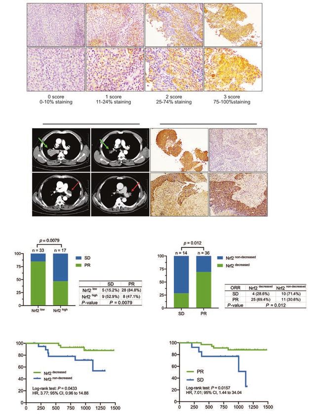
2                                                                         Huang et al. Metformin degrades Nrf2 to reverse chemoresistance

                 Keywords: Metformin; lung cancer; chemoresistance; Nrf2; posttranslational modification (PTM)

                 Submitted Aug 25, 2020. Accepted for publication Nov 27, 2020.
                 doi: 10.21037/tlcr-20-1072
                 View this article at: http://dx.doi.org/10.21037/tlcr-20-1072

Introduction                                                                 metabolism, metformin also regulates mitochondrial
                                                                             function. However, its effect on cellular ROS has not yet
Lung cancer is responsible for more deaths worldwide
                                                                             been fully elucidated.
than any other malignancy (1). Non-small cell lung cancer
                                                                                 The transcription factor nuclear factor erythoid-2-
(NSCLC) accounts for approximately 85% of lung cancer
                                                                             related factor 2 (NFE2L2/Nrf2), a master regulator of the
cases (2). In recent years, the tumor responses and clinical
                                                                             antioxidant response, plays a role in the most important
outcomes of patients with NSCLC have been dramatically
                                                                             endogenous defense mechanism by which ROS are
improved by targeted therapy and immunotherapy (3,4).
                                                                             maintained at low physiological levels. Nrf2 is essential
However, platinum-based systemic chemotherapy is still the
                                                                             to redox homeostasis, especially after cells have been
preferred first-line therapy for the remaining patients and
                                                                             exposed to chemotherapeutic agents (17,18). Nrf2 exerts
is the cornerstone in the treatment of resectable patients
                                                                             its detoxifying effect by binding to the antioxidant response
who undergo neoadjuvant chemotherapy (5). However,
various factors can result in the development of resistance                  element (ARE) and transactivating various cytoprotective
to chemotherapy (including reduced drug accumulation,                        genes, especially, heme oxygenase 1 (HO-1), which is one of
enhanced drug-mediated detoxification of oxidative stress,                   the strongest antioxidant phase II detoxifying enzymes.
and increased DNA adduct repair), which is a major                               “Nrf2 addiction” refers to hyperactivation of the
obstacle that limits our ability to treat advanced NSCLC                     Nrf2 pathway in lung cancer cells, which promotes
(6-8).                                                                       the development of NSCLC and can also enhance
   With the increasing number of combination strategies to                   chemoresistance (19,20). Emerging evidence has shown
attenuate tumor resistance and decrease chemotherapeutic                     that targeting Nrf2 is a potential therapeutic strategy for
toxicity (9), metformin, which is widely prescribed for the                  overcoming cisplatin resistance (21). Intriguingly, Truong
treatment of type II diabetes, has shown important anti-                     Do M revealed that metformin suppresses the expression
lung cancer effects in both in vitro and in vivo preclinical                 of Nrf2 at the transcriptional level by inhibiting Sirtuin 1
studies. The effect of metformin in combination with other                   (Sirt1) (22), while another study reported the opposite
treatment strategies has also been studied (10). Metformin                   result, with metformin also upregulating Sirt1 expression
was demonstrated to sensitize different cancer cell types to                 for decreasing the acetylation of Nrf2 and preventing its
cisplatin cytotoxicity, and various mechanisms have been                     nuclear distribution (23). Metformin somehow negatively
described, from mitochondrial apoptosis to the inhibition                    modulates Nrf2 expression in lung cancer, but there
of DNA synthesis (11). Although the signal transduction                      is complete lack of understanding of the underlying
mechanisms by which the combination of metformin                             mechanisms. Some Nrf2-ECH homology (Neh) domains
with cisplatin potentiates cytotoxicity in lung cancer are                   in Nrf2 are tightly regulated by various posttranslational
evidenced by a large body of research (12-14), fewer studies                 modifications (PTMs), such as phosphorylation and
have focused on the detoxification of reactive oxygen                        ubiquitylation (24), which effectively confer changes in Nrf2
species (ROS) under cisplatin-induced oxidative stress.                      expression. Effective PTMs in Nrf2 can change its location
Notably, mutagenic ROS is involved during carcinogenesis                     or expression level (17). Extracellular signal-regulated
and chemotherapy resistance (15). Conversely, high levels                    kinases 1/2 (ERK1/2) were shown to be involved in the
of ROS can further form DNA double-strand breaks,                            regulation of Nrf2 by metformin treatment (25). Butylated
resulting in a DNA catastrophe and subsequently inducing                     hydroxyanisole was reported to increase phosphorylation
apoptosis (16). Therefore, the increased cellular antioxidant                of the ERK1/2, thus promoting Nrf2 translocation into the
capacity may play a vital role in lung cancer cellular                       nucleus (26). However, the relationship between ERK1/2
adaptation to cisplatin-induced oxidative stress. ROS are                    and Nrf2-related PTMs still remains unclear and few
generated in mitochondria. As a drug regulating glucose                      studies have explored the effect of clinical agents on the

© Translational Lung Cancer Research. All rights reserved.                        Transl Lung Cancer Res 2020 | http://dx.doi.org/10.21037/tlcr-20-1072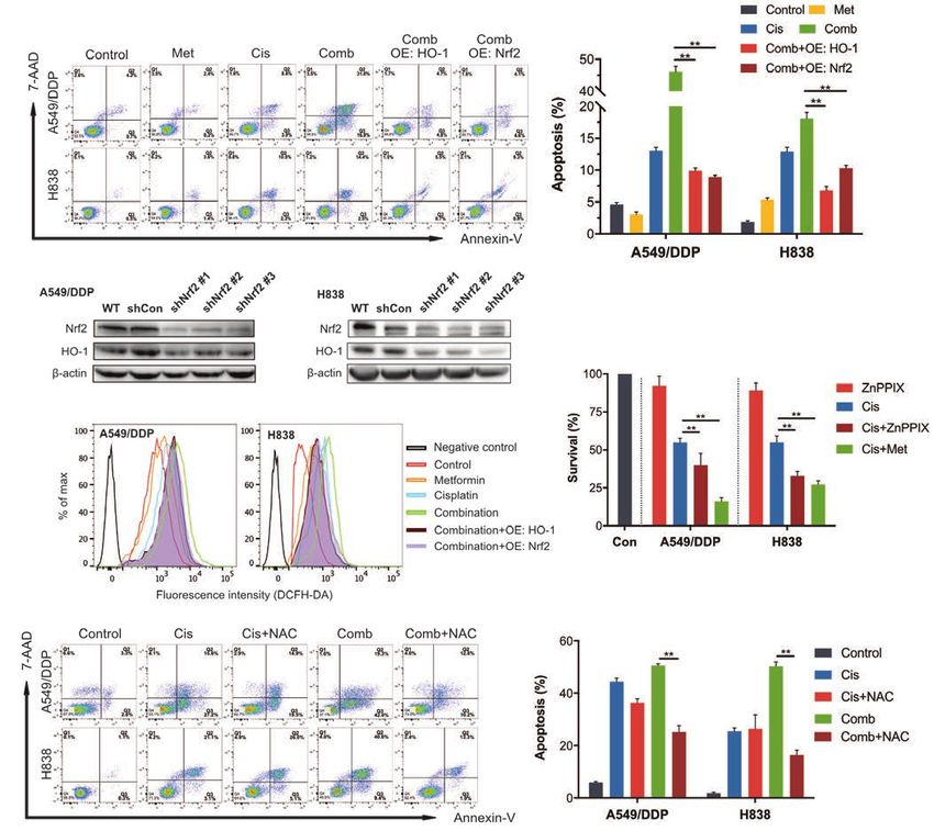
Translational Lung Cancer Research, 2020                                                                                               3

PTM status of Nrf2, which in turn affects Nrf2 activation.      infection, and the media were replaced with a medium
   In the current study, we investigated the role of Nrf2       (250 µL) containing lentivirus and polybrene (8 µg/mL).
in the regulation of cisplatin-induced ROS production           After infection for 4 h, another 250 µL of fresh medium
and chemoresistance in NSCLC cells with a more                  containing polybrene (8 µg/mL) was added into wells.
comprehensive analysis. Our data elucidate, for the             The original medium was replaced with fresh medium
first time, that the promotion effect of metformin on           24 h later. And the infected cells were selected with
mitochondrial ROS production plays a critical role in           puromycin (2 µg/mL) 72 h after infection. The mature
chemoresistance reversal in lung cancer. We explored the        antisense sequences targeting Nrf2 are as follows:
mechanism of metformin-mediated Nrf2 degradation                5'-CACCAGAACACTCAGTGGAAT-3' (shNrf2 #1),
through attenuating ERK-mediated Nrf2 phosphorylation           5'-GAGCAGTTCAATGAAGCTCAA-3' (shNrf2 #2),
to restore its polyubiquitination, which has a weakening        5'-AGCCATTCACTCTCTGAACTT-3' (shNrf2 #3).
effect on the ROS defense system in NSCLC. Importantly,
for the first time, we evaluated the difference in Nrf2
                                                                Reagents and antibodies
levels in NSCLC patients before and after neoadjuvant
chemotherapy as well as the correlation of this change with     Metformin (1,1-Dimethylbiguanide hydrochloride,
chemotherapeutic response and patient survival. We present      #D150959-5G), Protoporphyrin IX zinc (ZnPPIX,
the following article in accordance with the ARRIVE             #282820), N-Acetyl-L-cysteine (NAC, #A7250) and
reporting checklist (available at http://dx.doi.org/10.21037/   2’,7’-Dichlorofluorescein diacetate (DCFH-DA, #D6883)
tlcr-20-1072).                                                  were purchased from Sigma-Aldrich (St. Louis, MO, USA).
                                                                Cycloheximide (CHX, CAS 66-81-9) was purchased from
                                                                MedChemExpress LLC (Monmouth Junction, NJ, USA).
Methods                                                         Cisplatin and proteasome inhibitor MG132 was purchased
Cell culture and transfection                                   from Selleck Chemicals (Houston, TX, USA). The Annexin
                                                                V-PE/7-AAD Apoptosis Detection Kit (AP104) was
Human lung cancer cells A549/DDP (Bogoo                         purchased from Multi Sciences (Hangzhou, China). U0126
Biotechnology, Shanghai, China), a cisplatin-resistant A549     (#9903). The antibodies used for immunoblotting and co-
cell line, and H838 (Cell Bank of China Science Academy,        immunoprecipitation, β-actin (#58169), Nrf2 (#12721),
Shanghai, China) were maintained in RPMI 1640 medium            caspase-3 (#9662), poly ADP-ribose polymerase (PARP)
(CORNING, Mediatech, Inc., Manassas, VA, USA)                   (#9532), Phospho-c-Raf (#9431), p44/42 MAPK (Erk1/2,
supplemented with 10% fetal bovine serum (FBS, PAN-             #4695), Phospho-p44/42 MAPK (#8544), and ubiquitin
Biotech, Aidenbach, Germany) in a humidified atmosphere         (#3936), along with goat anti-rabbit immunoglobulin G
with 5% CO2. Cisplatin (800 ng/mL) was added in fresh           (IgG, #7074) and goat anti-mouse (#7076) HRP-linked
medium for A549/DDP to maintain its resistance. Cell            secondary antibodies, were purchased from Cell Signaling
lines were authenticated by Biowing Applied Biotechnology       Technology, Inc. (Danvers, MA, USA). Antibodies against
Co., Ltd and Cinoasia Institude (Shanghai, China) via short     Keap1 (AF5266) and HO-1 (AF5393) were obtained from
tandem repeat profiling.                                        Affinity Biosciences (Cincinnati, OH, USA).
   Commercial plasmids pcDNA3.1-mNrf2 and
pcDNA3.1-mHO-1 were supplied by Genechem Co.,
                                                                Cell viability assay
Ltd (Shanghai, China). The plasmids were transfected
into NSCLC cells using Lipofectamine 3000 reagent               Cell viability was measured with a Cell Counting Kit-8
(Invitrogen, CA, USA) according to the manufacturer’s           (CCK-8) assay (Dojindo, Japan). NSCLC cells were seeded
instructions. After 6 h, the original medium was replaced       in 96-well plates overnight at a density of 5,000 cells per
with fresh medium. At 48 h after transfection, the cells were   well, and then treated with drugs under the indicated
harvested or treated with drugs.                                conditions and times. Next, 10% of the final volume of
   For knockdown of Nrf2 expression, lentivirus containing      CCK-8 solution was added to each well and incubated at
an shRNA sequence targeting Nrf2 were supplied by               37 ℃ for 1 h. A SpectraMax M4 microplate reader
Genechem Co., Ltd (Shanghai, China). NSCLC cells were           (Molecular Devices, LLC, Sunnyvale, CA, USA) was used
seeded at 30% confluence in 24-well plates overnight before     to measure the absorbance at a wavelength of 450 nm.

© Translational Lung Cancer Research. All rights reserved.          Transl Lung Cancer Res 2020 | http://dx.doi.org/10.21037/tlcr-20-1072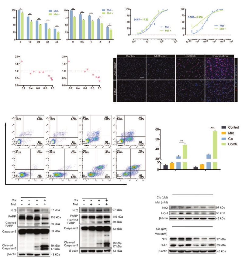
4                                                             Huang et al. Metformin degrades Nrf2 to reverse chemoresistance

   The IC50 curves were calculated using variable slope for     RIPA lysis buffer (Beyotime, Shanghai, China). Sonication
log(inhibitor) vs. normalized response (Graphpad Prism 8.3,     was used to reduce the viscosity of the lysate, and a BCA
La Jolla, CA). The combination index (CI) was calculated        assay (Solarbio, China) was carried out to measure the
using CompuSyn software (ComboSyn, Inc., Paramus, NJ,           protein concentration. The lysates were then mixed with
USA), with CI 1.1 indicating synergism,     3X reducing SDS loading buffer (#7723, Cell Signaling
additive effect, and antagonism, respectively.                  Technology, Inc., USA) and denatured at 100 ℃ for
                                                                5 min. The lysates were separated with 10–12% sodium
                                                                dodecyl sulfate polyacrylamide gel electrophoresis (SDS-
Cell apoptosis assay
                                                                PAGE) gels and then transferred onto a polyvinylidene
The distribution of apoptotic cells was evaluated using         difluoride (PVDF) membrane (Millipore, Billerica, MA).
Annexin V-PE/7-AAD staining. After indicated treatments,        The membrane was blocked with 5% nonfat milk for 1 h
NSCLC cells were trypsinized in 0.25% trypsin with              at room temperature and incubated overnight at 4 ℃ with
Ethylenediaminetetraacetic acid (EDTA)-free), washed with       primary antibodies. After that, the membrane was washed 3
cold phosphate-buffered saline (PBS), and then suspended        times with TBST (tris-buffered saline, 0.1% Tween-20) and
in 100 μL PBS. Subsequently, the cells were stained with        then probed with a secondary antibody for 1 h. After washing
5 μL of Annexin V-PE and 10 μL of 7-AAD, before being           with TBST, the blots were incubated with a high-sensitivity
incubated at room temperature for 30 min in the dark.           chemiluminescence detection system (Millipore) and
The stained cells were visualized by flow cytometry (FACS       visualized using the Amersham Biosciences ECL Detection
CantoII, BD Biosciences, USA) and analyzed using Flowjo         System (Amersham plc, GE Healthcare, Chicago, IL, USA).
(FlowJo 10.4, LLC, USA).                                           For the IP assay, the Pierce™ Direct Magnetic IP/
   Drug toxicity was assessed by Hoechst 33342/PI staining.     Co-IP Kit (Thermo Scientific, #88828) was used. The
NSCLC cells were seeded into 6-well plates at a density         main procedure was carried out in accordance with the
of 4×10 5/well. After incubation for 24 h, the cells were       manufacturer’s instructions. Briefly, Nrf2 antibody (5 μg)
treated with metformin and/or cisplatin under the indicated     was incubated with magnetic beads (25 μL) for 60 min. Cell
conditions. Next, the cells were trypsinized, resuspended       lysates were mixed with antibody-coupled magnetic beads
in PBS with Hoechst 33342 10 μg/mL and incubated for            and incubated for 2 h at room temperature on a rotator.
15 min at 37 ℃. The cells were then washed twice with           Precipitated proteins (60 μL) were eluted from beads and
PBS, and stained with PI (10 μg/mL) for 15 min at room          prepared for western blot analysis.
temperature. Finally, the cell suspension was dripped onto
the glass slides and observed by fluorescence microscopy.
                                                                Phos-tag analysis

                                                                For Phos-tag SDS-PAGE, 20 μM phosbind acrylamide
ROS detection
                                                                (F4002, APExBIO, Houston, TX, USA) and 80 μM
After the indicated treatments, adherent cells were             MnCl2 were added to 8% SDS-polyacrylamide gel before
harvested with trypsin-EDTA solution and washed once            polymerization. The gel was electrophoresed at 25 mA for
with PBS. For total cellular ROS detection, the cells were      2 h. Before transfer, the gel was first immersed three times
incubated with 10 µM ROS-sensitive probe (DCFH-DA)              in transfer buffer containing 10 mM EDTA for 10 min and
in PBS for 30 min at 37 ℃ in darkness. For mitochondrial        then in transfer buffer without EDTA twice for 10 min.
ROS accumulation detection, NSCLC cells were incubated          Protein transfer was performed for 3 h at 350 mA at 0 ℃,
with 5 µM mitochondrial superoxide indicator (MitoSOX           and then the membrane was analyzed by western blotting
Red, Thermo Scientific, M36008) in PBS for 10 min at            with anti-Nrf2 antibody.
37 ℃ in darkness. The cells were then washed twice with
PBS and suspended in a fresh medium for immediate
                                                                Dephosphorylation with lambda phosphatase
analysis by flow cytometer.
                                                                NSCLC cells were seeded in 10-cm culture dishes
                                                                overnight and collected using 400 μL IP Lysis Buffer. The
Western blot and immunoprecipitation (IP) assay
                                                                collected cells were incubated with 5 μL of lambda protein
Cells were first washed with ice-cold PBS and then lysed in     phosphatase (New England BioLabs, Ipswich, MA, USA)

© Translational Lung Cancer Research. All rights reserved.          Transl Lung Cancer Res 2020 | http://dx.doi.org/10.21037/tlcr-20-1072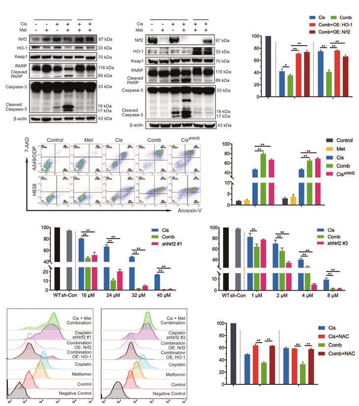
Translational Lung Cancer Research, 2020                                                                                             5

diluted in 50 μL of 10× Protein MetalloPhosphatases (PMP)     American Veterinary Medical Association Guidelines
and 50 μL of 10 mM MnCl2 for 30 min at 30 ℃. After            (Schaumburg, IL, USA).
incubation, the samples were analyzed by western blot or IP
as described above.
                                                              Terminal deoxynucleotidyl transferase dUTP nick-end
                                                              labeling assay
Quantitative real-time polymerase chain reaction
                                                              Detection of apoptotic cells was performed with a terminal
Total RNA was extracted using RNA-Quick Purification          deoxynucleotidyl transferase dUTP nick-end labeling
Kit (Yishan Biotech, Shanghai, China). cDNA was               (TUNEL) assay on paraffin-embedded sections of xenograft
synthesized using HiScript II Q RT SuperMix (Vazyme           tumor tissue using an In Situ Cell Death Detection Kit,
Biotech, Nanjing, China). Real-time polymerase chain          POD (Roche) according to the manufacturer’s instructions.
reaction (PCR) was performed on an ABI Prism 7500             Images were captured using a light microscope.
Fast Real-Time PCR System (Applied Biosystems, Foster
City, CA, USA) using ChamQ SYBR qPCR Master Mix
                                                              Immunohistochemical analysis
(Vazyme). The 2 −ΔΔCt method was used to calculate the
relative expression levels of Nrf2 with β-actin used for      Paraffin-embedded tumor tissue sections (4 μM) were
normalization. The sequences of the primers used for          deparaffinized and subjected to heated-induced epitope
quantitative real-time PCR (qRT-PCR) were as follows:         retrieval using citrate buffer (pH 6.0). The tissues
   • Nrf2: Forward 5'-TCAGCGACGGAAA                           were incubated with rabbit anti-Nrf2 (1:200 dilution,
      GAGTATGA-3'; Reverse 5'-CCACTGGTTT                      Abcam, #ab31163) or anti-Ki67 (1:200 dilution, Abcam,
      CTGACTGGATGT-3'.                                        #ab15580) antibody overnight at 4 ℃. Following that, the
   • β-actin: Forward 5'-TGGCACCCA                            tissues were incubated with the corresponding secondary
      GCACAATGAA-3'; Reverse 5'-CTAAGTCATAGT                  antibodies. Peroxidase activity was visualized with
      CCGCCTAGAAGCA-3'.                                       3,3'-diaminobenzidine (DAB; Darko, CA, USA) and then
                                                              counterstaining with hematoxylin was performed.
                                                                 The sections were examined by two senior pathologists
Xenograft model and treatments
                                                              in a double-blinded manner. The intensity of Nrf2 staining
All animal experiments were approved by the Committee         was quantified using a four-score grading system (0=0–10%
of Animal Experimental Ethical Inspection of the First        of stained cells; 1=11–24% of stained cells; 2=25–75% of
Affiliated Hospital, College of Medicine, Zhejiang            stained cells; 3=>75% of stained cells).
University (reference No. 2019-1232), and followed by the
institutional Guidelines for animal care and use.
                                                              Tumor specimens and patients
   Five-week-old male nude mice (Shanghai Sippr-bk
Laboratory Animal Co., Ltd, China; weight, 16 to 20 g)        All procedures performed in this study involving human
were injected subcutaneously in the left flanks with 5×10⁶    participants were in accordance with the Declaration of
A549/DDP cells mixed 1:1 with 100% Matrigel. On day 8,        Helsinki (as revised in 2013). Patients were informed that
when tumors had reached 40–80 mm 3, the mice were             the specimens were stored by the hospital and potentially
separated into four groups (5 mice per group) and given       used for scientific research, and signed informed consent
water (50 μL/10 g; qd) + intraperitoneal saline (100 μL/      to participants was waived by the Ethics Committee.
10 g; qod; control group) by oral gavage, metformin           This study was approved by the Clinical Research Ethics
(200 mg/kg; qd) + intraperitoneal saline (100 μL/10 g; qod;   Committee of the First Affiliated Hospital, College of
metformin group) by oral gavage, water (50 μL/10g; qd)        Medicine, Zhejiang University (Approval No.: 2020-13).
+ intraperitoneal cisplatin (2 mg/kg; qod; cisplatin group)   Clinical samples were obtained from 50 primary lung cancer
orally, or metformin (200 mg/kg; qd) + intraperitoneal        patients who received platinum-containing neoadjuvant
cisplatin (2 mg/kg; qod; combination group) orally.           chemotherapy followed by surgical resection. To be enrolled
The tumor volume was calculated using the following           in this study, patients must have been pathologically
formula: volume = 1/2(length × width 2). The animals          confirmed before neoadjuvant chemotherapy and obtained
were euthanized by cervical dislocation according to the      their final pathological report after surgery. Matched biopsy

© Translational Lung Cancer Research. All rights reserved.        Transl Lung Cancer Res 2020 | http://dx.doi.org/10.21037/tlcr-20-1072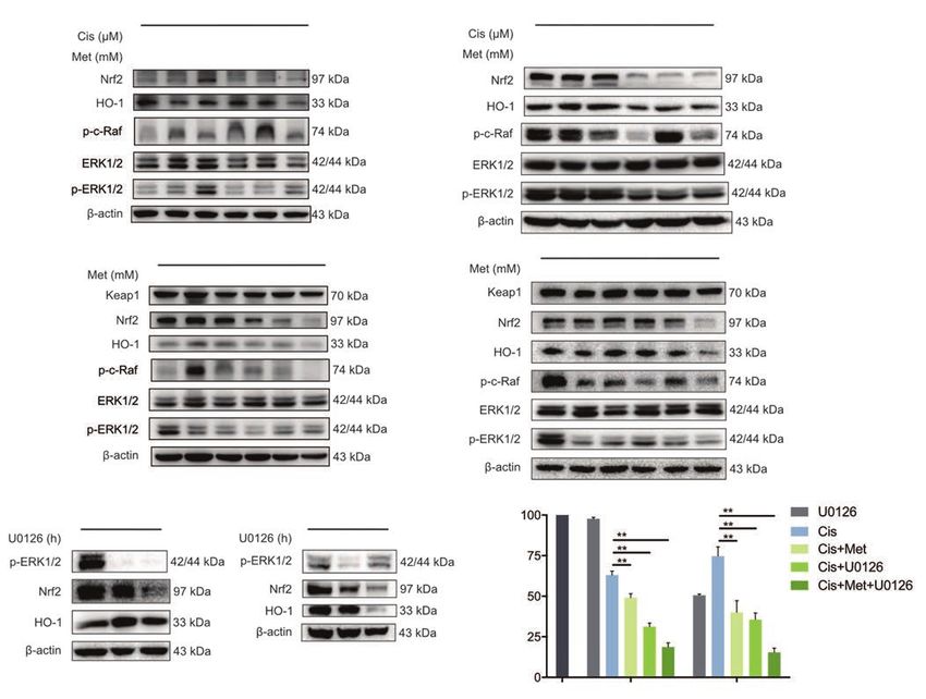
6                                                                   Huang et al. Metformin degrades Nrf2 to reverse chemoresistance

and surgical resection tissue samples from eligible patients          that metformin and cisplatin synergistically suppressed
were embedded in formalin-fixed paraffin, and IHC                     NSCLC proliferation (Figure 1C).
staining was used to confirm chemotherapy-related changes                Next, the effect of metformin on cisplatin-induced
in Nrf2 status. Patients were regularly followed and the              NSCLC cell death was evaluated. Cells in the late stage
chemotherapeutic response was clinically evaluated using              of apoptosis were stained with both Hoechst 33342 and
the Response Evaluation Criteria in Solid Tumors (RECIST              PI (pink). Combination treatment with metformin and
1.1). The objective response rate (ORR) used in this                  cisplatin significantly enhanced the induction of apoptosis
cohort was defined as follows: (I) complete response (CR):            (Figure 1D). Flow cytometry analysis showed that
disappearance of all known lesions; (II) partial response (PR),       combination treatment significantly increased the number
≥30% regression in the sum of the measurable diameters of             of apoptotic NSCLC cells (the total percentage of cells
target lesions; (III) progressive disease (PD), ≥20% increase         in quadrants Q2 + Q3) compared to that following
in the sum of the measurable diameters of target lesions; (IV)        treatment with cisplatin alone, while single treatment with
stable disease (SD), any response between PR and PD.                  metformin (3.2 and 12.8 mM for A549/DDP and H838
                                                                      cells, respectively) did not clearly promote cell apoptosis
                                                                      (Figure 1E). The protein levels of caspase-3 (cleaved
Statistical analysis                                                  caspase-3), a mediator of apoptosis signaling, and PARP
The two-tailed Student’s t-test was used for comparisons              (cleaved PARP) were also evaluated, which revealed similar
between two groups, and one-way or two-way analysis                   results in NSCLC cells treated with metformin and/or
of variance (ANOVA) followed by Tukey’s test was used                 cisplatin (Figure 1F). Intriguingly, metformin augmented
for multiple group comparisons. Categorical data were                 cisplatin-induced apoptosis by strongly inhibiting the
compared using Fisher’s exact text. For multi-classified              expression of Nrf2. With cisplatin stimulation, the levels of
variables, binary logistic regression analysis was adopted.           Nrf2 and HO-1 dose-dependently increased. Metformin
Survival curves were estimated using the Kaplan-Meier                 strongly inhibited cisplatin-mediated upregulation of Nrf2
method and analyzed using the log-rank test. Prism                    and HO-1 in both chemoresistant and normal NSCLC
8.3 (GraphPad, La Jolla, CA) and SPSS software 25.0                   cells (Figure 1G). These results therefore demonstrated
(IBM statistics, SPSS Inc., Chicago, IL, USA) were used               that metformin synergistically enhanced cisplatin-mediated
for statistical analyses. A P value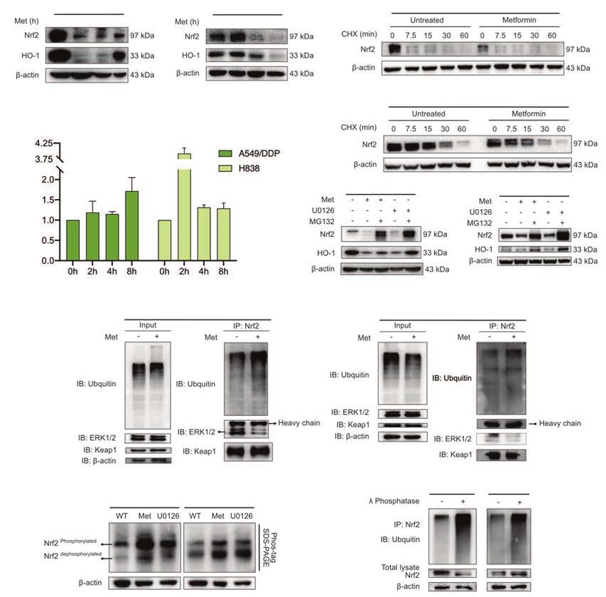
Translational Lung Cancer Research, 2020                                                                                                                                                                                                           7

          A                                         A549/DDP                                                    H838
                                                                                                                                  B
                                                                                                                                                       A549/DDP                                                            H838
                                                                                                                                                                                                                         IC50 Shift

                                                                                                                                  Inhibition (%)

                                                                                                                                                                                               Inhibition (%)
                                                                                                                                                          IC50 Shift
                     Survival (%)

                                                                                      Survival (%)
                                                   Cisplatin (μM)                                            Cisplatin (μM)                                            [Cis], μM                                                       [Cis], μM

          C                                         A549/DDP                                                  H838
                                                                                                                              D
                      Combination index (Cn)

                                                                    Combination index (Cn)
                                               Cis +3.2 mM Met                                       Cis +12.8 mM Met

                                                  Dose effect                                           Dose effect

           E
                                               Control                                 Met                              Cis                        Comb
            7-AAD
                    A549/DDP

                                                                                                                                                                        Apoptosis (%)
                    H838

                                                                                                                                                                                        A549/DDP                                  H838

                                                                                                                                                   Annexin-V

           F                                     A549/DDP                                                             H838
                                                                                                                                                                         G                                          A549/DDP

                                                                                                                                                                                           0               16       32       0        16   32
                                                                                                                                                                                           0                    0    0      3.2   3.2      3.2

                                                                                                                                                                                                                        H838
                                                                                                                                                                                           0                4       8        0        4     8
                                                                                                                                                                                           0                0       0      12.8 12.8 12.8

Figure 1 Synergistic effect of combination therapy with metformin and cisplatin on NSCLC cells. (A) Cell viability was assessed using
the CCK-8 assay. The number of surviving cells is expressed as the percentage of the control (100% survival). (B) Half maximal inhibitory
concentration (IC50) curves for single and combination therapy in NSCLC cells were determined using the CCK-8 assay. (C) The
combination index (CI 1.1 indicates antagonism; 0.9< CI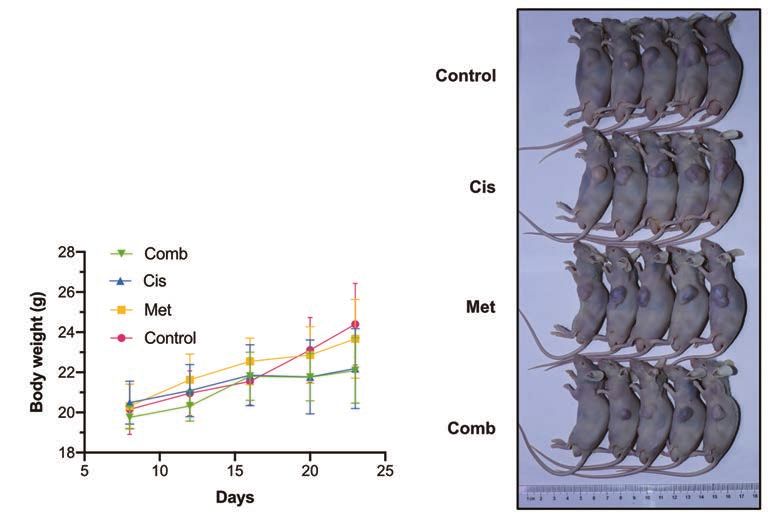
8                                                                                                       Huang et al. Metformin degrades Nrf2 to reverse chemoresistance

                        A                                                                                                                                      B

                                                                     O O-1

                                                                                                           O O-1
                                                                           rf2

                                                                                                                 rf2
                                                                         H

                                                                         N

                                                                                                               H

                                                                                                               N
                                                      A549/DDP                                   H838

                                                                   E:

                                                                      E:

                                                                                                          E:

                                                                                                            E:
                                                                  O

                                                                                                         O

                                                                                                                                                             Survival (%)
                                                                                                                                                                            Con       A549/DDP       H838

                        C

                                                                                                                                      Apoptosis (%)
                                                                                                                                                            A549/DDP                          H838

                        D                                                     A549/DDP                                                                                             H838
                                       Survival (%)

                                                                                                               Survival (%)

                        E                                                        Cisplatin                                                                                        Cisplatin

                            A549/DDP                                             H838
                                                                                                                     F
                                                                                                                              Survival (%)

                                                                                                                                                      Con            A549/DDP                 H838

                                                          Fluorescence intensity (MitoSox Red)

Figure 2 ROS-detoxification of the Nrf2/HO-1 axis mediates the antitumor effect of combination therapy. NSCLC cells (A549/DDP
and H838) were treated with metformin (3.2 and 12.8 mM, respectively) and/or cisplatin (32 and 2 μM, respectively) for 48 h after
overexpression of HO-1 or Nrf2 for 48 h. (A) Western blot showing the invalid effect of combination therapy in NSCLC cells after
overexpression of HO-1 or Nrf2. Protein levels of PARP, cleaved PARP, caspase-3, cleaved caspase-3, Keap1, Nrf2, HO-1 and β-actin are
shown. (B) Cell viability of HO-1- and Nrf2-overexpressing NSCLC cells after combination therapy was assessed by CCK-8 assay. (C)
Nrf2 knockdown enhanced cisplatin induced cellular apoptosis in NSCLC cells. A549/DDP and H838 cells were treated with metformin
(3.2 and 12.8 mM, respectively) and/or cisplatin (32 and 16 μM, respectively) for 24 h after pretreatment with metformin (12.8 mM) for
24 h. The stable knockdown of Nrf2 in A549/DDP and H838 cells were treated with cisplatin (32 and 16 μM, respectively) for 24 h. After
indicated treatment, cells were examined using Annexin-V/7-AAD staining, and the distribution of apoptotic cells was measured by flow
cytometry analysis. (D) Nrf2 knockdown promoted sensitivity of NSCLC cells to cisplatin. Cell viability of Nrf2-knockdown NSCLC cells
was assessed by CCK-8 assay. (E) Suppression of Nrf2/HO-1 axis caused the heavy mitochondrial ROS accumulation in NSCLC cells. After
indicated treatment, Nrf2-knockdown and Nrf2/HO-1-overexpressing NSCLC cells were examined using MitoSOX Red, a mitochondrial
superoxide indicator (the negative control was not treated with MitoSOX Red). (F) The effects of NAC on the NSCLC cell proliferation
after combination therapy were assessed by CCK-8 assay. Treatment with NAC (100 μM) and the other indicated drugs was carried out
for 48 h. All experiments were independently repeated at least three times. Bars represent the means ± SDs. *, P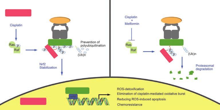
Translational Lung Cancer Research, 2020                                                                                               9

overexpression (Figure 2B, Figure S1A). Furthermore,            kinases upstream of metformin-abolished Nrf2 activation,
NSCLC cells were transfected with lentivirus containing an      the phosphorylation status of MAPKs with combination
shRNA sequence targeting Nrf2, and verified the inhibition      treatment with increasing concentrations of cisplatin was
efficiency (Figure S1B). Accordingly, the sensitivity and       evaluated first. Cisplatin activated the phosphorylation
cytotoxicity of the NSCLC cells to cisplatin was remarkably     of Raf and ERK1/2 in a dose-dependent manner, while
enhanced upon Nrf2 knockdown in NSCLC cells                     metformin at the indicated concentration strongly
(Figure 2C,D). The stable knockdown of Nrf2 showed              suppressed the Nrf2/HO-1 axis and the phosphorylation of
a similar effect of chemoresistance reversal to the             Raf-ERK1/2, which was activated by cisplatin (Figure 3A).
combination therapy. These results suggested that the           Metformin caused a dose-dependent reduction in the
detoxification ability of Nrf2/HO-1 axis toward cisplatin is    protein levels of Nrf2 and HO-1 in A549/DDP and H838
critical for NSCLC cells to develop drug resistance.            cells. Similarly, the phosphorylation of Raf and ERK1/2
   Next, the intracellular ROS level was detected in Nrf2-      was dose-dependently reduced by metformin. However,
knockdown and Nrf2/HO-1-overexpressing NSCLC                    the Keap1 protein level was not affected by metformin
cells using MitoSOX Red (Figure 2E). Evaluation of              (Figure 3B).
mitochondrial superoxide levels revealed that metformin            To further confirm the role of Raf-ERK signaling in
markedly increased the cisplatin-induced production             the regulation of Nrf2, the two cell lines were treated with
of ROS. Nrf2-knockdown NSCLC cells also exhibited               30 μM U0126, a highly selective inhibitor of mitogen-
remarkably increased mitochondrial production of ROS            activated protein kinase (MEK) 1 and MEK 2 that functions
in response to cisplatin treatment. With overexpression         upstream of ERK1/2, for 24 and 32 h. U0126 strongly
of Nrf2 and HO-1, the curve indicating ROS levels               suppressed the expression of Nrf2 in a time-dependent
moved substantially to the left, showing decreased              manner (Figure 3C). Further, the combination treatment
intracellular ROS accumulation under metformin                  with U0126 and cisplatin for 48 h was found to have
and cisplatin treatment. The total ROS detection by             synergistic inhibitory effects on NSCLC cell proliferation
2',7'-Dichlorofluorescein diacetate (DCFH-DA) probe             (Figure 3D). Single treatment with U0126 (30 μM) had
produces the similar results (Figure S1C). Then, the cells      almost no inhibitory effect on the chemoresistant cell line
were treated with the antioxidant N-acetyl cysteine (NAC)       (average survival rate, 97%), although it strongly inhibited
for 48 h and cisplatin or the combination of metformin          cell proliferation in a combination treatment with 16 μM
and cisplatin. NAC, a scavenger of ROS, inhibited the           cisplatin (average survival rate, 31%). Taken together,
synergistic effects of the combination therapy on cell death    these results indicate that inactivation of the Raf-ERK
(Figure S1D) and proliferation (Figure 2F), indicating that     signaling pathway is involved in the metformin-mediated
the synergistic effects of metformin and cisplatin depend on    downregulation of Nrf2 and HO-1.
intracellular ROS levels. Moreover, metformin and specific
HO-1 inhibitor ZnPPIX treatment (10 μM) significantly
                                                                Metformin accelerates Nrf2 degradation by increased
decreased the proliferation of A549/DDP and H838 cells
                                                                ubiquitin modification from its dephosphorylation
compared to that following treatment with cisplatin alone
(Figure S1E); this finding confirmed that HO-1 suppression      Nrf2 is mainly controlled through the regulation of
is responsible for the antiproliferative effect of metformin.   protein turnover by ubiquitin-mediated proteasomal
Overall, these results suggest that ROS-induced NSCLC           degradation and exhibits a half-life of approximately
cell death is regulated by the Nrf2/HO-1 signaling axis in      30 min (27). Given that metformin strongly suppressed
chemotherapy and that metformin weakens the Nrf2/HO-1           the protein level of Nrf2, even under cisplatin-activated
axis and the ROS-detoxification system to further reverse       conditions, we hypothesized that metformin can inhibit
chemoresistance.                                                the mRNA expression of Nrf2, thus reducing the protein
                                                                synthesis of Nrf2, and/or regulate Nrf2 protein at the
                                                                PTM level. On the basis that a mechanism must underlie
Metformin inhibits the cisplatin-activated Nrf2/HO-1 axis
                                                                metformin-mediated Nrf2 suppression, we investigated the
via Raf-ERK inactivation
                                                                possibilities. A549/DDP and H838 cells were maintained
ERK1/2 have been reported to regulate Nrf2 expression           in medium supplemented with cisplatin (8 and 1 μM,
via a keap1-independent mechanism (25). To evaluate the         respectively) to stably activate Nrf2 expression and simulate

© Translational Lung Cancer Research. All rights reserved.          Transl Lung Cancer Res 2020 | http://dx.doi.org/10.21037/tlcr-20-1072
10 Huang et al. Metformin degrades Nrf2 to reverse chemoresistance
A A549/DDP H838
0 16 32 0 16 32 0 4 8 0 4 8
0 0 0 3.2 3.2 3.2 0 0 0 12.8 12.8 12.8
H838
B A549/DDP
0 0.8 1.6 3.2 6.4 12.8
0 0.8 1.6 3.2 6.4 12.8
C D
A549/DDP H838
0 24 32 0 24 32
Survival (%)
Con A549/DDP H838
Figure 3 Metformin inhibits Nrf2 expression in NSCLC cells through the inactivation of Raf-ERK signaling. (A,B) Western blot showing
inactivation of the Raf-ERK-Nrf2 signaling pathway in NSCLC cells treated with different concentrations of drugs in combination or
alone. Protein levels of Nrf2, HO-1, keap1, p-c-Raf, ERK1/2, p-ERK1/2, and β-actin are shown. (C) NSCLC cells were treated with U0126
(30 μM), a MEK1/2 inhibitor, for 0, 24 and 32 h. Western blot showing the levels of Nrf2/HO-1 pathway protein and the phosphorylation
status of ERK1/2 in NSCLC cells. (D) The effects of metformin and U0126 on the proliferation of NSCLC cells were assessed by CCK-8
assay. A549/DDP and H838 cells were treated with cisplatin (16 and 2 μM, respectively) and/or metformin (12.8 mM) for 48 h after
pretreatment with U0126 (30 μM) for 2 h. All experiments were independently repeated at least three times. Bars represent the means ±
SDs. **, PTranslational Lung Cancer Research, 2020 11
A A549/DDP H838
C A549/DDP
0 2 4 8 0 2 4 8
H838
B
Cis + Met treatment
Relative Nrf2 mRNA to actin
D
(fold of control)
A549/DDP H838
E A549/DDP H838
F A549/DDP H838
G A549/DDP H838
Figure 4 Metformin regulates Nrf2 by posttranslational modification. NSCLC cells (A549/DDP and H838) were maintained in medium
supplemented with cisplatin (8 and 1 μM, respectively). (A) Western blot showing the time course of the effect of metformin (12.8 mM) on
Nrf2 and HO-1 protein levels. (B) Effects of metformin (12.8 mM) on Nrf2 mRNA expression in NSCLC cells determined by quantitative
real-time polymerase chain reaction. (C) NSCLC cells were pretreated with metformin (12.8 mM) or control diluent for 2 h, followed by
10 μg/mL cycloheximide (CHX) treatment for 0 to 60 min to block protein synthesis. Western blot showing the protein levels of Nrf2
and β-actin. (D) Metformin and U0126 strongly promoted the degradation of Nrf2. NSCLC cells were first treated with or without
20 μM MG132 (a proteasome inhibitor) for 2 h. Then, the cells were treated with metformin (12.8 mM) or U0126 (30 μM) for 48 h, and
the expression levels of Nrf2, HO-1, and β-actin were analyzed by Western blot. (E) The ubiquitination status of Nrf2 and proteins that
interact with Nrf2 with or without metformin (12.8 mM) treatment. Nrf2 ubiquitination was detected by immunoprecipitation (IP) with
anti-Nrf2 antibody and immunoblotting (IB) with anti-ubiquitin antibody. The binding of Nrf2 to Keap1 and Nrf2 to ERK1/2 was detected
by IP with anti-Nrf2 antibody and IB with anti-Keap1 and anti-ERK1/2 antibodies. The protein expression levels of ubiquitin, ERK1/2,
Keap1, and β-actin in whole-cell lysates were confirmed. (F) Effects of metformin and U0126 on the dephosphorylation of Nrf2 in NSCLC
cells were analyzed by Phos-tag assay. NSCLC cells were pretreated with MG132 (20 μM) for 2 h, followed by metformin (12.8 mM) and
U0126 (30 μM) treatment for 24 h. (G) Dephosphorylation of Nrf2 increased its ubiquitin modification. NSCLC cells were treated with
MG132 (20 μM) for 2 h before the collected cells were dephosphorylated with lambda phosphatase. Nrf2 ubiquitination was detected by IP.
All experiments were independently repeated at least three times. Bars represent the means ± SDs. NSCLC, non-small cell lung cancer; Cis,
cisplatin; Met, metformin; WT, wild-type.
© Translational Lung Cancer Research. All rights reserved. Transl Lung Cancer Res 2020 | http://dx.doi.org/10.21037/tlcr-20-107212 Huang et al. Metformin degrades Nrf2 to reverse chemoresistance
or without pretreatment with the proteasome inhibitor each group. In the combination group, the levels of cleaved
MG132 were compared in each group, a large gap in PARP were dramatically elevated and cisplatin-induced
protein expression was found, which suggested that Nrf2 activation of the Nrf2/HO-1 signaling axis was inhibited
degradation in A549/DDP and H838 cells is dependent on (Figure 5D,E). The results of IHC staining for Nrf2 in
the proteasome and that both metformin and U0126 have xenograft tissues correlated with those of western blot
a strong ability to promote the degradation of enormous analysis. Moreover, Ki-67 and TUNEL staining confirmed
amounts of Nrf2 protein (Figure 4D). that combination treatment inhibited proliferation and
To analyze the ubiquitination status of Nrf2 in NSCLC increased apoptosis in NSCLC cells in vivo (Figure 5F).
cells with and without metformin treatment, a co- The findings from our in vivo experiments recapitulated the
immunoprecipitation (co-IP) assay was performed. As results obtained through our in vitro experiments.
Raf-ERK signaling is involved in metformin-mediated
regulation of Nrf2, the amount of ERK1/2 that binds
Non-decreased Nrf2 expression in patients after
to Nrf2 was also analyzed. Nrf2 polyubiquitination
neoadjuvant chemotherapy is associated with poor survival
was found to be increased in NSCLC cells treated
and chemoresistance in NSCLC
with metformin compared with the control cells.
Intriguingly, metformin had no effect on the binding To elucidate whether the tumor Nrf2 status is related to
of Keap1 and Nrf2, although it significantly reduced neoadjuvant chemotherapeutic response and survival in
protein-protein interaction between ERK1/2 and Nrf2 NSCLC patients, Nrf2 expression was detected and scored
(Figure 4E). Further, metformin and U0126 promoted in matched tumor tissues using IHC staining. The matched
the dephosphorylation of Nrf2 in NSCLC cells tumor samples were obtained from 50 NSCLC patients
(Figure 4F). Next, fresh cell extract from NSCLC cells who underwent biopsy prior to receiving cisplatin-based
was treated with lambda phosphatase for 30 min, a protein neoadjuvant chemotherapy followed by surgical resection.
phosphatase with activity towards phosphorylated serine, The baseline characteristics of the patients with different
threonine, and tyrosine residues. Lambda phosphatase responses to chemotherapy are shown in Table S1. The
strongly increased ubiquitin modification of Nrf2 patients were divided into two groups according to their
(Figure 4G), revealing that phosphorylated Nrf2 prevented Nrf2 IHC staining scores (Figure 6A): the Nrf2 low group
its polyubiquitination. Overall, these results clearly (score of 0 or 1) and the Nrf2 high group (score of 2 or 3).
demonstrate that metformin accelerates the ubiquitin- Patients with decreased Nrf2 scores after neoadjuvant
mediated proteolysis of Nrf2 in a Keap1-independent chemotherapy were classified into the decreased Nrf2
mechanism through preventing ERK1/2 phosphorylating group, and those with increased or unchanged Nrf2
Nrf2 extensively to restore polyubiquitination of Nrf2. scores were classified into the non-decreased Nrf2 group
(Figure 6B). Low Nrf2 expression after chemotherapy was
associated with an improved chemotherapeutic response
Combination therapy overcomes resistance to cisplatin in
(PTranslational Lung Cancer Research, 2020 13
A Tumor-bearing
(A549/DDP) Euthanasia Con Cis
0 2 4 6 8 10 12 14 16 18 20 22 24 Day
Met Comb
B C
Control
Tumor volume (mm3)
Tumor weight (mg)
Cis
Met
Comb
Days Xenografts
D Xenografts E
F
Nrf2
Ki-67
TUNEL
Positive cells/quadrant
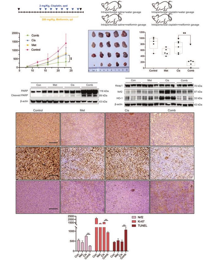
Figure 5 Metformin reversed chemoresistance in an A549/DDP xenograft model. (A) Schematic representation of the in vivo experiment.
The establishment of the xenograft model and treatments were described in the “Methods”. (B) Tumor sizes in the four groups were
recorded with a caliper every 4 days. (C) Gross morphology of subcutaneous tumors in the four groups and their net weights are shown
(n=5). (D) Three xenograft tumor tissues from each group were randomly selected and lysed. Western blot showing obvious apoptosis in
the xenograft combination group. The protein levels of PARP, cleaved PARP, and β-actin are shown. (E) Western blot showing inactivation
of the Nrf2/HO-1 signaling axis in the xenograft combination group. The protein levels of Keap1, Nrf2, HO-1, and β-actin are shown.
(F) IHC staining was used to detect the expression levels of Nrf2 and Ki-67 in the four xenograft groups (100× magnification; scale bar,
200 μm). Apoptotic cells in the four xenograft groups were determined by TUNEL staining (100× magnification; scale bar, 200 μm). The
bar graph indicates the results of quantification of cells positive for Nrf2, Ki-67, and TUNEL expression per quadrant. All experiments
were independently repeated at least three times. Points, error bars and connecting lines on the graph indicate the means and error ± SDs.
The box graph indicates the minimum, first quartile, median, third quartile, and maximum. The bar graph indicates the means ± SDs. **,
P14 Huang et al. Metformin degrades Nrf2 to reverse chemoresistance
A
100 ×
200 ×
B CT scan Nrf2
Patient 1 (PR)
Nrf2decreased
Patient 2 (SD)
Nrf2non-decreased
Befor neo After neo Befor neo After neo
C D
Nrf2 fluctuation
Percentage of total
Percentage of total
After neo-adjuvant
E Overall Survival F Overall Survival
Nrf2 fluctuation Different ORR
Percent survival (%)
Percent survival (%)
Time (days) Time (days)
Figure 6 Fluctuation in Nrf2 the level is associated with chemoresistance in NSCLC patients. Matched tumor samples were obtained from
50 NSCLC patients who underwent biopsy before neoadjuvant chemotherapy and received surgical resection after neoadjuvant therapy.
Tumor tissues were stained for Nrf2 protein using IHC staining. (A) Representative images of Nrf2 IHC staining from NSCLC cases
scored from 0 to 3 at 100× and 200× magnification. (B) Representative cases showing different chemotherapeutic responses. Left, computed
tomography images of patients with a partial response (PR, green arrows) and stable disease (SD, red arrows). Right, the results of Nrf2 IHC
at 100× magnification in matched tissues from patients before and after neoadjuvant chemotherapy. (C) A significant association was found
between Nrf2 expression (Nrf2 low = score of 0 or 1, Nrf2 high = score of 1 or 2) after chemotherapy and objective response rate (ORR)
in 50 patients. (D) The difference in Nrf2 expression in patients before and after neoadjuvant chemotherapy was correlated with the ORR.
(E) Kaplan-Meier analysis of overall survival (OS) in 50 cases with NSCLC based on IHC-detected fluctuations in Nrf2. (F) Kaplan-Meier
analysis of overall survival (OS) in 50 cases with NSCLC based on ORR. NSCLC, non-small cell lung cancer; SD, stable disease; PR, partial
response; Neo, neoadjuvant chemotherapy.
© Translational Lung Cancer Research. All rights reserved. Transl Lung Cancer Res 2020 | http://dx.doi.org/10.21037/tlcr-20-1072Translational Lung Cancer Research, 2020 15
Discussion the regulation of Nrf2 by metformin in the treatment
of lung cancer (25). However, the p38 MAPK and JNK
The biguanide metformin has been successfully used to
signaling pathways were not found to affect Nrf2 protein
treat millions of patients with type II diabetes worldwide.
expression (21). Our study produced similar results.
In addition to normalizing blood glucose levels, metformin
Accordingly, the mechanism underlying Nrf2 regulation
has shown antitumor properties, which has led to many
by metformin is complicated. Although metformin has
preclinical and clinical studies seeking to repurpose
been shown to reduce Nrf2 mRNA levels in human hepatic
metformin as an anticancer agent. Data obtained so far
carcinoma (25), no study to date has examined metformin-
indicate that metformin monotherapy shows strong
induced changes in Nrf2 mRNA expression in lung cancer
anticancer effects in various cancers, including lung cancer
cells. Intriguingly, we found that metformin elevated Nrf2
(28-32). Recent studies have tended to combine metformin
mRNA expression in a time-dependent manner, which was
with chemotherapeutic drugs to enhance their cytotoxicity,
consistent with the sensitivity of lung cancer to foreign
reduce side effects, and reverse tumor resistance to these
agents. However, metformin had entirely the opposite
drugs (11,33). Combination strategies allow metformin effect on Nrf2 protein and mRNA levels. PTMs, such as
to be used at a lower concentration and simultaneously ubiquitylation, often result in rapid changes in Nrf2 protein
improve the treatment efficacy of other agents. expression.
Combinations of metformin and chemotherapeutic Accumulating evidence suggests that several Nrf2
agents exert their effects through various mechanisms statuses, such as Nrf2 activation and subcellular localization,
(10,34). Lin et al. reported that metformin inhibited are tightly regulated by some types of PTM (17).
STAT3 phosphorylation and mammalian target of Unphosphorylated Nrf2 stays in the cytoplasm, whereas
rapamycin (mTOR) activity, thus enhancing sensitivity to the phosphorylated form is preferentially localized in the
cisplatin (12). They also found that metformin inhibited nucleus (39). Protein kinase C can phosphorylate Nrf2
cisplatin-induced ROS production. Interestingly, our at Ser-40 in its Neh2 domain and disrupt the association
research showed that metformin augmented the cisplatin- between Nrf2 and Keap1, thus resulting in the nuclear
induced increase in the ROS level, which was associated localization of Nrf2 (40). However, even when Nrf2
with decreased Nrf2 protein expression. The substantial was phosphorylated at Ser-40, the Nrf2 protein was
accumulation of ROS leads to an imbalance in “redox degraded by metformin or U0126 treatment (41). Ser-40
capacity”, which disrupts redox homeostasis (35). Cisplatin has been reported to not be necessary for the nuclear
remains a classic anticancer drug because it causes DNA accumulation and increased stability of Nrf2 (42). Unlike
crosslinking and is one of the strongest ROS generators. metformin, butylated hydroxyanisole was shown to increase
Elevated ROS levels can damage cellular components, phosphorylation of the ERK1/2 signaling pathway, which
resulting in a further increase in ROS levels, which is associated with Nrf2 release from Keap1 (26). Curiously,
induces cell cycle arrest and apoptosis (36). However, lung glycogen synthase kinase 3-beta was shown to phosphorylate
cancer cells have developed a strong antioxidant system to the Neh6 region of Nrf2, facilitating its ubiquitination and
manage endogenous or exogenous ROS overproduction subsequent proteasomal degradation (43).
(37,38). As the core element of this antioxidant defense Based on our results (Figure 7), the specific
system, Nrf2 was observed to be sensitive to cisplatin phosphorylation of Nrf2 at one or two phosphorylation
and elevated by cisplatin in a dose-dependent manner. sites does not affect the ubiquitination and subsequent
Metformin completely eliminated the detoxification degradation of Nrf2. Substantial phosphorylation of Nrf2
ability of cisplatin and induced ROS-mediated oxidative by ERK prevents the polyubiquitination of Nrf2, even
burst during cellular processes. We demonstrated that though it binds to Keap1. When the remaining Keap1
the mechanism of metformin to reverse chemoresistance is saturated by Nrf2, the phosphorylated Nrf2 becomes
is based on oxidative damage caused by the rapid more resistant to degradation and accumulates freely in
accumulation of ROS and a defective antioxidant system in the cytoplasm, thus indirectly promoting the translocation
lung cancer cell lines. of Nrf2 into the nucleus. Nrf2 contains over 100 serine,
Metformin was reported to suppress Nrf2 protein levels threonine, and tyrosine residues (44). Nrf2 contains many
in cancer cells via a Keap1-independent mechanism. Raf- putative PTM sites, which complicates the identification
ERK signaling attenuation was shown to be involved in of phosphorylation events. Changes to Nrf2 PTMs alter
© Translational Lung Cancer Research. All rights reserved. Transl Lung Cancer Res 2020 | http://dx.doi.org/10.21037/tlcr-20-107216 Huang et al. Metformin degrades Nrf2 to reverse chemoresistance
Cytoplasm
Cul3 Cul3
Keap1 Keap1
Nrf2 Nrf2
ER
K
ER
1/
K
2
1/
2
Nrf2
Nucleus
Figure 7 Proposed model of metformin-mediated Nrf2 degradation through posttranslational modifications. Cisplatin activates c-Raf
for ERK1/2 phosphorylation. Nrf2 is then phosphorylated by protein-protein interaction with ERK1/2. Substantial phosphorylation
of serine, threonine, and tyrosine residues on Nrf2 physically competes with ubiquitin binding to Nrf2, thereby preventing ubiquitin-
mediated proteasomal degradation of Nrf2 and resulting in the expression of antioxidant genes and chemoresistance. Metformin
extensively dephosphorylates Nrf2 by inhibiting c-Raf and attenuating the interaction between Nrf2 and ERK1/2, which then restores its
polyubiquitination and accelerated its proteasomal degradation.
the protein levels and intracellular location of Nrf2, which predicting the ORR and prognosis of patient who receive
should be carefully examined in lung cancer. cisplatin-based chemotherapy, in view of the dynamic
N r f 2 a c t i v a t i o n i s c o m m o n i n c a n c e r, a n d t h e changes in Nrf2 responses to foreign stimulation. The
transcription of its downstream genes promotes various emerging role of Nrf2 in NSCLC suggests that a strategy in
cancer markers (24). Continuous activation of Nrf2 which Nrf2 activation is inhibited could help to overcome
promotes malignant transformation, cancer progression chemoresistance and enhance the sensitivity of NSCLC to
and resistance to chemotherapy and is considered a poor chemotherapeutic drugs or even chemotherapy combined
prognostic factor (20). These malignant phenotypes have with immunotherapy (50,51).
been observed in lung cancer, leading to the definition
of Nrf2 as an oncogene (19,45-47). Solis et al.’s study
Conclusions
evaluated Nrf2 expression in 63 NSCLC patients who
received adjuvant treatment and reported that nuclear Nrf2 Combination therapy with metformin and cisplatin
expression was associated with worse progression-free strongly decreased the protein levels of Nrf2 by inhibiting
survival (48). Another study found a marginal but significant the Ras/Raf/ERK pathway in A549/DDP and H838
trend toward the benefit of chemotherapy in lung squamous NSCLC cells, thereby eliminating the detoxification
carcinoma in a group with low expression of Nrf2 and its effect of Nrf2 and promoting ROS-mediated apoptosis
associated genes (49). in NSCLC. Mechanistically, metformin increased
Our study demonstrates, for the first time, that Nrf2 polyubiquitination by attenuating ERK-mediated
variation in Nrf2 expression before and after neoadjuvant phosphorylation of Nrf2, subsequently facilitating its
chemotherapy is strongly associated with poor patient proteasomal degradation. Additionally, fluctuating levels
prognosis in NSCLC. As our research showed that of Nrf2 were discovered to confer a strong predictive
low Nrf2 expression in tumor tissues after neoadjuvant ability for chemotherapeutic response and survival
was correlated with a better ORR, fluctuations in Nrf2 outcomes in NSCLC patients who underwent neoadjuvant
expression should be an effective clinical biomarker for chemotherapy.
© Translational Lung Cancer Research. All rights reserved. Transl Lung Cancer Res 2020 | http://dx.doi.org/10.21037/tlcr-20-1072Translational Lung Cancer Research, 2020 17
Acknowledgments Open Access Statement: This is an Open Access article
distributed in accordance with the Creative Commons
The authors thank Dr. Qiaonan Shan and Dr. Sunbin Ling
Attribution-NonCommercial-NoDerivs 4.0 International
for technical assistance.
License (CC BY-NC-ND 4.0), which permits the non-
Funding: We gratefully acknowledge funding from the
commercial replication and distribution of the article with
National Natural Science Foundation of China (No.
the strict proviso that no changes or edits are made and the
81900099), the National Key R&D Program of China
original work is properly cited (including links to both the
(No. 2017YFC0113500), the Key Subject of Zhejiang
formal publication through the relevant DOI and the license).
Province Traditional Chinese Medicine (No. 2017-XK-
See: https://creativecommons.org/licenses/by-nc-nd/4.0/.
A33), the Special Project for Major Science and Technology
of Zhejiang Province (No. 2020C03058), the Research
Center for Diagnosis and Treatment of Lung Neoplasms References
of Zhejiang Province (JBZX-202007), and the Wu Jieping 1. Siegel RL, Miller KD, Jemal A. Cancer statistics, 2020.
Medical Foundation (320.320.2730.1869). CA Cancer J Clin 2020;70:7-30.
2. Torre LA, Siegel RL, Jemal A. Lung Cancer Statistics. Adv
Footnote Exp Med Biol 2016;893:1-19.
3. Liu S, Wang D, Chen B, et al. The safety and efficacy
Reporting Checklist: The authors have completed the of EGFR TKIs monotherapy versus single-agent
ARRIVE reporting checklist. Available at http://dx.doi. chemotherapy using third-generation cytotoxics as the
org/10.21037/tlcr-20-1072 first-line treatment for patients with advanced non-
small cell lung cancer and poor performance status. Lung
Data Sharing Statement: Available at http://dx.doi. Cancer 2011;73:203-10.
org/10.21037/tlcr-20-1072 4. Mok TSK, Wu Y-L, Kudaba I, et al. Pembrolizumab
versus chemotherapy for previously untreated, PD-L1-
Conflicts of Interest: All authors have completed the ICMJE expressing, locally advanced or metastatic non-small-cell
uniform disclosure form (available at http://dx.doi. lung cancer (KEYNOTE-042): a randomised, open-label,
org/10.21037/tlcr-20-1072). The authors have no conflicts controlled, phase 3 trial. Lancet 2019;393:1819-30.
of interest to declare. 5. Liao WY, Chen JH, Wu M, et al. Neoadjuvant
chemotherapy with docetaxel-cisplatin in patients with
Ethical Statement: The authors are accountable for all stage III N2 non-small-cell lung cancer. Clin Lung Cancer
aspects of the work in ensuring that questions related 2013;14:418-24.
to the accuracy or integrity of any part of the work are 6. Dasari S, Tchounwou PB. Cisplatin in cancer therapy:
appropriately investigated and resolved. All procedures molecular mechanisms of action. Eur J Pharmacol
performed in this study involving human participants 2014;740:364-78.
were in accordance with the Declaration of Helsinki 7. Galluzzi L, Senovilla L, Vitale I, et al. Molecular
(as revised in 2013). Patients were informed that the mechanisms of cisplatin resistance. Oncogene
specimens were stored by the hospital and potentially 2012;31:1869-83.
used for scientific research, and signed informed consent 8. Galluzzi L, Vitale I, Michels J, et al. Systems biology of
to participants was waived by the Ethics Committee. cisplatin resistance: past, present and future. Cell Death
This study was approved by the Clinical Research Ethics Dis 2014;5:e1257.
Committee of the First Affiliated Hospital, College 9. Ganesh S, Iyer AK, Weiler J, et al. Combination of
of Medicine, Zhejiang University (Approval number: siRNA-directed Gene Silencing With Cisplatin Reverses
2020-13). All animal experiments were approved by the Drug Resistance in Human Non-small Cell Lung Cancer.
Committee of Animal Experimental Ethical Inspection Mol Ther Nucleic Acids 2013;2:e110.
of the First Affiliated Hospital, College of Medicine, 10. Yousef M, Tsiani E. Metformin in Lung Cancer: Review
Zhejiang University (Reference number: 2019-1232), and of in Vitro and in Vivo Animal Studies. Cancers (Basel)
followed by the institutional Guidelines for animal care 2017;9:45.
and use. 11. Peng M, Darko KO, Tao T, et al. Combination of
© Translational Lung Cancer Research. All rights reserved. Transl Lung Cancer Res 2020 | http://dx.doi.org/10.21037/tlcr-20-1072You can also read