Dachshundencodes a nuclear protein required for normal eye and leg development in Drosophila
←
→
Page content transcription
If your browser does not render page correctly, please read the page content below
Development 120, 3473-3486 (1994)                                                                                                              3473
Printed in Great Britain © The Company of Biologists Limited 1994

dachshund encodes a nuclear protein required for normal eye and leg
development in Drosophila

Graeme Mardon*, Noah M. Solomon and Gerald M. Rubin
Howard Hughes Medical Institute, Department of Molecular and Cell Biology, University of California, Berkeley, CA 94720-3200,
USA
*Present address: Department of Pathology, Baylor College of Medicine, One Baylor Plaza, Houston, TX 77030, USA

SUMMARY

Neural specification and differentiation in the Drosophila                        tion signals such as hedgehog and furrow initiation does not
eye sweep across the unpatterned epithelial monolayer of                          occur. In contrast, cells in more anterior portions of the eye
the eye imaginal disc following a developmental wave                              disc are able to differentiate as retinal cells in the absence
termed the morphogenetic furrow. The furrow begins at                             of dachshund activity and respond normally to patterning
the posterior margin of the eye imaginal disc and moves                           signals. These results suggest that posterior margin cells
anteriorly as a linear front. Progression of the furrow                           are distinct from other cells of the eye imaginal disc by
requires the function of hedgehog, which encodes a secreted                       early stages of development. dachshund is also necessary
signaling protein. We characterize mutations in dachshund,                        for proper differentiation of a subset of segments in the
a gene that encodes a novel nuclear protein required for                          developing leg. Null mutations in dachshund result in flies
normal cell-fate determination of imaginal disc cells. In the                     with no eyes and shortened legs.
absence of dachshund function, cells at the posterior
margin of the eye disc fail to follow a retinal differentiation
pathway and appear to adopt a cuticle fate instead. These                         Key words: morphogenetic furrow, pattern propagation, cell-fate
cells are therefore unable to respond to pattern propaga-                         determination

INTRODUCTION                                                                      furrow (Ready et al., 1976). The morphogenetic furrow is char-
                                                                                  acterized by an apical-basal shortening of cells, synchroniza-
The adult epidermis in higher dipteran insects such as                            tion of the cell cycle, coordinated gene expression and
Drosophila is derived from specialized structures in larvae                       initiation of neural differentiation (Wolff and Ready, 1991;
known as imaginal discs. Set aside during mid-embryogenesis                       Heberlein et al., 1993; Ma et al., 1993). The furrow begins at
as small groups of 10 to 70 cells each, imaginal disc primordia                   the posterior margin of the eye disc and proceeds anteriorly as
grow and differentiate during larval development as epithelial                    a linear front, traversing the disc during a period of about 2
monolayers. Metamorphosis of imaginal discs during early                          days. Immediately posterior to the furrow, the first presump-
pupal life generates the adult cuticle and appendages, including                  tive photoreceptor cells become apparent by both morpholog-
the eyes, legs and wings (reviewed in Cohen, 1993). While a                       ical and molecular criteria (Tomlinson and Ready, 1987a;
morphological description of these events has been well-doc-                      Wolff and Ready, 1991). As the furrow progresses across the
umented, the genetic and molecular mechanisms controlling                         eye disc, photoreceptor cells are recruited in a stepwise fashion
pattern formation and morphogenesis of imaginal discs are just                    such that all stages of ommatidial assembly can be viewed in
now being unraveled.                                                              a single disc preparation (reviewed in Wolff and Ready, 1993).
   Eye imaginal disc development creates a highly ordered                         The molecular mechanisms controlling cell-fate determination
array of differentiated cells from a pool of equivalent, unde-                    and movement of the morphogenetic furrow in the eye
termined cells (Ready et al., 1976; Lawrence and Green, 1979).                    imaginal disc are the subjects of this report.
This process generates the adult eye, which consists of about                        Two genes that are involved in progression of the morpho-
800 unit eyes or ommatidia. Each ommatidium comprises eight                       genetic furrow, decapentaplegic (dpp) and hedgehog (hh), are
photoreceptors, termed R1-R8, as well as a dozen accessory                        also key determinants of pattern formation in other imaginal
cells, including lens-secreting cone cells, pigment cells and a                   discs as well as in the Drosophila embryo. Both dpp and hh
mechanosensory bristle. During late larval and early pupal                        encode phylogenetically conserved, secreted proteins that act
development, photoreceptor cell determination and differen-                       as concentration-dependent morphogens responsible for orga-
tiation sweep across the eye imaginal disc following an inden-                    nizing tissue polarity throughout development. dpp specifies
tation in the epithelial monolayer termed the morphogenetic                       dorsal-ventral pattern formation in the embryo (Ferguson and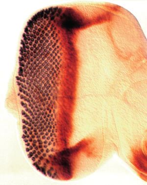
3474 G. Mardon, N. M. Solomon and G. M. Rubin

Anderson, 1992) and encodes a Drosophila homolog of the             initiation and anterior to the furrow during furrow progression.
mammalian transforming growth factor β (TGFβ) family of             In the absence of dac function, cells at the posterior margin of
cytokines that regulate cell proliferation and differentiation      the eye imaginal disc appear to follow a cuticle differentiation
(Padgett et al., 1987). The segment polarity gene hh is required    pathway and are unable to respond to the pattern propagation
for the specification of compartment boundaries in the              signals required for furrow movement. Furrow initiation fails
Drosophila embryo and imaginal discs (Ingham et al., 1991;          and little or no photoreceptor cell differentiation is observed.
Tabata and Kornberg, 1994; Basler and Struhl, 1994). Ver-           Curiously, the requirement for dac function in retinal cell-fate
tebrate homologs of hh exhibit organizing activity during           determination is not observed for cells located in more anterior
mouse central nervous system and chick limb development             portions of the eye disc. These results suggest that posterior
(Echelard et al., 1993; Riddle et al., 1993). During eye imaginal   margin cells possess an identity distinct from that of other cells
disc development, dpp is expressed specifically in the mor-         in the eye imaginal disc from an early stage of development.
phogenetic furrow and this expression is correlated with pro-
gression of the furrow. Mutations that stop furrow progression
also abolish dpp expression. In particular, hh function is          MATERIALS AND METHODS
required for both dpp expression and furrow progression
(Heberlein et al., 1993; Ma et al., 1993). hh is expressed          Drosophila stocks and mutagenesis
posterior to the furrow in determined photoreceptor cells and       The hypermorphic Egfr allele used was ElpB1(Baker and Rubin,
it has been proposed that hh protein diffuses anteriorly to pos-    1989). The recessive viable Egfr allele top1 was a gift of Kathryn
itively regulate dpp expression in the furrow. Consistent with      Anderson. The P-element insertion in the dachshund genomic locus
this interpretation, ectopic expression of hh in the anterior       was originally called rK364 and has been renamed dacP. The sup-
compartment of the wing imaginal disc is sufficient to induce       pressor of Elp phenotype did not separate meiotically from this P-
dpp expression in cells bordering the area of ectopic hh            element in over 1300 progeny scored. In situ hybridization of a probe
                                                                    representing P-element sequences to salivary gland polytene chromo-
expression (Basler and Struhl, 1994). In the eye disc, dpp
                                                                    somes placed the dacP element at cytological position 36A on the
and/or hh may diffuse several cell diameters further into the       second chromosome (not shown). An additional nine alleles of dac
unpatterned epithelium anterior to the furrow to regulate other     (dac1-dac9) were obtained from Iain Dawson, Yale University. The
genes required for undetermined cells to become competent to        dac locus was originally designated as the l(2)36Ae complementation
adopt a neural fate. Indeed, hh function may be required for        group (Ashburner et al., 1990) but was later renamed dac. Analysis
expression of at least three other genes involved in eye devel-     of polytene chromosomes from dac4 animals revealed a small deletion
opment: glass, scabrous and hairy (Ma et al., 1993). glass and      that removes the major band at 36A (not shown). This deletion was
scabrous are normally expressed in and posterior to the furrow      confirmed by DNA hybridization analysis with genomic probes
while hairy is expressed anterior to the furrow (Moses and          flanking the site of the P insertion (see below). The dac4 mutation is,
Rubin, 1991; Baker et al., 1990; Mlodzik et al., 1990; Brown        therefore, a complete loss-of-function or null mutation. dac1, dac3 and
et al., 1991). Since hh is expressed in differentiated photore-     dac4 are all null mutations for both the eye and leg phenotypes. The
                                                                    other dac mutant alleles are similar to the dacP allele with regard to
ceptors about three columns posterior to the furrow, these data
                                                                    both the eye and leg phenotypes and are assumed to be partial loss-
suggest that hh is required for expression of genes that are        of-function mutations in dac.
expressed earlier, in developmental terms, than hh itself. This
is possible because all stages of photoreceptor development are     Scanning electron microscopy and histology
in progress simultaneously in a single eye disc and hh is a         Adult flies were prepared for electron microscopy as described by
secreted protein capable of traveling backwards in develop-         Kimmel et al. (1990). Adult eyes were fixed, embedded and sectioned
mental time (i.e., anteriorly) by means of diffusion (Ma et al.,    as described by Tomlinson and Ready (1987b). Acridine orange
1993). In this manner, hh is thought to control progression of      staining of eye and leg imaginal discs was performed as described by
the morphogenetic furrow.                                           Bonini et al. (1993).
   In contrast to furrow progression, little is known about the     Cloning and sequencing
mechanisms controlling initiation of the morphogenetic
                                                                    Genomic DNA sequences flanking the P-element insertion in dac
furrow. The furrow begins moving (i.e., initiates) during the       were cloned by plasmid rescue (Mlodzik et al., 1990). Beginning with
late second or early third instar larval stage. However, the        a genomic rescue fragment as a probe, eight independent, overlapping
signal that causes the furrow to begin moving is not known. It      lambda phage clones containing genomic DNA surrounding the P-
seems likely that a distinct initiation mechanism precedes          element were then isolated from a Sau3A partial digestion genomic
propagation: while hh is essential for furrow progression, it       library cloned in λFix (provided by Kevin Moses). These clones span
cannot be involved in furrow initiation because it is not           approximately 30 kb. Ten independent, overlapping cDNA clones
expressed in the eye disc prior to neural differentiation. In       derived from this genomic region were isolated from a λgt10 cDNA
addition, not all cells of the eye imaginal disc differentiate as   library prepared from eye imaginal disc RNA (constructed by Alan
retina: the periphery of the eye disc monolayer gives rise to a     Cowman). All DNA sequences were obtained using a Pharmacia ALF
portion of the adult head cuticle surrounding the retinal field     automated sequencer and analyzed with the Staden software. Two
(Cohen, 1993). Again, the mechanisms controlling the distinc-       full-length cDNAs (5 kb) were sequenced on both strands using
                                                                    synthetic oligonucleotide primers. The corresponding sequences in
tion between retinal and cuticle fates in the eye disc are not      genomic DNA were also determined and the putative splice junctions
known. In this paper, we describe mutations in dachshund            deduced (Mount et al., 1992). Approximately 300 bp from both ends
(dac), which prevent cells at the posterior margin of the eye       of the ten independent cDNAs were also sequenced, as well as the
disc from following a retinal differentiation pathway. dac          splice junctions flanking exons two, three and four (see Fig. 2). The
encodes a novel nuclear protein which is expressed at the           site of the P-element insertion was determined by sequencing the ends
posterior margin of the eye imaginal disc prior to furrow           of the genomic rescue fragment.
dachshund function in eye and leg morphogenesis 3475

Antibody preparation and dac mutant analysis                               carried on the X chromosome. Eye imaginal discs were dissected from
The P insert in dac is an ‘enhancer trap’ element that contains the        wandering third instar larval animals and fixed as described above. β-
bacterial lacZ gene (Mlodzik and Hiromi, 1991). β-galactosidase            galactosidase activity in discs was detected using the X-Gal substrate
activity staining of discs prepared from third instar dacP heterozygote    as described by Simon et al. (1985). The position of homozygous dac
larvae reveals high levels of expression in both eye and leg imaginal      null mutant clones was determined by staining discs with anti-dac
discs (not shown). Monoclonal antibodies were raised against two           monoclonal antibodies or with anti-ELAV as described above.
non-overlapping, bacterially expressed dac proteins corresponding to
amino acids 149-368 and 378-599 (see Fig. 2). The monoclonal prepa-
rations are designated mAbdac1-1 and mAbdac2-3, respectively. Both         RESULTS
antibody preparations detect the same expression patterns as that seen
with β-galactosidase activity staining in the dacP line. Although dac      dachshund is a dominant suppressor of Ellipse
protein staining is still observed in imaginal discs prepared from
                                                                           Specification of appropriate spacing between ommatidial
larvae homozygous for the weak dacP allele, this staining is at sig-
nificantly lower levels as compared to wild-type (not shown). This is      founder cells in the morphogenetic furrow is crucial to the first
presumed to be full-length, wild-type dac protein, since the P-element     step of neural determination and pattern formation in the eye
is inserted well 5′ of the open reading frame (Fig. 2A). This result is    disc. Although this process is not understood, mutations in the
consistent with the partial loss-of-function phenotype of the dacP         Drosophila homolog of the epidermal growth factor receptor,
mutant. As expected, no dac protein is detectable in discs prepared        Egfr, prevent normal spacing and differentiation of photore-
from dac4 deletion mutant larvae or in dac null mutant clones (Fig.        ceptor cells in the developing eye (Clifford and Schüpbach,
5). The two other phenotypic dac null alleles, dac1 and dac3, are also     1989; Xu and Rubin, 1993). We reasoned that a genetic screen
protein nulls. In contrast to the dac4 deletion mutant, DNA blot           for modifiers of Egfr function might identify other genes
analysis indicates that the genomic DNA encompassing the dac1 and          involved in this process. Such a screen is facilitated by the
dac3 null alleles is grossly intact (not shown). These results suggest     dominant Ellipse (Elp) allele of Egfr (Baker and Rubin, 1989;
that loss-of-function mutations in the transcription unit defined by the
                                                                           1992). In contrast to the highly ordered arrangement of
P-element insertion (dacP) are responsible for the mutant phenotypes
described. In addition, the null phenotype for this locus is unambigu-     ommatidia in wild-type animals, Elp heterozygotes have
ously defined by these mutants.                                            reduced, rough eyes (Fig. 1A,B). Elp behaves as a hypermor-
                                                                           phic allele of Egfr because the Elp phenotype is suppressed by
Immunohistochemistry                                                       a deletion for this locus. Screens for mutations that dominantly
Imaginal discs from second or third instar larvae were dissected in        enhance or suppress the Elp rough eye phenotype are likely to
PBS (0.1 M phosphate pH 7.2, 150 mM NaCl), fixed for 15 minutes            identify gene products that affect the activity of Egfr.
in 4% formaldehyde in PBS, and washed three times 10 minutes in               A collection of 1170 P-element lines were generated in our
PBS. Primary antibody incubations were performed in PAXDG (PBS             laboratory and selected for expression in eye imaginal discs (U.
containing 1% BSA, 0.3% Triton X-100, 0.3% sodium deoxycholate,            Gaul, L. Higgins and G. M. R., unpublished data). A screen of
5% normal goat serum) for 2 hours at room temperature or overnight         these lines identified one strong suppressor of the Elp eye
at 4°C. mAbdac2-3 antibodies were used at a 1:5 dilution, rat mono-
clonal anti-ELAV antibodies were used at a 1:1 dilution and rabbit
                                                                           phenotype. A single copy of this P-element insert completely
anti-β-galactosidase polyclonal antibodies (Organon Teknica) were          suppresses both the external rough eye phenotype of Elp het-
used at a 1:5000 dilution. Following the primary antibody incubation,      erozygotes (Fig. 1C) as well as the disorganized ommatidial
discs were washed three times 10 minutes in PAXDG. Discs were              structure observed in sections of such eyes (not shown).
then incubated in PAXDG for 2 hours at room temperature with a             Excision of the P-element reverts both the suppression of Elp
1:200 dilution of one of the following horseradish peroxidase-conju-       and the semi-lethality of the chromosome. Additional alleles
gated secondary antibodies: goat anti-rat (for the ELAV primary),          that fail to complement the P-element mutant were identified
goat anti-mouse (for the mAbdac primary), or donkey anti-rabbit (for       in other genetic screens for modifiers of the Elp eye phenotype
the β-galactosidase primary). After washing three times 10 minutes         (Iain Dawson, personal communication). Due to the recessive
in PBS, discs were incubated 10 minutes in PBS with 0.5 mg/ml              leg phenotype associated with these mutants (described
diaminobenzidene (DAB), 0.003% hydrogen peroxide and 1.5 mM                below), this locus has been named dachshund (dac). One
NiCl2 for intensification. The double-label experiment for ELAV and
β-galactosidase shown in Fig. 4 was performed as follows: eye discs
                                                                           mutant allele, dac4, is a small deletion encompassing the entire
were incubated with both primary antibodies together as described          dac locus and is therefore a null mutation (data not shown).
above. Following the PAXDG washes, discs were incubated first with         These results suggest that loss-of-function mutations in dac
goat anti-rat secondary and then developed using DAB with NiCl2.           reduce the activity of the hyperactive Elp allele of Egfr. Two
Following three 10 minute washes in PAXDG, discs were then                 other dac mutants, dac1 and dac3, are also phenotypic and
incubated with donkey anti-rabbit secondary and then developed             molecular null mutations (see Materials and Methods). All of
using DAB without NiCl2 intensification.                                   the experiments described in this paper were performed with
                                                                           the dac4 deletion mutant and at least one of the other null
Mosaic analysis
                                                                           alleles. In all cases, identical results were obtained with each
Mitotic clones in eye imaginal discs were generated using the              of these null mutants.
FLP/FRT system essentially as described by Xu and Rubin (1993)
with some modifications. dac null alleles, dac3 and dac4, were recom-      dachshund is required for eye and leg
bined with an FRT sequence at cytological position 40A. In addition,       morphogenesis
these stocks carried a single copy of either the dpp-lacZ reporter
construct (Blackman et al., 1991) or a P-element containing the lacZ       Although dac mutants have greatly reduced viability, adult
gene inserted in hh (line rJ413 or P30; Ma et al., 1993). Mitotic recom-   homozygotes of both the P-element, dacP, and the null alleles
bination was induced during the first instar larval stage with a 1 hour    are routinely observed. The eyes of dacP homozygotes are
heat shock at 38°C to induce expression of the FLP recombinase             reduced and roughened (Fig. 1E), while the eyes of dac null
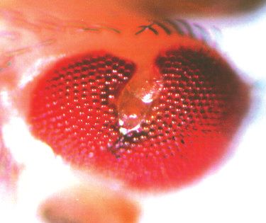
3476 G. Mardon, N. M. Solomon and G. M. Rubin

Fig. 1. The dac mutant phenotype. Scanning electron micrographs of adult eyes or prothoracic legs of the following genotypes are shown: wild-
type (A,L); EgfrE/+ (B); dacP Egfr+/dac+ EgfrE (C); dac4/dac4 (D); dacP/dacP (E,M); dacP top1/dacP top1 (F); top1/top1 (G); dac3/dac3 (H) and
(N-P); dacP/dacP ; hhrJ413/TM6B (I); +/CyO ; hh1/hh1 (J); dac3/CyO ; hh1/hh1 (K). All scale bars represent 100 µm. dacP top1/dacP top1 double
homozygote eyes (F) contain approximately half as many ommatidia as dacP/dacP homozygotes (E). For A-K, scale bar is shown in D; for L-N,
scale bar is shown in L; and for O-P, scale bar is in P. C, coxa; Cl, claw; F, femur; Ta, tarsi; Ti, tibia; Tr, trochanter; 4 and 5, fourth and fifth
tarsal segments, respectively.
dachshund function in eye and leg morphogenesis 3477

mutant homozygotes are either severely reduced or, in about          species seen by blot analysis of total imaginal disc RNA (not
50% of adults examined, absent (Fig. 1D, H). Although many           shown). DNA sequence comparison of genomic and cDNA
of the ommatidia in dacP homozygotes are normally con-               clones suggests that the locus spans approximately 20 kb and
structed, 50% have either too few or too many pho-
toreceptor cells. The overall array of ommatidia is
disrupted, contributing to the rough external eye
phenotype of these flies. None of the few
ommatidia in dac null mutant eyes has a normal
morphology (not shown). In contrast to the
compound eye, the external morphology of the
adult ocelli appears normal in all dac mutants.
   A second phenotype of dac flies inspired its
name: dachshund mutants have short, little legs
(Fig. 1L-P). The wild-type adult leg is composed
of ten discrete segments: the coxa, trochanter,
femur, tibia, five tarsal segments and the claw (Fig.
1L). While dacP homozygote legs appear to have
a normal proximal and distal morphology, the
intermediate segments are fused and condensed
(Fig. 1M). The same intermediate segments, the
femur, tibia and proximal three tarsi, are severely
condensed in null mutant legs. In contrast, the
coxa, trochanter, fourth and fifth tarsal segments,
and the claw appear to develop normally, even in
the null mutant (Fig. 1N-P). Upon eclosion from
their pupal cases, these mutants are unable to
locomote normally and quickly fall into the food
and die. However, if kept away from wet medium,
dac homozygotes can live for several days before
dying, presumably from dehydration. These
helpless homozygotes are able to flail their
misshapen legs, albeit to no avail, indicating that
at least a portion of the leg neuromusculature
develops normally and is functional.
   If loss-of-function mutations in dac reduce the
activity of the activated Elp allele of Egfr, then the
same mutations in dac should enhance the
phenotype of partial loss-of-function mutations in
Egfr. We looked for such an interaction with the
weak top1 allele of Egfr (Clifford and Schüpbach,
1989), which displays a very mild roughening of
the adult eye (Fig. 1G). Although we did not
observe a dominant effect by loss-of-function
mutations in dac on the top1 eye phenotype, we did
see what appears to be a recessive synergy between
                                                       Fig. 2. dac encodes a novel protein. (A) A map of the genomic dac locus is shown
the weak dacP allele and top1. That is, dacPtop1 with 5′ and 3′ non-coding exons depicted as open boxes and coding exons as black
double homozygotes have a reduced, rough eye boxes. The site of the P insertion in dac is shown by an arrow. Comparison of
phenotype that is more severe than either mutant partial sequences from ten independent cDNA clones indicate that exon 3, only 27
alone or what we would expect from simple base pairs (bp) in length, is alternately spliced and appears in six out of ten clones
additive effects (Fig. 1F). This result suggests that analyzed. In addition, exon 4 has three alternate splice acceptor sites, separated by
dac and Egfr may function in the same pathway or 21 and 6 bp. Each of these alternate acceptor sites appears to be used with
in parallel pathways during eye development.           approximately equal frequency. All of the predicted splice acceptor and donor sites
                                                          in the cDNA are in good agreement with the consensus splice sequences (Mount et
dachshund encodes a novel nuclear                         al., 1992; not shown). (B) The amino acid sequence of the predicted dac protein is
protein expressed in eye and leg imaginal                 shown beginning with the first ATG in the longest open reading frame. Splice sites
discs                                                     based on comparison of the genomic and cDNA sequences are indicated with solid
                                                          arrows. The second and third alternate acceptor sites for exon 4 are indicated with
DNA flanking the P-element insert in dacP was             open arrows. This sequence is characterized by multiple polyalanine,
isolated and used to screen cDNA libraries                polyasparagine, polyglycine and polyglutamine runs throughout the protein. In
prepared from eye imaginal disc RNA. The longest          addition, there are unusually high percentages of serine (11%) and proline (6%)
cDNAs obtained from our library were 5 kilobases          residues present in the predicted sequence. The entire cDNA sequence and splice
(kb) in length and correspond with the major RNA          junction sequences have been deposited in GenBank.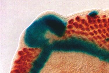
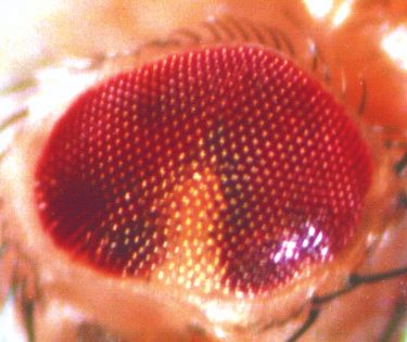
3478 G. Mardon, N. M. Solomon and G. M. Rubin

comprises 12 exons, some of which can be alternately spliced               preceding the morphogenetic furrow as it moves anteriorly
(Fig. 2). The dac P-element is inserted after a position corre-            across the eye disc (Fig. 3C,D). Posterior to the furrow, dac
sponding to base pair 11 of our longest cDNA clone (Fig. 2A).              expression is detected primarily in photoreceptors R1, R6 and
DNA sequence analysis reveals a single long open reading                   R7 as well as the cone cells (not shown). dac is expressed only
frame encoding a predicted protein of 1081 amino acids, which              in that part of the eye disc fated to become retina; the periphery
shares no significant similarity to any sequence in the databases          of the disc destined to become head cuticle does not express
(Fig. 2B).                                                                 dac (Fig. 3D). The ring pattern of dac expression in the leg
   Expression of dac is readily apparent prior to imaginal disc            disc is established at an early stage of leg disc development
morphogenesis. In the eye disc, dac is expressed at the                    (Fig. 3E), well before the characteristic epithelial folds of a
posterior margin prior to initiation of the morphogenetic                  mature leg disc are seen (Fig. 3F,G). In all cases, dac protein
furrow (Fig. 3A). This expression pattern is similar to that of            appears to be localized to the nucleus.
dpp at the same early stage of development (Fig. 3B). Strong                  In addition to strong expression in eye and leg discs, dac
dac expression is also detected in the unpatterned epithelium              protein is found in the third antennal disc segment and the wing

Fig. 3. dac is a nuclear protein expressed in the embryo and imaginal discs. All panels except B show expression of dac protein in wild-type
imaginal discs and embryos as detected by anti-dac monoclonal antibody mAbdac2-3. β-galactosidase activity staining of an eye-antennal disc
prepared from a second instar larva carrying the dpp-lacZ reporter is shown in B. Second (A,B,E) and third (C,D,F-H) instar larval discs of the
following types are shown: eye-antennal (A-D), with eye discs to the left; prothoracic leg (E-G) and wing (H). Expression of dac protein is
found in most or all nuclei anterior to the furrow and remains detectable primarily in photoreceptors R1, R6 and R7 posterior to the furrow (not
shown). MF, morphogenetic furrow. A side view of a leg disc similar to that in F is shown in G. The arrow in G indicates the portion of the disc
fated to give rise to the distal tarsal segments and claw of the adult leg. Ventral views of stage 13 (I) and stage 16 (J) embryos are shown.
Posterior is to the left for A-D; on top for E, F and H; and to the right for I, J. Scale bars for A-C and E-J are 50 µm. Scale bar in D is 10 µm.
dachshund function in eye and leg morphogenesis 3479

imaginal disc (Fig. 3C,H). dac expression is also detected in      account for the adult phenotype of dacP homozygote eyes.
the central nervous system of embryos (Fig. 3I,J) and in the       Moreover, null mutant eye discs have just a few ELAV-
optic lobe of the larval brain (see cover picture). In spite of    positive clusters or none at all (Fig. 4C). When present, these
these distinct patterns of expression, no obvious morphologi-      presumptive photoreceptors are always located near the
cal phenotype is observable in the embryonic CNS or the adult      posterior margin of the disc, where neural development
antennae and wings in dac null mutant animals. The function        normally begins (Fig. 4D). These results demonstrate that
of dac in these structures remains to be determined.               normal photoreceptor development is blocked in dac null
                                                                   mutants.
dachshund is essential for the development of
intermediate leg segments                                          dachshund is required for normal movement of the
The expression pattern of dac during larval leg disc develop-      morphogenetic furrow
ment is consistent with the mutant phenotype of the adult leg.     We examined furrow movement in wild-type and dac mutant
The leg imaginal disc is composed of concentric folds of           imaginal discs using a lacZ reporter construct for dpp
epithelia such that the outermost portions of the disc give rise   (Blackman et al., 1991). dpp is expressed specifically in the
to the most proximal segments (i.e. the coxa) in the adult leg     morphogenetic furrow and can serve as a marker for the
while the most distal structures (i.e. the tarsal segments and     position of the furrow in the eye disc (Heberlein et al., 1993).
claw) are derived from the central portion of the disc. As         While the morphogenetic furrow has normally progressed
shown by antibody staining, dac is strongly expressed specif-      about half-way across the eye disc by the late third instar larval
ically in the presumptive epithelium that is fated to give rise    stage, dpp-lacZ expression remains at the posterior margin of
to the femur, tibia and proximal tarsal segments (Fig. 3E-G),      dac null mutant discs (Fig. 4A,C). In about half of the dac null
the same structures most severely affected in dac mutant legs      mutant eye discs examined, there is a small amount of furrow
(Fig. 1M,N). There is no dac expression detected in either the     movement observed at the posterior midline of the disc, near
center or the periphery of the leg imaginal disc, and the struc-   the optic stalk. This movement is usually associated with the
tures derived from these portions of the disc appear to develop    appearance of a few ELAV-positive cells (Fig. 4C,D). In all
normally in dac mutant flies (Fig. 1O,P).                          other cases studied, there is neither furrow movement nor
   Although the adult legs of dac mutants are greatly shortened,   neural differentiation (not shown). Thus, dac does not appear
leg imaginal discs prepared from dac mutant third instar larvae    to be required specifically for neural differentiation. Instead,
appear morphologically normal, both in size and in their con-      dac function is required for normal movement of the morpho-
centric segmental structure (not shown). However, a significant    genetic furrow.
increase in cell death in dac mutant leg discs is revealed by         In the weak dacP mutant, furrow movement is uneven.
staining with acridine orange (Spreij, 1971), a dye that is        Normally, the furrow moves across the eye disc as a linear
actively excluded from living cells (Fig. 4G,H). This cell death   front with neural development beginning immediately in its
is limited primarily to the region of the disc where dac is        wake (Fig. 4A). In contrast, the partial loss-of-function dacP
expressed and most likely accounts for the failure of disc         mutation prevents the furrow from moving normally in lateral
eversion during early pupal stages. By 6 hours into pupal          regions of the eye disc. This results in a curved furrow that has
development, wild-type leg discs have undergone rapid              advanced further at the midline than at the periphery of the disc
elongation, primarily as a result of cell-shape change (Condic     (Fig. 4B). Nevertheless, neural development closely follows
et al., 1991). Such elongation does not occur in dac mutant        the furrow in all cases examined.
pupal legs, consistent with the adult leg phenotype (not              Eye discs of third instar dac null mutant larvae are approx-
shown). Thus, there is a failure of morphogenesis in dac           imately normal in size, suggesting that the failure of furrow
mutant leg discs during late larval and early pupal develop-       movement is not the result of inadequate cell proliferation (Fig.
ment.                                                              4C). Nevertheless, in the absence of furrow movement, cells
                                                                   in dac mutant eye discs fail to adopt a neural fate and are likely
Photoreceptor development is prevented in                          to remain in an undifferentiated state. The fate of these undif-
dachshund mutant eye discs                                         ferentiated cells is death. As detected by acridine orange
The near or total absence of ommatidia in complete loss-of-        staining, a large increase in cell death is seen in dac mutant
function dac mutant adult eyes (Fig. 1D,H) could be the result     eye discs (Fig. 4E,F).
of either an initial failure of photoreceptor determination           To analyze further the role that dac plays in furrow
during larval development or from a degenerative event             movement, we made homozygous dac null mutant clones of
following neural determination and differentiation, such as in     cells in a heterozygous dac background and examined the
the glass mutation (Moses et al., 1989). We examined the state     phenotype of such clones in both larvae and adults. Dramati-
of neural development in dac mutant eye discs using a mono-        cally different results were obtained depending on the position
clonal antibody that recognizes the nuclear ELAV antigen           of these clones in the developing eye. dac clones occurring
(Robinow and White, 1991). ELAV is expressed in all neurons        anywhere in the eye but not including the posterior margin
in Drosophila and is apparent immediately posterior to the         (‘interior’ clones) always give rise to ommatidia (Fig. 5A).
morphogenetic furrow in the eye imaginal disc. In contrast to      This suggests that the morphogenetic furrow is able to progress
the highly ordered array of photoreceptors seen in wild-type       through patches of dac mutant cells given the chance to first
eye discs, fewer neurons differentiate in dacP imaginal discs      initiate movement in wild-type tissue. In contrast to interior
(Fig. 4A,B). Close examination of these discs reveals that         clones, posterior margin dac mutant clones usually fail to
highly variable numbers of photoreceptors per ommatidial           develop into retinal tissue; head cuticle is found in its place
cluster are already present at this stage (not shown) and can      (Fig. 5B,C). In about one third of cases examined a few mutant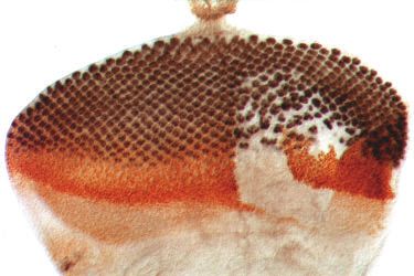
3480 G. Mardon, N. M. Solomon and G. M. Rubin

(white-minus) ommatidia are found at the borders of dac               hh is expressed in differentiating photoreceptors and is
clones (Fig. 5L) and about 5% of posterior margin clones will         required for progression of the morphogenetic furrow (Ma et
give rise to a substantial patch of mutant ommatidia (Table 1).       al., 1993; Heberlein et al., 1993). A lethal loss-of-function P-
The reason for this variability is not known.                         element insert in hh (rJ413) dominantly enhances the recessive
   dac mutant clones in larval discs are consistent with the          eye phenotype of the weak dacP allele (Fig. 1E,I). Similarly,
observed adult phenotypes. While neural differentiation is            loss of one copy of dac acts as a dominant enhancer of the
detected in interior dac clones (Fig. 5D), few or no ELAV-            recessive eye phenotype of the viable hh1 mutation (Fig. 1J,K),
positive cells are found in posterior margin dac
clones (Fig. 5E,F). Using the dpp-lacZ reporter
as an assay, we found that the furrow indeed pro-
gresses through dac null mutant clones, as long
as they do not include the posterior margin of the      A                                          B
eye disc (Fig. 5G). However, the rate of furrow
movement seems to be slowed in such clones as
compared to the surrounding wild-type tissue.
Interestingly, this effect on the rate of furrow
progression exhibits local domineering non-
autonomy: the furrow is also slowed in the geno-
typically wild-type epithelium immediately
adjacent to dac mutant clones (not shown). This
effect on furrow movement correlates with the
appearance of additional photoreceptors in geno-
typically wild-type ommatidia located next to
dac mutant clones in adult eyes (Fig. 5J). Con-
sistent with the phenotype of whole dac null
mutant eye discs (Fig. 4C), the furrow does not
initiate movement in clones that include the
posterior margin of the eye disc. Once the furrow
has moved past such a clone in the surrounding          C                                          D
wild-type tissue, normal furrow progression
anterior to the clone resumes (Fig. 5H,I,M).
Thus, our clonal analyses indicate that loss of
dac function prevents furrow initiation at the
posterior margin of the eye disc and slows but
does not prevent movement of the furrow during
progression.
   Although the morphogenetic furrow is able to
move through patches of dac mutant cells,
sections of dac mutant clones demonstrate that
dac is required for normal ommatidial assembly:
null mutant clones are clearly disrupted with
most ommatidia containing an abnormal number
and arrangement of photoreceptors (not shown).
Nonetheless, complete and properly formed
ommatidia composed entirely of dac mutant
cells are occasionally observed (Fig. 5K), sug-         E                      F                   G                     H
gesting that there is no absolute requirement for
dac in any given photoreceptor. In addition,
mosaic ommatidia at the border of clones – those
comprising both wild-type and dac mutant cells
– are most often abnormally constructed, sug-
gesting that dac is required cell autonomously
for ommatidial assembly (not shown). That is,
dac-dependent activity from surrounding wild-
                                                     Fig. 4. dac is required for movement of the morphogenetic furrow. Eye imaginal
type cells is unable to rescue the aberrant omma-    discs were double-labeled to reveal the position of the morphogenetic furrow and
tidial structure of dac mutant clones.               differentiating neurons. Wild-type (A,E,G), dacP (B) or dac3 (C,D,F,H) imaginal
                                                      discs were prepared from late third instar larvae also carrying the dpp-lacZ reporter
hedgehog and dachshund                                and stained with anti-ELAV (black) and anti-β-galactosidase (brown). Third instar
Additional support for a role for dac during          eye (E,F) and leg (G,H) imaginal discs were also stained with acridine orange to
furrow progression comes from genetic analyses        detect cell death. Dying cells are identified as brightly staining spots. Scale bar for A-
of interactions between dac and hedgehog (hh).        C is 50 µm. Scale bar in D is 10 µm. Scale bars for E,F and G,H are 100 µm.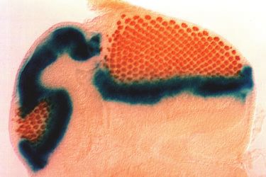
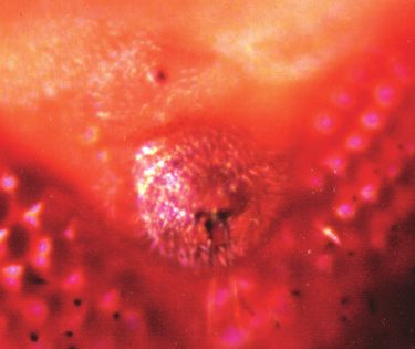
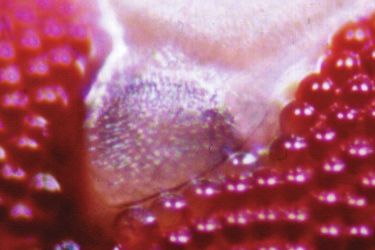
dachshund function in eye and leg morphogenesis 3481

 A                                                B                                                 C

 D                                                E                                                 F

 G                                                H                                                  I

 J                         K                      L                                                 M

Fig. 5. Mosaic analysis of dac function. Light microscope photographs of adult eyes with homozygous dac4 mutant clones (A-C,L) are
shown. Interior clones – those that do not include the posterior margin – always give rise to retinal tissue (A). In contrast, posterior
margin-containing clones (B,C,L) usually give rise to head cuticle and fail to respond to pattern propagation signals. Homozygous dac4
mutant clones in eye discs were detected by staining with anti-dac monoclonal antibody (D-G), shown in brown (D-F) or black (G).
Clones are located in areas that do not stain. Discs were also stained for the neuronal ELAV antigen, shown in black (D-F) or brown
(H,I,M). dpp-lacZ expression was detected by β-galactosidase activity staining, shown in blue (G-I,M). In most eye discs examined, the
level of dpp expression is not affected by loss of dac function. However, the position of expression of dpp is either slowed in interior dac
clones (G) or fails to move at all in posterior margin clones (H,I,M). In a minority of cases (less than 10%), dpp expression is reduced or
absent in both classes of clones (not shown). The basis for this variability of dpp expression is not known. The arrows in (L) indicate the
morphological boundary that is usually apparent at the border of posterior margin dac clones. This boundary is already apparent in larval
eye disc clones such as that shown in (F). Also visible in (L) are several white-minus dac mutant ommatidia bordering the clone. (J) A
section of an abnormal ommatidium containing one additional outer photoreceptor but composed entirely of wild-type cells is located near
a dac mutant clone. (K) A section of a normally constructed ommatidium composed entirely of dac mutant cells lacking pigment granules
is shown. Posterior is at the top for all panels. Scale bars represent 50 µm (A,B), 25 µm (C,F,I,L,M), 50 µm (D,E,G,H) or 2 µm (J,K).
3482 G. Mardon, N. M. Solomon and G. M. Rubin

          Table 1. Adult posterior margin dac clones                           Vertebrate homologs of hh have recently been shown to exhibit
dac             Cuticle          1-5 w−              >5 w−                     organizing activity in the chick limb and in central nervous
allele           only           ommatidia          ommatidia         Total     system development of mice (Riddle et al., 1993; Echelard et
dac1           82 (74%)          24 (22%)            5 (4%)           111      al., 1993). Similarly, nodal, a member of the TGFβ superfam-
dac3           65 (67%)          25 (26%)            7 (7%)            97      ily of secreted proteins, is expressed in the mouse node at the
dac4           67 (68%)          29 (30%)            2 (2%)            98      anterior of the primitive streak and is required for mesoderm
                                                                               formation (Zhou et al., 1993). A Drosophila homolog of
   Homozygous dac mutant clones for all three dac null alleles were
generated using the FLP-FRT system and were examined using a light
                                                                               TGFβ, dpp, acts as a morphogen controlling dorsal-ventral
dissecting microscope. Approximately three-quarters of the clones scored did   pattern formation in the embryo (Ferguson and Anderson,
not include the posterior margin of the eye (n=937) and always developed as    1992).
dac mutant (white) ommatidia. Clones including a portion of the posterior         Eye imaginal disc development in Drosophila provides an
margin were scored and categorized based on the number of white minus          excellent system for deciphering mechanisms of pattern
ommatidia found bordering each clone.
                                                                               formation and morphogenesis. Organization of the unpatterned
                                                                               epithelium of the eye disc into a highly ordered array of dif-
                                                                               ferentiated cells follows movement of an indentation across the
                                                                               epithelial monolayer termed the morphogenetic furrow.
which presents a significant reduction of the anterior portion                 Furrow progression is not associated with cell migration;
of the eye (Mohler, 1988). These genetic results suggest that                  instead, furrow movement is detected as a wave of changes in
dac and hh act in the same or in parallel pathways during eye                  cell shape, cell cycle, gene expression and neural differen-
development.                                                                   tiation. Progression of the furrow requires the function of hh
   We examined hh expression in wild-type and dac mutant                       and is likely to involve dpp. The developmental events sur-
imaginal discs using a lacZ enhancer trap insert in hh that faith-             rounding morphogenetic furrow movement in Drosophila may
fully reproduces the normal expression pattern of hh (Ma et al.,               be analogous to those associated with the Spemann organizer
1993). Since little or no neural development takes place in dac                in Xenopus and Hensen’s node and the zone of polarizing
null mutant eye discs and hh is expressed in the eye specifi-                  activity in chick: organizing centers determine cell fates of
cally in determined photoreceptors, it is not surprising that hh               adjacent tissues through the action of secreted morphogens
is not expressed in dac mutant eye discs (Fig. 6A,B). However,                 (reviewed in Slack, 1991; Riddle et al., 1993).
hh expression in other imaginal discs, including the antennal,                    hh plays a central role in progression of the morphogenetic
leg and wing discs, is not affected by loss of dac function (for               furrow during eye disc development (Ma et al., 1993;
example, compare the antennal discs shown in Fig. 6A,B).                       Heberlein et al., 1993). In this case, hh protein is expressed by
   Expression of hh in differentiating photoreceptors lacking                  determined neural cells posterior to the furrow and is thought
dac function was examined in homozygous dac null mutant                        to diffuse anteriorly, thereupon controlling expression of other
clones in eye imaginal discs. We found that hh expression is                   genes required for furrow movement. One such putative target
normal in the absence of dac activity using a single copy of                   is dpp (Heberlein et al., 1993). Removal of hh function during
the viable hh enhancer-trap insertion P30 as an assay (Fig. 6C).               furrow progression abolishes both the expression of dpp in the
Thus, dac function is not cell autonomously required for hh                    furrow as well as furrow propagation. dpp or hh, in turn, are
expression. The dominant enhancement of the adult dac mutant                   likely to regulate the expression of other genes in the unpat-
eye phenotype by a single copy of the lethal hh mutation rJ413                 terned epithelium anterior to the furrow. Transplantation
(Fig. 1I) is also apparent during larval development. As judged                experiments have suggested that positional information suffi-
by the near absence of ELAV or hh expression in dac null                       cient to direct neural development is present in the morpho-
mutant clones, loss-of-function of one copy of hh by the rJ413                 logically undifferentiated epithelium immediately anterior to
lethal insertion results in delayed or reduced neural differen-                the furrow in the eye disc (Lebovitz and Ready, 1986). More
tiation in such clones (Fig. 6D).                                              recently, molecular analyses have shown that several genes are
                                                                               indeed expressed just anterior to the furrow, including string,
                                                                               eyes absent, hairy and atonal (Alphey et al., 1992; Bonini et
DISCUSSION                                                                     al., 1993; Brown et al., 1991; Jarman et al., 1994). However,
                                                                               none of these genes expressed anterior to the furrow has been
Substantial progress has been made toward elucidating the                      shown to participate directly in either initiation or propagation
molecular mechanisms controlling pattern formation and mor-                    of the morphogenetic furrow.
phogenesis during development of both vertebrate and inver-                       While some of the genes involved in furrow progression
tebrate species. Genetic and molecular studies of embryonic                    have now been identified, little is known about the events sur-
development in Drosophila have been particularly fruitful in                   rounding initiation of the morphogenetic furrow. In this paper,
identifying and understanding the function of phylogenetically                 we present evidence that dachshund (dac) is expressed at the
conserved genes that are essential for organizing tissue                       posterior margin of the eye imaginal disc prior to movement
polarity, specifying segment boundaries and controlling cell-                  of the morphogenetic furrow and is required for cells at the
fate determination. In many cases, intercellular signaling is                  posterior margin to respond to pattern propagation signals such
achieved by diffusion of a secreted protein acting over several                as those encoded by dpp and hh. This may reflect a require-
cell diameters (Bryant, 1993). For example, the product of the                 ment for dac to control a cell-fate choice between retina and
segment polarity gene hh acts as a true morphogen in                           cuticle. Similar to other genes controlling pattern formation in
Drosophila, specifying cell fates in the embryo in a concen-                   multiple tissues, such as hh and dpp, dac is also required for
tration-dependent manner (Heemskerk and DiNardo, 1994).                        normal leg morphogenesis.
dachshund function in eye and leg morphogenesis 3483
dachshund and leg development Heberlein et al., 1993). In contrast, in weak dac mutants, dpp
In the absence of dac function, flies develop with severely expression is unaffected and the furrow progresses nearly
shortened legs. dac is expressed in early larval leg imaginal normally at the center of the eye disc but is delayed at the
discs and this expression continues into pupal development. periphery (Fig. 4B). This difference in furrow progression may
dac activity is necessary to specify the identity of a subset of reflect a requirement for dac, but not hh, during furrow
segments in the leg: the femur, tibia and proximal three tarsi. initiation.
Loss-of-function mutations in dac result in legs with these Comparison of the phenotype of dac and hh mutant clones
intermediate structures fused and condensed. Thus, dac is at the posterior margin of the eye disc suggests that dac plays
required in those leg segment primordia in which it is an essential role in either furrow initiation or retinal cell-fate
expressed for cells to adopt a fate appropriate to their position. determination: while a few ommatidial columns are observed
Greatly increased cell death is apparent in third instar dac in hh mutant clones located at the posterior margin of the eye
mutant leg discs, prior to the elongation that occurs during (Ma et al., 1993), dac mutant clones that include the posterior
early pupal development. Thus, dac function is required prior margin usually fail to give rise to retinal tissue (Fig. 5B,C,L).
to leg disc eversion. dac is likely to act downstream of a Consistent with this adult phenotype, the dpp-lacZ reporter
proximal-distal patterning signal, such as Distal-less, to expression remains at the posterior margin in dac mutant
establish segment boundaries or identities (Cohen and Jürgens, clones in the eye disc (Fig. 5H,I,M). Thus, our clonal analysis
1989). The dac mutant leg phenotype is consistent with a indicates that dac is cell autonomously required for initiation
model in which establishment of proximal and distal structures of movement of the morphogenetic furrow away from the
during limb development precedes elaboration of intermediate posterior margin of the eye imaginal disc. Moreover, the failure
positional information (Cohen, 1993). of furrow initiation in dac mutant clones is unlikely to result
from either a failure of photoreceptor differentiation or the
Morphogenetic furrow movement and dachshund absence of hh expression, since diffusion of hh protein from
function nearby wild-type photoreceptor cells is unable to rescue furrow
initiation in dac mutant clones. Thus, dac is clonally required
In the absence of dac activity, the morphogenetic furrow
for cells at the posterior margin of the eye disc to respond to
remains at the posterior margin of the eye imaginal disc.
pattern propagation signals normally sufficient to cause furrow
However, furrow movement is not prevented in dac mutants
movement. This may reflect a direct requirement for dac in
by a lack of sufficient epithelial tissue. Eye discs of dac null
furrow initiation. Alternatively, dac may be required for an
mutants are approximately normal in size, indicating that cell earlier step of cell-fate determination such that clones of dac
proliferation in the eye disc is not inhibited in the absence of mutant cells at the posterior margin of the eye disc are unable
dac function or furrow progression. This stands in contrast to to differentiate as retinal tissue.
other eyeless or reduced-eye mutants, such as eyes absent and In addition to an essential role in early furrow movement,
Lobe, where a failure of retinal development is associated with dac is also required for some aspect of ommatidial assembly.
larval eye discs that are significantly reduced in size (Bonini dac mutant clones in the adult eye are comprised of mostly
et al., 1993; Heberlein et al., 1993). While proliferation is abnormal ommatidia, even at the border of such clones. This
normal, cells of dac mutant eye discs either fail to receive or is in sharp contrast to hh mosaic studies, where even large hh
perhaps respond to the normal cues specifying a neural mutant clones are normally constructed (Ma et al., 1993;
identity. These cells eventually die, possibly as a result of inap- Heberlein et al., 1993). That is, unlike furrow progression, dac
propriate cell-fate specification. Alternatively, these dying function is required cell autonomously for ommatidial
cells may reflect the action of a programmed cell-death assembly. Since dac is a nuclear protein, it may be involved in
mechanism designed to remove undifferentiated cells during regulating other genes functioning in this process. One
development, thereby preventing disruption of normal tissue candidate is the Egfr gene. dac mutants were isolated by their
organization (Ma et al., 1993). ability to dominantly suppress the rough-eye phenotype of the
Although loss of dac function prevents all movement of the activated Ellipse allele of Egfr. We have also shown that loss-
morphogenetic furrow in posterior lateral regions of the eye of-function mutations in dac and Egfr display an apparent
disc, some furrow movement at the midline of the disc is recessive synergy during eye development. Both results
observed. Here, the furrow is often able to move a small suggest that dac may positively regulate Egfr. Consistent with
distance and this is sufficient to allow differentiation of a few the possibility that dac controls Egfr expression and that this
photoreceptor cells. These cells develop as apparently normal effects part of the dac mutant phenotype, Egfr is expressed
photoreceptors, able to support the determination of the cone, anterior to the morphogenetic furrow (Zak and Shilo, 1992)
pigment and bristle accessory cells found in wild-type and is required for normal photoreceptor determination
ommatidia. Thus, dac is not required specifically for photore- (Clifford and Schüpbach, 1989; Xu and Rubin, 1993).
ceptor differentiation. In support of this view, photoreceptor
development also occurs in interior dac mutant clones in the A model for movement of the morphogenetic furrow
eye, indicating that there is no cell-autonomous requirement Three lines of evidence suggest that a primary initiation signal
for dac in photoreceptor determination. acts at the posterior midline of the eye disc. First, as judged by
The eye phenotype of weak dac mutants is different from dpp expression, the furrow normally initiates movement at the
other mutants that affect furrow movement. For example, the midline of the eye disc. Second, neural-specific markers, such
partial loss-of-function hh1 allele eliminates dpp expression as ELAV and hh, first appear at the posterior midline. Third,
and furrow progression first in the middle of the eye disc, in the absence of dac function, the only movement of the
giving the adult eye a kidney-shaped appearance (Fig. 1J and furrow occurs at the posterior midline. Since this movement is3484 G. Mardon, N. M. Solomon and G. M. Rubin
A C D
B
Fig. 6. hedgehog enhances the dac clonal phenotype in eye discs. Third instar eye-antennal imaginal discs prepared from wild-type (A) and
dac3 mutant (B) larvae carrying a P-element insertion in hedgehog (line rJ413) were fixed and stained for β-galactosidase activity (blue). Eye
discs are to the left and antennal discs are to the right. hedgehog is not expressed in dac null mutant eye discs. Homozygous dac4 mutant clones
were generated in larvae carrying a single copy of either of two P-element insertions in hedgehog: viable insert P30 (C) or lethal insert rJ413
(D). Both eye discs were fixed and stained for β-galactosidase activity (blue) to show the expression pattern for hedgehog. Expression of the
hedgehog insert P30 is not affected by loss of dac function. The position of dac clones in (C) are revealed by staining with anti-dac monoclonal
antibody, shown in brown. Clones are located in areas that do not stain brown, seen as a white background behind the blue hh expression. The
disc in D was stained for the neuronal ELAV antigen, showing the delay in neural differentiation caused by the lethal hedgehog mutation rJ413.
Posterior is to the left for all panels.
observed in about half of the discs examined, dac may serve posterior margin dac clones, both in larvae and in adults, there
some role in receiving this primary initiation signal but is not is a morphological boundary that forms between dac mutant
absolutely required for this step. This initial step cannot require clones and the adjacent retinal field (Fig. 5F,L), suggesting that
hh function because hh is not expressed until several rows of dac mutant cells that fail to develop as retina possess different
ommatidia posterior to the furrow. The initial signal that adhesive properties than those of the surrounding cells. The
triggers furrow movement may be transmitted directly via the eye imaginal disc is fated to give rise not only to the retina but
optic stalk, thereby localizing initiation to the posterior to a portion of the surrounding head cuticle as well (Cohen,
midline. Alternatively, there may be a diffusable primary 1993). A boundary similar to that observed in posterior margin
initiator, such as an ecdysone pulse (Richards, 1981), that is dac clones is normally seen at the periphery of wild-type eye
received only by specialized cells at this position. discs (Fig. 3D), presumably representing a physical distinction
The expression patterns of dpp and ELAV indicate that the between cuticle and retinal fields. Such boundaries are not
furrow does not begin moving away from the entire posterior observed in interior dac clones. These observations suggest the
margin of the eye disc at once. Instead, the furrow advances as possibility that dac may be involved in controlling a choice
a linear front such that movement away from the curved between a cuticular versus a retinal fate. Specifically, dac may
posterior margin occurs over time as a function of the distance be required to suppress a cuticle fate in cells derived from the
from the midline of the eye disc. The simplest mechanism to posterior margin. In the absence of dac function, clones that
control propagation of pattern formation and neural differen- originate from a posterior margin cell follow a cuticle pathway
tiation in this manner would be by diffusion of a secreted and are usually unable to respond to the pattern propagation
signaling molecule such as hh. Transmission of this signal does signals, such as dpp and hh, that are present but apparently
not require dac activity since normal furrow movement ineffective. Consistent with this model, dac is not expressed in
resumes lateral to dac mutant clones located at the posterior the peripheral margin of the eye disc destined to become
margin of the eye (Fig. 5). That is, lateral propagation of cuticle (Fig. 3D). Thus, dac is at least indirectly required for
signals for furrow movement away from the posterior margin initiation of the morphogenetic furrow and the onset of neural
of the eye disc is not prevented by intervening clones of dac differentiation. However, this requirement is not absolute since
mutant cells. mutant ommatidia bordering posterior margin clones, although
Our clonal analysis demonstrates that dac function is cell usually few in number, are frequently observed. Moreover, if
autonomously required for cells at the posterior margin of the a dac clone is derived from an ‘interior’ cell of the eye disc,
eye disc to respond to pattern propagation signals from sur- these cells appear to respond relatively normally to differen-
rounding wild-type tissue. This requirement is not found in tiation signals. This difference in responsiveness to pattern
cells located in interior portions of the eye disc. In most propagation signals suggests that posterior margin cells mayYou can also read