Inhibition of kinase IKKβ suppresses cellular abnormalities induced by the human papillomavirus oncoprotein HPV 18E6
←
→
Page content transcription
If your browser does not render page correctly, please read the page content below
www.nature.com/scientificreports
OPEN Inhibition of kinase IKKβ
suppresses cellular abnormalities
induced by the human
papillomavirus oncoprotein HPV
18E6
Mojgan Padash Barmchi1*, Miranda Thomas2, Jayashree V. Thatte3, Arushi Vats2,
Bing Zhang4, Ross L. Cagan5 & Lawrence Banks2
Human papillomavirus (HPV) is the leading cause of cervical cancer and has been implicated in several
other cancer types including vaginal, vulvar, penile, and oropharyngeal cancers. Despite the recent
availability of a vaccine, there are still over 310,000 deaths each year worldwide. Current treatments
for HPV-mediated cancers show limited efficacy, and would benefit from improved understanding
of disease mechanisms. Recently, we developed a Drosophila ‘HPV 18 E6’ model that displayed loss
of cellular morphology and polarity, junctional disorganization, and degradation of the major E6
target Magi; we further provided evidence that mechanisms underlying HPV E6-induced cellular
abnormalities are conserved between humans and flies. Here, we report a functional genetic screen
of the Drosophila kinome that identified IKKβ —a regulator of NF-κB—as an enhancer of E6-induced
cellular defects. We demonstrate that inhibition of IKKβ reduces Magi degradation and that this
effect correlates with hyperphosphorylation of E6. Further, the reduction in IKKβ suppressed the
cellular transformation caused by the cooperative action of HPVE6 and the oncogenic Ras. Finally, we
demonstrate that the interaction between IKKβ and E6 is conserved in human cells: inhibition of IKKβ
blocked the growth of cervical cancer cells, suggesting that IKKβ may serve as a novel therapeutic
target for HPV-mediated cancers.
Human papillomaviruses (HPV) are small, double-stranded circular DNA viruses that are obligate epithelio-
trophs: they specifically infect either squamous or mucocutaneous epithelia. The latter HPV types can be cat-
egorized into two classes: low-risk HPVs and high-risk HPVs. The former induce large and obvious benign
lesions—condylomata—of the infected tissues. The latter are carcinogenic, causing most cervical cancers, as well
as being strongly implicated in oropharyngeal, anal, vulvar, vaginal and penile c ancers1. Among all high-risk
HPVs, HPV 16 and HPV 18 are the most prominent types, causing more than 70% of all invasive cervical can-
cers. According to World Health Organization (WHO), every year 570,000 women are diagnosed with cervical
cancer, with a current mortality rate of more than 50%2,3.
The availability of effective vaccines against the most prevalent high-risk HPVs is expected to eventually
reduce HPV-dependent tumors. However, the number of new HPV-induced cancer cases is not predicted to
decline appreciably for the next few decades. Economic and cultural barriers hinder widespread immuniza-
tion in the middle- and low-income countries that account for the majority of cervical c ancers4. Furthermore,
chronic HPV infection can require several decades to provoke transformation5,6. Existing vaccines are only
prophylactic against new HPV infections and are not effective against preexisting HPV infections, nor can they
inhibit cancer progression and malignancy7. As a result of these hurdles, effective treatment for HPV-induced
cancer in general remains a major unmet clinical need. Current treatments for invasive HPV-induced cancers are
1
Department of Biology, University of Oklahoma, Norman, OK, USA. 2International Centre for Genetic Engineering
and Biotechnology, Trieste, Italy. 3Biotech Research and Innovation Centre, University of Copenhagen,
Copenhagen, Denmark. 4Division of Biological Sciences, University of Missouri, Columbia, MO, USA. 5Institute of
Cancer Sciences, University of Glasgow, Wolfson Wohl Cancer Research Centre, Glasgow, Scotland, UK. *email:
mojgan.padash@ou.edu
Scientific Reports | (2021) 11:1111 | https://doi.org/10.1038/s41598-020-80193-5 1
Vol.:(0123456789)
www.nature.com/scientificreports/
primarily radiation and chemotherapy, which show limited effectiveness and the survival rates of, for example,
advanced-stage cervical cancer patients is l ow8,9.
HPV 16 and HPV 18 direct cellular transformation through the persistent expression of two viral early genes,
E6 and E710,11. E6 and E7 oncoproteins cause cellular transformation through elimination of key tumor sup-
pressors P53, and Rb r espectively12–14. High-risk HPV E6s contain a PDZ binding motif (PBM) at the extreme
C terminus that is absent in low-risk E6s15. Interaction of the PBM with the PDZ domains of key host cellular
PDZ domain proteins, including Magi, Dlg and Scribble, targets these proteins for ubiquitination and subsequent
proteasome-mediated destruction16–19. These PDZ domain-containing proteins are important components of
tight junctions and cell polarity-controlling c omplexes20–22. This action of E6 requires the assistance of the host
E3 ubiquitin ligase, UBE3A and is necessary for cellular transformation23,24. Transgenic mice expressing high-risk
E6 and E7 in the skin develop cancers at high frequency; those expressing E7 and an E6 deficient in the PBM
do not develop cancers. The failure to induce cellular transformation is independent of P53, as PBM-deleted E6
retains the ability to inactivate P5325. However, beyond the cellular targets of HPV oncogenes, we have a limited
understanding of how persistent expression of E6 and E7 can lead to dysplasia and cancer.
Drosophila has proven a strongly useful platform for modeling human diseases including cancer, owing in part
to high conservation of genes and signaling pathways, and the availability of a broad array of genetic t ools26–31.
One particular advantage of Drosophila disease models is their use in functional genetic screens designed to
discover novel targets and pathways that mediate human d isease32–34. In this study we used a recently developed
Drosophila model of HPV18 E6 + UBE3A in a screen to identify kinases active in aspects of E6/UBE3A-induced
35
transformation. We report that reduced activity of ‘inhibitor of nuclear factor kappa-B kinase subunit beta’
(IKKβ)—which regulates the innate immune response—strongly suppressed E6 + UBE3A-mediated cellular
abnormalities, as well as rescuing degradation of E6 targets. We provide evidence that the IKKβ-mediated sup-
pression of Magi degradation is due to phosphorylation of E6, which was previously shown to block the interac-
tion of E6 with PDZ domain proteins. Further, reduction in IKKβ suppressed the cellular transformation caused
by the cooperative action of HPVE6 and oncogenic Ras. Finally, we used a targeted inhibitor to demonstrate that
reduced IKKβ activity results in strongly reduced growth of cultured human cervical cancer cells of established
cell lines. Together, our results support the view that targeting IKKβ is a candidate strategy for developing novel
lead therapeutics for HPV-induced cancer.
Material and methods
Fly strains. The following fly stocks were used: UAS-hUBE3A36, UAS-HPV18-E635, GMR-Gal4, Kinome set,
IKKβ1/TM6B,Tb, UAS-IKKβ RNAi attp40, UAS-Ras64BV14 from Bloomington Drosophila stock center.
In vitro assays. To examine the effect of IMD 0354 on the growth of HaCat (HPV−ve), HeLa (HPV 18 +ve),
CaSki and SiHa (both HPV 16 +ve), the cells were plated out and allowed to adhere overnight. Next day their cul-
ture medium was replaced and different concentrations of IMD 0354 (Santa Cruz Biotechnologies, Santa Cruz,
CA) (final concentrations 250, 500, 750, 1000, 1250 nM, using DMSO to equalize input volumes and as negative
control 0 nM) were added to the plates. Cells were counted 48 h later using a haemocytometer. The assay was
repeated 3 times. One-way ANOVA test using the Prism program was used for statistical analysis.
Cell cycle and apoptosis analysis. HaCat, HeLa and CaSki cells were plated out at equal density, and
allowed to attach overnight. The next day they were treated with 500 nM IMD354 (or DMSO alone) for 3 h, then
harvested by typsinisation and centrifugation (the medium and all washes were also centrifuged to collect any
floating or dead cells). The cell pellet was resuspended in citrate buffer with 0.1% NP-40, RNase and Propidium
iodide, and incubated for 30 min in the dark at room temperature. FACS analyses were then performed (FAC-
Scalibur, Becton Dickinson) and the cell cycle profiles were analysed (10,000 events per run). The assays were
performed four times. Analysis: the % of events recorded in the subG1 (Essentially apoptosis), G1, S and G2
peaks for each run were compared between treated and untreated samples for each cell type and the fold change
with treatment was calculated.
Western blot analysis. HPV18-positive HeLa cells were seeded on 6 cm dishes and allowed to attach over-
night. Fresh medium was then added, containing either 0, 100 nM or 500 nM IMD 0345 (in DMSO). After 5 h
the cells were harvested in 2× SDS-PAGE gel loading buffer, run on SDS-PAGE, and analyzed by Western Blot.
The membrane was probed with antibody specific for phosphorylated E6, as described previously37,38. Following
incubation with primary antibody, the appropriate horseradish peroxidase (HRP)-conjugated secondary anti-
bodies (Dako) were used, followed by enhanced chemiluminescence (ECL) detection according to the manufac-
turer’s instructions (GE Healthcare). Original blots provided in supplementary information.
Immunohistochemistry. For immunolabeling pupal eyes, 40–42 h after puparium formation, were dis-
sected in PBS and fixed in 4% formaldehyde. Fixed tissues were washed three times in PBS solution containing
0.1% Triton-X-100 and blocked in 5% normal goat serum for 1 h before incubation with primary antibodies.
The primary antibodies used in this study were rabbit anti-Magi 1:20039, rabbit anti-Baz 1:1000, rat anti-DE-cad-
herin 1:50 (Developmental Studies Hybridoma Bank). The appropriate secondary antibodies were conjugated
Alexa488, Alexa594, and Alexa 647 (Invitrogen).
Scientific Reports | (2021) 11:1111 | https://doi.org/10.1038/s41598-020-80193-5 2
Vol:.(1234567890)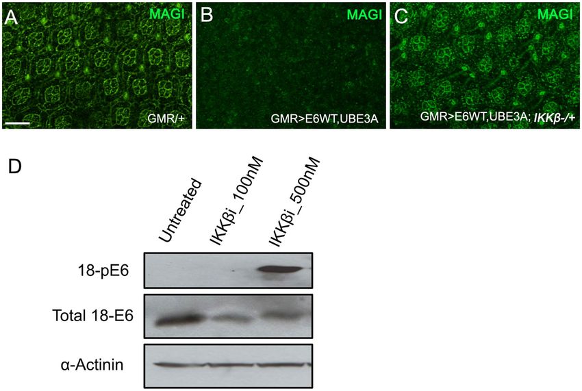
www.nature.com/scientificreports/
Figure 1. Reduction of IKKβ suppresses the E6 and UBE3A co-expression phenotypes. (A) Adult eye
expressing GMR-Gal4 showing an intact eye morphology. (B) GMR-Gal4-driven co-expression of E6 and
UBE3A causes rough eye morphology. (C) One mutated copy of IKKβ gene in GMR-Gal4 expressing eye has no
effect on the eye morphology. (D) One mutated copy of IKKβ gene suppresses the E6 + UBE3A-induced rough
eye defects. (E) Expression of IKKβ RNAi suppresses the E6 + UBE3A-induced rough eye defects.
Results
Kinome screen identified IKKβ as a mediator of E6 + hUBE3A‑mediated defects. The fly eye is a
compound eye consisting of 750 unit eyes. These ‘ommatidia’ are clusters of sensory neurons arranged in a pre-
cisely repeated hexagonal pattern formed by the precise arrangement of supporting pigment cells. Formation of
this highly organized pattern requires precisely regulated cell proliferation, cell differentiation and programmed
cell death; disruption of any of these processes leads to a disorganized, rough eye phenotype that is readily scored
under a light microscope40.
We have previously shown that co-expression of E6 and human UBE3A (hUBE3A) in the developing fly eye
leads to a disorganized, rough eye phenotype. To identify loci that modify the E6 + hUBE3A-induced eye defects,
we used mutations in the kinome to perform a dominant genetic modifier screen. Flies with stable integration
of the transgenes GMR-Gal4, UAS-E6, and UAS-hUBE3A (referred to as GMR>E6/hUBE3A) were crossed to
flies heterozygous for a mutant kinase. Comparing GMR>E6/hUBE3A; kinase+/− to GMR> E6/hUBE3A flies, we
screened 195 kinases and identified IKKβ as the strongest suppressor of the E6 + hUBE3A-mediated rough eye
Scientific Reports | (2021) 11:1111 | https://doi.org/10.1038/s41598-020-80193-5 3
Vol.:(0123456789)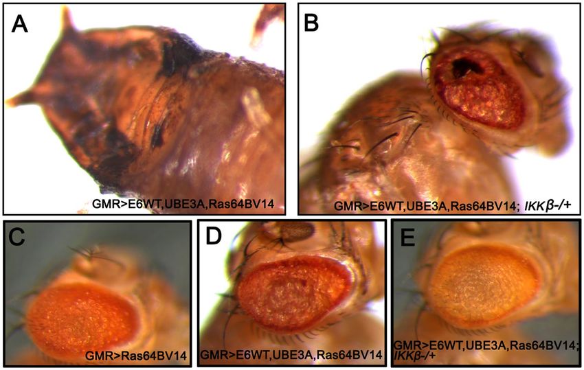
www.nature.com/scientificreports/
phenotype (Fig. 1D, compare with Fig. 1B; Fig. 1A,C are controls). Rescue was evident in 100% of flies (n = 40)
with GMR>E6/hUBE3A; IKKβ+/− genotype and to the same extent shown in Fig. 1D. The rescue by reduced IKKβ
activity was further confirmed with an RNA-interference transgene targeting IKKβ (Fig. 1E).
Inhibiting IKKβ in cervical cancer cells blocked their growth. IKKβ is a serine/threonine kinase that
is highly conserved between flies and humans. It regulates the innate immune pathway by activating NF-κB,
which in turn activates the expression of antimicrobial peptides to fight against pathogens41–43. To determine
whether IKKβ also has a functional link to E6 + UBE3A in human cells, we assessed the effect of the synthetic
IKKβ inhibitor IMD 0354 (N-(3,5-Bis-trifluoromethylphenyl)-5-chloro-2-hydroxybenzamide)44 on the growth
of HaCat (HPV−ve), HeLa (HPV 18 +ve), CasKi (HPV 16 +ve), and SiHa (HPV 16 +ve) cells. IMD 0354 is a
non–adenosine triphosphate–binding (ATP-binding) competitive selective IKKβ inhibitor that prevents ATP
attachment to IKKβ38. Different concentrations of the inhibitor were tested ranging from 250, 500, 750, 1000,
to 1250 nM. IMD 0354 reduced growth of all four cell types; HeLa cells were most strongly affected, with a
significant effect starting at the lowest concentration of 250 nM. While IMD 0354 had a significant effect on the
HPV 18 and 16 positive cells, it had a minor effect on the growth of HaCat cells that lack HPV (Fig. 2A). This
result suggested that the IKKβ-mediated mechanism of E6 + UBE3A-induced cellular abnormalities is conserved
between humans and fruit flies. To understand whether the reduced cell number resulting from inhibition of
IKKβ is due to arrest in cell cycle or to apoptosis, we treated these cells with 500 nM of IMD 0354 and analyzed
their cell cycle profile 3 h post treatment. We found no increase in apoptotic cell death for HeLa and CaSki
cells in comparison with HaCat cells. However, the majority of cells were found to exhibit a G1 cell cycle arrest
(Fig. 2B,C). This finding indicated that the reduction in cell numbers upon IKKβ inhibition is due to a halt in cell
growth and not to an increase in apoptotic cell death.
Reducing IKKβ suppressed the cellular defects caused by co‑expression of E6 and hUBE3A. As
reduction in IKKβ suppressed the rough eye phenotype caused by co-expression of E6 and hUBE3A, we exam-
ined the eye tissue of these flies 40 h after puparium formation, the time point at which the E6 + hUBE3A effect
becomes apparent. In the normal pupal eye, each ommatidium consists of eight photoreceptor cells covered by
four glial-like cells or cone cells and two primary pigment cells. Neighboring ommatidia are separated from each
other by a lattice of secondary and tertiary pigment cells and the sensory bristle cells. This interweaving lattice of
pigment cells organizes the ommatidial array into a precise pattern of repeated h exagons45.
Pupal eyes expressing E6 and hUBE3A exhibit severe morphological defects, including fusion of neighboring
ommatidia, increase in the number of pigment cells and cone cells, and a severe alteration in the stereotyped
pattern of ommatidia. In comparison, removing a single genomic copy of IKKβ (GMR>E6/hUBE3A; IKKβ+/−)
led to a strong phenotypic rescue: pigment and cone cell defects were reduced and the overall organization of the
ommatidial array improved (Fig. 3A–C). These observations suggest that E6 interferes with a molecular mecha-
nism involving IKKβ and that this mechanism plays an important role in E6-induced cellular abnormalities.
Reducing IKKβ suppressed the junctional and polarity defects caused by co‑expression of E6
and hUBE3A. We have previously shown that E6, in cooperation with UBE3A, perturbs the integrity of
junctional and polarity c omplexes35. Therefore, we hypothesized that reducing IKKβ activity might suppress
these defects. Immunolabeling for junctional marker E-cadherin and polarity marker Bazooka (the homolog
of human Par-3) revealed that, in comparison with pupal eyes expressing E6 and UBE3A, in which both the
junctional and polarity complexes were perturbed in ommatidia (Fig. 3 E–H′), GMR>E6/hUBE3A; IKKβ+/−
pupal eyes showed no disruption of junctional and polarity complexes and the integrity of these complexes was
restored to the extent seen in control eye tissues (Fig. 3F–I′ compared to D–G′). These observations suggest that
alterations in IKKβ significantly contribute to the E6-induced cellular junctional and polarity disorganization.
Reduced IKKβ activity suppressed E6 + hUBEA‑induced degradation of PDZ domain pro-
teins. Proteasomal degradation of PDZ domain-containing proteins, including Magi, Dlg, and Scribble, was
shown to be crucial for the cancerous effect of HPV 16 and 18 E 625. We have previously shown that HPV 18 E6,
with the addition of human UBE3A, targets the fly counterparts of these proteins for ubiquitin-mediated protea-
somal destruction35. As a reduction in IKKβ levels suppressed the cellular defects caused by E6 plus hUBE3A,
we asked whether the E6-mediated degradation of PDZ domain proteins was altered. To address this ques-
tion we examined the level of Magi, as Magi has been identified as a major degradation target of HPV18 E6 in
human and Drosophila19,35. Immunolabeling of pupal eyes for Magi revealed that, whereas GMR>E6/hUBE3A
eyes exhibited a complete loss of Magi, GMR>E6/hUBE3A; IKKβ+/− pupal eyes exhibited no detectable loss of
Magi (Fig. 4A–C). This result suggests that reducing IKKβ activity suppresses the E6-induced degradation of
Magi, and that rescue of Magi degradation is likely to play a role in suppression of E6-induced cellular defects.
Reducing IKKβ resulted in hyperphosphorylation of E6. Phosphorylation of the HPV 18 E6 PBM
was previously demonstrated to block its interaction with PDZ domain proteins Dlg and Magi37,46,47. To assess
whether phosphorylation-mediated regulation of E6 plays a role in suppressing Magi degradation, we treated
cells expressing HPV 18 E6 with the IKKβ inhibitor IMD 0354 (at 100 and 500 nM) and compared with untreated
cells for E6 phosphorylation. Western blot analysis was performed using an antibody to detect phosphorylated
E6. We found that inhibition of IKKβ resulted in extensive phosphorylation of E6, which was absent in untreated
cells (Fig. 4D). This result is consistent with previous findings, and suggests that the lack of Magi degradation in
cells expressing E6 + hUBE3A with mutated IKKβ could be due to the loss of PDZ recognition by E6.
Scientific Reports | (2021) 11:1111 | https://doi.org/10.1038/s41598-020-80193-5 4
Vol:.(1234567890)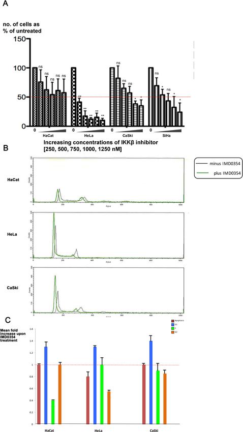
www.nature.com/scientificreports/

                                  Figure 2.  Inhibition of Human IKKβ in cervical cancer cells blocks their growth. (A) Five different concentrations (250, 500, 750,
                                  1000, 1250 nM) of human IKKβ inhibitor, IMD 0354, were applied to HPV-negative (HaCat), HPV 18-positive (HeLa), and HPV
                                  16-positive (CaSki and SiHa) cells and the effect was measured by number of surviving cells after 48 h. The inhibitor blocks the growth
                                  of HPV 16 and 18 cervical cancer cells without a significant effect on the HaCat cells, showing a most significant effect on HeLa cells:
                                  reducing the initial number of cells to 40%, at starting concentration of 250 nM and 10% at 1250 nM. Data are presented as mean ± SD
                                  (n = 3); not significance (n.s.) = P > 0.05; *P < 0.05, **P < 0.01. One-way ANOVA test using the Prism program was used for
                                  statistical analysis. (B) HPV-negative (HaCat), HPV 18-positive (HeLa), and HPV 16-positive (CaSki) cells were treated with 500 nM
                                  of human IKKβ inhibitor, IMD 0354, or with DMSO as control. After 3 h cells were trypsinized and stained with Propidium Iodide,
                                  and the cell cycle profiles were analysed by FACS. n = 4. Representative cell cycle profiles are shown for each cell type and condition.
                                  (C) The cell cycle FACS profiles were analysed to determine the number of cells in subG1 (i.e. apoptotic), G1, S and G2M phases of
                                  the cell cycle. The histogram shows the results from four experiments expressed as the fold change in cell number upon IMD 0354
                                  treatment, relative to DMSO treatment. Standard deviations are shown. The assay was repeated 4 times, n = 4.

Scientific Reports |   (2021) 11:1111 |                    https://doi.org/10.1038/s41598-020-80193-5                                                                    5

                                                                                                                                                                  Vol.:(0123456789)
www.nature.com/scientificreports/
Figure 3. Reduction of IKKβ suppresses the cellular abnormalities and restores the cell polarity and junctional
integrity disrupted by E6 + UBE3A co-expression. (A–C) Pupal eyes showing E-Cad immunolabeling. (A)
Expression of GMR-Gal4, showing a normal stereotype pattern of ommatidia. (B) Co-expression of E6 and
UBE3A causes severe abnormalities, including increase in number of pigment cells, bristle cells, and fusion
of neighboring ommatidia. (C) A mutated copy of the IKKβ gene suppresses the E6 + UBE3A abnormalities.
(E–H′) Co-expression of E6 and UBE3A causes disruption of cell polarity and junctional complex, as shown
by loss of Bazooka (Baz) and E-Cad from photoreceptor cells. This is in comparison with (D–G′) where Baz
and E-Cad are both localized correctly in each photoreceptor. (F–I′) A mutated copy of IKKβ gene in eyes
expressing E6 and UBE3A restores the polarity and junctional integrity as is shown by the correct localization of
Baz and E-Cad. Scale bars indicate 10 µm. Insets are digitally magnified 200%. Results shown are representative
of those of 5 different flies.
Reducing IKKβ suppressed the cooperative effect of Ras and E6 + hUBE3A. Previous studies
have shown that the HPV oncogenes E6 and E7 alone are insufficient to direct oncogenic transformation, and
that other factors including genetic alterations contribute to HPV-induced tumorigenesis in humans and mice48.
Mutations in Ras family proteins have been implicated in HPV-related cancers, and we have previously shown
that cooperation between E6 and oncogenic isoforms of Drosophila Ras (Ras64BV14) promotes cellular transfor-
mation and malignancy in fly e pithelia35,48–50. Therefore, we asked whether reducing IKKβ activity can suppress
Scientific Reports | (2021) 11:1111 | https://doi.org/10.1038/s41598-020-80193-5 6
Vol:.(1234567890)www.nature.com/scientificreports/
Figure 4. Reduced activity of IKKβ causes hyperphosphorylation of E6 and suppression of E6 + UBE3A-
mediated degradation of PDZ domain protein, Magi. (A–C) Pupal eyes showing Magi immunolabeling. (B)
Co-expression of E6 + UBE3A results in loss of Magi from cone cells and pigment cells compared with (A) in
which only the Gal4 driver is expressed. (C) A mutated copy of the IKKβ gene suppresses the E6 + UBE3A-
mediated loss of Magi. (D) Western blot of HeLa cell extracts shows that inhibition of IKKβ using a
concentration of 500 nM of the inhibitor IMD 0354 results in phosphorylation of E6; compare that to lack of E6
phosphorylation in HeLa cells grown with no inhibitor (untreated) or with a low concentration of 100 nM. Scale
bar indicates 10 µm. Results shown in (A–C) are representative of those of 5 different flies. Result shown in (D)
is a representative of n = 3.
the cellular transformation caused by the cooperation of oncogenic Ras and E6. Co-expression of oncogenic
Ras, Ras64BV14 with E6, and hUB3A in eye imaginal discs (GMR>E6/hUBE3A/Ras64BV14) at 25 °C resulted in
overgrowth and pupal lethality (Fig. 5A). However, when the level of IKKβ was reduced (GMR>E6/hUBE3A/
Ras64BV14; IKKβ+/−), the lethality was rescued and flies developed to adulthood. These adult flies, however, still
exhibited significant abnormalities in eye morphology, suggesting incomplete rescue (Fig. 5B).
Gal4 activity is reduced at lower temperatures51. At 22 °C, GMR>E6/hUBE3A/Ras64BV14 flies developed to
adulthood, exhibiting enhanced transformed eye morphology in comparison to expression of oncogenic Ras
alone (Fig. 5D compared to 5C). Reducing IKKβ activity significantly suppressed the transformed eye morphol-
ogy in these conditions (Fig. 5E), further demonstrating that IKKβ is important for the cooperative action of
E6 and oncogenic Ras.
Discussion
In this study we identify IKKβ as a mediator of the HPV 18 E6 and hUBE3A-induced cellular defects in both fly
and human cancer models. This is consistent with previous studies demonstrating that HPV 16 E6 interacts with
components of the innate immune pathway, including IKKβ, and it activates the NF-κB transcription f actor52,53.
The role of the innate immune system in HPV infection and cancer progression is not well understood. Activation
of the innate immune system, particularly the Toll pathway, has been associated with regression and clearance
of HPV 16 i nfection54. This is consistent with other studies demonstrating that HPV 16 oncogenes repress the
expression of a key innate immune sensor, Toll like receptor 9 (TLR9)55, suggesting that persistent infection may
reflect a defect in the host innate immune system. Conversely, activation of the innate immune system, and con-
sequent inflammation, contributes to HPV-induced tumor p rogression56. Several studies have shown an increase
in the level of inflammatory cytokines in individuals with reduced HPV clearance57–59. Additionally, upregulation
of several TLRs and components of the NF-κB signaling pathway have been implicated in HPV-related cancers.
It has been suggested that the malignant phenotype of cervical cancer cells requires the function and activation
of IKKβ/NF-κB signaling60,61. IKKβ phosphorylates the inhibitor of NF-κB, resulting in its ubiquitination and
Scientific Reports | (2021) 11:1111 | https://doi.org/10.1038/s41598-020-80193-5 7
Vol.:(0123456789)www.nature.com/scientificreports/
Figure 5. Reduction of IKKβ suppresses the cellular transformation caused by cooperative action of Ras
and E6 + UBE3A. (A,B) Expression of transgenes at 25 °C. (B) A mutated copy of IKKβ gene in eyes expressing
E6, UBE3A and an oncogenic Ras, suppresses pupal lethality and the severe defects caused by cooperative action
of E6, UBE3A and oncogenic Ras (A). (C–E) Expression of transgenes at 22 °C. (C) Expression of oncogenic Ras
in the eye causes cellular transformation. (D) Co-expression of E6 and UBE3A with oncogenic Ras enhances
the Ras phenotype. (E) A mutated copy of IKKβ gene suppresses the severe eye abnormalities mediated by the
synergistic effect of oncogenic Ras, E6 and UBE3A. Results shown are representative of those of 5 different flies.
proteasomal degradation. This action of IKKβ frees the NF-κB, which in turn enters the nucleus and activates
the transcription of pro-inflammatory, pro-cell proliferation and anti-apoptotic genes62,63. Increased expression
of IKKβ and its association with an aggressive phenotype has been reported in several types of cancers including
head-and-neck, ovarian and liver c ancers64–66. It is notable that IKKβ’s role in cancer is not only limited to its
function in regulation of the NF-κB pathway; IKKβ can also phosphorylate p53, resulting in its ubiquitination
and subsequent degradation. Inactivation or loss of p53 has been identified in more than 50% of cancers, includ-
ing HPV-induced cancers: high-risk HPV E6s target p53 for ubiquitination and proteasomal degradation14,67.
IKKβ-mediated loss of p53 can be suppressed by inhibition of IKKβ in cancer c ells68,69. The inhibitory function
of IKKβ on p53 was further found in HPV38E6E7 human keratinocytes. IKKβ phosphorylates and stabilizes a
dominant-negative inhibitor of p53, ∆Np73α, resulting in repression of p53-regulated genes such as p21. This
inhibitory effect of IKKβ on p53 can be suppressed upon its inactivation, which results in destabilization and
degradation of ∆Np73α70. Another phosphorylation target of IKKβ is fork-head transcription factor FOXO3a.
IKKβ has been found to stimulate cell cycle progression and proliferation of breast cancer cells, through phos-
phorylation of FOXO3a, leading to its exclusion from nucleus and subsequent degradation in the proteasome.
This function of IKKβ could be further overridden by the overexpression of FOXO3a71. All these findings sug-
gest that IKKβ may contribute to HPV-induced cellular abnormalities in several ways, some through the innate
immune pathway, and some independent of it.
IKKβ inhibitors have served as potent anti-tumor agents inhibiting cell proliferation and invasiveness, as
well as inducing cell death. Their activity has been demonstrated in several cancer types including breast, colon,
ovarian, oral, prostate, liver, melanoma, and leukemia65,66,72–76. One such inhibitor is IMD 0354, which selec-
tively inhibits IKKβ by preventing ATP attachment to IKKβ. This will in turn block phosphorylation of IκBα,
thus preventing nuclear translocation and activation of NF-κB38. Although IMD 0354 was initially designed to
inhibit inflammation77, it has recently proved to have strong anticancer p roperties78,79 in several cancers. IMD
0354 inhibits breast cancer cell proliferation, progression of mast cells, and it induces apoptosis in prostate, lung
and colon c ancers44,80–82. In our study we found that IMD 0354 inhibited the growth of HPV 16+ and 18+ cer-
vical cancer cell lines. This result is consistent with previously reported function of IMD 0354 in cancer cells
and hence suggests a therapeutic potential for IKKβ inhibitors to treat HPV-induced cancers. Although both
HPV16+ and HPV18+ cervical cancer cell lines responded to IMD 0354, however, IMD 0354-mediated inhibi-
tion of IKKβ exhibited greater effectiveness on HeLa cells (HPV 18+) compared with SiHa and CaSki cells (HPV
16+). It has previously been demonstrated that these cell lines differ in a number of molecular pathways, and
in their response to treatments such as therapeutic agents that induce cell death83–85. HeLa cells exhibit greater
apoptotic cell death in response to cisplatin than SiHa and CaSki cells do, a difference that is due to the higher
levels of p53 and p21 in HeLa cells84. Additionally, these cell lines show distinct proteomic profiles for pathways
connected to p53 activation, mitochondrial function and oxidative stress85,86. These findings suggest that the
stronger effect of IMD 0354 in HeLa relative to SiHa and CaSki cells could in part be due to their difference
Scientific Reports | (2021) 11:1111 | https://doi.org/10.1038/s41598-020-80193-5 8
Vol:.(1234567890)www.nature.com/scientificreports/
in molecular pathways affected by IKKβ. Additionally, IMD 0354 caused an extensive phosphorylation of E6,
which was absent in untreated cells. This finding is intriguing, as phosphorylation of E6 on the PDZ binding
motif results in disruption of its interaction with PDZ domain-containing proteins46,47. This is consistent with
our results, as degradation of Magi was inhibited when IKKβ was reduced. It would be of interest to understand
the mechanism by which inhibition of IKKβ results in E6 hyperphosphorylation.
In summary this study suggests an important role for IKKβ in E6-induced cellular abnormalities and that
targeting IKKβ could be a potential therapeutic option for cervical cancer and, perhaps, for other HPV-related
cancers for which there is currently no effective treatment.
Received: 18 December 2019; Accepted: 15 December 2020
References
1. de Martel, C., Plummer, M., Vignat, J. & Franceschi, S. Worldwide burden of cancer attributable to HPV by site, country and HPV
type. Int. J. Cancer 141, 664–670. https://doi.org/10.1002/ijc.30716 (2017).
2. de Sanjose, S. et al. Human papillomavirus genotype attribution in invasive cervical cancer: A retrospective cross-sectional world-
wide study. Lancet Oncol. 11, 1048–1056. https://doi.org/10.1016/S1470-2045(10)70230-8 (2010).
3. Arbyn, M. et al. Estimates of incidence and mortality of cervical cancer in 2018: A worldwide analysis. Lancet Glob. Health 8,
e191–e203. https://doi.org/10.1016/S2214-109X(19)30482-6 (2020).
4. Schiller, J. T. & Muller, M. Next generation prophylactic human papillomavirus vaccines. Lancet Oncol. 16, e217-225. https://doi.
org/10.1016/S1470-2045(14)71179-9 (2015).
5. Schiffman, M. & Wentzensen, N. Human papillomavirus infection and the multistage carcinogenesis of cervical cancer. Cancer
Epidemiol. Biomark. Prev. 22, 553–560. https://doi.org/10.1158/1055-9965.EPI-12-1406 (2013).
6. Skeate, J. G., Woodham, A. W., Einstein, M. H., Da Silva, D. M. & Kast, W. M. Current therapeutic vaccination and immunotherapy
strategies for HPV-related diseases. Hum. Vaccin Immunother. 12, 1418–1429. https://doi.org/10.1080/21645515.2015.1136039
(2016).
7. Thomas, T. L. Cancer prevention: HPV vaccination. Semin. Oncol. Nurs. 32, 273–280. https://doi.org/10.1016/j.soncn.2016.05.007
(2016).
8. Su, W. H., Chuang, P. C., Huang, E. Y. & Yang, K. D. Radiation-induced increase in cell migration and metastatic potential of cervi-
cal cancer cells operates via the K-Ras pathway. Am. J. Pathol. 180, 862–871. https://doi.org/10.1016/j.ajpath.2011.10.018 (2012).
9. Pectasides, D., Kamposioras, K., Papaxoinis, G. & Pectasides, E. Chemotherapy for recurrent cervical cancer. Cancer Treat. Rev.
34, 603–613. https://doi.org/10.1016/j.ctrv.2008.05.006 (2008).
10. Munger, K., Phelps, W. C., Bubb, V., Howley, P. M. & Schlegel, R. The E6-gene and E7-gene of the human papillomavirus type-16
together are necessary and sufficient for transformation of primary human keratinocytes. J. Virol. 63, 4417–4421 (1989).
11. Ho, G. Y. F. et al. Persistent genital human papillomavirus infection as a risk factor for persistent cervical dysplasia. J. Natl. Cancer
I(87), 1365–1371. https://doi.org/10.1093/jnci/87.18.1365 (1995).
12. Dyson, N., Howley, P. M., Munger, K. & Harlow, E. The human papilloma virus-16 E7-oncoprotein is able to bind to the retino-
blastoma gene-product. Science 243, 934–937. https://doi.org/10.1126/science.2537532 (1989).
13. Werness, B. A., Levine, A. J. & Howley, P. M. Association of human papillomavirus type-16 and type-18 E6 proteins with P53.
Science 248, 76–79. https://doi.org/10.1126/science.2157286 (1990).
14. Scheffner, M., Werness, B. A., Huibregtse, J. M., Levine, A. J. & Howley, P. M. The E6 oncoprotein encoded by human papillomavirus
types 16 and 18 promotes the degradation of p53. Cell 63, 1129–1136 (1990).
15. Spanos, W. C. et al. The PDZ binding motif of human papillomavirus type 16 E6 induces PTPN13 loss, which allows anchorage-
independent growth and synergizes with ras for invasive growth. J. Virol. 82, 2493–2500. https://doi.org/10.1128/JVI.02188-07
(2008).
16. Kiyono, T. et al. Binding of high-risk human papillomavirus E6 oncoproteins to the human homologue of the Drosophila discs
large tumor suppressor protein. Proc. Natl. Acad. Sci. USA 94, 11612–11616. https://doi.org/10.1073/pnas.94.21.11612 (1997).
17. Nakagawa, S. & Huibregtse, J. M. Human scribble (Vartul) is targeted for ubiquitin-mediated degradation by the high-risk papillo-
mavirus E6 proteins and the E6AP ubiquitin-protein ligase. Mol. Cell Biol. 20, 8244–8253. https: //doi.org/10.1128/Mcb.20.21.8244-
8253.2000 (2000).
18. Pim, D., Thomas, M., Javier, R., Gardiol, D. & Banks, L. HPV E6 targeted degradation of the discs large protein: Evidence for the
involvement of a novel ubiquitin ligase. Oncogene 19, 719–725. https://doi.org/10.1038/sj.onc.1203374 (2000).
19. Kranjec, C. & Banks, L. A systematic analysis of human papillomavirus (HPV) E6 PDZ substrates identifies MAGI-1 as a major
target of HPV type 16 (HPV-16) and HPV-18 whose loss accompanies disruption of tight junctions. J. Virol. 85, 1757–1764. https
://doi.org/10.1128/JVI.01756-10 (2011).
20. Laura, R. P., Ross, S., Koeppen, H. & Lasky, L. A. MAGI-1: A widely expressed, alternatively spliced tight junction protein. Exp.
Cell Res. 275, 155–170. https://doi.org/10.1006/excr.2002.5475 (2002).
21. Zihni, C., Mills, C., Matter, K. & Balda, M. S. Tight junctions: From simple barriers to multifunctional molecular gates. Nat. Rev.
Mol. Cell Biol. 17, 564–580. https://doi.org/10.1038/nrm.2016.80 (2016).
22. Javier, R. T. & Rice, A. P. Emerging theme: Cellular PDZ proteins as common targets of pathogenic viruses. J. Virol. 85, 11544–
11556. https://doi.org/10.1128/JVI.05410-11 (2011).
23. Scheffner, M., Huibregtse, J. M., Vierstra, R. D. & Howley, P. M. The Hpv-16 E6 and E6-Ap Complex Functions as a Ubiquitin-
Protein Ligase in the Ubiquitination of P53. Cell 75, 495–505. https://doi.org/10.1016/0092-8674(93)90384-3 (1993).
24. Beaudenon, S. & Huibregtse, J. M. HPV E6, E6AP and cervical cancer. BMC Biochem. https://doi.org/10.1186/1471-2091-9-S1-S4
(2008).
25. Nguyen, M. L., Nguyen, M. M., Lee, D., Griep, A. E. & Lambert, P. F. The PDZ ligand domain of the human papillomavirus type
16 E6 protein is required for E6’s induction of epithelial hyperplasia in vivo. J. Virol. 77, 6957–6964 (2003).
26. Halder, G. & Mills, G. B. Drosophila in cancer research: To boldly go where no one has gone before. Oncogene 30, 4063–4066.
https://doi.org/10.1038/onc.2011.128 (2011).
27. Gilbert, L. I. Drosophila is an inclusive model for human diseases, growth and development. Mol. Cell Endocrinol. 293, 25–31.
https://doi.org/10.1016/j.mce.2008.02.009 (2008).
28. Vidal, M. & Cagan, R. L. Drosophila models for cancer research. Curr. Opin. Genet. Dev. 16, 10–16. https://doi.org/10.1016/j.
gde.2005.12.004 (2006).
29. Crozatier, M. & Vincent, A. Drosophila: a model for studying genetic and molecular aspects of haematopoiesis and associated
leukaemias. Dis. Model Mech. 4, 439–445. https://doi.org/10.1242/dmm.007351 (2011).
30. Maitra, U. & Ciesla, L. Using Drosophila as a platform for drug discovery from natural products in Parkinson’s disease. Medchem-
comm 10, 867–879. https://doi.org/10.1039/c9md00099b (2019).
Scientific Reports | (2021) 11:1111 | https://doi.org/10.1038/s41598-020-80193-5 9
Vol.:(0123456789)www.nature.com/scientificreports/
31. Mirzoyan, Z. et al. Drosophila melanogaster: A model organism to study cancer. Front. Genet. 10, 51. https://doi.org/10.3389/fgene
.2019.00051(2019).
32. Ito, S. et al. A genetic screen in Drosophila for regulators of human prostate cancer progression. Biochem. Biophys. Res. Commun.
451, 548–555. https://doi.org/10.1016/j.bbrc.2014.08.015 (2014).
33. Rossi, F. et al. An in vivo genetic screen in Drosophila identifies the orthologue of human cancer/testis gene SPO11 among a
network of targets to inhibit lethal(3)malignant brain tumour growth. Open Biol. https://doi.org/10.1098/rsob.170156 (2017).
34. Anderson, A. M., Bailetti, A. A., Rodkin, E., De, A. & Bach, E. A. A genetic screen reveals an unexpected role for Yorkie signal-
ing in JAK/STAT-dependent hematopoietic malignancies in Drosophila melanogaster. G3 7, 2427–2438. https://doi.org/10.1534/
g3.117.044172 (2017).
35. Padash Barmchi, M. et al. A Drosophila model of HPV E6-induced malignancy reveals essential roles for magi and the insulin
receptor. PLoS Pathog. 12, e1005789. https://doi.org/10.1371/journal.ppat.1005789 (2016).
36. Reiter, L. T., Seagroves, T. N., Bowers, M. & Bier, E. Expression of the Rho-GEF Pbl/ECT2 is regulated by the UBE3A E3 ubiquitin
ligase. Hum. Mol. Genet. 15, 2825–2835. https://doi.org/10.1093/hmg/ddl225 (2006).
37. Boon, S. S., Tomaic, V., Thomas, M., Roberts, S. & Banks, L. Cancer-causing human papillomavirus E6 proteins display major
differences in the phospho-regulation of their PDZ interactions. J. Virol. 89, 1579–1586. https://doi.org/10.1128/JVI.01961-14
(2015).
38. Lennikov, A. et al. IkappaB kinase-beta inhibitor IMD-0354 beneficially suppresses retinal vascular permeability in streptozotocin-
induced diabetic mice. Investig. Ophthalmol. Vis. Sci. 55, 6365–6373. https://doi.org/10.1167/iovs.14-14671 (2014).
39. Padash Barmchi, M., Samarasekera, G., Gilbert, M., Auld, V. J. & Zhang, B. Magi is associated with the par complex and functions
antagonistically with bazooka to regulate the apical polarity complex. PLoS ONE 11, e0153259. https://doi.org/10.1371/journ
al.pone.0153259 (2016).
40. Tsachaki, M. & Sprecher, S. G. Genetic and developmental mechanisms underlying the formation of the Drosophila compound
eye. Dev. Dyn. 241, 40–56. https://doi.org/10.1002/dvdy.22738 (2012).
41. Ghosh, S., May, M. J. & Kopp, E. B. NF-kappa B and Rel proteins: Evolutionarily conserved mediators of immune responses. Annu.
Rev. Immunol. 16, 225–260. https://doi.org/10.1146/annurev.immunol.16.1.225 (1998).
42. Karin, M. & Delhase, M. The I kappa B kinase (IKK) and NF-kappa B: Key elements of proinflammatory signalling. Semin. Immu-
nol. 12, 85–98. https://doi.org/10.1006/smim.2000.0210 (2000).
43. Vallabhapurapu, S. & Karin, M. Regulation and function of NF-kappa B transcription factors in the immune system. Annu. Rev.
Immunol. 27, 693–733. https://doi.org/10.1146/annurev.immunol.021908.132641 (2009).
44. Tanaka, A. et al. A novel NF-kappaB inhibitor, IMD-0354, suppresses neoplastic proliferation of human mast cells with constitu-
tively activated c-kit receptors. Blood 105, 2324–2331. https://doi.org/10.1182/blood-2004-08-3247 (2005).
45. Cagan, R. Principles of Drosophila eye differentiation. Curr. Top. Dev. Biol. 89, 115–135. https://doi.org/10.1016/S0070
-2153(09)89005-4 (2009).
46. Kuhne, C., Gardiol, D., Guarnaccia, C., Amenitsch, H. & Banks, L. Differential regulation of human papillomavirus E6 by protein
kinase A: Conditional degradation of human discs large protein by oncogenic E6. Oncogene 19, 5884–5891. https: //doi.org/10.1038/
sj.onc.1203988 (2000).
47. Boon, S. S. & Banks, L. High-risk human papillomavirus E6 oncoproteins interact with 14–3-3 zeta in a PDZ binding motif-
dependent manner. J. Virol. 87, 1586–1595. https://doi.org/10.1128/Jvi.02074-12 (2013).
48. Greenhalgh, D. A. et al. Transgenic mice expressing targeted HPV-18 E6 and E7 oncogenes in the epidermis develop verrucous
lesions and spontaneous, rasHa-activated papillomas. Cell Growth Differ. 5, 667–675 (1994).
49. Schreiber, K. et al. Strong synergy between mutant ras and HPV16 E6/E7 in the development of primary tumors. Oncogene 23,
3972–3979. https://doi.org/10.1038/sj.onc.1207507 (2004).
50. Anwar, K., Nakakuki, K., Naiki, H. & Inuzuka, M. ras gene mutations and HPV infection are common in human laryngeal carci-
noma. Int. J. Cancer 53, 22–28. https://doi.org/10.1002/ijc.2910530106 (1993).
51. Duffy, J. B. GAL4 system in Drosophila: A fly geneticist’s Swiss army knife. Genesis 34, 1–15. https://doi.org/10.1002/gene.10150
(2002).
52. Oliveira, L. B., Haga, I. R. & Villa, L. L. Human papillomavirus (HPV) 16 E6 oncoprotein targets the Toll-like receptor pathway.
J. Gen. Virol. https://doi.org/10.1099/jgv.0.001057 (2018).
53. James, M. A., Lee, J. H. & Klingelhutz, A. J. Human papillomavirus type 16 E6 activates NF-kappaB, induces cIAP-2 expression, and
protects against apoptosis in a PDZ binding motif-dependent manner. J. Virol. 80, 5301–5307. https://doi.org/10.1128/JVI.01942
-05 (2006).
54. Daud, I. I. et al. Association between toll-like receptor expression and human papillomavirus type 16 persistence. Int. J. Cancer
128, 879–886. https://doi.org/10.1002/ijc.25400 (2011).
55. Hasan, U. A. et al. The human papillomavirus type 16 E7 oncoprotein induces a transcriptional repressor complex on the Toll-like
receptor 9 promoter. J. Exp. Med. 210, 1369–1387. https://doi.org/10.1084/jem.20122394 (2013).
56. Boccardo, E., Lepique, A. P. & Villa, L. L. The role of inflammation in HPV carcinogenesis. Carcinogenesis 31, 1905–1912. https://
doi.org/10.1093/carcin/bgq176 (2010).
57. Baker, R. et al. Increased plasma levels of adipokines and inflammatory markers in older women with persistent HPV infection.
Cytokine 53, 282–285. https://doi.org/10.1016/j.cyto.2010.11.014 (2011).
58. Kemp, T. J. et al. Elevated systemic levels of inflammatory cytokines in older women with persistent cervical human papillomavirus
infection. Cancer Epidemiol. Biomarkers Prev. 19, 1954–1959. https://doi.org/10.1158/1055-9965.EPI-10-0184 (2010).
59. Lam, J. O. et al. Association of serum cytokines with oral HPV clearance. Cytokine 83, 85–91. https://doi.org/10.1016/j.
cyto.2016.04.002 (2016).
60. Rich, A. M., Hussaini, H. M., Parachuru, V. P. & Seymour, G. J. Toll-like receptors and cancer, particularly oral squamous cell
carcinoma. Front. Immunol. 5, 464. https://doi.org/10.3389/fimmu.2014.00464 (2014).
61. Ma, X. F. et al. IKKbeta/NF-kappaB mediated the low doses of bisphenol A induced migration of cervical cancer cells. Arch.
Biochem. Biophys. 573, 52–58. https://doi.org/10.1016/j.abb.2015.03.010 (2015).
62. Silverman, N. & Maniatis, T. NF-kappaB signaling pathways in mammalian and insect innate immunity. Genes Dev. 15, 2321–2342.
https://doi.org/10.1101/gad.909001 (2001).
63. Ghosh, S. Regulation of inducible gene expression by the transcription factor NF-kappaB. Immunol. Res. 19, 183–189. https://doi.
org/10.1007/BF02786486 (1999).
64. Nottingham, L. K. et al. Aberrant IKKalpha and IKKbeta cooperatively activate NF-kappaB and induce EGFR/AP1 signaling to
promote survival and migration of head and neck cancer. Oncogene 33, 1135–1147. https://doi.org/10.1038/onc.2013.49 (2014).
65. Hernandez, L. et al. Activation of NF-kappaB signaling by inhibitor of NF-kappaB kinase beta increases aggressiveness of ovarian
cancer. Cancer Res. 70, 4005–4014. https://doi.org/10.1158/0008-5472.CAN-09-3912 (2010).
66. Jiang, R. et al. High expression levels of IKKalpha and IKKbeta are necessary for the malignant properties of liver cancer. Int. J.
Cancer 126, 1263–1274. https://doi.org/10.1002/ijc.24854 (2010).
67. Werness, B. A., Levine, A. J. & Howley, P. M. Association of human papillomavirus types 16 and 18 E6 proteins with p53. Science
248, 76–79 (1990).
68. Xia, Y. et al. Phosphorylation of p53 by IkappaB kinase 2 promotes its degradation by beta-TrCP. Proc. Natl. Acad. Sci. USA 106,
2629–2634. https://doi.org/10.1073/pnas.0812256106 (2009).
Scientific Reports | (2021) 11:1111 | https://doi.org/10.1038/s41598-020-80193-5 10
Vol:.(1234567890)www.nature.com/scientificreports/
69. Yang, P. M. et al. Loss of IKKbeta activity increases p53 stability and p21 expression leading to cell cycle arrest and apoptosis. J.
Cell Mol. Med. 14, 687–698. https://doi.org/10.1111/j.1582-4934.2009.00712.x (2010).
70. Accardi, R. et al. IkappaB kinase beta promotes cell survival by antagonizing p53 functions through DeltaNp73alpha phosphoryla-
tion and stabilization. Mol Cell Biol 31, 2210–2226. https://doi.org/10.1128/MCB.00964-10 (2011).
71. Hu, M. C. et al. IkappaB kinase promotes tumorigenesis through inhibition of forkhead FOXO3a. Cell 117, 225–237 (2004).
72. Cilloni, D. et al. The NF-kappaB pathway blockade by the IKK inhibitor PS1145 can overcome imatinib resistance. Leukemia 20,
61–67. https://doi.org/10.1038/sj.leu.2403998 (2006).
73. Yemelyanov, A. et al. Effects of IKK inhibitor PS1145 on NF-kappaB function, proliferation, apoptosis and invasion activity in
prostate carcinoma cells. Oncogene 25, 387–398. https://doi.org/10.1038/sj.onc.1209066 (2006).
74. Yang, J., Amiri, K. I., Burke, J. R., Schmid, J. A. & Richmond, A. BMS-345541 targets inhibitor of kappaB kinase and induces
apoptosis in melanoma: Involvement of nuclear factor kappaB and mitochondria pathways. Clin. Cancer Res. 12, 950–960. https
://doi.org/10.1158/1078-0432.CCR-05-1220 (2006).
75. Johnson, J., Shi, Z., Liu, Y. & Stack, M. S. Inhibitors of NF-kappaB reverse cellular invasion and target gene upregulation in an
experimental model of aggressive oral squamous cell carcinoma. Oral Oncol. 50, 468–477. https://doi.org/10.1016/j.oraloncolo
gy.2014.02.004 (2014).
76. Shao, L., Wu, L. & Zhou, D. Sensitization of tumor cells to cancer therapy by molecularly targeted inhibition of the inhibitor of
nuclear factor kappaB kinase. Transl. Cancer Res. 1, 100–108. https://doi.org/10.3978/j.issn.2218-676X.2012.05.04 (2012).
77. Sugita, A. et al. Antiallergic and anti-inflammatory effects of a novel I kappaB kinase beta inhibitor, IMD-0354, in a mouse model
of allergic inflammation. Int. Arch. Allergy Immunol. 148, 186–198. https://doi.org/10.1159/000161579 (2009).
78. Ochiai, T. et al. Inhibition of IkappaB kinase beta restrains oncogenic proliferation of pancreatic cancer cells. J. Med. Dent. Sci. 55,
49–59 (2008).
79. Uota, S. et al. An IkappaB kinase 2 inhibitor IMD-0354 suppresses the survival of adult T-cell leukemia cells. Cancer Sci. 103,
100–106. https://doi.org/10.1111/j.1349-7006.2011.02110.x (2012).
80. Hayakawa, Y. et al. Effectiveness of IkappaB kinase inhibitors in murine colitis-associated tumorigenesis. J. Gastroenterol. 44,
935–943. https://doi.org/10.1007/s00535-009-0098-7 (2009).
81. Kanduri, M., Tobin, G., Aleskog, A., Nilsson, K. & Rosenquist, R. The novel NF-kappaB inhibitor IMD-0354 induces apoptosis in
chronic lymphocytic leukemia. Blood Cancer J. 1, e12. https://doi.org/10.1038/bcj.2011.9 (2011).
82. Kim, S. et al. Anti-cancer activity of the novel 2-hydroxydiarylamide derivatives IMD-0354 and KRT1853 through suppression
of cancer cell invasion, proliferation, and survival mediated by TMPRSS4. Sci. Rep. 9, 10003. https://doi.org/10.1038/s41598-019-
46447-7 (2019).
83. Maliekal, T. T., Anto, R. J. & Karunagaran, D. Differential activation of Smads in HeLa and SiHa cells that differ in their response
to transforming growth factor-beta. J. Biol. Chem. 279, 36287–36292. https://doi.org/10.1074/jbc.M404568200 (2004).
84. Funaoka, K. et al. High-risk HPV-positive human cancer cell lines show different sensitivity to cisplatin-induced apoptosis cor-
related with the p21Waf1/Cip1 level. Cancer Lett. 108, 15–23. https://doi.org/10.1016/s0304-3835(96)04362-5 (1996).
85. Filippova, M. et al. Cellular levels of oxidative stress affect the response of cervical cancer cells to chemotherapeutic agents. Biomed.
Res. Int. 2014, 574659. https://doi.org/10.1155/2014/574659 (2014).
86. Pappa, K. I. et al. High resolution analysis of the intracellular proteome of cervical cancer cell lines unveils novel regulators of
cervical carcinogenesis. Oncol. Rep. https://doi.org/10.3892/or.2019.7269 (2019).
Acknowledgements
We would like to thank department of biology for their support and the Samuel Roberts Noble Microscopy
Laboratory for assistance in confocal imaging. This study was partially supported by Oklahoma Shared Clinical
and Translational Resources grant funded by NIH.
Author contributions
M.P.B. wrote the manuscript and prepared Figs. 1, 3, 4A–C and 5. M.T., J.V.T., and A.V. prepared Figs. 2 and 4D.
R.L.C. provided reagents. Experiments for Figs. 2 and 4D were performed in L.B. laboratory. B.Z., R.L.C. and
L.B. all intellectually contributed to the project and revised the manuscript. Additionally, all authors reviewed
the manuscript.
Competing interests
The authors declare no competing interests.
Additional information
Supplementary Information The online version contains supplementary material available at https://doi.
org/10.1038/s41598-020-80193-5.
Correspondence and requests for materials should be addressed to M.P.B.
Reprints and permissions information is available at www.nature.com/reprints.
Publisher’s note Springer Nature remains neutral with regard to jurisdictional claims in published maps and
institutional affiliations.
Open Access This article is licensed under a Creative Commons Attribution 4.0 International
License, which permits use, sharing, adaptation, distribution and reproduction in any medium or
format, as long as you give appropriate credit to the original author(s) and the source, provide a link to the
Creative Commons licence, and indicate if changes were made. The images or other third party material in this
article are included in the article’s Creative Commons licence, unless indicated otherwise in a credit line to the
material. If material is not included in the article’s Creative Commons licence and your intended use is not
permitted by statutory regulation or exceeds the permitted use, you will need to obtain permission directly from
the copyright holder. To view a copy of this licence, visit http://creativecommons.org/licenses/by/4.0/.
© The Author(s) 2021
Scientific Reports | (2021) 11:1111 | https://doi.org/10.1038/s41598-020-80193-5 11
Vol.:(0123456789)You can also read