CRISPR/Cas9 Ribonucleoprotein-Based Genome Editing Methodology in the Marine Protozoan Parasite Perkinsus marinus
←
→
Page content transcription
If your browser does not render page correctly, please read the page content below
ORIGINAL RESEARCH
                                                                                                                                              published: 09 April 2021
                                                                                                                                      doi: 10.3389/fbioe.2021.623278

                                            CRISPR/Cas9
                                            Ribonucleoprotein-Based Genome
                                            Editing Methodology in the Marine
                                            Protozoan Parasite Perkinsus
                                            marinus
                                            Raghavendra Yadavalli 1† , Kousuke Umeda 1,2 , Hannah A. Waugh 1,3 , Adrienne N. Tracy 1,4 ,
                                            Asha V. Sidhu 1,4 , Derek E. Hernández 1,4 and José A. Fernández Robledo 1*
                            Edited by:      1
                                             Bigelow Laboratory for Ocean Sciences, East Boothbay, ME, United States, 2 National Research Center for Protozoan
                             Tao Chen,
                                            Diseases, Obihiro University of Agriculture and Veterinary Medicine, Obihiro, Japan, 3 Southern Maine Community College,
               Tianjin University, China
                                            South Portland, ME, United States, 4 Colby College, Waterville, ME, United States
                        Reviewed by:
                       Theodore Roth,
                University of California,   Perkinsus marinus (Perkinsozoa), a close relative of apicomplexans, is an osmotrophic
         San Francisco, United States       facultative intracellular marine protozoan parasite responsible for “Dermo” disease in
                 Motomichi Matsuzaki,
          RIKEN Center for Advanced         oysters and clams. Although there is no clinical evidence of this parasite infecting
      Intelligence Project (AIP), Japan     humans, HLA-DR40 transgenic mice studies strongly suggest the parasite as a natural
                  *Correspondence:          adjuvant in oral vaccines. P. marinus is being developed as a heterologous gene
           José A. Fernández Robledo
     jfernandez-robledo@bigelow.org;
                                            expression platform for pathogens of medical and veterinary relevance and a novel
                robledo64@gmail.com         platform for delivering vaccines. We previously reported the transient expression of
                   † Present address:       two rodent malaria genes Plasmodium berghei HAP2 and MSP8. In this study, we
               Raghavendra Yadavalli,
                                            optimized the original electroporation-based protocol to establish a stable heterologous
Department of Chemical Engineering,
 University of Virginia, Charlottesville,   expression method. Using 20 µg of pPmMOE[MOE1]:GFP and 25.0 × 10⁶ P. marinus
                     VA, United States      cells resulted in 98% GFP-positive cells. Furthermore, using the optimized protocol, we
                                            report for the first time the successful knock-in of GFP at the C-terminus of the PmMOE1
                    Specialty section:
         This article was submitted to      using ribonucleoprotein (RNP)-based CRISPR/Cas9 gene editing methodology. The
                     Synthetic Biology,     GFP was expressed 18 h post-transfection, and expression was observed for 8 months
                a section of the journal
       Frontiers in Bioengineering and
                                            post-transfection, making it a robust and stable knock-in system.
                        Biotechnology
                                            Keywords: Perkinsus marinus, oral adjuvant, heterologous expression system, CRISPR/Cas9, protozoan,
         Received: 29 October 2020          transfection, oral vaccines
          Accepted: 09 March 2021
           Published: 09 April 2021
                             Citation:      INTRODUCTION
    Yadavalli R, Umeda K, Waugh HA,
   Tracy AN, Sidhu AV, Hernández DE         Perkinsus marinus (original name Dermocystidium marinum), first described in 1950 as infecting
    and Fernández Robledo JA (2021)         the eastern oyster (Crassostrea virginica), is still a constant threat to the oyster industry (Mackin
                        CRISPR/Cas9         et al., 1950; Andrews, 1996; Perkins, 1996). In North America, P. marinus and Perkinsus chesapeaki
   Ribonucleoprotein-Based Genome
                                            can coexist in the same bivalve host (McLaughlin and Faisal, 1998; Coss et al., 2001a,b; Pecher et al.,
    Editing Methodology in the Marine
          Protozoan Parasite Perkinsus
                                            2008; Reece et al., 2008; Arzul et al., 2012). In the oysters, the parasite is taken up by hemocytes and
                              marinus.      uses them as a vehicle for migration into other host tissues (Lau et al., 2018a; Schott et al., 2019;
 Front. Bioeng. Biotechnol. 9:623278.       Yadavalli et al., 2020). Studies based on intracellular structures and phylogeny suggest P. marinus
     doi: 10.3389/fbioe.2021.623278         as a close relative to the apicomplexan, a lineage leading to intracellular parasitism having shared

Frontiers in Bioengineering and Biotechnology | www.frontiersin.org               1                                              April 2021 | Volume 9 | Article 623278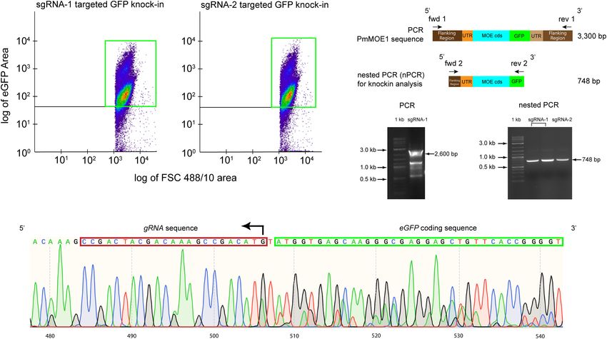
Yadavalli et al.                                                                                               Genome Editing in Perkinsus marinus

genomic and physiological affinities (Matsuzaki et al., 2008;             (Peng et al., 2014; Soares Medeiros et al., 2017). However, studies
Joseph et al., 2010; Bachvaroff et al., 2011; Fernández Robledo           have reported the toxicity and instability due to the transgenic
et al., 2011; Van Voorhis et al., 2016).                                  expression of Cas9 (Peng et al., 2014). Alternatively, the Cas9-
    Human exposure to Perkinsus spp. by consuming infected                gRNA ribonucleoprotein complex-based genome editing method
oysters/clams is likely to occur based on the high prevalence             was established in kinetoplastids (Beneke et al., 2017; Soares
of the parasite in oysters (Marquis et al., 2015, 2020).                  Medeiros et al., 2017; Verruto et al., 2018).
Nevertheless, to our knowledge, the effect of consumption of                 Here, we optimized electroporation-based transfection
P. marinus-infected oysters has not been investigated in humans.          methodology to improve heterologous gene expression
Interestingly, in studies using humanized mice expressing HLA-            in P. marinus. Furthermore, using the optimized
DR40 genes and lacking expression of mouse MHC-class II genes             transfection protocol, we successfully delivered Cas9-gRNA
(DR4.EA0 ), we reported that DR4.EA0 mice did not develop                 ribonucleoprotein coupled with dDNA into the P. marinus
any detectable pathology or systemic inflammation (Wijayalath             wild-type trophozoites and tagged the PmMOE1 gene with GFP
et al., 2014). Notably, naïve (unfed) DR4.EA0 mice had antibodies         at the C-terminus to achieve mutants phenotypically similar
(IgM and IgG) reacting against P. marinus, whereas parasite-              to previously reported P. marinus mutant strain (PRA-393)
specific T-cell responses were undetectable. Upon oral feeding            (Fernández Robledo et al., 2008).
with P. marinus, parasite-specific IgM and IgG antibodies were
boosted with parasite-specific cellular (INFγ) responses detected
in the spleen, suggesting P. marinus as a natural adjuvant                RESULTS
(Wijayalath et al., 2014).
    Our group is focused on developing molecular tools to                 Plasmid Amount and Cell Number
establish P. marinus as a heterologous expression system to               Optimization Experiments
express genes of pathogens of medical and veterinary relevance.           To increase the heterologous gene expression efficiency, we
Previously, we built the plasmid pMOE[MOE1]:GFP (formerly                 used the previously developed Lonza-based electroporation
known as pMOE:GFP) by expanding 1 kb each of 50 and                       method (Fernández Robledo et al., 2008). In the first round of
30 flanking regions for PmMOE1 coding sequence tagged                     optimization, we maintained 50.0 × 10⁶ P. marinus trophozoites
with GFP, developing an electroporation-based transfection                per transfection. We optimized the pPmMOE[MOE1]:GFP
protocol to deliver the plasmid, and successfully showing a               plasmid amount with a twofold increase (5.0, 10.0, 20.0,
single integration event into the genome via non-homologous               and 40.0 µg). In all the cases, green fluorescent cells were
recombination (Fernández Robledo et al., 2008). Recent studies            observed under a UV-microscope as early as 24 h post-
using developed electroporation-based transfection protocol and           transfection. The flow cytometer was used to detect the number
pMOE[MOE1]:GFP plasmid reported possibilities of plasmid                  of GFP-positive cells. The trophozoites transfected with 5
fragmentation and transposable element-dependent genome                   and 10 µg of pPmMOE[MOE1]:GFP were detected as 0.002
integration (Faktorová et al., 2020). Considering the phylogenetic        and 0.03%, respectively. Parasites transfected with 5.0 µg of
relationship of P. marinus with apicomplexans, we transfected             pPmMOE[MOE1]:GFP yielded 0.05 and 0.2% GFP-positive
P. marinus with plasmids carrying Plasmodium berghei HAP2                 cells at 72 and 120 h time points, respectively (Figure 1A,
and MSP8 and observed transient expression of both genes                  black bar). Furthermore, parasites transfected with 10.0 µg of
(Cold et al., 2016). However, we fell short of replicating 37.8%          pPmMOE[MOE1]:GFP yielded 1 and 7.7% GFP-positive cells
efficiency when the transfection methodology was developed                at 72 and 120 h time points, respectively (Figure 1A, purple
(Fernández Robledo et al., 2008).                                         bar). Interestingly, at 72 h post-transfection, parasites transfected
    To our knowledge, other than the transfection using                   with 40 µg (Figure 1A, orange bar) of pPmMOE[MOE1]:GFP
pMOE[MOE1]:GFP-derived plasmids, currently, there are no                  yielded 2 × higher GFP-positive cells compared with parasites
systems for functional studies of P. marinus genes like gene              that received 20 µg (Figure 1A, blue bar). However, to our
knock-out. The clustered regularly interspaced short palindromic          surprise, at 120 h post-transfection, we have detected 9 and 11%
repeats (CRISPR) and CRISPR-associated protein 9 (Cas9)                   of GFP-positive cells in the cases of parasites transfected with
system is a powerful tool for editing genomes (Jinek et al.,              20 and 40 µg of the plasmid, respectively (Figure 1A, blue and
2012; Lander, 2016). CRISPR/Cas9 technology utilizes machinery            orange bars). Observing the plateau of GFP-positive cells when
such as Cas9 protein, an RNA-guided endonuclease protein, as              parasites were transfected with 20 and 40 µg, we have decided to
well as a guide RNA (gRNA) for the nuclease to generate a                 move on with the 20 µg of plasmid for cell number optimization.
double-strand break, which is repaired by nonhomologous end                  In the second round of optimization, keeping the amount of
joining (NHEJ) and random mutations incorporated to disrupt               pPmMOE[MOE1]:GFP plasmid constant at 20 µg/transfection,
the target gene (Mali et al., 2013; Bortesi and Fischer, 2015).           we varied P. marinus trophozoites cell number by a twofold
However, to knock-in a gene of interest, a donor DNA (dDNA)               increase between 1.56 × 10⁶ and 50.0 × 10⁶ cells/transfection.
molecule with homologous templates on either side of the knock-           In this case, using the confocal microscope, we observed that 25.0
in sequence is required in addition to Cas9 and gRNA. The                 million parasites transfected with 20 µg of the plasmid yielded
incorporation of the gene of interest into the genome happens             the highest levels of GFP-expressing cells qualitatively at 24- and
via a homologous-dependent repair mechanism. Most studies                 120 h post-transfection (Figure 1B). We took advantage of the
utilize plasmid-based endogenous expression of Cas9 and gRNA              flow cytometer and detected 2% of GFP-positive cells (Figure 1C,

Frontiers in Bioengineering and Biotechnology | www.frontiersin.org   2                                        April 2021 | Volume 9 | Article 623278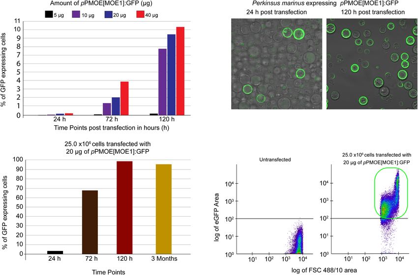
Yadavalli et al.                                                                                                                  Genome Editing in Perkinsus marinus

  FIGURE 1 | Plasmid amount and cell number optimization studies. (A) Fifty million parasites transfected with 5 µg (black bar), 10 µg (purple), 20 µg (blue), and
  40 µg (orange) of pPmMOE[MOE1]:GFP, respectively. Bar graphs showing that the %GFP-positive cells (y-axis) were detected by flow cytometry at 24, 72, and
  120 h post-transfection time points (x-axis). (B) Twenty-five million parasites transfected with 20 µg of pPmMOE[MOE1]:GFP 24 and 120 h post-transfections.
  (C) Twenty-five million parasites were transfected with 20 µg of pPmMOE[MOE1]:GFP, respectively. Bar graphs showing the %GFP-positive cells (y-axis) detected by
  flow cytometry at 24 (black bar), 72 (brown bar), 120 h (dark red bar), and 3 month (yellow bar) post-transfection time points (x-axis). (D) The scattered plot from
  FCM showing no GFP expression in untransfected controls and 98% GFP-positive cells in 25.0 × 10⁶ cells transfected with 20 µg of pPmMOE[MOE1]:GFP
  indicated in the green box.

black bar) as early as 24 h and 68% in 72 h (Figure 1C, brown bar)                     using 3R-buffer and BTX cuvette (Figures 2C,E, light gray bar).
and achieved 98% of GFP-positive cells 120 h post-transfection                         By using a 3R buffer in combination with a Lonza cuvette, we
(Figure 1C, red bar). The trophozoites were monitored for                              detected 48% of GFP-positive cells (Figures 2D,E, dark gray
3 months, where we detected a constant 95% GFP-positive cells                          bar). Finally, transfection utilizing Lonza–buffer and BTX cuvette
[Figure 1C, light brown bar and Figure 1D (highlighted in                              yielded a meager 2% GFP-positive cells (Figure 2E, white bar).
the green box)].
                                                                                       SpCas9-RNP and sgRNA Mediated GFP
Comparison of Proprietary and                                                          Knock-In in Perkinsus marinus
Non-proprietary Transfection Reagents                                                  Trophozoites
and Materials                                                                          To establish an HDR-based gene-editing method, we generated a
To establish an affordable and reliable transfection methodology,                      dDNA plasmid containing 396 bp of PmMOE1 coding sequence
we tested non-proprietary protocols, such as using 3R buffer                           lacking a start codon on the 50 of the GFP coding sequence.
with Lonza and commercial cuvettes (BTX Disposable Cuvettes                            Furthermore, there are 396 bp of 30 UTR of PmMOE1 at the
Plus), and Lonza buffer-based transfection utilizing Lonza cuvette                     30 of the GFP-coding sequence. The dDNA with GFP and
and commercial cuvette. In all the cases, 25 million cells were                        templates was amplified using PCR from previously reported
transfected with 20 µg of plasmid, and GFP-positive cells were                         plasmid pPmMOE[MOE1]:GFP (schematic representation in
detected 120 h post-transfection using flow cytometry (Figure                          Figure 3A; Fernández Robledo et al., 2008). The sgRNA targeting
2A). As expected, we identified 98% of GFP-positive cells using                        at position 314 on the top strand (sgRNA-1) and another
a proprietary Lonza system (Figures 2B,E, black bar). To our                           sgRNA targeting position 395 on the bottom strand (sgRNA-2)
surprise, flow cytometry evaluated 90% of GFP-positive cells                           of the PmMOE1 coding sequence were designed using the

Frontiers in Bioengineering and Biotechnology | www.frontiersin.org                3                                              April 2021 | Volume 9 | Article 623278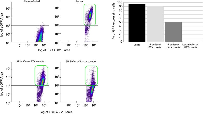
Yadavalli et al. Genome Editing in Perkinsus marinus
FIGURE 2 | Comparison of proprietary and non-proprietary transfection. Cells, 25 × 106 cells transfected with 20 µg of pPmMOE[MOE1]:GFP plasmid using
proprietary and non-proprietary protocols. (A) Flow cytometry scattered plot of untransfected (wild-type) cells, no GFP expression detected. (B) The scattered plot of
flow cytometry, identifying GFP-positive cells in transfection performed using the proprietary Lonza method. (C) Scatterplot representation of GFP-positive cells in
transfection performed using 3R buffer and BTX cuvette. (D) Scatterplot showing the GFP-positive cells when transfected with 3R buffer utilizing Lonza cuvette.
(E) Bar graph showing the % of GFP-positive cells when transfected with the Lonza system (black bar), 3R buffer in combination with BTX cuvette (gray bar), 3R
buffer using Lonza cuvette (dark gray), and Lonza buffer with BTX cuvette (white bar).
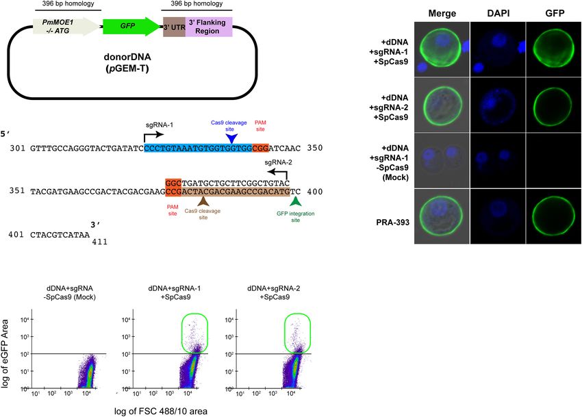
Benchling software (Figure 3B). Twenty-five million P. marinus 3,300 bp) resulted in around 2,600 bp amplicon, which would
trophozoites were transfected with 20 µg of SpCas9 and sgRNA include the 50 flanking, 50 UTR, PmMOE1, but not the GFP
(1:1) along with 20 µg of dDNA. The parasites transfected with knock-in (Figure 4C, 2,600 bp arrow and Supplementary
sgRNA-1/SpCas9 and sgRNA-2/SpCas9 and dDNA exhibited Figure 2A) suggesting that the knock-in of GFP was less
GFP expression 24 h post-transfection (Figure 3C, +sgRNA- represented compared with the wild type. Consequently, we
1+dDNA+SpCas9 and +dDNA+sgRNA-2+SpCas9), showing diluted the PCR product and run a nested PCR with specific
a similar pattern of GFP mutant strain PRA-393 (Figure 3C, primers targeting the putative knock-in; sequencing of the nested
PRA-393 panel). The parasites transfected without SpCas9, only PCR product (748 bp) confirmed the accurate integration by
with dDNA and sgRNA used (i.e., +dDNA+sgRNA) as mock HDR (Figure 4C, 748 bp arrow and Supplementary Figure 2B).
transfection, did not show GFP expression (Figure 3C, mock The chromatogram validated the successful knock-in of GFP at
transfection panel). Two months post-transfection, using the flow the C-terminus PmMOE1 (Figure 4D).
cytometer, we detected 0.2% GFP-positive cells in the experiment
transfected with sgRNA-1+dDNA+SpCas9. Furthermore,
parasites transfected with sgRNA-2+dDNA+SpCas9 complex DISCUSSION
yielded 0.26%. GFP-positive cells were not detected in mock
transfections (Figure 3D). Perkinsus marinus, a marine protozoan parasite, causing
devastating infection to eastern oysters, is currently under
Sorting Perkinsus marinus GFP-Positive development as a model organism for the protozoan parasite of
Cells for Genotyping Validation mollusks (Yadavalli et al., 2020). The availability of axenic culture
Ten thousand GFP-positive cells from the sgRNA-1 and sgRNA- and transfection methodology, the parasite’s ability to naturally
2 transfections were sorted and cultured for 3 months. Flow trigger an immune response in mice, and phylogenetic affinities
cytometry analysis detected approximately 81% of GFP-positive drove us to use it to express apicomplexan genes. However,
cells transfected with sgRNA-1 (Figure 4A) and 87% of GFP- these attempts were met with variable success (Wijayalath et al.,
positive cells transfected with sgRNA-2 (Figure 4B), respectively. 2014; Cold et al., 2017). The other laboratories often report the
Attempts of amplification of the knock-in (expected size inconsistency of the gene expression.
Frontiers in Bioengineering and Biotechnology | www.frontiersin.org 4 April 2021 | Volume 9 | Article 623278Yadavalli et al. Genome Editing in Perkinsus marinus FIGURE 3 | SpCas9-RNP and sgRNA-mediated GFP knock-in in P. marinus trophozoites. (A) Schematic representation of dDNA with 396 bp homology on the 50 and 30 of the GFP coding sequence. (B) Schematic representation of showing the guide RNA target sites on PmMOE1 coding sequence sgRNA-1 targets the top strand indicated by the arrow direction; sgRNA-2 targets the bottom strand indicated by the arrow direction. (C) Confocal microscopy panel showing successful GFP expression in cells transfected with sgRNA-1/SpCas9 and sgRNA-2/SpCas9, showing localization pattern similar to the PRA-393 MOE-GFP mutant strain. (D) The scattered plot from FCM showing no GFP expression in mock (dDNA+sgRNA alone) control and 0.2% GFP-positive cells knocked in using sgRNA-1, and 0.35% in case of sgRNA-2 indicated in the green box. The original transfection method uses 5 µg of plasmid (Supplementary Figure 1). We also could transfect P. marinus and 50.0 × 10⁶ cells; we started by increasing the plasmid cells with the non-proprietary transfection buffer (3R buffer), amount by 2-fold to 40 µg. The plasmid amount increased in which provides an efficiency above 40% and provides savings higher GFP-positive cells, especially cells transfected with 20 and when the research budgets are tight. Experiments utilizing 40 µg of the plasmid. In all the cases, fluorescent cells were the combination of non-proprietary transfection buffer and observed as early as 24 h post-transfection. Cells need 3 days BTX cuvettes were successful, although with a low number of to recover and for the GFP expression to be quantifiable. The transfectants. We observed more than 90% of GFP-expressing cells transfected with 40 µg yielded twofold higher GFP-positive cells even at 3 months post-transfection in all the cases, cells than cells transfected with 20 µg of plasmid after 72 h. suggesting a stable GFP expression. However, at 120 h, the number of GFP-expressing cells plateaued The CRISPR/Cas9 methodology is broadly adopted by to 10% in both cases, indicating that the number of cells also numerous parasitology labs around the world (Mali et al., 2013; affects transfection efficiency. We determined that 25.0 × 10⁶ Ghorbal et al., 2014; Peng et al., 2014; Shen et al., 2014; Sollelis cells transfected with 20 µg of plasmid resulted in 98% GFP- et al., 2015; Janssen et al., 2018; Lin et al., 2019). Utilizing the positive cells 120 h post-transfection. The NucleofectorTM 2b optimized conditions, we took a step further to develop the uses cuvettes, and the transfection occurs in a 100 µl reaction, CRISPR/Cas9-based gene editing methodology for P. marinus. and it appears that the delivery of the electrical pulse is optimal For the proof of concept, we targeted the PmMOE1 gene that has when 25.0 × 10⁶ cells are used. a defined phenotype when tagged with GFP (Fernández Robledo Interestingly, cell numbers above and below 25.0 × 10⁶ et al., 2008). We were able to detect fluorescent trophozoites cells resulted in quite a low transfection efficiency within 18 h of delivering the CRISPR/Cas9 system components. Frontiers in Bioengineering and Biotechnology | www.frontiersin.org 5 April 2021 | Volume 9 | Article 623278
Yadavalli et al.                                                                                                                 Genome Editing in Perkinsus marinus

  FIGURE 4 | Sorting P. marinus GFP-positive cells for endogenous PmMOE1 C-terminus GFP tagging analysis. (A) Scattered plot showing 81% GFP-positive cells
  indicated with a green box in the experiment where cells transfected with sgRNA-1-Cas9. (B) Scattered plot showing 87% GFP-positive cells indicated with a green
  box in the experiment where cells transfected with sgRNA-1-Cas9. (C) The PCR intended to amplify the knock-in (expected sized 3,300 bp) using Fwd 1 and Rev 1
  primers resulting in the amplification of the wildtype 2,600 bp amplicon (left panel). This PCR product was used as a template in the nested PCR (nPCR) to confirm
  the GFP knock-in using Fwd 2 and Rev 2 primers, which yielded the expected 748 bp amplicon (right panel), 1 kb Plus DNA Ladder (New England Biolabs, Ipswich,
  MA, United States). (D) Sequencing results of the nPCR product from the sgRNA-2 targeted GFP knock-in experiment.

Lack of GFP expression in the dDNA alone transfection (lacking                        RNA and utilization of single-stranded linear vs. double-stranded
CRISPR/Cas9 components) rules out the possibilities of non-                           dDNA, would likely result in an optimized protocol (Beneke
homologous recombination in frame with any expressed gene;                            et al., 2017; Markus et al., 2019). With this CRISPR/Cas9 system
however, with this fluorescence screening, plasmid fragmentation                      and several Perkinsus spp. genomes being available (Bogema
and integration at the transposable element sites cannot be                           et al., 2020), we now have the tools to interrogate these
excluded. The GFP expression pattern in the transfectants was                         genomes and improve the experimental design of sgRNA to target
similar to that of P. marinus PRA393 (Figure 3C). GFP-positive                        additional genes.
cells sorted from sgRNA-1/Cas9 and sgRNA-2/Cas9 experiments                               Genome editing tools like CRISPR/Cas9 in parasite biology
were PCR amplified to check for the knock-in of the GFP.                              is used for gene disruption, fluorescent tagging, and single
Interestingly, the PCR in the sorted cells did not result in the                      nucleotide mutation incorporation to study genes involved in the
3,300 bp amplicon. However, the nested PCR produced the                               parasite growth, invasion, and drug resistance (Wagner et al.,
expected size amplicons whose direct sequences confirmed the                          2014; Di Cristina et al., 2017). For example, in Plasmodium
successful knock-in of GFP at the C-terminus of PmMOE1.                               falciparum study development of artemisinin-resistant parasite
   In the protozoan parasites with a large trajectory of genetic                      by single-nucleotide substitution, identification of the multidrug
manipulation, the trend is to build a plasmid vector that                             resistance mutation 1 (PfMDR1) in response to the drug
incorporates both the expression of Cas9 and the sgRNA or                             ACT-451840 and incorporation of a point mutation in the
even generate a mutant conditionally expressing Cas9. We chose                        PfATP4 gene to generate the drug-resistant strain were all
to deliver the CRISPR/Cas9 components, including the SpCas9                           possible by utilizing the CRISPR/Cas9 system (Ghorbal et al.,
nuclease, directly by electroporation. The data reported here                         2014; Ng et al., 2016; Crawford et al., 2017). In Toxoplasma
are from a single trial targeting PmMOE1 using 25.0 × 10⁶                             gondii, CRISPR/Cas9 is widely used in high-throughput and
log-phase trophozoites, 20 µg of dDNA, 10 µg of sgRNAs,                               genome screening studies to identify essential genes involved in
and SaCas9 nuclease chosen based on Beneke et al. (2017) and                          parasite invasion and antiparasitic drug candidates (Di Cristina
Soares Medeiros et al. (2017) resulted in a successful knock-                         et al., 2017; Di Cristina and Carruthers, 2018). CRISPR/Cas9-
in. The optimization was outside of the scope of this study;                          based knock-out studies in Cryptosporidium parvum are used
more robust optimization focusing on the amount of guide                              to understand the mechanism of the parasite’s resistance to

Frontiers in Bioengineering and Biotechnology | www.frontiersin.org               6                                             April 2021 | Volume 9 | Article 623278
Yadavalli et al.                                                                                                   Genome Editing in Perkinsus marinus

antifolate drugs and nutrient acquisition pathways (Vinayak et al.,           as reported elsewhere (Gauthier and Vasta, 1995). Trophozoites
2015; Pawlowic et al., 2017, 2019).                                           in the log phase (OD595 = 0.4–0.5) were aliquoted in Eppendorf
    P. marinus genome encodes for 23,454 genes embedded in                    tubes to contain 1.56 × 10⁶ , 3.13 × 10⁶ , 6.25 × 10⁶ , 12.50 × 10⁶ ,
17,000 supercontigs. However, tetra-polyploidy pose a significant             25.0 × 10⁶ , and 50.0 × 10⁶ .
bottleneck for the assembly (El-Sayed et al., 2007; Bogema et al.,
2020). Proteome studies identified that P. marinus possess 4,073              Perkinsus marinus Transfection
non-redundant hypothetical proteins, of which 36 and 27%                      The transfection vector pPmMOE[MOE1]:GFP (former
are involved in metabolic and cellular processes, respectively                pPmMOE-GFP) (Fernández Robledo et al., 2008) was propagated
(Marcia et al., 2017). Additionally, the rhoptry proteins such                in Escherichia coli JM109. Plasmid minipreps were prepared using
as serine–threonine kinases, protein phosphatases, proteosomes,               a commercial kit (E.Z.N.A. Plasmid mini Kit I, Omega-Tek,
                                                                                                               R

and a virulent candidate merozoite surface protein 3, which are               Norcross, GA, United States), and DNA concentration and purity
known to play a crucial role in parasite invasion and cell–cell               were estimated with a NanodropTM 1000 spectrophotometer
communication during the invasion in P. falciparum were also                  (Thermo Fisher Scientific, Waltham, MA, United States). The
identified in P. marinus. Studies so far reported that P. marinus             isolated plasmid DNA was air dried using speedVac for all the
possess extracellular proteins such as high molecular weight cell             experiments. P. marinus cells were prepared following the Cell
wall protein 1 (Montes et al., 2002); glycosylation, mucin, and               Line Optimization Nucleofector Kit before electroporation using
sugar-binding domain protein Pmar_XP_002783417.1 encoded                      the NucleofectorTM 2b (Lonza, Walkersville, MD, United States).
by Pmar_PMAR006943; sensory signal transduction-related                       For all the experiments, we used the pre-set program D-023
histidine kinase encoded by Pmar_PMAR009211; and a family of                  and Lonza’s solution V (Fernández Robledo et al., 2008).
cysteine-rich modular proteins whose function in the parasite life            Briefly, dried plasmid was resuspended in 100 µl of Solutio V
cycle are yet to be investigated (Montes et al., 2002). Furthermore,          containing supplement 1. We tested 5, 10, 20, and 40 µg of
apoptotic genes such as apoptosis inhibitory molecule (Fas),                  pPmMOE[MOE1]:GFP with 50 million P. marinus cells. Once
apoptosis-inducing factor (Tadesse et al., 2017; Lau et al.,                  the optimal plasmid amount was established (20 µg), we tested
2018b), peroxiredoxin, and superoxide dismutase are shown to                  it with variable P. marinus cell number (1.56 × 10⁶ , 3.13 × 10⁶ ,
favor parasite survival by reducing the host cell (Schott and                 6.25 × 10⁶ , 12.5 × 10⁶ , 25.0 × 10⁶ , and 50.0 × 10⁶ ). Immediately
Vasta, 2003; Schott et al., 2003; Box et al., 2020). The function             after electroporation, the individual electroporation cuvettes’
of these apoptotic genes responsible for the disease in the                   contents were transferred to a 24-well plate, each well containing
oysters is limited.                                                           1 ml of DME:Ham’s F12 (1:2) supplemented with 5% FBS
    The CRISPR/Cas9 method developed here can be used to                      (Gauthier and Vasta, 1995). The cuvettes were gently washed
understand the localization and protein–protein interactions in               with 500 µl of fresh culture medium and pooled with those wells
the parasite’s life cycle and generate the transgenic parasite                corresponding to each original sample. We also tested the non-
model. The natural adjuvant ability of the P. marinus and                     proprietary transfection buffer (3R buffer)-based transfection
its potential as a novel oral vaccine platform, efforts for the               protocol (Faktorová et al., 2020). The 3R-transfection buffer,
expression of heterologous antigens, always relied on one plasmid             composed of 200 mM Na2 HPO4 , 70 mM NaH2 PO4 , 15 mM KCl,
p[MOE]:EK-His-GFP (Cold et al., 2017), with potential for a                   and 150 mM HEPES, was prepared and pHed to 7.3. Dried 20 µg
monovalent vaccine expression. With the availability of fast and              of circular pPmMOE[MOE1]:GFP plasmid was resuspended in
robust CRISPR/Cas9, we can now express multiple heterologous                  60 µl of milliQ water. Once dissolved, 35 µl of 3R transfection
genes and develop P. marinus as a polyvalent oral vaccine                     buffer and 10 µl of 1.5 mM CaCl2 were added (Protocols.io).
delivery system. Considering the precision and efficiency of                  Twenty-five million P. marinus trophozoites were transfected.
CRISPR/Cas9 in gene editing, this opens doors for discovering
new treatments and therapeutic discoveries. The system can                    Protospacer Adjacent Motif-Target Site
also be used in clinical and population validation studies to                 Selection and Donor DNA Construction
identify new antiparasitic agents (Visscher et al., 2017). Finally,           PAM-target site selection was identified using the PmMOE1
the CRISPR/Cas9 system can be applied to generate non-                        sequence (Pmar_PMAR027036) and the software (Benchling,
pathogenic and immunogenic parasites for the immunization                     Inc.)1 . The output sequences were searched using BLASTx
and vaccination studies (Hollingdale and Sedegah, 2017).                      (NCBI-Blast, 2021) against the P. marinus nr database
                                                                              (RefSeq assembly: GCF_000006405.1), which predicted
                                                                              Pmar_PMAR025337 as another possible target. Based on
MATERIALS AND METHODS                                                         the string searches, the PAM sites were rated according to
                                                                              their“uniqueness.” Single-guide RNA (sgRNA) targeting positive
Perkinsus marinus Cell Culture                                                strand at position 339 of PmMOE1 CDS (sgRNA-1) 50 -CCC
Experiments were carried out with cultures of the wild-type                   TGT AAA TGT GGT GGT GG-30 and sgRNA targeting negative
P. marinus CB5D4 (ATCC#PRA-240) (Shridhar et al., 2013)                       strand at position 382 of PmMOE CDS (sgRNA-2) 50 -CAT
maintained in DME:Ham’s F12 (1:2) supplemented with 5%                        GTC GGC TTC GTC GTA GT CGG-30 with unique PAM
fetal bovine serum (FBS), in a 25 cm2 (5–8 ml) polystyrene                    sequence “CGG” were synthesized (Synthego, Silicon Valley, CA,
canted neck cell culture flasks with vent caps (Corning , Corning,    R

New York, United States) at 24–28◦ C in a microbiology incubator              1
                                                                                  https://www.benchling.com/

Frontiers in Bioengineering and Biotechnology | www.frontiersin.org       7                                        April 2021 | Volume 9 | Article 623278
Yadavalli et al. Genome Editing in Perkinsus marinus
United States). Although the sgRNA-2 sequence identified three flanking region, PmMOE1 CDS, and 201 bp of the GFP sequence
target sites on PmMOE1 CDS, the sequence reported here is the (Fwd 2 50 -TGT TGT AAG GCG AGA CGC TA–30 and Rev 2 50 -
only fragment that exhibited 100% complementarity. The dDNA GTA GGT CAG GGT GGT CAC GA–30 ), respectively. Briefly,
was amplified from pPmMOE[MOE1]:GFP (Fernández Robledo 50 ng of the gDNA and primers mentioned above were used
et al., 2008) using primers forward 50 -CGC TTC ATT GTT GGT to amplify by polymerase chain reaction. The amplicons were
CTG TAC–30 and reverse 50 -CAG TAC GAA ATT ACG CGA purified from the 1% agarose gel using the ZymocleanTM Gel
GAT G–30 . The amplicon was cloned into the pGEM -T vector R
DNA Recovery kit (Tustin, CA, United States).
by T-A cloning (pGEM-T Vector Systems, Promega Corporation,
Madison, WI), propagated in Escherichia coli JM109 (L1001, Confocal Microscopy
Promega), and sent for sequencing. Plasmid minipreps were Parasites were fixed with 3% paraformaldehyde (Thermo
prepared using a commercial kit (E.Z.N.A. Plasmid Mini Kit
R
Fisher Scientific, preserved 37% reagent) for 15 min at room
I, Omega Bio-Tek, Norcross, GA, United States), and DNA temperature. Parasites were washed three times at 1,000 × g
concentration and purity were estimated with a Nanodrop 1000 for 5 min using 1 × phosphate-buffered saline (1 × PBS).
spectrophotometer. Following the washes, parasites were treated with 0.1% Triton
X-100 for 15 min and washed three times with 1 × PBS. The
RNP Complex and Donor DNA Delivery cells were stained with 25 µg/ml concentration of 40 ,6-diamidino-
Into Perkinsus marinus 2-phenylindole (DAPI) (Vector Laboratories, Burlingame, CA,
Perkinsus marinus cells were prepared following the Cell Line United States). Excess DAPI was washed with 1 × PBS,
Optimization Nucleofector Kit protocol before electroporation. and parasites were resuspended in the fresh 1 × PBS and
Using the NucleofectorTM 2b, 10 µg of Streptococcus pyrogenes placed in NunC Lab-Tek II (Millipore Sigma, Darmstadt,
R R
Cas9 (SpCas9) nuclease TrueCutTM Cas9 protein v2 (Thermo Germany) for live-cell imaging. Parasites were imaged at a total
Fisher Scientific, Vilnius, Lithuania) and 10 µg of sgRNAs magnification of 630 × on Carl Zeiss LSM-700 multiphoton
(Synthego, Silicon Valley, CA, United States) were mixed in scanning laser microscope.
100 µl of Lonza’s solution V and incubated at room temperature
for 15 min for hybridization of sgRNA and Cas9 protein (Beneke Flow Cytometry Sorting and Analysis
et al., 2017). Twenty micrograms of dried dDNA plasmid were The flow cytometry experiments were performed on the live
resuspended with a SpCas9–gRNA complex and electroporated parasite, using ZE5 Cell Analyzer; data was collected using
into 25.0 × 10⁶ P. marinus trophozoites, using pre-set D-023 Everest software version 2.0 and analyzed. A minimum of 100,000
program (Fernández Robledo et al., 2008). Immediately after events was collected for parasites based on forward and side
electroporation, the individual electroporation cuvettes’ contents scatterplot, and a singlet gate was applied to collect a minimum
were transferred to a 12-well plate, each well containing 1 ml of 30,000 cells. BD Sciences Influx Cell Sorter (BD Sciences, NJ,
of DME:Ham’s F12 (1:2) supplemented with 5% FBS, to allow United States) was used for cell sorting, and cells were sorted
cells to recover (Gauthier and Vasta, 1995). Upon identifying the based on eGFP-positive gates.
fluorescent cells, the cells were spun down at 1,000 × g for 5 min
at room temperature and resuspended into fresh media. Cells DATA AVAILABILITY STATEMENT
were screened for green fluorescence at 24, 72, and 120 h, and
2, 4, and 6 weeks post-transfection using confocal microscopy The raw data supporting the conclusions of this article will be
and flow cytometry. made available by the authors, without undue reservation.
DNA Isolation and Genotyping for GFP
Knock-In by DNA Sequencing AUTHOR CONTRIBUTIONS
Upon observing green fluorescent P. marinus trophozoites,
RY and JF: conceptualization, methodology, writing—original
cells were allowed to recover for 1 week. The genomic DNA
draft preparation, and supervision. JF: validation, project
from P. marinus:wild-type (PRA-240), GFP-mutant (PRA-393),
administration, and funding acquisition. RY, KU, HW, AT, AS,
parasites transfected with dDNA alone, lacking CRISPR elements
DH, and JF: formal analysis, investigation, writing—review and
(sgRNA and Cas9), and parasites transfected with sgRNA-1 and
editing, and visualization. RY and KU: resources. RY: data
sgRNA-2 with Cas9 were isolated using E.Z.N.A. tissue DNA R
curation. All authors contributed to the article and approved the
kit (Norcross, GA, United States) according to the manufacturer
submitted version.
protocol. The purity and concentration of isolated DNA were
analyzed using NanodropTM 1000 spectrophotometer. For DNA
genotyping, the primer pair Fwd 1 50 -CTC GTA ATG AGC CCA FUNDING
ACC AT–30 and Rev 1 50 -GGA GGA CTT GAG GCT CTG
TG 30 (Fernández Robledo et al., 2008) were designed using This research was funded by the National Science Foundation
the available supercontig (Ensembl, 2021) results in 2,600 bp of Grant No. 1701480, National Science Foundation Research
PmMOE1 (wildtype) and would yield 3,300 bp after successful Experiences for Undergraduates, Grant No. 1460861, JSPS
GFP knock-in. To identify the GFP knock-in site at the 30 KAKENHI Grant No. 19J00148, and Bigelow Laboratory
PmMOE CDS, we designed primers spanning 136 bp of the 50 institutional funds.
Frontiers in Bioengineering and Biotechnology | www.frontiersin.org 8 April 2021 | Volume 9 | Article 623278Yadavalli et al. Genome Editing in Perkinsus marinus
ACKNOWLEDGMENTS Supplementary Figure 1 | Cell number optimization studies.
Supplementary Figure 2 | PCR and nested PCR based genotyping of
We thank the manuscript’s reviewers for the exhaustive and PMAR_Pmar027036 for the GFP knock in analysis. The genomic DNA was
comprehensive review of the manuscript. isolated from two different clones labeled as 4 and 5, which were hand-picked 3
months after the cell sorting from the experiments involved in the utilization of
SgRNA-1 and SgRNA-2, respectively. (A) The PCR product showing the
SUPPLEMENTARY MATERIAL successful amplification of 2,600 bp of DNA sequencing containing flanking
regions, MOE1 CDS encoded by PMAR_Pmar027036 and the GFP. PCR product
was diluted 100 times and used in the nested PCR to identify the knock in of the
The Supplementary Material for this article can be found GFP, which successfully amplified the 748 bp of DNA fragment. The fragment was
online at: https://www.frontiersin.org/articles/10.3389/fbioe. sequenced to confirm the successful knock in of GFP. (B) Repeat of the gel
2021.623278/full#supplementary-material analysis of the nested PCR products from (A), showing the DNA size of 748 bp.
REFERENCES Faktorová, D., Nisbet, R. E. R., Fernández Robledo, J. A., Casacuberta, E., Sudek,
L., Allen, A. E., et al. (2020). Genetic tool development in marine protists:
Andrews, J. D. (1996). History of Perkinsus marinus, a pathogen of oysters in emerging model organisms for experimental cell biology. Nat. Methods 17,
Chesapeake Bay 1950-1984. J. Shellfish Res. 15, 13–16. 481–494.
Arzul, I., Chollet, B., Michel, J., Robert, M., Garcia, C., Joly, J. P., et al. (2012). Fernández Robledo, J. A., Caler, E., Matsuzaki, M., Keeling, P. J., Shanmugam,
One Perkinsus species may hide another: characterization of Perkinsus species D., Roos, D. S., et al. (2011). The search for the missing link: a relic plastid in
present in clam production areas of france. Parasitology 139, 1757–1771. doi: Perkinsus? Int. J. Parasitol. 41, 1217–1229. doi: 10.1016/j.ijpara.2011.07.008
10.1017/s0031182012001047 Fernández Robledo, J. A., Lin, Z., Vasta, G. R., Fernández-Robledo, J. A., Lin,
Bachvaroff, T. R., Handy, S. M., Place, A. R., and Delwiche, C. F. (2011). Alveolate Z., and Vasta, G. R. (2008). Transfection of the protozoan parasite Perkinsus
phylogeny inferred using concatenated ribosomal proteins. J. Eukaryot. marinus. Mol. Biochem. Parasitol. 157, 44–53. doi: 10.1016/j.molbiopara.2007.
Microbiol. 58, 223–233. doi: 10.1111/j.1550-7408.2011.00555.x 09.007
Beneke, T., Madden, R., Makin, L., Valli, J., Sunter, J., and Gluenz, E. (2017). Gauthier, J. D., and Vasta, G. R. (1995). In vitro culture of the eastern oyster parasite
A CRISPR Cas9 high-throughput genome editing toolkit for kinetoplastids. Perkinsus marinus: optimization of the methodology. J. Invertebr. Pathol. 66,
R. Soc. Open Sci. 4:170095. doi: 10.1098/rsos.170095 156–168. doi: 10.1006/jipa.1995.1079
Bogema, D. R., Yam, J., Micallef, M. L., Kanani, H. G., Go, J., Jenkins, C., et al. Ghorbal, M., Gorman, M., Macpherson, C. R., Martins, R. M., Scherf, A., and
(2020). Draft genomes of Perkinsus olseni and Perkinsus chesapeaki reveal Lopez-Rubio, J. J. (2014). Genome editing in the human malaria parasite
polyploidy and regional differences in heterozygosity. Genomics 113(Pt 2), Plasmodium falciparum using the CRISPR-Cas9 system. Nat. Biotechnol. 32,
677–688. doi: 10.1016/j.ygeno.2020.09.064 819–821. doi: 10.1038/nbt.2925
Bortesi, L., and Fischer, R. (2015). The CRISPR/Cas9 system for plant genome Hollingdale, M. R., and Sedegah, M. (2017). Development of whole sporozoite
editing and beyond. Biotechnol. Adv. 33, 41–52. doi: 10.1016/j.biotechadv.2014. malaria vaccines. Expert. Rev. Vaccines 16, 45–54. doi: 10.1080/14760584.2016.
12.006 1203784
Box, A., Capó, X., Tejada, S., Sureda, A., Mejías, L., and Valencia, J. M. (2020). Janssen, B. D., Chen, Y. P., Molgora, B. M., Wang, S. E., Simoes-Barbosa, A.,
Perkinsus mediterraneus infection induces oxidative stress in the mollusc and Johnson, P. J. (2018). CRISPR/Cas9-mediated gene modification and gene
Mimachlamys varia. J. Fish Dis. 43, 1–7. doi: 10.1111/jfd.13085 knock out in the human-infective parasite Trichomonas vaginalis. Sci. Rep.
Cold, E. R., Freyria, N. J., Martínez Martínez, J., and Fernández Robledo, J. A. 8:270.
(2016). An Agar-based method for plating marine protozoan parasites of the Jinek, M., Chylinski, K., Fonfara, I., Hauer, M., Doudna, J. A., and Charpentier,
genus Perkinsus. PLoS One 11:e0155015. doi: 10.1371/journal.pone.0155015 E. (2012). A programmable dual-RNA-guided DNA endonuclease in adaptive
Cold, E. R., Vasta, G. R., and Robledo, J. A. F. (2017). Transient expression bacterial immunity. Science 337, 816–821. doi: 10.1126/science.1225829
of Plasmodium berghei MSP8 and HAP2 in the marine protozoan parasite Joseph, S. J., Fernández-Robledo, J. A., Gardner, M. J., El-Sayed, N. M., Kuo, C. H.,
Perkinsus marinus. J. Parasitol. 103, 118–122. doi: 10.1645/16-88 Schott, E. J., et al. (2010). The alveolate Perkinsus marinus: biological insights
Coss, C. A., Robledo, J. A., Ruiz, G. M., and Vasta, G. R. (2001a). Description of from EST gene discovery. BMC Genomics 11:228. doi: 10.1186/1471-2164-11-
Perkinsus andrewsi n. sp. isolated from the baltic clam (Macoma balthica) by 228
characterization of the ribosomal RNA locus, and development of a species- Lander, E. S. (2016). The heroes of CRISPR. Cell 164, 18–28. doi: 10.1016/j.cell.
specific PCR-based diagnostic assay. J. Eukaryot. Microbiol. 48, 52–61. doi: 2015.12.041
10.1111/j.1550-7408.2001.tb00415.x Lau, Y. T., Gambino, L., Santos, B., Espinosa, E. P., and Allam, B. (2018a).
Coss, C. A., Robledo, J. A., and Vasta, G. R. (2001b). Fine structure of clonally Regulation of oyster (Crassostrea virginica) hemocyte motility by the
propagated in vitro life stages of a Perkinsus sp. isolated from the baltic clam intracellular parasite Perkinsus marinus: a possible mechanism for host
Macoma balthica. J. Eukaryot. Microbiol. 48, 38–51. doi: 10.1111/j.1550-7408. infection. Fish Shellfish Immunol. 78, 18–25. doi: 10.1016/j.fsi.2018.04.019
2001.tb00414.x Lau, Y. T., Santos, B., Barbosa, M., Espinosa, E. P., and Allam, B. (2018b).
Crawford, E. D., Quan, J., Horst, J. A., Ebert, D., Wu, W., and DeRisi, J. L. (2017). Regulation of apoptosis-related genes during interactions between oyster
Plasmid-free CRISPR/Cas9 genome editing in Plasmodium falciparum confirms hemocytes and the alveolate parasite Perkinsus marinus. Fish Shellfish Immunol.
mutations conferring resistance to the dihydroisoquinolone clinical candidate 83, 180–189. doi: 10.1016/j.fsi.2018.09.006
SJ733. PLoS One 12:e0178163. doi: 10.1371/journal.pone.0178163 Lin, Z. Q., Gan, S. W., Tung, S. Y., Ho, C. C., Su, L. H., and Sun, C. H. (2019).
Cristina, M. D., and Carruthers, V. B. (2018). New and emerging uses of Development of CRISPR/Cas9-mediated gene disruption systems in Giardia
CRISPR/Cas9 to genetically manipulate apicomplexan parasites. Parasitology lamblia. PLoS One 14:e0213594. doi: 10.1371/journal.pone.0213594
145, 1119–1126. doi: 10.1017/s003118201800001x Mackin, J. G., Owen, H. M., and Collier, A. (1950). Preliminary note on the
Cristina, M. D., Dou, Z., Lunghi, M., Kannan, G., Huynh, M. H., McGovern, occurrence of a new protistan parasite, Dermocystidium marinum n. sp., in
O. L., et al. (2017). Toxoplasma depends on lysosomal consumption of Crassostrea virginica (Gmelin). Science 111, 328–329. doi: 10.1126/science.111.
autophagosomes for persistent infection. Nat. Microbiol. 2:17096. 2883.328
El-Sayed, N., Caler, E., Inman, J., Amedeo, P., Hass, B., Wortman, J., et al. (2007). Mali, P., Esvelt, K. M., and Church, G. M. (2013). Cas9 as a versatile tool for
Perkinsus Genome Project. Bethesda, MD: U.S. National Library of Medicine. engineering biology. Nat. Methods 10, 957–963. doi: 10.1038/nmeth.2649
Ensembl (2021). Ensembl. Available online at: https://protists.ensembl.org/ Marcia, P. L., Tavares, G. C., Pereira, F. L., Perdigão, C., Azevedo, V., Figueiredo,
Perkinsus_marinus_atcc_50983_gca_000006405/Info/Index H. C. P., et al. (2017). Shotgun label-free proteomic analyses of the oyster
Frontiers in Bioengineering and Biotechnology | www.frontiersin.org 9 April 2021 | Volume 9 | Article 623278Yadavalli et al. Genome Editing in Perkinsus marinus parasite Perkinsus marinus. J. Proteom. Genomics Res. 2, 13–22. doi: 10.14302/ oxygen species. Exp. Parasitol. 105, 232–240. doi: 10.1016/j.exppara.2003. issn.2326-0793.jpgr-17-1571 12.012 Markus, B. M., Bell, G. W., Lorenzi, H. A., and Lourido, S. (2019). Optimizing Schott, E. J., and Vasta, G. R. (2003). The PmSOD1 gene of the protistan parasite systems for Cas9 expression in Toxoplasma gondii. mSphere 4:e00386-19. doi: Perkinsus marinus complements the Sod2Delta mutant of Saccharomyces 10.1128/mSphere.00386-19 cerevisiae, and directs an iron superoxide dismutase to mitochondria. Mol. Marquis, N. D., Bishop, T. J., Record, N. R., Countway, P. D., and Fernández Biochem. Parasitol. 126, 81–92. doi: 10.1016/s0166-6851(02)00271-2 Robledo, J. A. (2020). A QPCR-based survey of Haplosporidium nelsoni and Shen, B., Brown, K. M., Lee, T. D., and Sibley, L. D. (2014). Efficient gene disruption Perkinsus spp. in the eastern oyster, Crassostrea virginica in Maine, USA. in diverse strains of Toxoplasma gondii using CRISPR/CAS9. mBio 5:e1114. Pathogens 9:256. doi: 10.3390/pathogens9040256 Shridhar, S., Hassan, K., Sullivan, D. J., Vasta, G. R., and Fernández Robledo, Marquis, N. D., Record, N. R., Fernández Robledo, J. A. (2015). Survey for J. A. (2013). Quantitative assessment of the proliferation of the protozoan protozoan parasites in eastern oysters (Crassostrea virginica) from the Gulf of parasite Perkinsus marinus using a bioluminescence assay for ATP content. Int. Maine using PCR-Based Assays. Parasitol. Int. 64, 299–302. doi: 10.1016/j. J. Parsitol. Drug Drug Resist 3, 85–92. doi: 10.1016/j.ijpddr.2013.03.001 parint.2015.04.001 Soares Medeiros, L. C., South, L., Peng, D., Bustamante, J. M., Wang, W., Matsuzaki, M., Kuroiwa, H., Kuroiwa, T., Kita, K., and Nozaki, H. (2008). A cryptic Bunkofske, M., et al. (2017). Rapid, selection-free, high-efficiency genome algal group unveiled: a plastid biosynthesis pathway in the oyster parasite editing in protozoan parasites using CRISPR-Cas9 Ribonucleoproteins. mBio Perkinsus marinus. Mol. Biol. Evol. 25, 1167–1179. doi: 10.1093/molbev/ 8:e01788-17. doi: 10.1128/mBio.01788-17 msn064 Sollelis, L., Ghorbal, M., MacPherson, C. R., Martins, R. M., Kuk, N., Crobu, McLaughlin, S. M., and Faisal, M. (1998). In vitro propagation of two Perkinsus L., et al. (2015). First efficient CRISPR-Cas9-mediated genome editing in species from the Softshell clam Mya arenaria. Parasite 5, 341–348. Leishmania parasites. Cell Microbiol. 17, 1405–1412. doi: 10.1111/cmi.12456 Montes, J. F., Durfort, M., Llado, A., and Garcia-Valero, J. (2002). Characterization Tadesse, F. G., Lanke, K., Nebie, I., Schildkraut, J. A., Goncalves, B. P., Tiono, and immunolocalization of a main proteinaceous component of the cell wall A. B., et al. (2017). Molecular markers for sensitive detection of Plasmodium of the protozoan parasite Perkinsus atlanticus. Parasitology 124(Pt 5), 477–484. falciparum asexual stage parasites and their application in a malaria clinical trial. doi: 10.1017/s0031182002001415 Am. J. Trop. Med. Hygiene 97, 188–198. doi: 10.4269/ajtmh.16-0893 NCBI-Blast (2021). NCBI-Blast. Available online at: https://blast.ncbi.nlm.nih. Van Voorhis, W. C., Adams, J. H., Adelfio, R., Ahyong, V., Akabas, M. H., Alano, gov/Blast.cgi?PROGRAM=blastn&PAGE_TYPE=BlastSearch&LINK_LOC= P., et al. (2016). Open source drug discovery with the malaria box compound blasthome collection for neglected diseases and beyond. PLoS Pathog. 12:e1005763. doi: Ng, C. L., Siciliano, G., Lee, M. C., de Almeida, M. J., Corey, V. C., Bopp, S. E., 10.1371/journal.ppat.1005763 et al. (2016). CRISPR-Cas9-modified Pfmdr1 protects Plasmodium falciparum Verruto, J., Francis, K., Wang, Y., Low, M. C., Greiner, J., Tacke, S., et al. (2018). asexual blood stages and gametocytes against a class of piperazine-containing Unrestrained markerless trait stacking in Nannochloropsis gaditana through compounds but potentiates artemisinin-based combination therapy partner combined genome editing and marker recycling technologies. Proc. Natl. Acad. drugs. Mol. Microbiol. 101, 381–393. Sci. U.S.A. 115, E7015–E7022. Pawlowic, M. C., Somepalli, M., Sateriale, A., Herbert, G. T., Gibson, A. R., Cuny, Vinayak, S., Pawlowic, M. C., Sateriale, A., Brooks, C. F., Studstill, C. J., Bar- G. D., et al. (2019). Genetic ablation of purine salvage in Cryptosporidium Peled, Y., et al. (2015). Genetic modification of the diarrhoeal pathogen parvum reveals nucleotide uptake from the host cell. Proc. Natl. Acad. Sci. U.S.A. Cryptosporidium parvum. Nature 523, 477–480. doi: 10.1038/nature14651 116, 21160–21165. doi: 10.1073/pnas.1908239116 Visscher, P. M., Wray, N. R., Zhang, Q., Sklar, P., McCarthy, M. I., Brown, M. A., Pawlowic, M. C., Vinayak, S., Sateriale, A., Brooks, C. F., and Striepen, B. (2017). et al. (2017). 10 Years of GWAS discovery: biology, function, and translation. Generating and maintaining transgenic Cryptosporidium parvum parasites. Am. J. Hum. Genet. 101, 5–22. doi: 10.1016/j.ajhg.2017.06.005 Curr. Protoc. Microbiol. 46, 20B 2 1–20B 2 32. Wagner, J. C., Platt, R. J., Goldfless, S. J., Zhang, F., and Niles, J. C. (2014). Pecher, W. T., Alavi, M. R., Schott, E. J., Fernández Robledo, J. A., Roth, L., Efficient CRISPR-Cas9-mediated genome editing in Plasmodium falciparum. Berg, S. T., et al. (2008). Assessment of the northern distribution range of Nat. Methods 11, 915–918. doi: 10.1038/nmeth.3063 selected Perkinsus species in eastern oysters (Crassostrea virginica) and hard Wijayalath, W., Sai, M., Kleschenko, Y., Pow-Sang, L., Brumeanu, T. D., Villasante, clams (Mercenaria mercenaria) with the use of PCR-based detection assays. E. F., et al. (2014). Humanized HLA-DR4 mice fed with the protozoan pathogen J. Parasitol. 94, 410–422. doi: 10.1645/ge-1282.1 of oysters Perkinsus marinus (Dermo) do not develop noticeable pathology Peng, D., Kurup, S. P., Yao, P. Y., Minning, T. A., and Tarleton, R. L. but elicit systemic immunity. PLoS One 9:e87435. doi: 10.1371/journal.pone. (2014). CRISPR-Cas9-mediated single-gene and gene family disruption in 0087435 Trypanosoma cruzi. mBio 6:e02097-14. Yadavalli, R., Umeda, K., and Fernández Robledo, J. A. (2020). Perkinsus marinus. Perkins, F. O. (1996). The structure of Perkinsus marinus (Mackin, Owen and Trends Parasitol. 36, 1013–1014. Collier, 1950) Levine, 1978 with comments on taxonomy and phylogeny of Perkinsus spp. J. Shellfish Res. 15, 67–87. Conflict of Interest: The authors declare that the research was conducted in the Reece, K. S., Dungan, C. F., and Burreson, E. M. (2008). Molecular epizootiology of absence of any commercial or financial relationships that could be construed as a Perkinsus marinus and P. chesapeaki infections among wild oysters and clams potential conflict of interest. in Chesapeake Bay, USA. Dis. Aquat. Organ. 82, 237–248. doi: 10.3354/dao 01997 Copyright © 2021 Yadavalli, Umeda, Waugh, Tracy, Sidhu, Hernández and Schott, E. J., Di Lella, S., Bachvaroff, T. R., Amzel, L. M., and Vasta, G. R. (2019). Fernández Robledo. This is an open-access article distributed under the terms Lacking catalase, a protistan parasite draws on its photosynthetic ancestry to of the Creative Commons Attribution License (CC BY). The use, distribution or complete an antioxidant repertoire with ascorbate peroxidase. BMC Evol. Biol. reproduction in other forums is permitted, provided the original author(s) and the 19:146. doi: 10.1186/s12862-019-1465-5 copyright owner(s) are credited and that the original publication in this journal Schott, E. J., Pecher, W. T., Okafor, F., and Vasta, G. R. (2003). The is cited, in accordance with accepted academic practice. No use, distribution or protistan parasite Perkinsus marinus is resistant to selected reactive reproduction is permitted which does not comply with these terms. Frontiers in Bioengineering and Biotechnology | www.frontiersin.org 10 April 2021 | Volume 9 | Article 623278
You can also read