Loss of Basal Forebrain Cholinergic Neurons Following Adolescent Binge Ethanol Exposure: Recovery With the Cholinesterase Inhibitor Galantamine ...
←
→
Page content transcription
If your browser does not render page correctly, please read the page content below
ORIGINAL RESEARCH
                                                                                                                                             published: 26 February 2021
                                                                                                                                        doi: 10.3389/fnbeh.2021.652494

                                            Loss of Basal Forebrain Cholinergic
                                            Neurons Following Adolescent Binge
                                            Ethanol Exposure: Recovery With the
                                            Cholinesterase Inhibitor Galantamine
                                            Fulton T. Crews 1,2 , Rachael Fisher 1 , Chloe Deason 1 and Ryan P. Vetreno 1,2*
                                            1
                                             Bowles Center for Alcohol Studies, School of Medicine, University of North Carolina at Chapel Hill, Chapel Hill, NC,
                                            United States, 2 Department of Psychiatry, School of Medicine, University of North Carolina at Chapel Hill, Chapel Hill, NC,
                                            United States

                                            Binge drinking and alcohol abuse are common during adolescence and cause
                                            both cognitive deficits and lasting cholinergic pathology in the adult basal forebrain.
                                            Acetylcholine is anti-inflammatory and studies using the preclinical adolescent
                                            intermittent ethanol (AIE; 5.0 g/kg, i.g., 2 day on/2 day off from postnatal day
                                            [P]25 to P54) model of human adolescent binge drinking report decreased basal
                                            forebrain cholinergic neurons (BFCNs) and induction of proinflammatory genes that
                            Edited by:
                       Tiffany A. Wills,
                                            persist long into adulthood. Recent studies link AIE-induced neuroimmune activation
            Louisiana State University,     to cholinergic pathology, but the underlying mechanisms contributing to the persistent
                        United States       loss of BFCNs are unknown. We report that treatment with the cholinesterase inhibitor
                        Reviewed by:        galantamine (4.0 mg/kg, i.p.) administered during AIE (i.e., P25–P54) or following the
                      Giuseppe Talani,
National Research Council (CNR), Italy      conclusion of AIE (i.e., P57–P72) recovered the persistent loss of cholinergic neuron
                  Mary-Louise Risher,       phenotype markers (i.e., ChAT, TrkA, and p75NTR ) and somal shrinkage of residual
     Marshall University, United States
                                            ChAT + neurons known to persist in AIE-exposed adults. Galantamine treatment
                  *Correspondence:
                      Ryan P. Vetreno
                                            also recovered the AIE-increased expression of the proinflammatory receptors TLR4
              rvetreno@email.unc.edu        and RAGE, the endogenous TLR4/RAGE agonist HMGB1, and the transcription
                                            activation marker pNF-κB p65. Interestingly, we find BFCNs express TLR4 and
                   Specialty section:
         This article was submitted to
                                            RAGE, and that AIE treatment increased pNF-κB p65 expression in adult ChAT + IR
              Pathological Conditions,      neurons, consistent with intracellular HMGB1-TLR4/RAGE signaling within BFCNs. AIE
                a section of the journal
                                            increased epigenetic transcription silencing markers (i.e., H3K9me2 and H3K9me3)
 Frontiers in Behavioral Neuroscience
                                            in the adult basal forebrain and H3K9me2 occupancy at cholinergic phenotype gene
         Received: 12 January 2021
        Accepted: 10 February 2021          promoters (i.e., ChAT and TrkA). The finding of no AIE-induced changes in total basal
        Published: 26 February 2021         forebrain NeuN + neurons with galantamine reversal of AIE-induced ChAT + neuron
                            Citation:       loss, TLR4/RAGE-pNF-κB p65 signals, and epigenetic transcription silencing markers
        Crews FT, Fisher R, Deason C
 and Vetreno RP (2021) Loss of Basal
                                            suggests that AIE does not cause cell death, but rather the loss of the cholinergic
       Forebrain Cholinergic Neurons        phenotype. Together, these data suggest that AIE induces HMGB1-TLR4/RAGE-pNF-
  Following Adolescent Binge Ethanol
                                            κB p65 signals, causing the loss of cholinergic phenotype (i.e., ChAT, TrkA, and p75NTR )
             Exposure: Recovery With
          the Cholinesterase Inhibitor      through epigenetic histone transcription silencing that result in the loss of the BFCN
                        Galantamine.        phenotype that can be prevented and restored by galantamine.
  Front. Behav. Neurosci. 15:652494.
    doi: 10.3389/fnbeh.2021.652494          Keywords: adolescence, alcohol, binge drinking, epigenetic, neuroimmune, choline acetyltransferase

Frontiers in Behavioral Neuroscience | www.frontiersin.org                          1                                         February 2021 | Volume 15 | Article 652494
Crews et al.                                                                                        Galantamine Recovery of Cholinergic Pathology

INTRODUCTION                                                             with neuroimmune signals contributing to reductions of BFCNs
                                                                         (Vetreno and Crews, 2018). However, it is possible that the loss
Adolescence is a conserved neurodevelopmental period                     of anti-inflammatory acetylcholine also contributes to increases
characterized by increased social interactions and risky behavior        in neuroimmune signaling. In the hippocampus, AIE induces
that coincide with refinement of brain neurotransmitter systems          HMGB1, RAGE, TLRs, and other neuroimmune genes in
(Shoval et al., 2014), including the cholinergic system of the           association with reductions of adult neurogenesis (Crews et al.,
basal forebrain (Semba, 2004; Coleman et al., 2011), that parallel       2017b; Vetreno et al., 2018). Recent studies find that the AIE-
the transition from adolescent to adult characteristic behaviors         induced loss of hippocampal neurogenesis and neuroimmune
(Spear, 2009). Basal forebrain cholinergic neurons (BFCNs)               gene induction are blocked by exercise, indomethacin, and
are critically involved in cognitive function and modulation             donepezil, which is a cholinesterase inhibitor used to treat
of cortical networks (Ballinger et al., 2016; Blake and Boccia,          AD (Vetreno et al., 2018; Swartzwelder et al., 2019). In this
2017; Peeters et al., 2020) due to their extensive projections to        study, we extend these findings to BFCNs and find that AIE
the hippocampus and multiple cortical regions (Mesulam et al.,           combined with the cholinesterase inhibitor galantamine prevents
1983; Baxter and Chiba, 1999). Adolescent binge drinking is              the AIE induction of adult neuroimmune signaling molecules
associated with lasting consequences that persist into adulthood,        and loss of BFCNs.
including increased risk for alcohol use disorder (AUD) (Grant              Emerging studies suggest neuroimmune induction and
and Dawson, 1997) and neurodegenerative diseases, such as                ethanol exposure can elicit long-lasting changes in chromatin
Alzheimer’s disease (AD) (Viner and Taylor, 2007; Kamal et al.,          programming through chromatin remodeling (Montesinos
2020). Indeed, degeneration of BFCNs and ChAT + somal                    et al., 2016; Chastain and Sarkar, 2017; Pandey et al.,
shrinkage are features of several neurodegenerative disorders,           2017; Wolstenholme et al., 2017; Nestler and Luscher, 2019;
including AD and AUD (Lehericy et al., 1993; Vetreno et al.,             Vetreno et al., 2019). Epigenetic modifications involve histone
2014), and may contribute to the cognitive deficits associated           acetylation and methylation, which can enhance or repress gene
with these disorders (Vogels et al., 1990; De Rosa et al., 2004;         transcription without changing the underlying DNA sequence
Mufson et al., 2008), all of which are also associated with              (Kouzarides, 2007; Berger et al., 2009; Fernandes et al., 2017).
increased expression of neuroimmune genes. Employing                     Acetylation and methylation of histone 3 lysine 9 (H3K9)
the preclinical adolescent intermittent ethanol (AIE) model              can activate and repress gene transcription, respectively (Wang
of human adolescent binge drinking, we find that AIE, but                et al., 2008). Recent studies have found that AIE exposure
not an identical adult intermittent ethanol treatment, causes            increases adult anxiety as well as alcohol drinking and preference
long-lasting partial reductions of BFCN markers (i.e., choline           through epigenetic programming within the amygdala that
acetyltransferase [ChAT] and vesicular acetylcholine transporter         is reversed with inhibitors and other molecular tools (Kyzar
[VAChT] as well as the nerve growth factor [NGF] receptors               et al., 2016; Pandey et al., 2017). Similarly, AIE-induced loss
tropomyosin receptor kinase A [TrkA] and p75 neurotrophin                of BFCN markers is accompanied by increased histone 3
receptor [p75NTR ]) that are accompanied by somal shrinkage of           acetyl 9 dimethylation (H3K9me2) occupancy at cholinergic
the remaining ChAT + neurons. Studies in adult AIE-treated               gene promoters that is reversed by post-AIE exercise exposure
rats as well as in human AD and AUD find reductions of BFCNs             (Vetreno et al., 2019). In hippocampus, Swartzwelder et al. (2019)
and increased expression of proinflammatory neuroimmune                  found AIE-induced reductions of hippocampal neurogenesis
signals, including Toll-like receptor 4 (TLR4), receptor for             are accompanied by increased expression of RAGE and pNF-
advanced glycation end-products (RAGE), and the endogenous               kBp65 as well as the transcriptional-silencing marker H3K9me2
TLR4/RAGE cytokine-like agonist high-mobility group box                  that is reversed by treatment with the cholinesterase inhibitor
1 (HMGB1) (Crews et al., 2013; Vetreno et al., 2013; Paudel              donepezil in adulthood. Although loss of cholinergic neurons was
et al., 2020). HMGB1 binds to and activates TLR4, RAGE, and              initially thought to involve ChAT + neuron loss and cognitive
other receptors, leading to nuclear translocation of NF-κB,              deficits, our exercise reversal studies support a reversal of
thereby contributing to complex proinflammatory signaling                epigenetic programming. Interestingly, early studies of fimbria-
(Mayfield et al., 2013; Crews et al., 2017b). The AIE-induced loss       fornix lesion-induced loss of BFCNs and cholinergic neuron
of BFCNs is accompanied by persistent increases of HMGB1,                shrinkage found NGF infusions reverse pathology (Hagg et al.,
TLR4, RAGE, phosphorylated (activated) NF-κB p65 (pNF-κB                 1988, 1989). We therefore tested the hypothesis that treatment
p65), and downstream proinflammatory neuroimmune genes                   with galantamine, which is a cholinesterase inhibitor with FDA
throughout the adult brain, including the prefrontal cortex              approval for the treatment of AD (Lilienfeld, 2002; Hampel
and hippocampus (Vetreno and Crews, 2012; Vetreno et al.,                et al., 2018; Haake et al., 2020), would recover the AIE-
2013, 2018; Vetreno and Crews, 2018). In rodent studies,                 induced cholinergic pathology in the adult basal forebrain. We
administration of the TLR4 ligand lipopolysaccharide (LPS)               find that galantamine treatment following AIE recovers the
induces lasting upregulation of proinflammatory NF-κB target             loss of cholinergic neuron markers (i.e., restores ChAT, TrkA,
genes and reductions of BFCNs (Qin et al., 2007, 2008; Vetreno           and p75NTR expression) and restores the large BFCN somal
and Crews, 2018). The anti-inflammatory drug indomethacin                size. These findings provide additional support for epigenetic
and voluntary wheel running exercise exposure during AIE                 programming having a long-lasting impact on brain function
treatment block the AIE increase of proinflammatory genes and            and behavior, with the exciting possibility that neurobiological
prevent BFCN ChAT immunoreactive (+ IR) loss, consistent                 changes may be reversible.

Frontiers in Behavioral Neuroscience | www.frontiersin.org           2                                  February 2021 | Volume 15 | Article 652494
Crews et al.                                                                                         Galantamine Recovery of Cholinergic Pathology

MATERIALS AND METHODS                                                    t-test, p > 0.30). While subjects in the galantamine prevention
                                                                         experiment evidenced dramatic body weight increases across age
Animals                                                                  (main effect of Age: p < 0.01, repeated measures ANOVA), we did
Male Wistar rats bred and reared at the University of North              not observe an effect of AIE (main effect of Treatment: p > 0.30,
Carolina at Chapel Hill were used in this study. We previously           repeated measures ANOVA) or galantamine (main effect of Drug:
reported that AIE treatment, which models human adolescent               p > 0.20) on body weight.
binge drinking, causes a persistent reduction of ChAT (BFCNs                In the galantamine restoration experiment (N = 76; n = 38
in both male and female rats (Vetreno et al., 2014, 2019;                subjects per treatment), CON- and AIE-treated subjects (N = 60;
Vetreno and Crews, 2018). On the day following birth (postnatal          n = 15 subjects per group) received a single dose of galantamine or
day [P]1), litters remained with their dams in standard clear            vehicle daily from P57 to P72 (see Figure 1B). The remainder of
plastic tubs with shavings until group housing with same-sex             subjects (N = 16; n = 8/CON, n = 8/AIE) were sacrificed on P57 to
littermates at the time of weaning on P21. Subjects were housed          verify ChAT + loss following AIE. Prior to galantamine treatment
in a temperature −20◦ C) and humidity-controlled vivarium                in the restoration experiment, there were no differences in
on a 12/12 h light/dark cycle (light onset at 7:00 a.m.), and            BECs (± SEM) at either P38 (AIE/Vehicle: 142 mg/dL [±22],
provided ad libitum access to food and water. This study was             AIE/Galantamine: 171 mg/dL [±18]; Student’s t-test, p > 0.30)
performed in an AAALAC-accredited facility and conducted in              or P54 (AIE/Vehicle: 188 mg/dL [±25], AIE/Galantamine:
strict accordance with NIH regulations for the care and use of           195 mg/dL [±40]; Student’s t-test, p > 0.80). While subjects
animals in research. Experimental procedures reported in this            in the galantamine restoration experiment evidenced dramatic
study were approved by the Institutional Animal Care and Use             body weight increases across age (main effect of Age: p < 0.01,
Committee of the University of North Carolina at Chapel Hill             repeated measures ANOVA), we did not observe an effect of
(Protocol #: 20-189).                                                    AIE (main effect of Treatment: p > 0.07, repeated measures
                                                                         ANOVA) or galantamine (main effect of Drug: p > 0.82) on body
                                                                         weight. Across studies, CONs did not evidence detectable BECs,
Adolescent Intermittent Ethanol (AIE)                                    consistent with water treatment.
Paradigm
On P21, Wistar rats (N = 108) were randomly assigned to either:          Perfusion and Brain Tissue Preparation
(i) AIE (n = 54) or (ii) water control (CON; n = 54) conditions.         Subjects were sacrificed on P70 (prevention experiment) or
To minimize the impact of litter variables, no more than one             P73 (restoration experiment), and brain tissue collected for
subject from a given litter was assigned to a single experimental        assessment. At the conclusion of each experiment, subjects
condition. From P25 to P54, AIE subjects received a single daily         were anesthetized with a lethal dose of sodium pentobarbital
intragastric (i.g.) administration of ethanol (EtOH; 5.0 g/kg, 25%       (100 mg/kg, i.p.). For immunohistochemical studies, rats (n = 8
EtOH, w/v) in the morning on a 2 day on/2 day off schedule,              subjects per group) were transcardially perfused with 0.1 M
and CON subjects received comparable volumes of water on an              PBS followed by 4.0% paraformaldehyde in PBS. Brains were
identical schedule as previously described (Vetreno et al., 2019).       excised and post-fixed in 4.0% paraformaldehyde for 24 h at
Body weight increases dramatically during rat adolescence (i.e.,         4◦ C followed by 4 d fixation in 30% sucrose solution. Coronal
approximately fourfold [∼80 to 400 g]), so we treat with g/kg            sections were cut (40 µm) on a sliding microtome (MICROM
to match body weight. Tail blood was collected from AIE- and             HM450; ThermoScientific, Austin, TX), and sections sequentially
CON-treated subjects 1 h after treatment on P38 and P54 to assess        collected into well plates and stored at −20◦ C in a cryoprotectant
blood ethanol concentrations (BECs) using a GM7 Analyzer                 solution (30% glycol/30% ethylene glycol in PBS). For DNA
(Analox; London, United Kingdom). Body weights were assessed             extraction, rats in the galantamine restoration experiment (n = 7
through AIE to the conclusion of experimentation.                        subjects per group) were transcardially perfused with 0.1 M
                                                                         PBS, and basal forebrain tissue dissected, rapidly frozen in liquid
Galantamine Treatment                                                    nitrogen, and stored at −80◦ C.
To determine if the cholinesterase inhibitor galantamine prevents
or restores AIE-induced basal forebrain neuropathology, subjects         Immunohistochemistry (IHC)
received treatment with galantamine HCl (4.0 mg/kg, i.p.                 Free-floating basal forebrain tissue samples (every 6th section;
in sterile phosphate-buffered saline [PBS]; Sigma-Aldrich, St.           approximate Bregma: 1.60–0.20 mm based on the atlas of
Louis, MO, Cat. #1287755) or vehicle (sterile PBS) during AIE            Paxinos and Watson, 1998; see Figure 1C) were washed in
treatment (preventative) or post-AIE treatment (restorative). In         0.1 M PBS, incubated in 0.6% H2 O2 to inhibit endogenous
the galantamine prevention experiment (N = 32; n = 8 subjects            peroxidases, and blocked with normal serum (MP Biomedicals,
per group), CON- and AIE-treated subjects received a single              Solon, OH). Sections were then incubated in a primary
dose of galantamine or vehicle 30 min prior to each EtOH                 antibody solution for 24 h at 4◦ C. Primary antibodies, dilutions,
administration from P25 to P54 (see Figure 1A). Treatment                and validation information are included in Table 1. Sections
with galantamine during AIE did not affect BECs (± SEM) at               were washed with PBS, incubated for 1 h in a biotinylated
either P38 (AIE/Vehicle: 172 mg/dL [±27], AIE/Galantamine:               secondary antibody (Vector Laboratories, Burlingame, CA),
158 mg/dL [±25]; Student’s t-test, p > 0.70) or P54 (AIE/Vehicle:        and incubated for 1 h in avidin-biotin complex solution
124 mg/dL [±23], AIE/Galantamine: 150 mg/dL [±13]; Student’s             (Vectastain ABC Kit; Vector Laboratories). The chromogen,

Frontiers in Behavioral Neuroscience | www.frontiersin.org           3                                  February 2021 | Volume 15 | Article 652494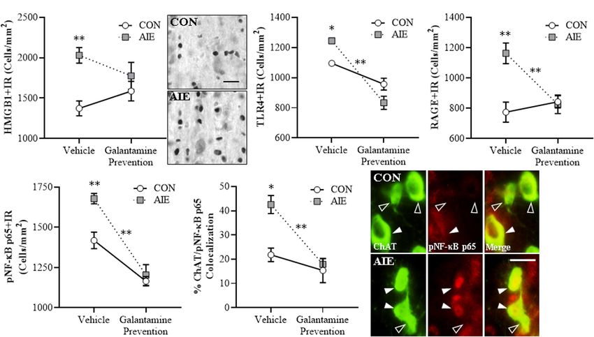
Crews et al.                                                                                                                  Galantamine Recovery of Cholinergic Pathology

  FIGURE 1 | Schematic of the galantamine prevention and restoration experimental design. On postnatal day (P)21, male Wistar rats were randomly assigned to
  either (i) water control (CON) or (ii) adolescent intermittent ethanol (AIE) treatment conditions. From P25 to P54, AIE subjects received a single daily intragastric (i.g.)
  gavage administration of ethanol (EtOH; 5.0 g/kg, 25% EtOH, w/v) in the AM on a 2 day on/2 day off schedule, and CONs received comparable volumes of water on
  an identical schedule. (A) In the galantamine prevention experiment, adolescent CON and AIE subjects received a single dose of galantamine HCl (4.0 mg/kg, i.p. in
  sterile PBS) or vehicle 30 min prior to each EtOH administration from P25 to P54. At the conclusion of treatment on P54, subjects were left undisturbed until sacrifice
  on P70. (B) In the galantamine restoration experiment, adult CON- and AIE-treated subjects received a single daily dose of galantamine HCl (4.0 mg/kg, i.p. in sterile
  PBS) or vehicle beginning 72 h following the conclusion of AIE treatment from P57 to P72. At the conclusion of treatment, subjects were sacrificed 24 h later on P73.
  (C) Depicted are coronal sections of the medial septum and vertical limb of the diagonal band of the basal forebrain between Bregma 1.60 and 0.20 mm according
  to the atlas of Paxinos and Watson (1998) used for immunohistochemical analysis. Gray shaded regions indicate regions of interest.

nickel-enhanced diaminobenzidine (Sigma-Aldrich), was used                                 values relative to CONs (Crews et al., 2004). The outlined regions
to visualize immunoreactivity. Tissue was mounted onto slides,                             of interest were determined and data expressed as cells/mm2 .
dehydrated, and cover slipped. Negative control for non-specific                           ChAT (+IR somal size was assessed using BioQuant Nova
binding was conducted on separate sections employing the                                   Advanced Image Analysis software (R&M Biometric).
abovementioned procedures omitting the primary antibody.
                                                                                           Fluorescent IHC and Microscopy
Microscopic Quantification and Image                                                       To assess ChAT colocalization with pNF-κB p65, TLR4, and
Analysis                                                                                   RAGE, free-floating basal forebrain sections were processed
Across experiments, BioQuant Nova Advanced Image Analysis                                  similar to previously reported methods (Vetreno et al., 2019).
software (R&M Biometric, Nashville, TN) was used for image                                 Briefly, sections were washed in 0.1 M Tris-buffered saline (TBS),
capture and quantification of IHC. Representative images were                              antigen retrieval performed by incubation in Citra solution
captured using an Olympus BX50 microscope and Sony DXC-                                    (BioGenex, Fremont, CA) for 1 h at 70◦ C, and blocked with
390 video camera linked to a computer. For each measure, the                               normal horse serum (MP Biomedicals). Sections were incubated
microscope, camera, and software were background corrected                                 for 48 h at 4◦ C in a primary antibody cocktail of goat anti-
and normalized to preset light levels to ensure fidelity of data                           ChAT (Millipore) in combination with either mouse anti-
acquisition. Microscopic quantification, which was performed                               TLR4 (Abcam), rabbit anti-RAGE (Abcam), or rabbit anti-
by experimenters blind to treatment conditions, was conducted                              pNF-κB p65 (Abcam) (see Table 1). Sections were washed
in the medial septum and vertical limb of the diagonal band,                               in TBS and incubated for 2 h at room temperature in a
as we previously discovered that AIE causes a persistent loss                              secondary antibody cocktail (Alexa Fluor 594, Alexa Fluor 488;
of ChAT + IR cells throughout the cholinergic nuclei of the                                Invitrogen, Carlsbad, CA). Tissue was mounted onto slides and
brain (Vetreno et al., 2014). A modified unbiased stereological                            cover slipped using Prolong Gold Anti-Fade mounting media
quantification method was used to quantify immunoreactivity in                             (Life Technologies, Grand Island, NY). Immunofluorescent
the basal forebrain. While there is variability in immunoreactive                          images were obtained using a DS-RiZ scope (Nikon Inc.,
cell density across basal forebrain coronal position, IHC staining                         Melville, NY) and colocalization quantified using NIS Elements
for all groups within studies were processed at the same time                              AR46 (Nikon Inc.).
with identical coronal sections to reduce variability. However,
although baseline values of basal forebrain IHC staining vary                              Chromatin Immunoprecipitation (ChIP)
across studies and cohorts, this variation does not affect                                 The procedure used is similar to methods reported previously
group comparisons. We previously reported that comparison                                  (Kyzar et al., 2017; Vetreno et al., 2019). Briefly, basal forebrain
of traditional unbiased stereological methodology with our                                 tissue samples from vehicle- and galantamine-treated CON- and
modified unbiased stereological approach yielded nearly identical                          AIE-treated subjects (n = 7 subjects per group) in the galantamine

Frontiers in Behavioral Neuroscience | www.frontiersin.org                             4                                          February 2021 | Volume 15 | Article 652494
Crews et al. Galantamine Recovery of Cholinergic Pathology
TABLE 1 | List of primary antibodies used for immunohistochemistry.
Antibody Isotype Dilution Source/purification Company, catalog number Validation
ChAT Goat IgG 1:200 Polyclonal Millipore, AB144P WB (Millipore)
TrkA Rabbit IgG 1:200 Polyclonal Millipore, 06-574 WB (Millipore)
p75NTR Mouse IgG 1:200 Monoclonal Millipore, MAB365 Binding assaya
HMGB1 Rabbit IgG 1:1,000 Polyclonal Abcam, ab18256 KO Validated (Abcam)
TLR4 Mouse IgG 1:100 Monoclonal Abcam, ab22048 KO Validated (Abcam)
RAGE Rabbit IgG 1:1,000 Polyclonal Abcam, ab3611 WB (Abcam)
NF-κB p65 (phospho S536) Rabbit IgG 1:2,000 Polyclonal Abcam, ab86299 WB (Abcam)
H3K9ac Rabbit IgG 1:500 Polyclonal Abcam, ab10812 WB/Peptide validated (Abcam)
H3K9me2 Mouse IgG 1:1,000 (IHC) 5 µL/sample (ChIP) Monoclonal Abcam, ab1220 WB (Abcam)
H3K9me3 Rabbit IgG 1:300 Polyclonal Abcam, ab8898 WB/Peptide validated (Abcam)
NeuN Mouse IgG 1:500 Monoclonal Millipore, MAB377 IHC (Millipore)
Validation information includes references as well as methods used for validation and source.
a Chandler et al. (1984).
restoration study were homogenized, fixed in 1.0% methanol- relation to outcomes of Tukey’s HSD post hoc tests. All values
free formaldehyde, quenched with 1.0 M glycine, lysed with lysis are reported as mean ± SEM, and significance was defined as
buffer (1.0% [v/v] SDS, 10 mM EDTA, 50 mM Tris-HCl [pH 8.0]), p ≤ 0.05.
and chromatin sheared to fragments of < 1,000 bp on a Covaris
ME220. Input DNA fractions were removed from the sheared
chromatin to be processed separately and the remaining sheared RESULTS
chromatin was incubated overnight at 4◦ C with an antibody
against H3K9me2 (5 µl/sample; Abcam) (see Table 1). Protein Adolescent Galantamine Treatment
A Dynabeads (ThermoFisher Scientific, Austin, TX) were added Combined With AIE Prevents the Loss of
and rotated at 4◦ C for 1 h followed by six washes in ChIP Cholinergic Neuron Markers and
wash buffer. Both immunoprecipitated DNA and input DNA
Shrinkage of Residual ChAT+ Neurons in
were eluted in 10% (w/v) Chelex by boiling at 95◦ C for 10 min
followed by centrifugation. The resulting DNA was quantified the Adult Basal Forebrain
using qPCR with SSOAdvanced Universal SYBR Green Supermix BFCNs are characterized by expression of ChAT and the NGF
(Bio-Rad, Berkeley, CA) using primers targeted against the ChAT receptors TrkA and p75NTR (Sobreviela et al., 1994; Vetreno
and TrkA promoters (see Table 2). The 11Ct method was used and Crews, 2018; Vetreno et al., 2019). Our laboratory and
to determine fold change relative to CON and was normalized to others previously reported that populations and size of BFCNs
the Input DNA fraction. do not differ between male and female rats, and that AIE
treatment causes a comparable loss of BFCNs across sexes in
adulthood (Gibbs, 1998; Vetreno and Crews, 2018). In the
Statistical Analysis present study, we sought to determine if treatment during AIE
Statistical analysis was performed using GraphPad Software
(i.e., P25–P54) with the cholinesterase inhibitor galantamine,
(Prism 9; San Diego, CA). Two-tailed Student’s t-tests were
which is approved for the treatment of AD (Lilienfeld, 2002;
used to assess BECs (P38 and P54) and ChAT + IR at P57.
Haake et al., 2020), would prevent the loss of BFCN markers
Repeated measure ANOVAs were used to assessed body weight.
in the adult (i.e., P70) basal forebrain. We report an 18%
The immunohistochemical and ChIP data were analyzed using
(±3%) reduction of ChAT + IR (Tukey’s HSD: p < 0.05),
2 × 2 ANOVAs. Post hoc analyses were performed using Tukey’s
a 24% (±6%) reduction of TrkA + IR [one-way ANOVA:
HSD where appropriate. Significant interactions are discussed in
F (1, 14) = 9.2, p < 0.01], and a 22% (±4%) reduction of
p75NTR + IR (Tukey’s HSD: p < 0.05) cells in the basal forebrain
of adult AIE-treated subjects, relative to CON subjects (see
TABLE 2 | List of primer sequences for ChIP analysis.
Figures 2A–C). Adolescent galantamine treatment alone did not
Primer Forward Reverse affect expression of BFCN markers in CONs but prevented the
AIE-induced loss of ChAT + IR (Tukey’s HSD: p < 0.05) and
ChAT Promoter ACT TGA TTG CTG GGG ATG GTG GAA TrkA + IR cells [one-way ANOVA: F (1, 14) = 7.0, p < 0.05],
CCT CTC TC GAT ACA GAA G
and blunted the loss of p75NTR + IR cells in the adult basal
ChAT Promoter TGC ATC TGG AGC GGG GAT AGT GGT
CpG Island TCA AAT CGT GAC GTT GT
forebrain, relative to vehicle-treated AIE subjects. Assessment
TrkA Promoter CCT CAC CGT GCA AGG GTC TGG AGA
of ChAT + BFCNs revealed a significant AIE-induced 11%
CTT TAC CT GCG TAC AT (±3%) reduction in somal size of the residual ChAT + IR
TrkA Promoter CpG TCA AGC AAG GCT CAC AGG GTG GCG neurons in the adult basal forebrain (Tukey’s HSD: p < 0.05),
Island CCG AAC AG CTA GAA G relative to CON subjects (see Figure 2D). Similar to the BFCN
Frontiers in Behavioral Neuroscience | www.frontiersin.org 5 February 2021 | Volume 15 | Article 652494Crews et al. Galantamine Recovery of Cholinergic Pathology
markers, adolescent galantamine treatment alone did not affect CON subjects. Galantamine treatment alone during adolescence
ChAT + somal size in CONs, but prevented the AIE-induced decreased constitutive expression of pNF-κB p65 + in CONs
somal shrinkage of ChAT + IR BFCNs in the adult basal (Tukey’s HSD: p < 0.01) and prevented the AIE-induced
forebrain (Tukey’s HSD: p < 0.01), relative to vehicle-treated increase of pNF-κB p65+, relative to vehicle-treated AIE
AIE subjects. Together, these data indicate that treatment with subjects (Tukey’s HSD: p < 0.01; see Figure 3D). Emerging
galantamine during AIE provided long-lasting prevention of the studies suggest both glia and neurons are involved in brain
BFCN marker loss and ChAT + somal shrinkage in the adult neuroimmune signaling (Crews et al., 2013, 2017a; Erickson et al.,
basal forebrain. 2019), prompting a determination of co-localization of pNF-κB
p65 + with ChAT + IR BFCNs. Interestingly, AIE increased
Adolescent Galantamine Treatment ChAT + /pNF-κB p65 + colocalized cells by approximately
During AIE Prevents the AIE-Induced twofold (Tukey’s HSD: p < 0.01) in the adult AIE-treated basal
forebrain, relative to CON subjects (see Figure 3E). These data
Increases of Proinflammatory suggest that direct neuroimmune signaling within BFCNs is
Neuroimmune Signals and NF-κB increased by AIE. Adolescent galantamine treatment alone did
activation Within Adult BFCNs not affect ChAT + /pNF-κB p65 + colocalization in CONs,
Emerging studies find HMGB1 release contributes to induction but galantamine treatment combined with AIE prevented the
of TLR and RAGE neuroimmune receptors as well as other AIE-induced increase of ChAT + /pNF-κB p65 + colocalization
neuroimmune genes, contributing to neurodegeneration in brain in the adult basal forebrain (Tukey’s HSD: p < 0.01), relative
diseases, including AD and AUD (Crews et al., 2013; Vetreno to vehicle-treated AIE subjects. Together, these data suggest
et al., 2013, 2018; Heneka et al., 2015; Paudel et al., 2020). that the AIE-induced increase of HMGB1-TLR4/RAGE-pNF-κB
We find AIE causes persistent upregulation of HMGB1, RAGE, p65 expression, including increased pNF-κB p65 colocalization
TLR4, NF-κB, and other neuroimmune signaling molecules in within ChAT + BFCNs, contributes to the loss of ChAT, TrkA,
brain with associated regional adult neuropathology (Vetreno and p75NTR expression. Further, these findings are consistent
and Crews, 2012, 2018; Vetreno et al., 2013, 2018, 2019). with galantamine increasing anti-inflammatory acetylcholine by
We assessed whether AIE combined with galantamine, which blocking cholinesterase, which prevents increases of HMGB1-
is reported to exert anti-inflammatory effects (Liu et al., TLR4/RAGE-pNF-κB p65 signaling and the concomitant loss
2018), would prevent the lasting activation of proinflammatory and somal shrinkage of BFCNs.
neuroimmune signaling in the adult basal forebrain. We find an
AIE-induced 48% (±7%) increase of HMGB1 + IR in the adult Adolescent Galantamine Treatment
basal forebrain [one-way ANOVA: F (1, 14) = 24.4, p < 0.01;
see Figure 3A], relative to CON subjects. While adolescent
During AIE Prevents the AIE-Induced
galantamine treatment alone did not affect HMGB1+, it blunted Increase of Epigenetic Histone Silencing
the AIE-induced increase of HMGB1 + IR cells in the adult basal Markers in the Adult Basal Forebrain
forebrain. HMGB1 is an endogenous agonist at TLR4, RAGE, Emerging studies find epigenetic mechanisms are related to
and other proinflammatory neuroimmune receptors (Andersson neuroimmune gene induction and persistent AIE pathology
et al., 2018; Paudel et al., 2020). We report a 14% (±2%) increase (Montesinos et al., 2016; Pandey et al., 2017; Crews et al.,
of TLR4 + IR cells (Tukey’s HSD: p < 0.05; see Figure 3B) and a 2019). We previously found AIE-induced increases of histone 3
50% (±9%) increase of RAGE + IR cells (Tukey’s HSD: p < 0.01; lysine 9 dimethylation (H3K9me2) occupancy at ChAT and TrkA
see Figure 3C) in the basal forebrain of adult AIE-treated rats, gene promoters, which is known to silence gene transcription,
relative to CON subjects. Adolescent galantamine treatment that appears to contribute to the loss of ChAT + neurons
alone decreased constitutive expression of TLR4 + (Tukey’s HSD: (Wang et al., 2008; Vetreno et al., 2019). In the present
p < 0.05) but not RAGE+, and prevented the AIE-induced experiment, we find AIE induced a 45% (±6%) increase
increase of TLR4 + IR (Tukey’s HSD: p < 0.01) and RAGE + IR of H3K9me2 + IR cells and a 21% (±6%) increase of
cells (Tukey’s HSD: p < 0.01) in the adult basal forebrain, H3K9me3 + IR cells (Tukey’s HSD: p < 0.01) in the adult
relative to vehicle-treated AIE subjects. Thus, galantamine, a basal forebrain, relative to CON subjects (see Figures 4A,B).
cholinesterase inhibitor that increases acetylcholine, blocks the Adolescent galantamine treatment alone did not affect expression
AIE-induced increases of proinflammatory HMGB1, TLR4, and of these epigenetic gene silencing markers in CONs, but
RAGE expression as well as somal shrinkage and loss of BFCNs. AIE combined with galantamine prevented the AIE-induced
HMGB1 activation of TLR4 and RAGE leads to downstream increases of H3K9me2 + IR (Tukey’s HSD: p < 0.05) and
activation of NF-κB (Chavakis et al., 2004; Tobon-Velasco et al., H3K9me3 + IR (Tukey’s HSD: p < 0.05) in the adult basal
2014; Liu et al., 2017; Aucott et al., 2018). NF-κB is a nuclear forebrain, relative to vehicle-treated AIE subjects. We did
transcription factor known to induce multiple neuroimmune not observe an effect of AIE or galantamine treatment on
genes (Pahl, 1999), and activated pNF-κB p65 provides insight expression of acetylated H3K9 (H3K9ac + IR cells: CON/Vehicle:
into neuroimmune signaling in brain (Vetreno et al., 2017, 5,186 ± 124, CON/Galantamine: 5,112 ± 73, AIE/Vehicle:
2018; Vetreno and Crews, 2018). We report a significant AIE- 5,153 ± 138, AIE/Galantamine: 5,136 ± 110). Taken together,
induced 18% (±2%) increase of pNF-κB p65 + IR cells in these experiments suggest that blockade of AIE induction of
the adult basal forebrain (Tukey’s HSD: p < 0.01), relative to HMGB1-TLR4/RAGE-pNF-κB p65 signaling and increases in
Frontiers in Behavioral Neuroscience | www.frontiersin.org 6 February 2021 | Volume 15 | Article 652494Crews et al. Galantamine Recovery of Cholinergic Pathology
FIGURE 2 | Adolescent galantamine treatment prevented the adolescent intermittent ethanol (AIE)-induced loss and somal shrinkage of cholinergic neurons in the
adult basal forebrain. (A) Modified unbiased stereological assessment revealed an 18% (±3%) reduction of choline acetyltransferase immunoreactive (ChAT + IR)
neurons in the adult (P70) basal forebrain of AIE-treated subjects, relative to CONs. Adolescent galantamine treatment from P25 to P54 did not affect ChAT + IR in
CONs, but prevented the AIE-induced loss of ChAT + IR neurons in adulthood, relative to vehicle-treated AIE subjects. (B) Modified unbiased stereological
assessment revealed a 24% (±6%) reduction of tropomyosin receptor kinase A immunoreactive (TrkA + IR) neurons in the adult (P70) basal forebrain of AIE-treated
subjects, relative to CONs. Adolescent galantamine treatment from P25 to P54 did not affect TrkA + IR in CONs, but prevented the AIE-induced loss of TrkA + IR
neurons in adulthood, relative to vehicle-treated AIE subjects. (C) Modified unbiased stereological assessment revealed a 22% (±4%) reduction of p75 neurotrophin
receptor immunoreactive (p75NTR + IR) neurons in the adult (P70) basal forebrain of AIE-treated subjects, relative to CONs. Adolescent galantamine treatment from
P25 to P54 did not affect p75NTR + IR in CONs and blunted the AIE-induced loss of p75NTR + IR neurons in adulthood, relative to vehicle-treated AIE subjects.
(D) Analysis of ChAT + IR neuron somal size revealed an 11% (±3%) reduction in somal size of the residual ChAT + neurons in the adult basal forebrain of AIE-treated
subjects, relative to CONs. Galantamine treatment alone did not affect ChAT + somal size, but prevented the AIE-induced somal shrinkage of ChAT + cholinergic
neurons in the adult basal forebrain, relative to vehicle-treated AIE subjects. Data are presented as mean ± SEM (n = 8/group). ∗ p < 0.05, ∗∗ p < 0.01.
H3K9me2 + and H3K9me3 + by adolescent galantamine underlie lasting changes in adult gene expression, hippocampal
treatment prevents the somal shrinkage and loss of BFCN neurogenesis, ethanol drinking and preference, anxiety, cognitive
markers in the adult basal forebrain (see Figure 4C). These deficits, and reduced behavioral flexibility (Pandey et al., 2015;
studies are consistent with AIE-induced ChAT + neuron Sakharkar et al., 2016; Mulholland et al., 2018; Kyzar et al.,
loss involving epigenetic gene silencing mechanisms that were 2019; Vetreno et al., 2019). Interestingly, anti-inflammatory
initially thought to reflect cell death. indomethacin, the cholinesterase inhibitor donepezil, and the
histone deacetylase inhibitor TSA have been found to reverse
Galantamine Treatment Post-AIE adult AIE-induced brain cellular and behavioral pathology
Restores Cholinergic Neuron Markers (Crews et al., 2019). We sought to determine if adult treatment
with galantamine following the conclusion of AIE (i.e., P57–
and Somal Shrinkage of Residual P72) would restore the loss of BFCN markers in the adult
ChAT + IR Neurons in the Adult Basal (i.e., P73) basal forebrain. In this replicate, we included an
Forebrain additional group of AIE subjects that we sacrificed at P57
Emerging studies find that persistent AIE-induced adult and find AIE caused a 22% (±5%) reduction of ChAT + IR
pathology includes complex epigenetic mechanisms that neurons (t [14] = 3.3, p < 0.01; ChAT + IR cells: CON: 208 ± 9,
Frontiers in Behavioral Neuroscience | www.frontiersin.org 7 February 2021 | Volume 15 | Article 652494Crews et al. Galantamine Recovery of Cholinergic Pathology FIGURE 3 | Adolescent galantamine treatment prevented the adolescent intermittent ethanol (AIE)-induced increase of proinflammatory neuroimmune signaling molecules in the adult basal forebrain. (A) Modified unbiased stereological assessment revealed a 48% (±7%) increase of high-mobility group box 1 immunoreactive (HMGB1 + IR) cells in the adult (P70) basal forebrain of AIE-treated subjects, relative to CONs. Adolescent galantamine treatment from P25 to P54 did not affect HMGB1 + IR in CONs, but blunted the AIE-induced increase of HMGB1 + IR cells in adulthood, relative to vehicle-treated AIE subjects. Representative photomicrographs of HMGB1 + IR cells in the adult basal forebrain of CON- and AIE-treated subjects. Scale bar = 50 µm. (B) Modified unbiased stereological assessment revealed a 14% (±2%) increase of Toll-like receptor 4 immunoreactive (TLR4 + IR) cells in the adult basal forebrain of AIE-treated subjects, relative to CONs. Adolescent galantamine treatment alone from P25 to P54 decreased constitutive expression of TLR4 + IR cells in CONs and prevented the AIE-induced increase of TLR4 + IR cells in adulthood, relative to vehicle-treated AIE subjects. (C) Modified unbiased stereological assessment revealed a 50% (±9%) increase of receptor for advanced glycation end-products immunoreactive (RAGE + IR) cells in the adult basal forebrain of AIE-treated subjects, relative to CONs. Adolescent galantamine treatment from P25 to P54 did not affect RAGE + IR in CONs, but prevented the AIE-induced increase of RAGE + IR cells in adulthood, relative to vehicle-treated AIE subjects. (D) Modified unbiased stereological assessment revealed an 18% (±2%) increase of phosphorylated nuclear factor kappa-light-chain-enhancer of activated B cells p65 immunoreactive (pNF-κB p65 + IR) cells in the adult basal forebrain of AIE-treated subjects, relative to CONs. Adolescent galantamine treatment alone from P25 to P54 decreased constitutive expression of pNF-κB p65 + IR cells in CONs and prevented the AIE-induced increase of pNF-κB p65 + IR cells in adulthood, relative to vehicle-treated AIE subjects. (E) Immunofluorescent co-labeling analysis revealed a 95% (±17%) increase of choline acetyltransferase immunoreactive (ChAT + IR) neurons that co-expressed pNF-κB p65 in the adult basal forebrain of AIE-treated subjects, relative to CONs. Galantamine treatment alone did not affect ChAT colocalization with pNF-κB p65 in CONs, but prevented the AIE-induced increase of ChAT colocalization with pNF-κB p65 in the adult basal forebrain, relative to vehicle-treated AIE subjects. Representative fluorescent photomicrographs of ChAT (green) and pNF-κB p65 (red) colocalization in the adult basal forebrain of CON- and AIE-treated subjects. Closed arrowheads = ChAT + IR neuron colocalization with pNF-κB p65 + (yellow); open arrowheads = ChAT + IR neurons that did not co-express pNF-κB p65. Scale bar = 50 µm. Data are presented as mean ± SEM (n = 8/group). ∗ p < 0.05, ∗∗ p < 0.01. AIE: 162 ± 11) in the late adolescent basal forebrain, relative forebrain, relative to vehicle-treated AIE subjects. Similarly, to age-matched CON subjects. In the adult (i.e., P73) basal we report a significant 18% (±2%) reduction in somal size forebrain following AIE treatment, we find a persistent AIE- of the residual ChAT + neurons in the adult basal forebrain induced 18% (± 4%) reduction of ChAT + IR cells [one-way (Tukey’s HSD: p < 0.01), relative to CON subjects. Galantamine ANOVA: F (1, 14) = 6.4, p < 0.05], a 23% (±3%) reduction of treatment alone did not affect ChAT + somal size, but reversed TrkA + IR cells (Tukey’s HSD: p < 0.05), and a 30% (± 6%) the AIE-induced somal shrinkage of ChAT + BFCNs in the reduction of p75NTR + IR cells (Tukey’s HSD: p < 0.01), adult basal forebrain (Tukey’s HSD: p < 0.01; see Figure 5D), relative to age-matched CON subjects (see Figures 5A–C). relative to vehicle-treated AIE subjects. Thus, AIE caused a Adult galantamine treatment alone did not affect expression loss of ChAT + BFCNs in the late adolescent (i.e., P57) basal of BFCN markers in CONs but restored the AIE-induced forebrain that persisted into adulthood (i.e., P73), and post-AIE loss of ChAT + IR cells [one-way ANOVA: F (1, 14) = 9.6, galantamine treatment reversed the AIE-induced loss of BFCN p < 0.01], TrkA + IR cells (Tukey’s HSD: p < 0.05), and markers and somal shrinkage of the residual ChAT + neurons in p75NTR + IR cells (Tukey’s HSD: p < 0.05) in the adult basal the adult basal forebrain. Frontiers in Behavioral Neuroscience | www.frontiersin.org 8 February 2021 | Volume 15 | Article 652494
Crews et al.                                                                                                            Galantamine Recovery of Cholinergic Pathology

  FIGURE 4 | Adolescent galantamine treatment prevented the adolescent intermittent ethanol (AIE)-induced increase of histone methylation markers in the adult
  basal forebrain. (A) Modified unbiased stereological assessment revealed a 45% (± 6%) increase of histone 3 lysine 9 dimethylation immunoreactive (H3K9me2 + IR)
  cells in the adult (P70) basal forebrain of AIE-treated subjects, relative to CONs. Adolescent galantamine treatment alone from P25 to P54 did not affect expression
  of H3K9me2 + IR cells in CONs, but prevented the AIE-induced increase of H3K9me2 + IR cells in adulthood (P70), relative to vehicle-treated AIE subjects.
  Representative photomicrographs of H3K9me2 + IR cells in the adult basal forebrain of CON- and AIE-treated subjects. (B) Modified unbiased stereological
  assessment revealed a 21% (±6%) increase of histone 3 lysine 9 trimethylation immunoreactive (H3K9me3 + IR) cells in the adult basal forebrain of AIE-treated
  subjects, relative to CONs. Adolescent galantamine treatment alone from P25 to P54 did not affect expression of H3K9me3 + IR cells in CONs, but prevented the
  AIE-induced increase of H3K9me3 + IR cells in adulthood, relative to vehicle-treated AIE subjects. Representative photomicrographs of H3K9me3 + IR cells in the
  adult basal forebrain of CON- and AIE-treated subjects. Scale bar = 50 µm. Data are presented as mean ± SEM (n = 8/group). ∗ p < 0.05, ∗∗ p < 0.01. (C) Simplified
  schematic depicting the proposed neuroimmune and epigenetic mechanism underlying the persistent AIE-induced loss of basal forebrain cholinergic neurons
  (BFCNs). (Left) AIE treatment from P25 to P54 causes the loss of BFCN markers and somal shrinkage of the residual cholinergic neurons in the adult (P70) basal
  forebrain. (Small panel). Representative photomicrograph of ChAT + IR neurons in the adult (P70) AIE-treated basal forebrain. (Large panel) Schematic depicting
  AIE-induced decreased expression of BFCN markers (i.e., ChAT, TrkA, p75NTR ), somal shrinkage of the residual ChAT + neurons, and increased expression of
  activated pNF-κB p65 within ChAT + BFCNs. In the proposed mechanism, AIE treatment increases expression of the endogenous proinflammatory TLR4/RAGE
  ligand HMGB1 and the HMGB1 receptors TLR4 and RAGE leading to increased BFCN expression of activated pNF-κB p65 and increased expression of the histone
  3 lysine 9 (H3K9) methylation markers (i.e., dimethylation and trimethylation) resulting in loss of BFCN markers. (Right) Galantamine treatment combined with AIE
  prevents the AIE-induced loss of BFCN markers and somal shrinkage of the residual cholinergic neurons in the adult basal forebrain. (Small panel) Representative
  photomicrograph of ChAT + IR neurons in the adult AIE-treated basal forebrain following adolescent galantamine treatment. (Large panel) Adolescent galantamine
  treatment (i.e., P25–P54) combined with AIE blocks the induction of HMGB1-TLR4/RAGE-pNF-κB p65 signaling, increase of histone 3 methylation markers, and
  provides long-lasting recovery of the somal shrinkage and loss of BFCN markers in the adult basal forebrain.

Galantamine Reverses the AIE-Induced                                                   HMGB1-TLR4/RAGE-pNF-κB p65 signal expression in the adult
                                                                                       (i.e., P73) basal forebrain. Consistent with our earlier studies,
Increase of Proinflammatory
                                                                                       AIE treatment caused a 20% (± 5%) increase of HMGB1 + IR
Neuroimmune Signaling Molecules in the                                                 cells [one-way ANOVA: F (1, 14) = 11.7, p < 0.01], a 46%
Adult Basal Forebrain                                                                  (±5%) increase of TLR4 + IR cells (Tukey’s HSD: p < 0.01),
The post-AIE galantamine reversal of AIE-induced reductions                            a 28% (±5%) increase of RAGE + IR cells [one-way ANOVA:
of BFCN markers prompted determination of neuroimmune                                  F (1, 14) = 15.2, p < 0.01], and a 15% (±4%) increase of

Frontiers in Behavioral Neuroscience | www.frontiersin.org                         9                                        February 2021 | Volume 15 | Article 652494
Crews et al. Galantamine Recovery of Cholinergic Pathology FIGURE 5 | Adult galantamine treatment restored the adolescent intermittent ethanol (AIE)-induced loss and somal shrinkage of cholinergic neurons in the adult basal forebrain. (A) Modified unbiased stereological assessment revealed an 18% (±4%) reduction of choline acetyltransferase immunoreactive (ChAT + IR) neurons in the adult (P73) basal forebrain of AIE-treated subjects, relative to CONs. Adult galantamine treatment alone from P57 to P73 did not affect ChAT + IR in CONs, but restored the AIE-induced loss of ChAT + IR neurons in adulthood, relative to vehicle-treated AIE subjects. (B) Modified unbiased stereological assessment revealed a 23% (±3%) reduction of tropomyosin receptor kinase A immunoreactive (TrkA + IR) neurons in the adult (P73) basal forebrain of AIE-treated subjects, relative to CONs. Adult galantamine treatment alone did not affect TrkA + IR in CONs, but restored the AIE-induced loss of TrkA + IR neurons in adulthood, relative to vehicle-treated AIE subjects. (C) Modified unbiased stereological assessment revealed a 30% (±6%) reduction of p75 neurotrophin receptor immunoreactive (p75NTR + IR) neurons in the adult (P73) basal forebrain of AIE-treated subjects, relative to CONs. Adult galantamine treatment alone did not affect p75NTR + IR in CONs, but restored the AIE-induced loss of p75NTR + IR neurons in adulthood, relative to vehicle-treated AIE subjects. (D) Analysis of ChAT + neuron somal size revealed an 18% (±2%) reduction in somal size of the residual ChAT + neurons in the adult basal forebrain of AIE-treated subjects, relative to CONs. Adult galantamine treatment alone did not affect ChAT + somal size, but restored the AIE-induced somal shrinkage of ChAT + cholinergic neurons in the adult basal forebrain, relative to vehicle-treated AIE subjects. Data are presented as mean ± SEM (n = 8/group). ∗ p < 0.05, ∗∗ p < 0.01. pNF-κB p65 + IR cells [one-way ANOVA: F (1, 14) = 10.3, signal expression in the adult basal forebrain that paralleled the p < 0.01] in the adult basal forebrain, relative to CON subjects restoration of BFCNs. (see Figure 6). Adult galantamine treatment alone did not affect expression of proinflammatory neuroimmune markers Galantamine Reverses the AIE-Induced in CONs, but post-AIE treatment with galantamine reversed the AIE-induced increase of HMGB1 + [one-way ANOVA: Increases of Cellular and Cholinergic F (1, 14) = 8.5, p < 0.05], TLR4 + (Tukey’s HSD: p < 0.01), Promoter Histone Silencing Markers in RAGE + [one-way ANOVA: F (1, 14) = 8.0, p < 0.05], and pNF- the Adult Basal Forebrain κB p65 + [one-way ANOVA: F (1, 14) = 9.1, p < 0.01] cells in We further tested our hypothesis that AIE increases of HMGB1- the adult basal forebrain, relative to vehicle-treated AIE subjects. TLR4/RAGE-pNF-κB p65 signaling within BFCNs contribute to Double immunofluorescent IHC for ChAT with TLR4 + and silencing of cholinergic phenotype genes by determining if post- RAGE + suggests that some of the increases in signaling occur AIE galantamine treatment reverses the AIE-induced increase of within ChAT + BFCNs (see Figures 6C,E). Thus, treatment H3K9 methylation markers. with galantamine following AIE-induced pathology reversed We find an AIE increase of H3K9me2 + IR cells [22% the AIE-induced increase of HMGB1-TLR4/RAGE-pNF-κB p65 (±5%), one-way ANOVA: F (1, 14) = 7.5, p < 0.05] and Frontiers in Behavioral Neuroscience | www.frontiersin.org 10 February 2021 | Volume 15 | Article 652494
Crews et al.                                                                                                            Galantamine Recovery of Cholinergic Pathology

  FIGURE 6 | Adult galantamine treatment reversed the adolescent intermittent ethanol (AIE)-induced increase of proinflammatory neuroimmune signaling molecules in
  the adult basal forebrain. (A) Modified unbiased stereological assessment revealed a 20% (±5%) increase of high-mobility group box 1 immunoreactive (HMGB1 + IR)
  cells in the adult (P73) basal forebrain of AIE-treated subjects, relative to CONs. Adult galantamine treatment from P57 to P73 did not affect HMGB1 + IR in CONs,
  but reversed the AIE-induced increase of HMGB1 + IR cells in adulthood, relative to vehicle-treated AIE subjects. (B) Modified unbiased stereological assessment
  revealed a 46% (±5%) increase of Toll-like receptor 4 immunoreactive (TLR4 + IR) cells in the adult (P73) basal forebrain of AIE-treated subjects, relative to CONs.
  Adult galantamine treatment alone did not affect expression of TLR4 + IR cells in CONs, but reversed the AIE-induced increase of TLR4 + IR cells in adulthood,
  relative to vehicle-treated AIE subjects. Representative photomicrographs of TLR4 + IR cells in the adult basal forebrain of CON- and AIE-treated subjects. Scale
  bar = 50 µm. (C) Immunofluorescent co-labeling revealed TLR4 (red) colocalization with choline acetyltransferase immunoreactive (ChAT + IR) neurons in the adult
  basal forebrain. Closed arrowheads = TLR4 + IR colocalization with ChAT + IR neurons (yellow). Scale bar = 50 µm. (D) Modified unbiased stereological assessment
  revealed a 28% (±5%) increase of receptor for advanced glycation end-products immunoreactive (RAGE + IR) cells in the adult (P73) basal forebrain of AIE-treated
  subjects, relative to CONs. Adult galantamine treatment alone did not affect RAGE + IR in CONs, but reversed the AIE-induced increase of RAGE + IR cells in
  adulthood, relative to vehicle-treated AIE subjects. Representative photomicrographs of RAGE + IR cells in the adult basal forebrain of CON- and AIE-treated
  subjects. Scale bar = 50 µm. (E) Immunofluorescent co-labeling revealed RAGE (red) colocalization with ChAT + IR neurons in the adult basal forebrain. Closed
  arrowheads = RAGE + IR colocalization with ChAT + IR neurons (yellow). Scale bar = 50 µm. (F) Modified unbiased stereological assessment revealed a 15% (±4%)
  increase of phosphorylated nuclear factor kappa-light-chain-enhancer of activated B cells p65 immunoreactive (pNF-κB p65 + IR) cells in the adult (P73) basal
  forebrain of AIE-treated subjects, relative to CONs. Adult galantamine treatment alone did not affect pNF-κB p65 + IR cells in CONs, but reversed the AIE-induced
  increase of pNF-κB p65 + IR cells in adulthood, relative to vehicle-treated AIE subjects. Data are presented as mean ± SEM (n = 8/group). ∗ p < 0.05, ∗∗ p < 0.01.

H3K9me3 + IR cells [24% (±8%), one-way ANOVA: F (1,                                    (Vetreno et al., 2019). To determine cholinergic gene-specific
14) = 5.4, p < 0.05] in the adult basal forebrain, relative to CON                     information, we assessed histone methylation silencing within
subjects (see Figures 7A,B). While adult galantamine treatment                         ChAT and TrkA gene promoters, which are two cholinergic
alone did not affect expression of H3K9 methylation markers                            phenotype genes. We report that AIE treatment increased levels
in CONs, post-AIE galantamine treatment reversed the AIE-                              of H3K9me2 occupancy by approximately 1.9-fold at the ChAT
induced increase of H3K9me2 + IR cells [one-way ANOVA:                                 promoter (Tukey’s HSD: p < 0.05), 1.7-fold at the CpG island of
F (1, 14) = 16.0, p < 0.01] and H3K9me3 + IR cells [one-                               the ChAT promoter (Tukey’s HSD: p < 0.01), and 2.6-fold at the
way ANOVA: F (1, 14) = 5.3, p < 0.05], relative to vehicle-                            CpG island of the TrkA promoter (Tukey’s HSD: p < 0.01) in the
treated AIE subjects. Although BFCN markers decreased and                              adult basal forebrain, relative to CON subjects (see Figures 7D–
epigenetic silencing markers increased following AIE treatment,                        F). While adult galantamine treatment alone did not affect levels
there were no changes in neuronal density across treatment                             of H3K9me2 occupancy in CONs, it reversed the AIE-induced
conditions as determined by expression of the neuronal marker                          increase of H3K9me2 occupancy at the ChAT promoter (Tukey’s
NeuN (see Figure 7C), consistent with our earlier studies                              HSD: p < 0.05), the CpG island of the ChAT promoter (Tukey’s

Frontiers in Behavioral Neuroscience | www.frontiersin.org                        11                                        February 2021 | Volume 15 | Article 652494
Crews et al.                                                                                           Galantamine Recovery of Cholinergic Pathology

HSD: p < 0.01), and the CpG island of the TrkA promoter                     data support the hypothesis that AIE increases HMGB1+,
(Tukey’s HSD: p < 0.05), relative to vehicle-treated AIE subjects.          TLR4+, and RAGE+, and downstream pNF-κB p65 expression
Levels of H3K9me2 were unchanged at the TrkA promoter (data                 within ChAT + BFCNs. As shown in Figure 8, relaxed
not shown). The loss of BFCN markers along with increases of                chromatin within cholinergic phenotype genes allow high levels
H3K9me2 at both ChAT and TrkA promoters, but no change                      of expression in the healthy adult brain that are reduced by AIE
in total neurons, supports epigenetic programming leading to                along with ChAT + cholinergic neuron shrinkage. The increases
long-lasting silencing of the cholinergic phenotype.                        in HMGB1, TLR4, RAGE, and pNF-κB p65 are associated
                                                                            with increases in both cellular histone 3 silencing markers
                                                                            and cholinergic gene promoter methylation, consistent with
DISCUSSION                                                                  AIE inducing condensed chromatin that decreases expression
                                                                            of cholinergic phenotype genes, causes shrinkage and loss of
The current study tested the hypothesis that treatment with the             BFCNs. Galantamine combined with or following AIE reverses
cholinesterase inhibitor galantamine, which has FDA approval                increases of HMGB1, TLR4, RAGE, pNF-κB p65, and histone
for the treatment of AD (Lilienfeld, 2002; Hampel et al., 2018;             3 silencing markers, opening chromatin and restoring gene
Haake et al., 2020), would recover the lasting adult basal forebrain        expression and BFCN markers. Neuroimmune-linked epigenetic
cholinergic pathology associated with adolescent binge ethanol              alterations in neuronal phenotype may represent a previously
exposure. Degeneration of BFCNs and ChAT + somal shrinkage                  unappreciated mechanism of plasticity. Galantamine restoration
is a feature of several neurodegenerative disorders, including              of the AIE-induced persistent loss of BFCNs through reversal
AD and AUD (Lehericy et al., 1993; Vetreno et al., 2014), and               of persistent neuroimmune-induced changes in chromatin
may contribute to the cognitive deficits associated with these              epigenetic silencing may offer great promise for reversing AIE
disorders (Vogels et al., 1990; De Rosa et al., 2004; Mufson                pathology as well as other pathologies initially thought to
et al., 2008). Galantamine treatment during AIE (i.e., P25–P54)             be irreversible.
or following the conclusion of AIE (i.e., P57–P72) recovered the                Galantamine is an herbal alkaloid that inhibits cholinesterase
persistent loss of cholinergic neuron markers (i.e., ChAT, TrkA,            and is used to treat cholinergic hypofunction in AD. By
and p75NTR ) and somal shrinkage of residual BFCNs in the adult             inhibiting cholinesterase, galantamine increases acetylcholine
basal forebrain. However, similar to previous findings (Vetreno             bioavailability, which is known to block induction of
et al., 2019), we found no loss of NeuN + IR, a global neuronal             proinflammatory genes. While the mechanisms underlying
marker, consistent with no loss of neurons, only cholinergic                the loss of BFCNs remain to be fully elucidated, accumulating
phenotype markers. We and others (Pascual et al., 2007; Vetreno             evidence implicates activation of the proinflammatory
and Crews, 2018; Vetreno et al., 2018) have previously found                neuroimmune signaling system (Vetreno and Crews, 2018).
the anti-inflammatory drug indomethacin can prevent ethanol                 HMGB1-RAGE/TLR4-NF-κB p65 neuroimmune signaling has
induction of brain proinflammatory neuroimmune genes and                    been implicated in neuropathology associated with AD and AUD
neuropathology, including loss of ChAT + IR neurons, consistent             (Boissiere et al., 1997; Crews et al., 2013; Vetreno et al., 2013;
with loss of BFCNs being mediated by proinflammatory                        Fujita et al., 2016; Paudel et al., 2020). The anti-inflammatory
neuroimmune and epigenetic gene silencing histone (H3K9)                    effects of galantamine have been reported in rodent models of
methylation mechanisms. We found galantamine treatment                      endotoxemia, obesity, colitis, and other disorders (Pavlov et al.,
during AIE or initiated 72 h post-AIE blocked the AIE-induced               2009; Satapathy et al., 2011; Wazea et al., 2018). Acetylcholine
increased expression of the proinflammatory neuroimmune                     is anti-inflammatory as ACh suppresses LPS-induced release
receptors TLR4 and RAGE, the endogenous TLR4/RAGE ligand                    of HMGB1 and nicotinic ACh receptor (nAChR) activation
HMGB1, and activation of pNF-κB p65 in the adult basal                      prevents activity of the NF-κB signaling pathway (Wang et al.,
forebrain. Further, we found BFCNs express TLR4, RAGE, and                  2004). Galantamine is not only a cholinesterase inhibitor, but
activated pNF-κB p65, and that AIE treatment increased pNF-κB               also acts as a positive allosteric ligand at nAChRs, including
p65 expression in adult ChAT + IR neurons that was reversed                 α7 nAChRs (Wazea et al., 2018), and potentiates cholinergic
by galantamine treatment consistent with galantamine inhibiting             transmission by positively modulating the response of nAChR to
neuroimmune signaling within BFCNs. Previous studies found                  ACh and their agonists (Dajas-Bailador et al., 2003; Samochocki
AIE increased HMGB1, TLRs, RAGE, pNF-κB p65, and other                      et al., 2003). α7 nAChRs are present on microglia and neurons,
neuroimmune genes that persist into adulthood across multiple               including BFCNs (Azam et al., 2003; Ulloa, 2005), and play a
brain regions, including the forebrain, cortex, hippocampus,                role in modulating neuroinflammation (Sitapara et al., 2014; Ren
and cerebellum (Vetreno and Crews, 2012; Vetreno et al.,                    et al., 2017; Wazea et al., 2018). Alternatively, galantamine may
2013, 2018, 2019; Crews et al., 2017b). We previously reported              exert neuroprotective effects on BFCNs through interactions
that exercise during or following AIE treatment can restore                 with the high-affinity NGF receptor TrkA. Studies suggest that
ChAT +, TrkA +, and p75NTR + loss as well as associated                     the AIE-induced loss of trophic factor receptors, such as the
adult cognitive deficits (Vetreno et al., 2019). In the present             high affinity NGF receptor TrkA, might contribute to the loss
study, we report that galantamine treatment recovered the AIE-              of ChAT + IR neurons. NGF and TrkA are critical for the
induced increase of cellular histone 3 methylation markers                  survival and maintenance of BFCNs (Hagg et al., 1988, 1989;
(i.e., H3K9me2 and H3K9me3) as well as increased H3K9me2                    Fagan et al., 1997; Kim et al., 2014; Allaway and Machold, 2017),
occupancy at ChAT and TrkA gene promoters. Together, these                  and galantamine has been reported to activate TrkA receptors,

Frontiers in Behavioral Neuroscience | www.frontiersin.org             12                                  February 2021 | Volume 15 | Article 652494
You can also read