HHS Public Access Author manuscript Sci Signal. Author manuscript; available in PMC 2019 September 05.
←
→
Page content transcription
If your browser does not render page correctly, please read the page content below
HHS Public Access
                                Author manuscript
                                Sci Signal. Author manuscript; available in PMC 2019 September 05.
Author Manuscript

                    Published in final edited form as:
                     Sci Signal. ; 12(590): . doi:10.1126/scisignal.aav7259.

                    Application of a MYC degradation screen identifies sensitivity to
                    CDK9 inhibitors in KRAS-mutant pancreatic cancer
                    Devon R. Blake1, Angelina V. Vaseva4, Richard G. Hodge4, McKenzie P. Kline2, Thomas S.K.
                    Gilbert1,5, Vikas Tyagi6, Daowei Huang6, Gabrielle C. Whiten6, Jacob E. Larson6, Xiaodong
                    Wang4,6, Kenneth H. Pearce6, Laura E. Herring1,5, Lee M. Graves1,4,5, Stephen V. Frye4,6,
                    Michael J. Emanuele1,4, Adrienne D. Cox1,3,4, Channing J. Der1,4,*
Author Manuscript

                    1Department   of Pharmacology, Eshelman School of Pharmacy, University of North Carolina at
                    Chapel Hill, Chapel Hill, NC 27599, USA
                    2Department   of Biology, Eshelman School of Pharmacy, University of North Carolina at Chapel
                    Hill, Chapel Hill, NC 27599, USA
                    3Department   of Radiation Oncology, Eshelman School of Pharmacy, University of North Carolina
                    at Chapel Hill, Chapel Hill, NC 27599, USA
                    4Lineberger Comprehensive Cancer Center, Eshelman School of Pharmacy, University of North
                    Carolina at Chapel Hill, Chapel Hill, NC 27599, USA
                    5UNC  Michael Hooker Proteomics Center, Eshelman School of Pharmacy, University of North
                    Carolina at Chapel Hill, Chapel Hill, NC 27599, USA
Author Manuscript

                    6Center for Integrative Chemical Biology and Drug Discovery, Eshelman School of Pharmacy,
                    University of North Carolina at Chapel Hill, Chapel Hill, NC 27599, USA

                    Abstract
                         Stabilization of the MYC oncoprotein by KRAS signaling is a requirement for growth of
                         pancreatic ductal adenocarcinomas (PDAC). However, current understanding of how MYC protein
                         stability is regulated in MYC-dependent cancers such as KRAS-mutant PDAC is incomplete. We
                         developed a flow cytometry-based assay, screened a library of >800 protein kinase inhibitors, and
                         identified compounds that promoted either stability or degradation of MYC. We validated
                         compounds that stabilize or destabilize MYC, and then focused on one compound,
                         UNC10112785, that induced significant loss of MYC protein. We determined that this compound
                         is a potent CDK9 inhibitor with a novel scaffold, and that it causes MYC loss through both
Author Manuscript

                         transcriptional and post-translational mechanisms. We discovered that CDK9 regulates MYC
                         protein stability through a novel KRAS-independent mechanism involving direct phosphorylation

                    *
                     Corresponding author: cjder@med.unc.edu.
                    Author contributions: D.R.B., A.V.V., K.H.P., and C.J.D. designed the experiments; D.R.B., K.H.P., A.D.C., and C.J.D. wrote the
                    manuscript; D.R.B., A.V.V., M.P.O., R.G.H., T.S.K.G., and J.E.L. performed experiments; V.T., D.H., G.C.W., and X.W. synthesized
                    compounds for the experiments; L.E.H., L.M.G., K.H.P., A.D.C., S.V.F., M.J.E. and C.J.D. provided scientific guidance.
                    Competing interests: The authors declare no competing interests.
                    Data and materials availability: Plasmids generated for this study will be deposited at Addgene. The PKIS compound set is available
                    from the Structural Genomics Consortium (SGC) at UNC (SGC-UNC@unc.edu).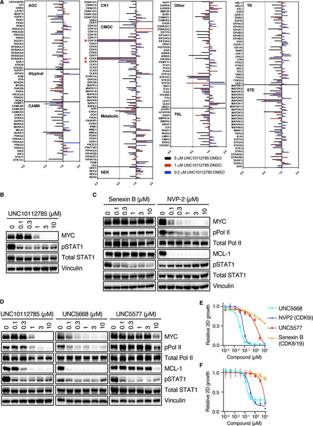
Blake et al.                                                                                                 Page 2

                             of MYC Ser62. Our studies present the application of an innovative screening strategy that can be
Author Manuscript

                             adapted to identify novel regulators of protein stability.

                       One Sentence Summary:
                             We applied a MYC oncoprotein degradation screen and identified CDK9 as a novel regulator of
                             MYC protein degradation.

                       INTRODUCTION
                                       In 2017, pancreatic cancer surpassed breast cancer to become the third leading cause of
                                       cancer deaths in the U.S (1). By around 2020, pancreatic cancer is projected to surpass
                                       colorectal cancer and become the second leading cause of U.S. cancer deaths (2). Currently,
                                       the 5-year overall survival rate is at an abysmal 8% (1). Despite a well-defined genetic
Author Manuscript

                                       profile of pancreatic ductal adenocarcinoma (PDAC) (3), clinically effective targeted
                                       therapies remain to be developed, with current treatments limited to conventional cytotoxic
                                       drugs (4).

                                       The main genetic driver of PDAC initiation, progression and maintenance is mutational
                                       activation of the KRAS oncogene, which is found in ~95% of PDAC (3). Although KRAS
                                       was the first cancer gene identified in human cancers over 35 years ago (5), the effort to
                                       effectively target RAS-driven cancers is still ongoing (6, 7).

                                       The interplay and interdependency of the RAS and the MYC oncogenes in driving cancer
                                       development and maintenance is well-established. This association was first demonstrated
                                       when it was shown that MYC overexpression was necessary to support RAS transformation
                                       of rodent fibroblasts (8). MYC expression is frequently increased in many cancers, most
Author Manuscript

                                       commonly by gene amplification or increased gene transcription (9). Subsequent studies in
                                       genetically engineered mouse models of cancer demonstrated the essential role of MYC in
                                       KRAS-driven oncogenesis (10, 11) and genetic suppression of MYC impairs KRAS-driven
                                       PDAC (12–15). Moreover, overexpression of MYC alone was sufficient to phenocopy
                                       mutant KRAS and drive development of metastatic PDAC (16). Thus, targeting MYC could
                                       be a novel therapeutic strategy for MYC-dependent cancers such as KRAS-mutant PDAC.
                                       However, similar to RAS, MYC has also been considered undruggable due to a surface
                                       topology that lacks deep pockets for design of potent small molecule binders (17).

                                       The MYC transcription factor drives a multitude of proliferative and pro-growth phenotypes
                                       (9). Strategies to inhibit MYC function have included inhibition of MYC transcription (for
                                       example, bromodomain inhibitors, JQ1) (18, 19), inhibition of MYC/MAX dimerization (20,
Author Manuscript

                                       21), targeting expression of MYC-regulated genes (22) or MYC-associated metabolic
                                       vulnerabilities (23). Of these strategies, only bromodomain inhibitors have entered clinical
                                       trials, but their relative lack of selectivity for MYC transcription remains a concern (24).

                                       Mutationally activated KRAS promotes increased MYC expression by stimulating MYC
                                       gene transcription and by promoting MYC protein stability (14, 25). KRAS effector
                                       signaling promotes MYC protein stability through ERK mitogen-activated protein kinase
                                       phosphorylation on MYC residue Ser62 (26). Phosphorylated Ser62 then facilitates GSK3β

                                           Sci Signal. Author manuscript; available in PMC 2019 September 05.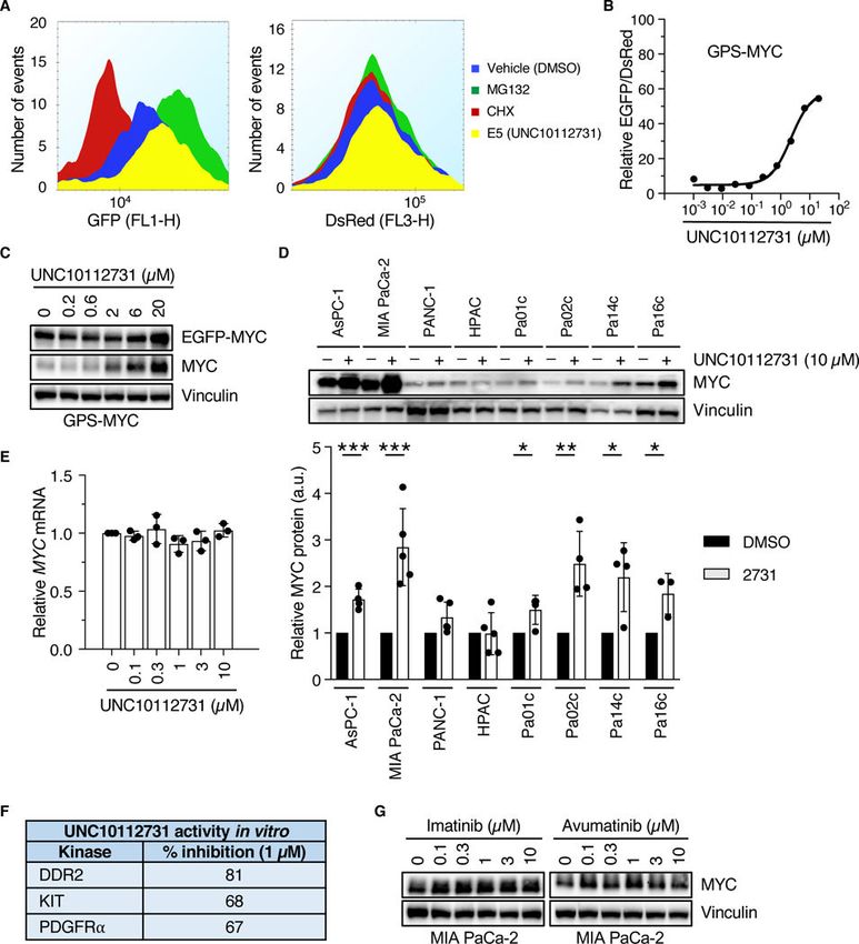
Blake et al.                                                                                             Page 3

                                   phosphorylation of MYC at Thr58, and subsequent dephosphorylation of Ser62 by the tumor
Author Manuscript

                                   suppressor PP2A promotes E3 ligase FBXW7-dependent MYC degradation. KRAS
                                   signaling through the PI3K effector pathway, leading to activation of AKT and concomitant
                                   inactivation of GSK3β, represents a second effector signaling mechanism by which KRAS
                                   can regulate MYC protein stability. Pharmacologic inhibition of SET, a negative regulator of
                                   PP2A, increased MYC degradation and impaired PDAC tumorigenic growth, supporting the
                                   therapeutic value of targeting MYC protein degradation (27).

                                   Our previous studies found that KRAS regulation of MYC protein stability in KRAS-mutant
                                   PDAC involved both ERK-dependent and -independent mechanisms but not PI3K-AKT
                                   signaling (14, 25). To further elucidate the mechanisms by which KRAS regulates MYC
                                   protein stability, we developed and applied a MYC protein degradation screen in KRAS-
                                   mutant PDAC cells (14). To identify novel protein kinase-dependent mechanisms that
                                   regulate MYC protein stability, we then screened the Published Kinase Inhibitor Set (PKIS)
Author Manuscript

                                   of ATP-competitive protein kinase inhibitors (28, 29). This approach, together with two
                                   other screening strategies, identified a MEK5-ERK5 compensatory mechanism induced by
                                   inhibition of KRAS-ERK1/2 function (14). In this study, we now focus on the methodology
                                   for application of the screen and the experimental strategies to validate kinase inhibitors that
                                   either stabilize MYC protein or promote its degradation. Our evaluation of one compound
                                   that stimulated loss of MYC protein identified cyclin-dependent kinase 9 (CDK9) as a novel
                                   regulator of MYC protein stability.

                       RESULTS
                       Establishment of a MYC protein degradation screen
                                   We have described our generation and validation of a MYC degradation reporter for use in a
Author Manuscript

                                   cell-based screen to identify protein kinases that regulate MYC protein stability (14). We
                                   utilized the pGPS-LP lentiviral reporter plasmid in which a CMV promoter regulates
                                   expression of a single bicistronic mRNA transcript that encodes both DsRed and EGFP-
                                   tagged proteins, separated by an internal ribosome entry site (IRES) (30). To construct a
                                   reporter capable of monitoring MYC protein expression, we introduced the cDNA sequence
                                   encoding human MYC into pGPS-LP to encode an EGFP-MYC fusion protein (designated
                                   GPS-MYC; Fig. 1A, (14)). We then stably infected the KRAS-mutant PDAC cell line MIA
                                   PaCa-2 with the GPS-MYC vector and established populations of cells stably expressing
                                   DsRed-IRES-EGFP-MYC (hereafter referred to as GPS-MYC cells). Importantly, EGFP has
                                   a long half-life, but partially adopts the degradation characteristics of MYC when expressed
                                   as a fusion protein (see below). Further, since DsRed is expressed from the same transcript
                                   as EGFP-MYC, we can normalize for fluctuations in transcription and proteasome activity
Author Manuscript

                                   on a per cell basis. Thus, we can infer the relative stability of MYC on a per cell basis by
                                   examining the EGFP/DsRed ratio (31).

                                   In this study, we first determined whether the exogenously expressed EGFP-MYC displayed
                                   properties of endogenous MYC. Like endogenous MYC, EGFP-MYC localization was
                                   restricted to the nucleus (Fig. 1B). We showed previously that acute suppression of KRAS
                                   caused similar loss of endogenous MYC and EGFP-MYC (14). Although the half-life of
                                   EGFP-MYC is slightly longer than that of endogenous MYC (Fig. 1C), demonstrating that

                                       Sci Signal. Author manuscript; available in PMC 2019 September 05.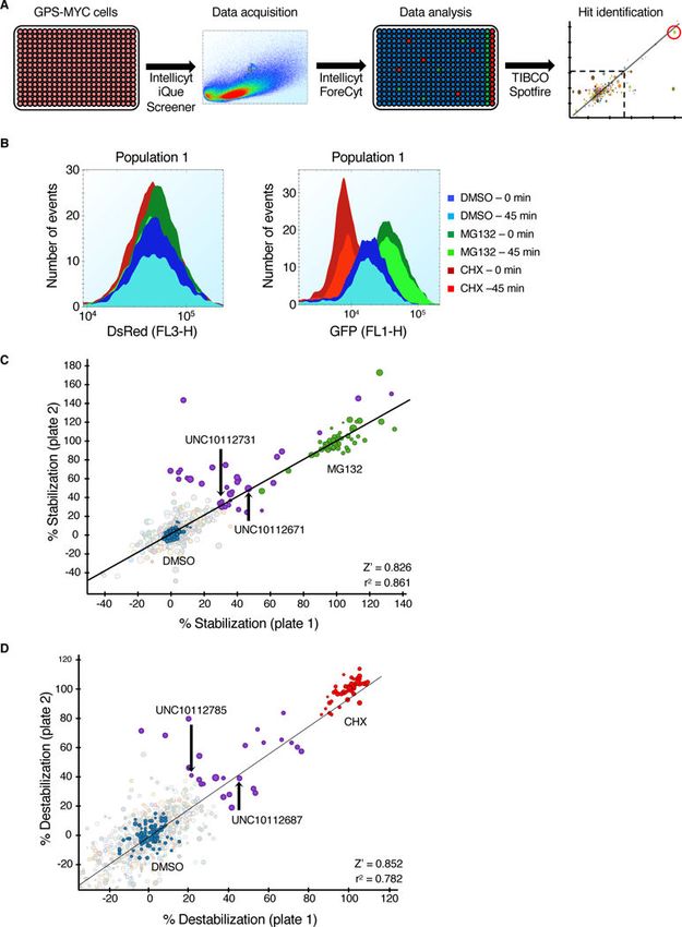
Blake et al.                                                                                             Page 4

                                   EGFP-MYC protein stability is not regulated exactly like endogenous MYC, we reasoned
Author Manuscript

                                   that compounds potent enough to cause loss of the more stable EGFP-MYC would also be
                                   able to cause loss of the less stable endogenous MYC. In the GPS-MYC cells, DsRed serves
                                   as an internal control for differential expression levels of the cassette amongst GPS-MYC
                                   cells. Importantly, treatment of GPS-MYC cells with either cycloheximide (CHX) to inhibit
                                   protein synthesis or MG132 to inhibit proteasome-dependent protein degradation altered the
                                   EGFP but not the DsRed signal (Fig. 1D). Thus, the ratio of EGFP/DsRed signals provides
                                   an accurate determination of MYC protein stability in GPS-MYC cells.

                                   After validating GPS-MYC cells as a cell model for monitoring MYC protein stability, we
                                   optimized the assay in a 384-well format using liquid handing automation (Fig. 2A). We first
                                   evaluated several sources of commercially available 384-well plates for retention of GPS-
                                   MYC cells during the automated washing steps in the screen. Although all the plates were
                                   cell culture-treated, not all of them retained cells well during the washing process. Next,
Author Manuscript

                                   because MIA PaCa-2 cells have a propensity to aggregate, we optimized the type and
                                   volume of cell dissociation reagents as well as shake speed and frequency during the screen
                                   to ensure that cells remained in a single cell suspension throughout the assay. Since MYC
                                   has a short half-life in MIA PaCa-2 cells (t1/2 ~50 min) (32), a potential confounding assay
                                   variable could be differences in the EGFP-MYC signal over the course of assaying an entire
                                   plate. To minimize this potential variable, we optimized the sip time (analysis time per well).
                                   At the conclusion of our optimization efforts, the assay duration for each 384-well plate was
                                   ~45 min, during which the EGFP and DsRed signals remained constant (Fig. 2B). Since the
                                   expression level of EGFP-MYC was only ~5-fold above that of endogenous MYC, the
                                   resulting EGFP signal was relatively weak, and, coupled with a high background signal, the
                                   dynamic range between the vehicle (DMSO) and CHX controls was also relatively low (~2-
                                   fold). Fixation of GPS-MYC cells reduced the EGFP signal even further, rendering the
Author Manuscript

                                   dynamic range too small, so we used live cells in the assay. Despite the relatively narrow
                                   dynamic range of the assay between the CHX and DMSO controls, the Z-factors were
                                   consistently greater than 0.7, indicating that the assay was robust. All of our screening was
                                   then done on the optimized conditions as described in the Materials and Methods section.

                                   Given the important nature of protein kinases in regulating MYC protein stability, we
                                   screened the GPS-MYC cells with the Published Kinase Inhibitor Set (PKIS) of ATP-
                                   competitive protein kinase inhibitors (28, 29, 33) to identify novel kinases that regulate
                                   MYC protein stability in PDAC. PKIS was generated to maximize structural diversity and
                                   the spectrum of kinases inhibited, and the activity profiles of the compounds is published
                                   (28, 29, 33), enabling target hypothesis generation as soon as hits are discovered. In total, we
                                   screen GPS-MYC cells with this ~800 compound set, treating cells for 6 hours at a
Author Manuscript

                                   concentration of 20 μM, using the Intellicyt iQue Screener. The screen was run in duplicate
                                   to ensure reproducibility.

                                   To process the primary screening data, we first separated the data into two parts, percent
                                   stabilization and percent destabilization. We calculated percent stabilization by normalizing
                                   the results to the DMSO (0%) and MG132 (100%) treatment controls, and we similarly
                                   calculated percent destabilization by normalizing the results to the DMSO (0%) and CHX
                                   (100%) treatment controls. This enabled determination of the percent effect of the test

                                       Sci Signal. Author manuscript; available in PMC 2019 September 05.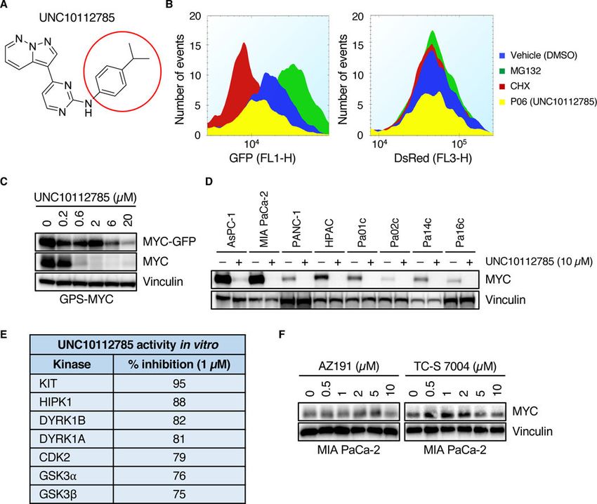
Blake et al.                                                                                           Page 5

                                   compounds compared to maximum stabilization or destabilization of EGFP-MYC with
Author Manuscript

                                   MG132 and CHX, respectively. With the cut-off for a hit set at a 30% stabilization or
                                   destabilization, we identified 30 compounds that stabilized EGFP-MYC (Fig. 2C, Data File
                                   S1) and 26 compounds that destabilized EGFP-MYC (Fig. 2D, Data File S1). Supporting the
                                   validation of the GPS-MYC screen to identify regulators of MYC protein stability, we found
                                   that UNC10112687 (SB-590885-AAD), which inhibits BRAF and CRAF (29), was
                                   identified as a compound that destabilized MYC, whereas a GSK3α/β inhibitor,
                                   UNC10112671 (SB-739452) stabilized MYC (fig. S1, A and B). These results are expected
                                   because RAF activation of ERK phosphorylates MYC at Ser62 and blocks degradation,
                                   whereas GSKβ phosphorylates MYC at Thr58 and promotes degradation (26).

                       UNC10112731 increases levels of endogenous MYC protein
                                   We prioritized the top stabilizing compounds based on published specificity profiles (28, 29,
Author Manuscript

                                   33), novelty of the kinases inhibited, and ease of synthesis, and selected and resynthesized
                                   UNC10112731 (GW694590A) (29) for further validation (fig. S1C). Consistent with its
                                   ability to enhance MYC protein stability, in the initial screen UNC10112731 increased
                                   EGFP-MYC without affecting DsRed (Fig. 3A). In a secondary validation screen, treatment
                                   of GPS-MYC cells with UNC10112731 showed a dose-dependent increase in EGFP-MYC
                                   and endogenous MYC levels when measured by flow cytometry (Fig. 3B) and immunoblot
                                   analyses (Fig. 3C). Next, we sought to determine if UNC10112731 treatment also stabilized
                                   endogenous MYC protein in other PDAC lines, and found that endogenous MYC protein
                                   was increased to varying degrees (2- to 20-fold) in three of eight cell lines analyzed (Fig.
                                   3D). As expected from its identification using the GPS-MYC reporter, this effect was not
                                   due to increased transcription (Fig. 3E). Interestingly, the published kinase targets of
                                   UNC10112731 include the KIT and PDGFRα receptor tyrosine kinases (28) (Fig. 3F and
Author Manuscript

                                   fig. S1C). However, treatment of PDAC cells with the multi-tyrosine kinase inhibitors
                                   imatinib and amuvatinib, both of which have activities against KIT and PDGFRα (34), did
                                   not increase endogenous MYC in a dose-dependent manner (Fig. 3G). Therefore, we
                                   speculated that the mechanism whereby UNC10112731 stabilizes MYC protein may involve
                                   inhibition of additional kinases not identified in the kinase profiling reported for the PKIS
                                   compounds.

                       UNC10112785 causes loss of endogenous MYC protein
                                   Six of the destabilizer hits from the primary screen shared the same 7-azaindole
                                   pharmacophore (fig. S1, D to H), so we synthesized one of those compounds,
                                   UNC10112785 (Fig. 4A), for further analysis. We found that treatment of GPS-MYC cells
                                   with UNC10112785 did not affect DsRed levels (Fig. 4B) but did decrease the levels of
Author Manuscript

                                   EGFP-MYC and endogenous MYC (Fig. 4C). Analyses in additional KRAS-mutant PDAC
                                   cell lines showed loss of endogenous MYC in all eight cell lines evaluated (Fig. 4D).
                                   Examination of the published specificities of five of the 7-azaindole compounds revealed
                                   that the shared kinase targets included KIT and DYRK1A/1B (Fig. 4E, and fig. S1, D to G).
                                   However, we found that commercially available inhibitors of either KIT (imatinib and
                                   amuvatinib) or DYRK1A/1B (AZ191 and TC-S 7004) failed to cause a dose-dependent loss
                                   of endogenous MYC protein (Fig. 3G, 4F), suggesting that, like UNC10112731,

                                       Sci Signal. Author manuscript; available in PMC 2019 September 05.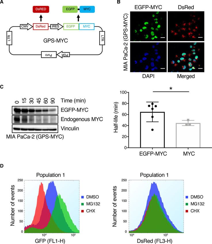
Blake et al. Page 6
UNC10112785 may also have additional kinase targets that regulate MYC protein
Author Manuscript
expression.
In 2017, a quantitative, mass spectrometry-based chemical proteomic assay was used to
profile the specificity and potency of 243 clinical kinase inhibitors (35). The sensitive
technology of this assay, which evaluates full length kinases expressed in a cellular
environment, enables detection of more kinases (~300) than the kinome coverage of typical
in vitro kinase profiling panels. Moreover, it has been shown to be more relevant to inhibitor
potency and selectivity assessment than traditional kinase profiling using recombinant
proteins. The profiling of PKIS compounds was performed in vitro at only a few
concentrations against an incomplete panel of recombinant protein kinases: 196 for PKIS1
and 232 for PKIS2 (28, 29). We reasoned that the kinase targets of UNC10112785 involved
in regulation of MYC stability may not have been included in the original profiling of PKIS
compounds. Therefore, to further evaluate the kinase selectivity profile of UNC10112785,
Author Manuscript
we applied the Multiplex Kinase Inhibitor Bead (MIB)-Mass Spectrometry (MS) chemical
proteomic assay (36) in MIA PaCa-2 cells (fig. S2A). This assay is based on kinase inhibitor
competition assays to assess kinome-wide inhibitor specificity, similar to that described
previously (35, 37).
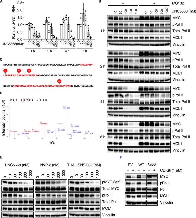
MIB-MS in MIA-PaCa2 cells revealed that UNC10112785 appeared to inhibit CDK8 and
the closely related paralog CDK19 (80% identity), and to a lesser degree CDK9 (Fig. 5A).
These kinases were not included in the panel evaluated previously with PKIS compounds
(28, 29). To validate these activities, we chose CDK8 and CDK19, as well as additional
kinases identified by PKIS (Fig. 4E) or MIB-MS (Fig. 5A) for biochemical analyses using
recombinant kinases (Table 1). We found that the kinases most potently inhibited in vitro by
UNC10112785 were CDK8, CDK19 and CDK9, with IC50s of 1.05, 2.67 and 19.9 nM,
Author Manuscript
respectively, and ~10-fold selectivity over other kinases analyzed.
Consistent with our MIB-MS and biochemical analyses, we found that treatment of MIA
PaCa-2 cells with UNC10112785 led to a reduction in phosphorylation of STAT1 at S727
(pSTAT1), a marker of CDK8/19 activity (Fig. 5B) (38). However, pSTAT1 was maximally
reduced by 100 nM, whereas MYC loss was not observed until 1 μM, suggesting that
CDK8/19 inhibition may not be the basis for MYC loss. To address this possibility, we
treated MIA PaCa-2 cells with Senexin B, a structurally distinct potent and selective
CDK8/19 inhibitor (39), which resulted in a reduction in pSTAT1 but not MYC protein
levels (Fig. 5C). Senexin B is not a CDK9 inhibitor, as indicated by the absence of a
reduction in MCL-1, a well-validated transcriptional target of CDK9 (40). Thus, we
conclude that, although UNC10112785 is a potent inhibitor of CDK8/19 in vitro and in
Author Manuscript
cells, MYC loss was not caused by inhibition of CDK8/19.
Inhibition of CDK9 drives MYC loss caused by UNC10112785
Since UNC10112785 inhibited CDK9 in the MIB-MS analyses, and this was verified by
biochemical analyses, we next addressed the possibility that inhibition of CDK9 was
responsible for UNC10112785-driven loss of MYC. First, we evaluated the activity of a
structurally distinct CDK9-selective inhibitor, NVP2 (41). NVP2 treatment reduced
phosphorylation of RNA polymerase II (pPol II) at S2, another marker of CDK9 activity
Sci Signal. Author manuscript; available in PMC 2019 September 05.Blake et al. Page 7
(40), as well as protein levels of MCL-1 and MYC. However, NVP2 did not reduce pSTAT1,
Author Manuscript
indicating that it inhibited CDK9 but not CDK8/19 (Fig. 5C).
We then synthesized 10 structurally related analogues of UNC10112785 to determine the
relative contributions of their CDK8/19 and CDK9 inhibitory activities to driving MYC loss
(Table S1). Some analogues lost the ability to decrease MYC, whereas others were more
potent at this than the parent compound (Fig. 5D and fig. S2B). Retention of the ability to
suppress pPol II and MCL-1 (CDK9 inhibition) correlated with the loss of MYC, whereas
some analogues retained the ability to inhibit pSTAT1 (CDK8/19 inhibition) yet lost the
ability to cause loss of MYC. These data support the possibility that UNC10112785
inhibition of CDK9 rather than CDK8/19 is the basis for its reduction of MYC protein.
To further explore the relative contribution of CDK8/19 versus CDK9 inhibition to loss of
MYC, we chose two analogues for additional characterization. While both analogues
Author Manuscript
retained low nM activities against CDK8/19 (fig. S2C), the potency of UNC5668 to inhibit
CDK9 in vitro was increased by 7.5-fold compared with parent compound UNC10112785,
whereas that of UNC5577 was decreased by 5.3-fold. MIB-MS inhibitor competition
analyses verified their altered CDK9 activities in MIA PaCa-2 cells (fig. S2D). Consistent
with their relative CDK9 potencies in vitro, we found that UNC5668 exhibited 10-fold
increased potency over UNC10112785 in cells. Treatment of MIA PaCa-2 cells with 100 nM
of UNC5668 suppressed both CDK9 signaling (pPol II and MCL-1) and MYC protein levels
(Fig. 5D). In contrast, treatment with 100 nM UNC5577 suppressed CDK8/19 signaling
(pSTAT1) but not pPol II, MCL-1 or MYC (Fig. 5D). Finally, since we showed previously
that MYC is essential for the growth of PDAC cell lines (14), we next evaluated the ability
of these analogues to suppress cell proliferation. Consistent with their ability to reduce MYC
levels, all compounds with CDK9 but not CDK8/19 inhibitory activity reduced proliferation
Author Manuscript
of MIA PaCa-2 cells on plastic (Fig. 5E) and colony formation in soft agar (Fig. 5F), at
concentrations where potent inhibition of CDK9 signaling was observed. Taken together,
these data indicate that inhibition of CDK9 rather than CDK8/19 is responsible for
UNC10112785-driven MYC loss.
CDK9 promotes phosphorylation of MYC on Ser62
CDK9 can regulate MYC transcription by direct phosphorylation of Pol II (40). Consistent
with this, we found that treatment of PDAC cells with UNC5668 decreased MYC mRNA
(Fig. 6A) as well as MYC protein levels (Fig. 6B). Consistent with our identification of a
CDK9 inhibitor as a MYC destabilizer in the GPS-MYC assay, concurrent inhibition of the
proteasome using MG132 was able to partially rescue the loss of MYC protein (Fig. 6B,
upper panel). Thus, CDK9 regulates both MYC transcription and MYC protein stability,
Author Manuscript
Given these two effects of CDK9 on MYC levels, we then speculated that inhibiting the
proteasome would be able to rescue MYC protein levels only in the short term, because
suppression of transcription would eventually result in suppression of MYC protein
regardless of proteasomal activity. Consistent with this possibility, we found that MG132
clearly rescued MYC protein levels at early time points (1 and 2 hours) but not at later time
points (4 and 6 hours, Fig. 6B). Thus, inhibition of CDK9 both suppresses MYC
transcription and promotes MYC protein degradation.
Sci Signal. Author manuscript; available in PMC 2019 September 05.Blake et al. Page 8
Scansite analysis (42) of the MYC protein sequence revealed several putative serine-proline
Author Manuscript
(SP) CDK9 consensus motifs. Therefore, we reasoned that CDK9 might control MYC
protein stability through direct phosphorylation. To address this possibility, we performed an
in vitro kinase assay coupled with mass spectrometry and found that CDK9 phosphorylates
MYC on several residues in vitro, including Ser62 (Fig. 6, C and D). To determine if CDK9
can also promote MYC phosphorylation at Ser62 in cells, we treated MIA PaCa-2 cells for
30 min with three structurally distinct CDK9 inhibitors. Consistent with the in vitro kinase
assay results, we found that inhibition of CDK9 activity in PDAC cells, including by THAL-
SNS-032 targeted CDK9 degradation (43), decreased MYC phosphorylation at Ser62 (Fig.
6E and fig. S3A) while not affecting total MYC levels. Since Ser62 phosphorylation
attenuates degradation of MYC (26), we conclude that CDK9 can enhance MYC protein
stability at least in part by promoting Ser62 phosphorylation. In support of this possibility,
we found that exogenous expression of the MYC S62A phospho-deficient mutant partially
Author Manuscript
rescued MYC degradation caused by CDK9i (Fig. 6F and fig. S3B). That MYC S62A did
not fully prevent MYC loss caused by CDK9i may indicate that there are additional
phosphorylation sites, possibly including those we identified by mass spectrometry (Fig.
6C), that also contribute to CDK9 regulation of MYC protein stability.
Since we previously identified ERK5 as a regulator of MYC degradation through
phosphorylation at MYC Ser62 phosphorylation (14), we wanted to eliminate the possibility
that CDK9 regulated MYC stability through MEK5-ERK5 signaling. Unlike CDK9i, the
ERK5 inhibitor XMD8–92 (ERK5i) did not reduce phosphorylation of the CDK9 substrate
Pol II, and conversely CDK9i did not reduce phosphorylation of the MEK5 substrate ERK5
(Fig. 7A). Furthermore, MIB-MS analysis of UNC5668 did not demonstrate a decrease in
MEK5/MAP2K5 activity (Fig. 5A). Thus, we conclude that CDK9 and MEK5-ERK5
comprise distinct signaling networks that each regulate MYC protein phosphorylation and
Author Manuscript
stability.
Finally, we previously determined that MEK5 activity comprised a compensatory activity
caused by KRAS suppression (14). We therefore determined if CDK9 activity is also linked
with mutant KRAS activity. Whereas acute KRAS suppression in KRAS-mutant MIA
PaCa-2 cells caused an increase in MEK5 activity (14), no increase in CDK9 activity was
observed (Fig. 7B). Additionally, CDK9i treatment of HPNE immortalized human
pancreatic ductal cells, that are KRAS wild-type, also caused a reduction in MYC (Fig. 7C).
We conclude that, unlike MEK5, CDK9 activity is not regulated by mutant KRAS activity.
DISCUSSION
Although RAS and MYC are well-validated cancer drivers implicated in the growth of a
Author Manuscript
diverse spectrum of cancers, both have largely been considered ‘undruggable’ cancer targets
(17). Renewed efforts have identified promising directions for inhibiting RAS (6), but the
development of selective anti-MYC therapies remains a significant challenge (44, 45). One
promising but relatively underexplored strategy to target MYC involves disruption of the
signaling mechanisms that promote MYC protein stability (26). For example, we previously
found that mutant KRAS prevents MYC degradation in PDAC through both ERK-dependent
and -independent signaling mechanisms (14). These findings prompted us to identify
Sci Signal. Author manuscript; available in PMC 2019 September 05.Blake et al. Page 9
additional protein kinase-dependent mechanisms that regulate MYC protein stability in
Author Manuscript
KRAS-mutant PDAC. Targeting of these kinases to promote MYC degradation may then
serve as a novel, indirect therapeutic strategy in KRAS-driven cancers. In this study, we
developed a MYC degradation screen in the KRAS-mutant PDAC cell line MIA PaCa-2 and
identified kinase inhibitors that either increased or decreased MYC protein levels. We then
used these compounds as tools to identify new protein kinase-dependent mechanisms that
regulate MYC protein stability. We identified CDK9 as a novel regulator of MYC protein
stability through enhanced phosphorylation at Ser62, a modification that prevents MYC
degradation (26). We propose that our screening approach can be adapted to investigate the
mechanisms that regulate the degradation of other clinically important proteins.
Our GPS-MYC screen identified multiple kinase inhibitors that either stabilize or destabilize
MYC protein. However, the dynamic range of the assay as currently constituted is better
suited for identifying stabilizing compounds than destabilizing compounds. Thus, this assay
Author Manuscript
could more easily be adapted to identify novel compounds that stabilize important proteins
such as tumor suppressors. Although stabilizing compounds would not be useful for
therapeutically targeting oncogenic proteins such as MYC, they are nonetheless valuable
tools for understanding signaling mechanisms that regulate MYC activity in PDAC and
other cancers.
While our GPS-MYC screen illustrates the potential power of this strategy to identify novel
regulators of MYC protein stability, it also reveals some challenges associated with using the
PKIS library of protein kinase inhibitors. For example, a number of the strongest stabilizers
that we identified targeted multiple kinases, making delineation of the relevant kinase(s) a
daunting task. Similarly, in our characterization of UNC10112731 as a compound that
stabilized MYC protein, we were not able to ascribe this activity to the published targets of
Author Manuscript
this compound. Instead, we suspect that additional kinase(s) may be responsible for
stabilizing MYC. We faced a similar challenge in our identification of the protein kinase(s)
that account for the ability of UNC10112785 to cause loss of MYC protein. Initially, we
focused on a set of MYC-destabilizing compounds that shared the same core chemotype,
with the rationale that they targeted the same kinase. Analysis of the published PKIS data
available for their specificities led us to investigate the role of the DYRK1A/1B family of
kinases. After failing to validate DYRK1A/1B upon using additional commercially available
DYRK1A/1B inhibitors, we then speculated that UNC10112785 had activities not identified
previously. To address this possibility, we applied MIB-MS analyses that identified potent
activity against CDK8 and the related paralog CDK19, and against CDK9, all of which are
kinases that were not included in the original PKIS biochemical profiling dataset. We then
performed in vitro assays with recombinant kinases and validated that UNC10112785
Author Manuscript
exhibited potent inhibition of CDK8/19 and CDK9 in vitro. Further evaluation of this
compound in PDAC lines demonstrated its ability to inhibit CDK8/19 and CDK9. As
demonstrated in another study (35), we suggest that cell-based proteomic profiling of protein
kinase inhibitor specificity is an effective approach to provide a more comprehensive
determination of the cellular targets of protein kinase inhibitors. Based on our studies, we
recommend that researchers using the PKIS set for screening should not rely solely on the
published specificity dataset and instead should also perform further independent profiling
of inhibitor specificity in their studies. Ongoing profiling of the PKIS libraries using wide-
Sci Signal. Author manuscript; available in PMC 2019 September 05.Blake et al. Page 10
spectrum kinase profiling in live cells (46) will also help to address this concern for future
Author Manuscript
researchers.
We were initially surprised that we identified CDK9 as a regulator of MYC protein stability,
since to date there has been no evidence of such regulation. Instead, CDK9 is known to be
part of the P-TEFb complex that is recruited to the MYC promoter by BRD4, where CDK9
then phosphorylates Pol II to stimulate transcriptional elongation and MYC expression (47).
Since MYC expression from our GPS-MYC reporter is controlled from a heterologous
CMV promoter that is not subject to the same mechanisms that control endogenous MYC
transcription, our reporter is able to reveal nontranscriptional mechanisms that regulate
MYC protein levels. Collectively, our data are in agreement with CDK9 regulation of MYC
transcription, and additionally suggest that CDK9 may directly phosphorylate MYC on
Ser62 to block MYC protein degradation. Ser62 can also be phosphorylated upon KRAS
activation of ERK1/2, and we previously identified ERK5 as another kinase that can
Author Manuscript
phosphorylate MYC Ser62 (14). Thus, the roster of kinases that can promote MYC protein
stability through the Thr58/Ser62 phosphodegron continues to expand.
There has been a renewed interest in developing selective CDK9 inhibitors for cancer
treatment (48–51), due in large part to their inhibition of MCL-1 and MYC transcription. We
now propose that destabilization of MYC protein is a third anti-tumor activity of CDK9
inhibitors. This supports their clinical evaluation in MYC-dependent cancers, particularly
where MYC expression is deregulated, such as in KRAS-mutant PDAC. In conclusion, our
study presents the development and application of a MYC protein degradation screen which
we used to identify novel regulators of MYC protein stability. We propose that this screen
can be readily adapted to study the stability of many other important proteins.
Author Manuscript
MATERIALS AND METHODS
Cell culture
Human pancreatic cancer cell lines were obtained from ATCC (MIA PaCa-2, PANC-1,
HPAC, AsPC-1) or were a gift from Dr. Jason Fleming at MD Anderson Cancer Center
(Pa01c, Pa02c, Pa14c, Pa16c). Cells were maintained in DMEM (GPS-MYC, MIA PaCa-2,
PANC-1, HPAC, Pa01c, Pa02c, Pa14c, Pa16c) or RPMI-1640 (AsPC-1) supplemented with
10% fetal bovine serum (FBS) and antibiotics (Pen Strep; Sigma 15070063). STR (short
tandem repeat) analyses were performed to verify cell line identity, and all cell lines tested
negative for mycoplasma contamination.
Antibodies and reagents
Author Manuscript
The following antibodies were used, from Cell Signaling Technology: anti-MYC (5605),
anti-Pol II (14958), anti-pSTAT1Ser727 (8826), anti-STAT1 (14995), anti-pERK (4370),
anti-ERK (9201), anti-CDK9 (2316); from AbCam: anti-pMYC Ser62 (ab106952), anti-pPol
II Ser2 (ab5095); from Sigma-Aldrich: anti-KRAS (WH0003845M1), anti-vinculin (V9131)
from Santa Cruz Biotechnology: anti-MCL1 (sc-12756). The protein kinase inhibitors
NVP-2 (HY-12214A), amuvatinib (HY-10206) and imatinib (HY-15463) were obtained from
MedChemExpress, TC-S 7004 (5088) from Tocris Bioscience, and AZ 191 (5232) from
Sci Signal. Author manuscript; available in PMC 2019 September 05.Blake et al. Page 11
Sigma-Aldrich. MG132 (M7449) and cycloheximide (C7698) were also obtained from
Author Manuscript
Sigma-Aldrich. THAL-SNS-032 was a gift from Dr. Nathanael Gray (Harvard/DFCI).
Synthetic schemes for UNC10112731 and UNC10112785 and derivatives are shown in
Supplemental Methods.
Expression vectors
To generate the GPS-MYC plasmid, the human MYC DNA was amplified by PCR using the
following Gateway cloning primers:
attB1:5’-
GGGGACAAGTTTGTACAAAAAAGCAGGCTTAGAAGGAGATAGAACCATGCCCCT
CAACGTTAGCTTCAC-3’
attB2:5’-
Author Manuscript
GGGGACCACTTTGTACAAGAAAGCTGGGTCCTACGCACAAGAGTTCCGTAGC-3’
The resultant PCR product was cloned into pDONR223 (Addgene) with the BP reaction and
subsequently cloned into the pGPS-LP vector with the LR reaction.
MIA PaCa-2 GPS-MYC Screen
Lentivirus particles were generated by transfecting HEK293T cells with GPS-MYC and the
packaging plasmids pMD2.G and pSPAX2 (Addgene) using Fugene 6 transfection reagent
(Promega). MIA PaCa-2 cells were infected with lentivirus and selected with 2 μg/ml
puromycin to establish mass populations of cells stably expressing EGFP-MYC (designated
GPS-MYC cells). To minimize variability during the screening and validation processes,
large amounts of GPS-MYC cultures were expanded and then frozen at a concentration of
106 cells/ml in Recovery Cell Culture Freezing Medium (Gibco). Cells were thawed and
Author Manuscript
allowed to grow for two days prior to the screen, and then were plated at 20,000 cells/well
into a clear-bottom, white 384-well plate (Corning 3707) using a Multidrop Combi Reagent
Dispenser (Thermo Fisher). The following day, growth medium was removed from the assay
plate and replaced with inhibitor-containing growth medium using a Multimek Automated
Liquid Handler (NanoScreen).
To prepare the assay plates, 1 μl DMSO was added to columns 1 and 2, 1 μl of 375 μg/ml
cycloheximide (final concentration 7.5 μg/ml) was added to column 23, and 1 μl of 187.5
μM MG132 (final concentration 3.75 μM) was added to column 24. Columns 2–22
contained PKIS (Published Kinase Inhibitor Sets) compounds. Fifty μl DMEM
supplemented with 2% FBS was added per well, and 20 μl of the resultant PKIS compound-
containing media were added to each plate using a Multimek Automated Liquid Handler
Author Manuscript
(NanoScreen).
After 6 hours the plates were washed with 75 μl PBS per well, 10 μl Accumax Cell
Dissociation Solution (Innovative Cell Technologies) was added per well, and the plates
were centrifuged briefly (5 s at 500 rpm). After 20 min incubation at 37ºC, 10 μl DMEM
supplemented with 2% FBS was added to each well to quench the dissociation reaction. All
liquids were added with a Multidrop Combi Reagent Dispenser (Thermo Fisher) at slow
speed. The plates were then analyzed on an iQue Screener PLUS (Intellicyt) using medium
Sci Signal. Author manuscript; available in PMC 2019 September 05.Blake et al. Page 12
sip speed, 4 s sip time, shaking initially at 2000 rpm for 30 s and then at 2000 rpm for 10 s
Author Manuscript
after every 12 wells. The data were imported into the CICBDD database (ScreenAble
Solutions) and each compound was normalized to control wells. The normalized data were
then imported into TIBCO Spotfire for visualization and analysis. Some analyses were also
performed in ForeCyt (Intellicyt).
Immunoblotting
Cells were lysed in 1% Triton buffer (25 mM TRIS, pH 7.4, 100 mM NaCl, 10% glycerol, 1
mM EDTA, 1% Triton-X 100), supplemented with protease (Roche) and phosphatase
inhibitors (Sigma) for 10 min, with brief vortexing. Lysates were clarified by centrifugation
at 18,000 x g for 15 min at 4ºC, and protein concentrations were measured using the
Bradford reagent. Standard immunoblotting procedures were performed as we described
previously (14).
Author Manuscript
Confocal microscopy
Cells were plated on MatTek dishes (MatTek Corporation) and fixed for 5 min in ice-cold
paraformaledehye (4%), washed 3X with cold PBS, stained with DAPI (0.1 μg/ml) for 10
min, and washed 3X with cold PBS. Images were collected on an Olympus FV1000
microscope using a 40X objective.
Gene silencing
All siRNA experiments were performed using 10 nM siRNA and Lipofectamine RNAiMAX
(Life Technologies) according to the manufacturer’s protocol.
Quantitative real-time PCR
Author Manuscript
mRNA was isolated using an RNeasy RNA isolation kit (Qiagen) and reverse transcription
was performed using a High-Capacity cDNA Reverse Transcription Kit (Applied
Biosystems). Samples were run on a QuantStudio 6 Flex (Applied Biosystems) and analyzed
using the ddCT method.
Growth assays
To measure anchorage-dependent growth, cells were plated at low density in 96 well plates
and allowed to attach overnight. Inhibitors were then added with a D300e Digital Dispenser
(Tecan). After 72 hours, cells were labeled with calcein AM (Thermo Fisher) according to
the manufacturer’s protocol, and counted on a SpectraMax MiniMax 300 Imaging
Cytometer (Molecular Devices).
Author Manuscript
To measure anchorage-independent growth, soft agar colony formation assays were
performed. In a 96-well plate, wells were coated with 50 μl 2% SeaPrep Agarose (Lonza)
mixed with cell culture medium. Next, 100 μl of 5000 cells mixed with 1% SeaPrep Agarose
were added to each well and allowed to solidify. Fifty μl of medium were added to each
well, and drugs were dispensed with a D300e Digital Dispenser (Tecan). After 72 hours, the
alamarBlue cell viability reagent (Thermo Fisher) was added and incubated for 3 hours.
Fluorescence was quantified on a SpectraMax MiniMax 300 Imaging Cytometer (Molecular
Devices).
Sci Signal. Author manuscript; available in PMC 2019 September 05.Blake et al. Page 13
Multiplexed Inhibitor Beads – Mass Spectrometry (MIB-MS)
Author Manuscript
Lysates were prepared and MIB-MS was performed as described previously (36). Briefly,
cells were harvested on ice in lysis buffer [50 mM HEPES (pH 7.5), 0.5% Triton X-100, 150
mM NaCl, 1 mM EDTA, 1 mM EGTA, 10 mM sodium fluoride, 2.5 mM sodium
orthovanadate, 1X protease inhibitor cocktail (Roche), 1% phosphatase inhibitor cocktail 2
(Sigma-Aldrich), and 1% of phosphatase inhibitor cocktail 3 (Sigma-Aldrich)]. Lysates were
sonicated for 3×10 sec on ice and centrifuged at 10,000 x g for 10 min at 4°C. Supernatant
was collected and syringe-filtered through a 0.2 μm SFCA membrane (Corning).
Approximately 5 mg of lysate was brought to 1 M NaCl and passed through a column of
multiplexed inhibitor-conjugated beads (MIBs) consisting of Sepharose-conjugated
UNC2147A, CTx-0294885, UNC8088A, purvalanol B, PP58 and VI16832. The kinase-
bound MIBs were washed with 5 ml of high-salt buffer and 5 ml of low-salt buffer [50 mM
HEPES (pH 7.5), 0.5% Triton X-100, 1 mM EDTA, 1 mM EGTA, and 10 mM sodium
Author Manuscript
fluoride, and 1 M NaCl or 150 mM NaCl, respectively]. The columns were washed a final
time with 1 ml 0.1% SDS wash buffer [50 mM HEPES (pH 7.5), 0.5% Triton X-100, 1 mM
EDTA, 1 mM EGTA, 10 mM sodium fluoride, 150 mM NaCl, 0.1% SDS] then eluted with 1
ml of elution buffer [0.5% SDS, 1% 2 mercaptoethanol, and 0.1 M Tris (pH 6.8)] (100°C, 10
min). Eluted kinases were reduced (dithiothreitol) and alkylated (iodoacetamide) then
concentrated with Amicon 10k Ultra centrifugal filters (Millipore). Detergent was removed
from the eluate by chloroform/methanol precipitation. Protein pellets were resuspended in
50 mM HEPES (pH 8.0) and digested overnight at 37ºC with sequencing grade modified
trypsin (Promega). Digested peptides were desalted using PepClean C18 spin columns
(Thermo Scientific) and filtered with centrifugal filter columns (Nest). Peptides were
extracted and dried by vacuum centrifugation. All peptide samples were stored at −80˚C
until further analysis.
Author Manuscript
LC-MS/MS analysis was performed as described below. Raw data files were processed
using MaxQuant version 1.5.3.17 and searched against the reviewed Uniport human
database (containing 20,203 entries), using Andromeda within MaxQuant. Enzyme
specificity was set to trypsin, up to two missed cleavage sites were allowed,
carbamidomethylation of Cys was set as a fixed modification and oxidation of Met was set
as a variable modification. A 1% FDR was used to filter all data. Match between runs was
enabled (2 min match time window, 20 min alignment window), and a minimum of two
peptides was required for label-free quantitation using the LFQ intensities. Further analyses
were performed in Perseus version 1.6.0.2 and R.
In vitro MYC phosphorylation
Author Manuscript
Recombinant MYC (Abcam; ab169901) at 1000 nM was incubated with 50 nM recombinant
CDK9 together with cyclin T1 required for kinase activity (Thermo Fisher; PV4131) and
100 μM ATP for 30 min at 30°C. The reaction was stopped by adding SDS sample buffer
and boiling for 5 min at 95°C. After separation by SDS-PAGE, the gel was stained for 30
min with SimplyBlue Coomassie (Thermo Fisher) and destained overnight in water. Protein
bands corresponding to MYC were excised and the proteins were reduced, alkylated, and in-
gel digested with trypsin overnight at 37˚C. Peptides were extracted and dried by vacuum
centrifugation. All peptide samples were stored at −80˚C until further analysis.
Sci Signal. Author manuscript; available in PMC 2019 September 05.Blake et al. Page 14
LC-MS/MS analysis was performed as described below. Raw data files were processed
Author Manuscript
using Proteome Discoverer version 2.1 (Thermo Scientific). Peak lists were searched against
a reviewed Uniprot human database, appended with a common contaminants database, using
Sequest. The following parameters were used to identify tryptic peptides for protein
identification: 10 ppm precursor ion mass tolerance; 0.02 Da product ion mass tolerance; up
to two missed trypsin cleavage sites. Carbamidomethylation of Cys was set as a fixed
modification and oxidation of Met and phosphorylation of Ser, Thr and Tyr were set as
variable modifications. The phosphoRS node was used to localize the sites of
phosphorylation. Peptide false discovery rates (FDR) were calculated by the Percolator node
using a decoy database search and data were filtered using a 1% FDR cutoff.
LC-MS/MS analysis
Samples were analyzed by LC/MS/MS using an Easy nLC 1000 coupled to a QExactive HF
Author Manuscript
mass spectrometer (Thermo Scientific). Samples were injected onto an Easy Spray PepMap
C18 column (75 μm id × 25 cm, 2 μm particle size; Thermo Scientific) and separated over a
45 min or 2 hour method. The gradient for separation consisted of 5–32% mobile phase B at
a 250 nl/min flow rate, where mobile phase A was 0.1% formic acid in water and mobile
phase B consisted of 0.1% formic acid in ACN. The QExactive HF was operated in data-
dependent mode where the 15 most intense precursors were selected for subsequent
fragmentation. Resolution for the precursor scan (m/z 400–1600) was set to 120,000 with a
target value of 3 × 10⁶ ions. MS/MS scans resolution was set to 15,000 with a target value of
2 × 10⁴ ions. The normalized collision energy was set to 27% for HCD. Peptide match was
set to preferred, and precursors with unknown charge or a charge state of 1 and ≥ 8 were
excluded.
Author Manuscript
Supplementary Material
Refer to Web version on PubMed Central for supplementary material.
Acknowledgments:
We thank Dr. Nathanael Gray for THAL-SNS-032. We also thank the UNC Flow Cytometry Core Facility and Dr.
Nancy Cheng of the UNC CICBDD for help in optimizing the GPS-MYC assay. We thank Dr. Naim Rashid for
ensuring proper statistical analyses were performed. Research reported in this publication was supported in part by
the North Carolina Biotech Cancer Institutional Support Grant 2015-IDG-1001.
Funding: The UNC Flow Cytometry Core Facility and the Michael Hooker Proteomics Center are supported in
part by P30 CA016086 Cancer Center Core Support Grant to the UNC Lineberger Comprehensive Cancer Center.
K.H.P. was supported by the UNC University Cancer Research Fund. D.R.B. was supported by NCI fellowships
(T32CA071341 and F31CA216965). A.V.V. was supported by fellowships from the NCI (T32CA009156) and
American Cancer Society (128014-PF-15–059-01-TBE). R.G.H. was supported by a grant from Debbie’s Dream
Author Manuscript
Foundation. This work was supported by grants (C.J.D. and A.D.C.) from the NIH/NCI (CA42978, CA179193,
CA175747, CA199235) and the Pancreatic Cancer Action Network-AACR. C.J.D. was also supported by a grant
from the DoD (W81XWH-15–1-0611) and from the Lustgarten Pancreatic Cancer Foundation (388222). Work
carried out in the CICBDD was also supported by the University Cancer Research Fund.
REFERENCES
1. Siegel RL, Miller KD, Jemal A, Cancer statistics, 2018. CA Cancer J Clin 68, 7–30 (2018).
[PubMed: 29313949]
Sci Signal. Author manuscript; available in PMC 2019 September 05.Blake et al. Page 15
2. Rahib L et al., Projecting cancer incidence and deaths to 2030: the unexpected burden of thyroid,
liver, and pancreas cancers in the United States. Cancer Res 74, 2913–2921 (2014). [PubMed:
Author Manuscript
24840647]
3. Waters AM, Der CJ, KRAS: the critical driver and therapeutic target for pancreatic cancer. Cold
Spring Harb Perspect Med 8, (2018).
4. Ryan DP, Hong TS, Bardeesy N, Pancreatic adenocarcinoma. N Engl J Med 371, 2140–2141 (2014).
5. Cox AD, Der CJ, Ras history: The saga continues. Small GTPases 1, 2–27 (2010). [PubMed:
21686117]
6. Papke B, Der CJ, Drugging RAS: Know the enemy. Science 355, 1158–1163 (2017). [PubMed:
28302824]
7. Ledford H, Cancer: The Ras renaissance. Nature 520, 278–280 (2015). [PubMed: 25877186]
8. Land H, Parada LF, Weinberg RA, Tumorigenic conversion of primary embryo fibroblasts requires
at least two cooperating oncogenes. Nature 304, 596–602 (1983). [PubMed: 6308472]
9. Dang CV, MYC on the path to cancer. Cell 149, 22–35 (2012). [PubMed: 22464321]
10. Soucek L et al., Modelling Myc inhibition as a cancer therapy. Nature 455, 679–683 (2008).
[PubMed: 18716624]
Author Manuscript
11. Soucek L et al., Inhibition of Myc family proteins eradicates KRas-driven lung cancer in mice.
Genes Dev 27, 504–513 (2013). [PubMed: 23475959]
12. Mazur PK et al., Notch2 is required for progression of pancreatic intraepithelial neoplasia and
development of pancreatic ductal adenocarcinoma. Proc Natl Acad Sci U S A 107, 13438–13443
(2010). [PubMed: 20624967]
13. Saborowski M et al., A modular and flexible ESC-based mouse model of pancreatic cancer. Genes
Dev 28, 85–97 (2014). [PubMed: 24395249]
14. Vaseva AV et al., KRAS Suppression-Induced Degradation of MYC Is Antagonized by a MEK5-
ERK5 Compensatory Mechanism. Cancer Cell 34, 807–822 e807 (2018). [PubMed: 30423298]
15. Walz S et al., Activation and repression by oncogenic MYC shape tumour-specific gene expression
profiles. Nature 511, 483–487 (2014). [PubMed: 25043018]
16. Lin WC et al., Dormant cancer cells contribute to residual disease in a model of reversible
pancreatic cancer. Cancer Res 73, 1821–1830 (2013). [PubMed: 23467612]
17. Dang CV, Reddy EP, Shokat KM, Soucek L, Drugging the ‘undruggable’ cancer targets. Nat Rev
Author Manuscript
Cancer 17, 502–508 (2017). [PubMed: 28643779]
18. Filippakopoulos P et al., Selective inhibition of BET bromodomains. Nature 468, 1067–1073
(2010). [PubMed: 20871596]
19. Mazur PK et al., Combined inhibition of BET family proteins and histone deacetylases as a
potential epigenetics-based therapy for pancreatic ductal adenocarcinoma. Nat Med 21, 1163–1171
(2015). [PubMed: 26390243]
20. Berg T et al., Small-molecule antagonists of Myc/Max dimerization inhibit Myc-induced
transformation of chicken embryo fibroblasts. Proc Natl Acad Sci U S A 99, 3830–3835 (2002).
[PubMed: 11891322]
21. Stellas D et al., Therapeutic effects of an anti-Myc drug on mouse pancreatic cancer. J Natl Cancer
Inst 106, (2014).
22. Kota J et al., Therapeutic microRNA delivery suppresses tumorigenesis in a murine liver cancer
model. Cell 137, 1005–1017 (2009). [PubMed: 19524505]
23. Gouw AM, Toal GG, Felsher DW, Metabolic vulnerabilities of MYC-induced cancer. Oncotarget
Author Manuscript
7, 29879–29880 (2016). [PubMed: 26863454]
24. Andrieu G, Belkina AC, Denis GV, Clinical trials for BET inhibitors run ahead of the science.
Drug Discov Today Technol 19, 45–50 (2016). [PubMed: 27769357]
25. Hayes TK et al., Long-Term ERK Inhibition in KRAS-mutant pancreatic cancer is associated with
MYC degradation and senescence-like growth suppression. Cancer Cell 29, 75–89 (2016).
[PubMed: 26725216]
26. Farrell AS, Sears RC, MYC degradation. Cold Spring Harb Perspect Med 4, (2014).
27. Farrell AS et al., Targeting inhibitors of the tumor suppressor PP2A for the treatment of pancreatic
cancer. Mol Cancer Res 12, 924–939 (2014). [PubMed: 24667985]
Sci Signal. Author manuscript; available in PMC 2019 September 05.Blake et al. Page 16
28. Drewry DH et al., Progress towards a public chemogenomic set for protein kinases and a call for
contributions. PLoS One 12, e0181585 (2017).
Author Manuscript
29. Elkins JM et al., Comprehensive characterization of the Published Kinase Inhibitor Set. Nat
Biotechnol 34, 95–103 (2016). [PubMed: 26501955]
30. Emanuele MJ et al., Global identification of modular cullin-RING ligase substrates. Cell 147, 459–
474 (2011). [PubMed: 21963094]
31. Yen HC, Elledge SJ, Identification of SCF ubiquitin ligase substrates by global protein stability
profiling. Science 322, 923–929 (2008). [PubMed: 18988848]
32. Vaseva AV et al., KRAS suppression-induced degradation of MYC is antagonized by a MEK5-
ERK5 compensatory mechanism. Cancer Cell in press, (2018).
33. Drewry DH, Willson TM, Zuercher WJ, Seeding collaborations to advance kinase science with the
GSK Published Kinase Inhibitor Set (PKIS). Curr Top Med Chem 14, 340–342 (2014). [PubMed:
24283969]
34. Tibes R et al., A phase I, first-in-human dose-escalation study of amuvatinib, a multi-targeted
tyrosine kinase inhibitor, in patients with advanced solid tumors. Cancer Chemother Pharmacol 71,
463–471 (2013). [PubMed: 23178951]
Author Manuscript
35. Klaeger S et al., The target landscape of clinical kinase drugs. Science 358, (2017).
36. Duncan JS et al., Dynamic reprogramming of the kinome in response to targeted MEK inhibition
in triple-negative breast cancer. Cell 149, 307–321 (2012). [PubMed: 22500798]
37. Krulikas LJ et al., Application of integrated drug screening/kinome analysis to identify inhibitors
of gemcitabine-resistant pancreatic cancer cell growth. SLAS Discov 23, 850–861 (2018).
[PubMed: 29742358]
38. Rzymski T, Mikula M, Wiklik K, Brzozka K, CDK8 kinase--An emerging target in targeted cancer
therapy. Biochim Biophys Acta 1854, 1617–1629 (2015). [PubMed: 26006748]
39. Porter DC et al., Cyclin-dependent kinase 8 mediates chemotherapy-induced tumor-promoting
paracrine activities. Proc Natl Acad Sci U S A 109, 13799–13804 (2012). [PubMed: 22869755]
40. Sonawane YA et al., Cyclin dependent kinase 9 inhibitors for cancer therapy. J Med Chem 59,
8667–8684 (2016). [PubMed: 27171036]
41. Wang J, Gray NS, SnapShot: kinase inhibitors i. Mol Cell 58, 708 e701 (2015). [PubMed:
26000854]
Author Manuscript
42. Obenauer JC, Cantley LC, Yaffe MB, Scansite 2.0: Proteome-wide prediction of cell signaling
interactions using short sequence motifs. Nucleic Acids Res 31, 3635–3641 (2003). [PubMed:
12824383]
43. Olson CM et al., Pharmacological perturbation of CDK9 using selective CDK9 inhibition or
degradation. Nat Chem Biol 14, 163–170 (2018). [PubMed: 29251720]
44. Whitfield JR, Beaulieu ME, Soucek L, Strategies to inhibit Myc and their clinical applicability.
Front Cell Dev Biol 5, 10 (2017). [PubMed: 28280720]
45. Chen H, Liu H, Qing G, Targeting oncogenic Myc as a strategy for cancer treatment. Signal
Transduct Target Ther 3, 5 (2018). [PubMed: 29527331]
46. Vasta JD et al., Quantitative, wide-spectrum kinase profiling in live cells for assessing the effect of
cellular ATP on target engagement. Cell Chem Biol 25, 206–214 e211 (2018). [PubMed:
29174542]
47. Franco LC, Morales F, Boffo S, Giordano A, CDK9: A key player in cancer and other diseases. J
Cell Biochem 119, 1273–1284 (2018). [PubMed: 28722178]
Author Manuscript
48. Dey J et al., Voruciclib, a clinical stage oral CDK9 inhibitor, represses MCL-1 and sensitizes high-
risk Diffuse Large B-cell Lymphoma to BCL2 inhibition. Sci Rep 7, 18007 (2017). [PubMed:
29269870]
49. Bragelmann J et al., Systematic kinase inhibitor profiling identifies cdk9 as a synthetic lethal target
in NUT midline carcinoma. Cell Rep 20, 2833–2845 (2017). [PubMed: 28930680]
50. Narita T et al., Cyclin-dependent kinase 9 is a novel specific molecular target in adult T-cell
leukemia/lymphoma. Blood 130, 1114–1124 (2017). [PubMed: 28646117]
51. Yin T et al., A novel CDK9 inhibitor shows potent antitumor efficacy in preclinical hematologic
tumor models. Mol Cancer Ther 13, 1442–1456 (2014). [PubMed: 24688048]
Sci Signal. Author manuscript; available in PMC 2019 September 05.Blake et al. Page 17
Author Manuscript
Author Manuscript
Author Manuscript
Fig. 1. Validation of a MYC degradation reporter.
Author Manuscript
(A) Overview of the GPS-MYC vector. (B) Confocal images of GPS-MYC cells to
determine EGFP-MYC subcellular localization to the nucleus, which was visualized by
DAPI staining. Scale bar = 20 μm. (C) GPS-MYC cells were treated with CHX for the
indicated times and EGFP-MYC and MYC levels were measured by immunoblotting (left
panel). The half-lives of EGFP-MYC and endogenous MYC were calculated by fitting the
data to a one-phase decay curve. (D) GPS-MYC cells were treated with MG132 and
Sci Signal. Author manuscript; available in PMC 2019 September 05.You can also read