Pore properties of Orai1 calcium channel dimers and their activation by the STIM1 ER calcium sensor
←
→
Page content transcription
If your browser does not render page correctly, please read the page content below
JBC Papers in Press. Published on June 28, 2018 as Manuscript RA118.003424
       The latest version is at http://www.jbc.org/cgi/doi/10.1074/jbc.RA118.003424

     Pore properties of Orai1 calcium channel dimers and their
            activation by the STIM1 ER calcium sensor
      Xiangyu Cai1, Robert M. Nwokonko1, Natalia A. Loktionova1, Raz Abdulqadir1,
      James H. Baraniak, Jr.1, Youjun Wang2, Mohamed Trebak1, Yandong Zhou1*,
                                  and Donald L. Gill1*

From the 1Department of Cellular and Molecular Physiology, The Pennsylvania State University College
     of Medicine, Hershey, PA 17033; 2Beijing Key Laboratory of Gene Resources and Molecular
    Development College of Life Sciences, Beijing Normal University, Beijing 100875, P.R. China

                                  Running title: Function of Orai1 dimers

* To whom correspondence should be addressed: Donald L. Gill or Yandong Zhou, Department
of Cellular and Molecular Physiology, The Pennsylvania State University College of Medicine,

                                                                                                                Downloaded from http://www.jbc.org/ by guest on October 5, 2018
500 University Drive, Hershey, PA 17033, USA. Tel: 717-531-8567; Fax: 717-531-7667; E-mail:
dongill@psu.edu or zhouyd@psu.edu

Keywords: Calcium, calcium channel, ion channel, cell signaling, Orai1, STIM1, store-operated channel,
channel gating, selectivity filter
_____________________________________________________________________________________

ABSTRACT                                                negative over Orai-WT in heterodimers. Hetero-
                                                        dimers containing the inhibitory K85E mutation
     Store-operated Ca2+ entry signals are mediated     extending outward from the pore-helix, gave an
by plasma membrane Orai channels activated              interesting partial effect on both channel-activation
through intermembrane coupling with Ca2+-sens-          and STIM1-binding, indicating an important allo-
ing STIM proteins in the endoplasmic reticulum.         steric link between the cytosolic Orai1 domains.
The nature of this elaborate Orai gating mechanism      The Orai1 C-terminal STIM1-binding domain mu-
has remained enigmatic. Based on the Drosophila         tation, L273D, powerfully blocked STIM1-
Orai structure, mammalian Orai1 channels are            induced channel activation. The Orai1-L273D/WT
hexamers comprising three dimeric subunit pairs.        heterodimer had drastically impaired STIM1-
We utilized concatenated Orai1 dimers to probe the      induced channel-gating, but, unexpectedly, retain-
function of key domains within the channel pore         ed full STIM1-binding. This reveals the critical
and gating regions. The Orai1-E106Q selectivity         role of Leu-273 in transducing the STIM1 binding
filter mutant, widely considered a dominant pore        signal into the allosteric conformational change
blocker, was surprisingly non-dominant within           that initiates channel gating. Overall, our results
concatenated heterodimers with Orai1-WT. The            provide important new insights into the role of key
Orai1-E106Q/WT heterodimer formed STIM1-                functional domains that mediate STIM1-induced
activated nonselective cation channels with signifi-    gating of the Orai1 channel.
cantly enlarged apparent pore-diameter. Other Glu-      _____________________________________
106 substitutions entirely blocked the function of
heterodimers with Orai1-WT. The hydrophobic                 The ubiquitously expressed Orai channels are
pore-lining mutation, V102C that constitutively         an evolutionarily conserved family of Ca2+ entry
opens channels, was suppressed by Orai1-WT in           channels mediating “store-operated” Ca2+ signals.
the heterodimer. In contrast, the naturally occurring   These signals play a crucial role in regulating key
R91W pore-lining mutation associated with human         cellular responses including gene expression,
immunodeficiency, was a completely dominant-            growth, secretion, and motility (1-4). Orai channels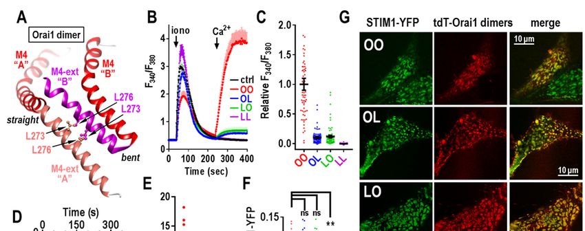
Cai et al                                                                               Function of Orai1 dimers

function in the plasma membrane (PM) but are               symmetry (see Fig. 1A). Unclear is whether these
activated through a dynamic intermembrane coup-            two M4-ext helices dissociate to form separate
ling process between the ER and PM, mediated by            STIM1-binding sites (17,27).
STIM proteins. These ER membrane proteins are
sensors of ER luminal Ca2+ changes and undergo                 Because of the C-terminal interactions between
an intricate unfolding process when Ca2+ stored in         adjacent pairs of M4-ext helices, the Orai channel
the ER becomes depleted (1-3,5-7). The unfolded            comprises three dimers. Our studies therefore
STIM1 protein extends from the ER membrane                 focused on the function of concatenated versions of
surface and becomes trapped in ER-PM junctions             these Orai1 dimers. Using such dimers, we were
by attaching to the PM where it tethers and                able to mutate either one or both of the separate
activates PM Orai1 channels (1,2,5-7). A consider-         Orai1 monomers, allowing us to gain important
able number of immunological, muscular and                 new information on the functional properties of the
inflammatory disease states are related to changes         N-terminal pore-forming helix and the C-terminal
in the operation of STIM proteins and Orai cha-            STIM1-binding domain.
nnels (1,8-10). Hence, our understanding of the
molecular coupling and function of the two pro-            Results and Discussion
teins has major translational significance. Al-
though we have recently determined much on the

                                                                                                                    Downloaded from http://www.jbc.org/ by guest on October 5, 2018
                                                                Orai1 dimers – reliable probes for Orai1
function and organization of STIM and Orai                 structure/function. In a recent study, we compared
proteins (11-14), there is still considerable              the function of concatenated Orai1 channels of
uncertainty about how the proteins associate within        varying length (25). Surprisingly we observed that
junctions and how these interactions mediate               concatemers containing 2 to 6 Orai1 subunits, all
opening of Orai channels and the generation of             give rise to authentic CRAC currents, similar to the
Ca2+ signals (1-4,15,16).                                  expressed Orai1 monomer. Although Orai1 exists
                                                           and functions as a hexamer, we concluded that the
     The highly Ca2+-selective mammalian Orai1             activity of concatemers containing 3-5 Orai1
channel bears close sequence homology with the             subunits, results from hexamers assembling from
Drosophila Orai (dOrai) channel from which the             only the first two N-terminal subunits in the
crystal structure was recently resolved (17). The          concatemer. This results in the formation of a
dOrai channel protein has four transmembrane-              hexameric “trimer of dimers”, with the additional
helices and is organized as a hexameric assembly           C-terminal Orai1 subunits in each concatemer (that
of subunits (see Fig. 1A). Although earlier studies        is, subunits beyond the initial N-terminal dimer)
were interpreted to indicate a tetrameric structure        extending out from the hexamer and not
(18-24), recent evidence indicates the Orai1               contributing to function. The exceptions to this
channel functions as a hexamer (25,26). Based on           anomalous behavior, were concatenated Orai1
the dOrai crystal structure, most of the hexameric         dimers and hexamers. The hexamer appeared to
channel has 6-fold symmetry. The central pore is           largely form hexameric rings. The Orai1 dimers
created from the six pore-forming N-terminal               reliably assembled into hexameric Orai1 channels.
transmembrane helices (M1). Surrounding this are           We therefore constructed a series of Orai1 dimers
the tightly packed M2 and M3 helices, and at the           (Fig. 1B) containing point mutations at function-
periphery of the channel, the C-terminal M4 helices        ally key residues, placed within either one or both
form STIM1-binding sites on the cytoplasmic face           subunits in the dimer (see Fig. 1C). For each con-
of the channel. The six M4 helices each have a             struct, we assessed both the expression level (Fig.
small cytoplasmic extension (M4-ext; 39 amino              S1) and cellular location of expression (Fig. S2). In
acids in Orai1) that folds in two different                all cases, mutations did not alter either the level or
configurations – one is almost straight from the           the PM-localization of Orai1 dimers. We expressed
M4-helix, the other is bent almost 180° back on            each mutant in HEK cells in which endogenous
itself. Each adjacent pair of M4-ext helices closely       Orai1 was eliminated through CRISPR/Cas9 gene
interact with one another in an antiparallel config-       editing, and STIM1-YFP was stably expressed
uration. Thus, the STIM1-binding domain made up            (referred to as HEK-O1koS1+ cells) (25). The func-
of the pair of M4-ext helices has three-fold               tional consequences of hetero- and homodimer

                                                       2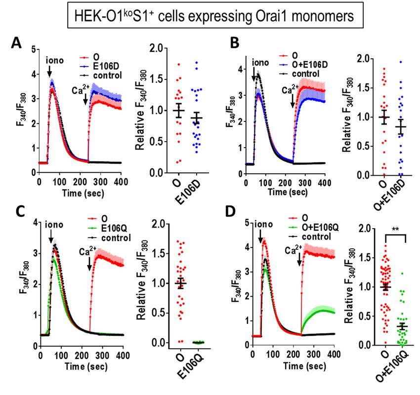
Cai et al                                                                               Function of Orai1 dimers

mutations provided important new mechanistic
information on Orai1 channel operation.                          This is important information since the action
                                                            of E106Q has been widely described as both a
      Dominance of selectivity filter mutations in          pore-dead mutation and one that is a dominant-
Orai1 dimers. The E106 residue is well established          negative over Orai1-WT subunits (1,19,29-32).
to function as the critical selectivity filter in the       Indeed, the dominance of E106Q has been
Orai1 channel (28-30). We initially examined the            described in Orai1 competition studies as providing
expression of Orai1 monomers containing the                 evidence to support a purported tetrameric assemb-
E106D and E106Q mutations, measuring their                  ly of Orai1 channels (19). The dominance of
function either expressed alone or co-expressed             E106Q was also the basis for its conditional
with Orai1-WT (Fig. 2). The Orai1-E106D muta-               transgenic expression in animal studies to assess
tion has a shortened side chain which results in            the role of Orai1 in muscle tissue (33). Based on
altered pore geometry and decrease channel                  our studies here, although E106A and E106Q are
selectivity (28-30). As expected, this mutation is          clearly both pore-inactive mutations, their relative
still able to mediate Ca2+ entry (Fig. 2A) and co-          dominance in blocking Orai1-WT channels is very
expressed with Orai1-WT the mutant has little               different. Clearly, therefore, such dominance stud-
effect (Fig. 2B). In contrast, removing the charge          ies would have been more conclusive using E106A
on the E106 by replacing with Gln (but retaining            instead of E106Q. The descriptions in earlier

                                                                                                                    Downloaded from http://www.jbc.org/ by guest on October 5, 2018
side-chain length) gives a non-conducting pore              papers of the dominance of E106Q over Orai-WT
(Fig. 2C). In this case, co-expression of Orai1-WT          (1,19,29-32) was in each case based on co-
with Orai1-E106Q, clearly causes inhibition of              expression studies in which the relative expression
Ca2+ entry (Fig. 2D). This result was expected since        of the Orai1 subunits could not be controlled. Only
the E106Q mutant has been recognized earlier as a           in concatemer studies can the ratio of expression be
“pore-dead” mutant (19,29-32).                              defined. Indeed, we used similar concatemeric con-
                                                            structs to reveal that E106L, E106V, E106G, and
     In such co-expression studies, the stoichio-           E106K all have complete dominance over Orai1-
metry of subunits in hexamers is of course uncer-           WT (Fig. S3).
tain. Therefore, in further experiments, we com-
pared the function of the E106D and E106Q                        Probing the anomalous functional character-
subunits when incorporated into Orai1 dimer                 istics of E106Q. The unexpected finding that OQ
constructs. Initially we combined Orai1-WT and              and QO heterodimers are functional prompted us to
Orai1-E106D subunits (Fig. 3A-C). The Orai1-                undertake an electrophysiological characterization
E106D homodimer (DD) gave rise to Ca2+ entry                to assess their selectivity and conductance.
that was little different to the Orai1-WT                   Important was to compare their function with
homodimer, consistent with the data in Fig. 2 for           constructs containing E106D since this mutation is
monomer expression. The heterodimers of Orai1-              known to convert the highly Ca2+-selective Orai1
WT and Orai1-E106D (OD, DO) also behaved                    channel into a non-selective cation channel (28-
similarly for Ca2+ entry. Using the Orai1-E106Q             30). As expected, the Orai1-WT homodimer (OO)
homodimer (QQ), there was no detectable Ca2+                gave typical CRAC current with full inward
entry (Fig. 3D), again consistent with the monomer          rectification and a reversal potential slightly above
in Fig. 2C. However, the heterodimers of Orai1-             50 mV (Fig. 4A,B). In contrast, the Orai1-E106D
WT and Orai1-E106Q (OQ, QO) unexpectedly                    homodimer (DD) gave rise to a non-selective cur-
revealed strong Ca2+ entry, similar to that mediated        rent with a strong outward component and much
by Orai1-WT (Fig. 3E,F). This result is in stark            reduced reversal potential (approximately 5 mV;
contrast to heterodimers comprising Orai1-WT                Fig. 4B). The reduction of a single methylene
together with the Orai1-E106A mutant. For this              group by replacing the wildtype Glu-106 residue
mutant, the homodimer (AA) is clearly pore-dead             with Asp, is reported to cause widening of the pore
(Fig. 3G), just like the Orai1-E106Q mutant (QQ).           sufficient to make it cation nonselective (34).
However, the heterodimers of Orai1-WT and                   Surprisingly, the presence of just a single aspartate
Orai1-E106A (OA, AO) were also almost without               in the E106D heterodimers (OD, DO) gave an I/V
function (Fig. 3H,I).                                       curve very similar to DD, with reversal potentials

                                                        3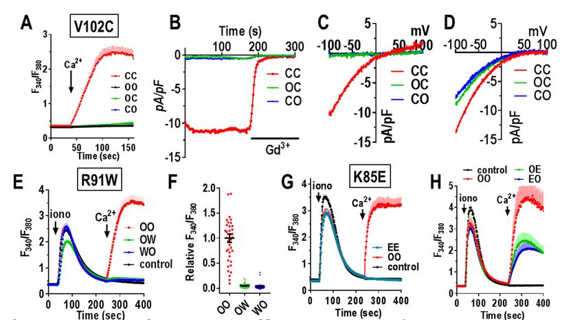
Cai et al                                                                               Function of Orai1 dimers

of approximately 10 mV (Fig 4B). We would have             replacing three glutamate residues with glutamines
expected that the properties of the heterodimeric          in the Orai1 selectivity filter prompted us to
channels (OD and DO) would be somewhere                    investigate whether this altered the channel pore
between the two homodimers. Thus our results               diameter. For this we measured the permeation of
indicate that replacement of just three Glu residues       methylated ammonium derivatives of increasing
with Asp in the selectivity filter of the hexameric        size. We compared permeation of the OQ or QO
channel, is still sufficient to render it a non-           Orai1 heterodimers with that of the OO wildtype
selective channel.                                         homodimer expressed in HEK-O1koS1+ cells (Fig.
                                                           5). Consistent with previous studies expressing
    The effects of the E106Q mutation are also             wildtype monomeric Orai1 (34,35), we observed
surprising (Fig. 4C,D). Clearly, the homodimeric           that the OO dimer had almost no current after
replacement in QQ results in a channel with no             switching from 20 mM Ca2+ to 150 mM methylam-
conductance, as expected. However, the large but           monium (Fig. 5A,B). Increasing the methyl groups
non-selective current seen with both the OQ and            on methylammonium to two, three and four, also
QO heterodimers, was unexpected. In both cases             revealed no measurable currents (Fig. 5C,D).
the reversal potential was close to zero mV.               These results are similar to earlier results from
Assuming the channel operates as a hexamer, this           which the Orai1 pore size was calculated to be
reveals that the full configuration of 6 charges in        ~3.9Å (34,35). In contrast, using cells expressing

                                                                                                                    Downloaded from http://www.jbc.org/ by guest on October 5, 2018
the selectivity filter is not required for cation          the OQ heterodimer, switching from Ca2+ to
conductance. Thus, the presence of just 3 negative         methylammonium resulted in a massive increase in
charges in the pore filter is sufficient for strong        current (Fig. 5E,F). Moreover, large currents were
cation permeation, albeit non-selectively.                 still observed following consecutive switching
                                                           from Ca2+ to dimethylammonium or even trimeth-
    We also examined the function of a concaten-           ylammonium. The same current measurements
ated hexameric Orai1 construct containing three            using the QO construct provided very similar
pairs of OQ or QO dimers (Fig. 4E-G). The                  results (Fig. 5G,H). In both cases, the decreased
function of both of these two hexamers was very            currents and increasingly negative reversal poten-
comparable to that of their respective contributing        tials seen with increasing number of methyl groups
heterodimers. For Ca2+ entry, both the OQOQOQ              on ammonium ion, allowed us to estimate an ap-
and QOQOQO hexamers gave similar function as               parent pore diameter for the OQ and QO constructs
the wildtype homohexamer (OOOOOO) (Fig. 4E).               considerably greater the WT pore (see Experi-
Current measurements with the hexamers revealed            mental Procedures and Fig. S4; this estimate is
that both heterohexamers behaved as nonselective           based on the assumption that permeability is lim-
cation channels, similar to heterodimers. Reversal         ited only by the steric properties of the pore). We
potential for hexamers were slightly elevated (Fig.        also constructed equivalent Orai1 heterodimers
4G), and currents mediated by the hexamers were            containing E106 residues mutated to asparagine
slightly smaller perhaps due to less efficient             instead of glutamine (ON, NO and NN). Ca2+
expression compared to dimers. The similar func-           measurements revealed a similar functional
tion of both hexamers and dimers, supports the             pattern; NN gave no Ca2+ entry whereas ON and
view that the Orai1 channel is hexameric and               NO gave Ca2+ entry identical to the wildtype, OO
comprises three pairs of dimers. In our previous           (Fig. S5A,B). Current measurements revealed the
paper, we concluded that a hexamer-concatemer              ON and NO dimers had similar ion non-selectivity
could create a functional channel either as a single       (Fig. S5C-E), and currents measured with
hexameric ring or as a “trimer of dimers” in which         methylated ammonium ions (Fig. S5F-M) indicat-
three concatemeric hexamers each contribute one            ed similarly enlarged estimated pore size.
dimer to form a single functional hexamer (25).
The results we show here for the two hetero-                   These surprising results clearly reveal that
hexamers would be consistent with either scheme.           reducing the number of negatively charged resi-
                                                           dues in the selectivity filter of Orai1 from 6 (in the
  Pore properties of the non-selective OQ and              wildtype hexamer formed from OO) to 3 (formed
QO concatemer dimers. The surprising effect of             by the OQ, QO, ON, or NO dimers), causes a

                                                       4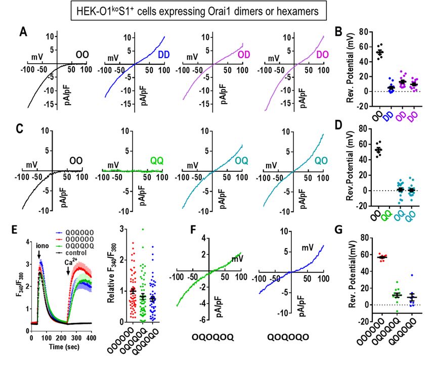
Cai et al                                                                                Function of Orai1 dimers

substantial increase in apparent pore diameter and           expressing the CC homodimer revealed a typical
a major alteration in ion channel selectivity. This          CRAC-like store-dependent current with reversal
change in pore properties was even larger than that          potential greater than +50 mV (Fig. 6D, Fig. S6D),
resulting from substitution of all six E106 residues         in close agreement with the earlier results with the
within the hexamer with Asp residues, indicating             Orai1-V102C monomer (13,37). In similar experi-
that both charge density and steric influence of             ments, the two Orai1 heterodimers, OC and CO,
head-group size contribute to the geometry of the            gave current properties almost identical to those
pore. The results with the OQ and ON heterodimers            seen with CC. The results are interesting in reveal-
also gives support to a recent hypothesis on the ion-        ing that the wildtype Val at position 102 is domi-
selectivity mechanism for the Orai channel. Thus,            nant over the Cys mutant in the heterodimer. We
in an interesting new crystallography study on               might have expected that the heterodimer would
dOrai channel structure, Hou et al (36) suggest that         have some constitutive function, but instead the
glutamates comprising the selectivity filter, have           hydrophobic gating function of this residue appears
sidechain flexibility that allows two Ca2+ ions to be        to be maintained with just three hydrophobic Val
positioned one above the other. Repulsion between            residues lining the pore. Conversely, the constit-
the two Ca2+ ions allows one Ca2+ to be displaced            utive activation of Ca2+ entry by V102C appears to
and move through the pore and be replaced by a               require mutation of all six residues.
new Ca2+ ion. Thus in the presence of external Ca2+,

                                                                                                                     Downloaded from http://www.jbc.org/ by guest on October 5, 2018
the channel is highly Ca2+ selective. In the case that            This result is an interesting contrast to
three glutamates are replaced with three similarly           experiments to assess the function of a different
sized but uncharged glutamines, we would suggest             pore-lining residue, R91, residing below the hydro-
that the selectivity filter loses the ability to bind        phobic pore region (Fig. 1C). The R91 residue lies
two Ca2+ ions, Ca2+ selectivity would be lost, and           within the highly positively charged region of the
monovalent cations would permeate in the                     pore toward the cytosolic side of the channel. The
presence of external Ca2+.                                   naturally occurring R91W mutation has been
                                                             reported to cause severe combined imunodefici-
    Orai1 dimers mutated elsewhere in the Orai1              ency in homozygous patients (28). We constructed
M1 pore-forming helix: In further experiments, we            Orai1 mutant heterodimers with one R91W
assessed the role of substituting other pore-lining          mutation (OW and WO). Expressed in HEK-
residues within the Orai1 heterodimers.                      O1koS1+ cells, we observed that the Orai1
Immediately below the E106 selectivity filter, the           heterodimers, OW and WO, each gave no store-
hexameric pore is lined with three rings of                  dependent Ca2+ entry (Fig. 6E,F). Indeed, their
hydrophobic residues, thought to act as a hydro-             action was virtually the same as non-transfected
phobic gate (37). Mutating the first of these res-           cells. This result is quite different to that earlier
idues (V102; see Fig. 1C) to Cys causes the channel          reported by (38) in which co-expression of Orai1-
to be constitutively active although with altered            WT and Orai1-R91W resulted in only a modest
cation selectivity (37). Using an Orai1 V102C                slowing of the activation of ICRAC. In that study, it
homodimer (CC), we observed substantial                      is likely that expression of WT and mutant R91W
constitutive Ca2+ entry in HEK-O1ko cells (not               were not equal, in contrast to the OW and WO
over-expressing STIM1) (Fig. 6A, Fig. S6A).                  heterodimers. Another earlier study examined
Patch-clamp measurements also revealed a                     tetramer-concatemers of Orai1 in which 1, 2, 3, or
substantial current (Fig. 6B, Fig. S6B) with a rever-        all 4 WT Orai1 subunits were substituted with
sal potential of approximately +25 mV (Fig. 6C,              Orai1-R91W subunits (39). In that study, a single
Fig. S6C). These results all agree closely with              R91W residue caused 50% loss of activity, and
studies expressing monomer Orai1-V102C (13,                  with 2 or 3 mutant subunits, there was almost 100%
37). In contrast, the heterodimers containing one            loss of activity. Our results would concur with this
Orai1-WT and one Orai1-V102C subunit (OC or                  dominance observed with R91W. However, the use
CO) displayed no constitutive Ca2+ entry or current          of the tetrameric construct is difficult to reconcile
(Fig. 6A-C). Using HEK-O1koS1+ we measured                   since the Orai1 channel functions as a hexamer
currents after depletion of stores following break-          (17,25,26). Likely the expressed tetramers form
in with 20 mM BAPTA in the pipette. Cells                    hexamers through trimers of dimers, as described

                                                         5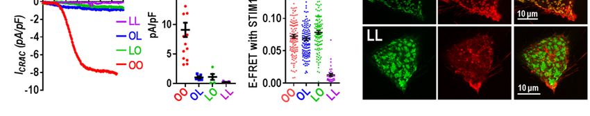
Cai et al                                                                              Function of Orai1 dimers

above. Given the almost complete dominance of              Nevertheless, our current results do show that
the R91W in our heterodimers, patients hetero-             altering the cytosolic M1 extension (K85E) does
zygous for the R91W gene would be expected to              affect binding of STIM1. Although the OE and EO
have substantially reduced Orai1-mediated Ca2+             heterodimers have reduced current, the pore-
entry. Such heterozygous R91W carriers had T               properties have not changed. The effects on
cells with approximately 50% of WT Ca2+ entry              STIM1-binding of mutating the K85 sites in the
(28). It is possible that compensatory expression of       dimer, supports a model of close coupling between
other Orai subtypes may have occurred in such              the M1-ext and the M4-ext, as suggested earlier
patients.                                                  (41). These two domains exist in a closely
                                                           associated complex together with the 16 amino
    We next used Orai1 dimers to examine the               acid 2-3 loop joining the M2 and M3 helices (see
effects of mutating one of the residues of the pore-       Fig. 1C). Recent evidence indicates that the 2-3
forming M1 helix which is not lining the pore (see         loop mediates important communication with the
Fig. 1C). The K85 residue has been the center of           M1-ext helix (43). Although the C-terminal M4-ext
much attention since the K85E mutation com-                is the primary site for STIM1 binding to gate the
pletely blocks store-dependent channel activation          channel, we would speculate that the entire
(13,40-42). This residue faces outward from the            cytosolic domain (M1-ext, 2-3 loop, and M4-ext)
pore-forming helix with its head group likely              exists as a conformationally coupled entity, altera-

                                                                                                                  Downloaded from http://www.jbc.org/ by guest on October 5, 2018
extending into the cytosol close to the cytosolic          tion of which affects both STIM1 binding and gat-
membrane surface. Using Orai1 homodimers with              ing of the channel. However, even though K85E
both K85 residues mutated to E, we determined              alters STIM1 binding, we do not consider that the
that there was no Ca2+ entry function, consistent          N-terminus is a STIM1 binding site required for
with the blocking action determined earlier (40)           gating. Thus, we revealed earlier that a C-terminal
(Fig. 6G,I). Interestingly, we observed that the           mutation in Orai1 that mimics STIM1-induced
heterodimers with a single K85E mutation (OE and           channel activation without any STIM1 present, is
EO), each displayed Ca2+ entry that was approx-            similarly sensitive to the K85E mutation (13).
imately 50% of the OO-WT homodimer (Fig. 6H).
Current analysis confirmed that OE and EO had                   Probing the STIM1-Orai1 coupling interface
similarly diminished channel function and that the         at the C-terminus of Orai1: We shifted our
I/V relationship revealed each mediated otherwise          exploration of the function of Orai1 dimers to
normal CRAC current (Fig. 6J-K, Fig. S6E). Since           understanding more about the C-terminal M4-ext
the K85 site has been implicated in binding STIM1          helix, the site at which STIM1 binds strongly and
to possibly gate the channel (40-42), we also ex-          initiates activation of the channel. Unlike the N-
amined FRET between CFP-tagged Orai1 dimers                terminus which has 6 identical helices, the dOrai
(C-terminally labeled) and STIM1-YFP. In these             crystal structure shows that the short 45-amino acid
studies, we observed FRET with each heterodimer            C-terminal M4-ext exists in two different con-
(OE or EO) was decreased approximately 30%                 figurations (Fig. 1A,C). Thus, each dimer of Orai
compared to the WT homodimer (Fig. 6L). The EE             contains one M4-ext that is in an extended
mutant homodimer gave a slight further decrease in         conformation, and one in a bent conformation (17).
FRET.                                                      Based on the dOrai1 structure, the two M4-ext
                                                           helices in Orai1 are likely attached in an anti-
    The K85E mutation has been the subject of              parallel configuration through inter-helical hydro-
considerable speculation. It clearly has a profound        phobic interactions between the L273 and L276
effect on the store-operated activation of Orai1           residues in each helix (see Fig. 7A). Mutation of
channels. It was speculated that K85 mediates              L273 in Orai1 to either D or S is known to abolish
gating through possible interactions of STIM1 with         binding of STIM1 and any store-dependent active-
the cytosolically-protruding extension of the pore-        tion of the channel (27,38,41,44-46). Likely this
forming M1 helix (M1-ext) (40-42). However,                stems from disruption of the two bound M4-ext
recent studies suggest that K85 is necessary for           helices, although how STIM associates with these
maintaining the configuration of the channel rather        helices is still unclear (47,48). We constructed
than directly mediating channel gating (13).               tdTomato-tagged Orai1 dimers in which either one

                                                       6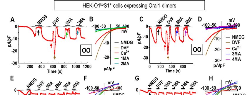
Cai et al                                                                              Function of Orai1 dimers

or both monomers contained the L273D mutation.             phospholipids in the ER-PM junctions (49,50).
We tested the function of these dimers in HEK-             Indeed, as seen in Fig. 7G (top row) the lower right
O1koS1+ cells. As expected, the L273D mutant               cell did not express Orai1, yet the STIM1 still
Orai1 homodimer (LL) gave no store-operated                entered puncta exactly the same as in the cell co-
Ca2+ entry (Fig. 7B,C), nor any current through the        expressing Orai1.
channel (Fig. 7D,E). In contrast, the two Orai1
heterodimers (OL and LO) both gave very small                   These results provide important new infor-
Ca2+ entry (Fig. 7B,C). Current measurements also          mation about the critical nature of the Orai1 C-
revealed that the conductance of the OL and LO             terminal configuration in mediating gating of the
heterodimers was extremely low (Fig. 7D,E).                Orai1 channel. The single L273D mutation in the
Summary data for both the Ca2+ and current                 OL or LO Orai1 heterodimers, drastically impedes
measurements reveal that the OL and LO                     the ability of Orai1 to be gated by STIM1, yet, the
heterodimers have less than 10% of the activity of         same heterodimers have not lost any significant
the wildtype OO homodimer (Fig. 7C,E).                     ability to bind STIM1. Recent studies indicate that
                                                           binding of STIM1 to the Orai1 C-terminal M4-ext
    We considered that the drastically decreased           is sufficient to gate the channel (13). This
function of the OL and LO heterodimer channels             interaction causes an allosteric change in the Orai1
might reflect a loss of the ability to bind STIM1.         channel that is propagated from the M4-ext through

                                                                                                                  Downloaded from http://www.jbc.org/ by guest on October 5, 2018
To address this question we undertook FRET                 the M3, M2 and M1 helices resulting in certain
studies using another set of Orai1-dimers (OO, OL,         pore-lining M1 residues undergoing a structural
LO and LL) with C-terminal CFP-tags. These were            alteration that constitutes gating of the channel
transiently expressed in cells stably expressing           (13,51). From the LL-Orai1 homodimer result, the
STIM1-YFP. Surprisingly, we observed that the              two M4-ext helices have likely undergone
Orai1 OL and LO heterodimers did not have a                unfolding sufficient to completely prevent STIM1
defect in STIM1-binding activity. Thus, after store-       binding. With the single L273D mutation in the OL
depletion, the FRET values for OO, OL, and LO              or LO heterodimers, the rearrangement must be
with STIM1-YFP were not significantly different            much more subtle such that STIM1 binding is
(Fig. 7F). As expected, the FRET value for the LL          largely intact. Yet, the propagation of allosteric
homodimer was much decreased indicating that               change across the protein to gate the channel, is
STIM1 binding was effectively abolished. In                almost completely lost. Thus, it seems that the M4-
further experiments, we undertook direct visualiza-        ext may undergo only a small conformational
tion of cells co-expressing Orai-tdT dimers and            change to initiate gating. This conclusion agrees
STIM1-YFP (Fig. 7G). In these studies we utilized          with studies on cross-linking of the M4-ext helices
HEK-O1koS1+ cells and transiently transfected              by Tirado-Lee et al (27). Recent structural analysis
each of the Orai1-tdT-tagged dimers. The results           has suggested that the interacting pairs of M4-ext
are highly consistent with the FRET studies. In            helices act as latches on the outside of the channel
cells transfected with the wildtype Orai1-OO               that must be opened to allow the M1-ext helices to
homodimer (Fig. 7G, top row, upper-left cell),             expand outward permitting the cytosolic mouth of
there was almost complete co-localization of OO-           the pore to open (36). This model was based on a
tdT with STIM1-YFP. The two molecules co-                  crystalized dOrai structure that stabilized the
localized within the same clearly observable               unlatched channel with straightened M4-ext
punctal regions in cells which represent the ER-PM         helices. However, this dOrai configuration was not
junctional areas. An almost identical pattern of co-       based on any input from STIM proteins. Our results
localization was also seen in cells expressing the         indicate that the initial STIM1-induced alteration
OL and LO heterodimers (Fig. 7G, 2nd and 3rd               of the M4-ext helices is extremely subtle and
rows). These results contrast dramatically with            unlikely to be a large unlatching of the entire Orai
cells expressing the LL mutant homodimer (Fig.             outer structure.
7G, bottom row). In this case, there is clearly no
co-localization of the LL dimer with the STIM1                 Concluding remarks: The use of concatenated
protein at the PM junctions, even though STIM1             Orai1 dimers has proven highly useful in determin-
can still form puncta by interacting with PM               ing the role of key residues in the channel pore and

                                                       7
Cai et al                                                                              Function of Orai1 dimers

STIM1 binding site of Orai1. Using a CRISPR/                tdT-tag was changed to ECFP with BamHI and
Cas9-derived Orai1-free background cell system,             NotI restriction sites.
the dimer concatemers are reliable probes for the
function of the hexameric Orai1 channel. Two                     Cell culture and transfection – HEK-O1ko
highlights of these findings are particularly               cells and the HEK-O1koS1+ cell line were generated
prominent. First, our studies provide some impor-           using the CRISPR-Cas9 nickase system as des-
tant new understanding on the properties of the             cribed earlier (25). Cells were cultured in DMEM
pore selectivity filter that normally functions with        (Corning Cellgro) supplemented with 10% fetal
six charged Glu residues. We speculate that these           bovine serum, penicillin and streptomycin (Gemini
residues may have sufficient flexibility to allow           Bioproducts, CA) containing puromycin (2 μg/ml)
formation of two Ca2+ binding sites within the              at 37      with 5% CO2. All transfections were
selectivity filter, as recently suggested (36). The         performed by electroporation at 180 V, 25 ms in 4
symmetrical and somewhat conservative replace-              mm cuvettes (Molecular Bio-Products) using the
ment of three Glu residues with three intervening           Bio-Rad Gene Pulser Xcell system in OPTI-MEM
Gln residues (formed with OQ or QO dimers), may             medium as previously described (52). Experiments
prevent formation of the putative second site and           were all performed 18-24 h after transfection.
render the pore as a nonselective cation channel.
Functionally this construct operates the same as six             Cytosolic Ca2+ measurements – Cytosolic Ca2+

                                                                                                                  Downloaded from http://www.jbc.org/ by guest on October 5, 2018
Asp residues in the selectivity filter, however, in         levels were measured as described earlier (53) by
that case the shortened head groups lack the                ratiometric imaging using fura-2 (Molecular
dimension and flexibility to form two sites. Future         Probes). Cells were used 18-24 hours after trans-
structure and modeling studies might examine this           fection, and incubated with 2 μM fura-2 in buffer
possibility. Second, our studies also throw new             containing: 107 mM NaCl, 7.2 mM KCl, 1.2 mM
light on the Orai1 C-terminal binding site for              MgCl2, 1 mM CaCl2, 11.5 mM Glucose, 0.1%
STIM1. The OL and LO Orai1 heterodimers appear              BSA, 20 mM HEPES pH 7.2 for 30 min at room
to have little change in their STIM1-binding                temperature, followed by treatment with fura-2 free
properties, yet they have almost entirely lost the          solution of another 30 mins. Fluorescence ratio
ability to mediate gating of the channel. This result       imaging was measured utilizing the Leica DMI
emphasizes the exquisitely precise nature of the            6000B fluorescence microscope and Hamamatsu
initial interacting site for STIM1 on Orai1 (the M4-        camera ORCA-Flash 4 controlled by Slidebook 6.0
ext) through which it induces channel activation.           software (Intelligent Imaging Innovations; Denver,
The two M4-ext sites likely propagate this                  CO) as described previously (54). Consecutive
allosteric gating of the channel across the tightly         excitation at 340nm (F340) and 380nm (F380) was
packed M3, M2 and M1 helices as recently                    undertaken every 2 sec and emission fluorescence
suggested (13).                                             was collected at 505 nm. Intracellular Ca2+ levels
                                                            are shown as F340/F380 ratios obtained from
Experimental Procedures                                     groups of 8-25 single cells on coverslips. For the
                                                            Orai1 concatemer experiments, data is shown for
     DNA constructs – Concatenated Orai1 dimers             cells expressing a narrow range of tdTomato fluor-
were constructed as described earlier. The 36 aa            escence in order to maximize consistency of
linker sequence is TSSSTMLHPGLSPGLLPLH                      responses between cells. For each concatemer ex-
PASIGGSGGSGGSGRAT (25). For Orai1 hetero-                   periment, we simultaneously examined each set of
dimers, point mutations in the M1 or M4-ext helic-          mutants with the corresponding wildtype concate-
es (E106, V102, R91, K85, L273), were performed             mer. The expression levels for wildtype and mutant
on Orai1-WT monomers using the QuickChange                  concatemers were within a similar narrow range of
Lightning Site-Directed Mutagenesis Kit (Agilent;           fluorescence intensity. All Ca2+ imaging experi-
210518), then inserted into the ptdTomato-N1-               ments were performed at room temperature and re-
Orai1 plasmid with XhoI and BamHI restriction               presentative traces of at least three independent
sites. For C terminal-CFP tagged constructs, the            repeats are shown. Scatter plots with means ± SEM
                                                            are shown for all the cells recorded.

                                                        8
Cai et al                                                                               Function of Orai1 dimers

    Deconvolved fluorescence image analysis -              10 amplifier (HEKA). Glass electrodes with a
For imaging of cells, we used the inverted Leica           typical resistance of 2-4 MΩ were pulled using a P-
DMI 6000B automated fluorescence microscope                97 pipette puller (Sutter Instruments). A 50-ms step
and Hamamatsu ORCA-Flash 4 camera controlled               to -100 mV from a holding potential of 0 mV,
by Slidebook software to collect and analyze high          followed by a 50-ms ramp from -100 to 100 mV,
resolution fluorescence images. For the PM locali-         was delivered every 2 seconds. Currents were
zation studies of tdTomato-tagged Orai1 concate-           filtered at 3.0 kHz and sampled at 20 kHz. A +10
mers, stacks of 10–20 3-D Z-axis image-planes              mV junction potential compensation was applied to
close to the cell-glass interface were collected at        correct the liquid junction potential between the
0.35 μm steps. The no-neighbor deconvolution fun-          bath and pipette solutions. The current measure at
ction of the Slidebook 6.0 software was used to            -100 mV was used in I-V curves. All data were
analyze images and derive enhanced deconvolved             acquired with Patch Master and analyzed using
images with minimized fluorescence contamina-              FitMaster and Graph Pad Prism 6.
tion from out-of-focus planes. The tdT-tagged
Orai1 concatemer images shown were typical of at               Using the Goldman-Hodgkin-Katz (GHK)
least three independent analyses.                          voltage equation, relative permeabilities of the
                                                           different methylated ammonium ions and Na+
    Confocal Imaging – Cells transfected with              (Px/PNa) were calculated from changes in the

                                                                                                                    Downloaded from http://www.jbc.org/ by guest on October 5, 2018
tdT-tagged constructs were imaged 14-20 hr after           reversal potential:
transfection. Live cell images were collected on the
inverted Leica TCS SP8 confocal microscope using                                            ∆
a 63x/1.40 Oil objective. Images shown are typical
of at least three independent experiments.
                                                           in which [X]o and [Na]o are the external ionic con-
     Electrophysiological measurements and pore            centrations, ∆Erev is the change in reversal potential
size estimation – Whole-cell patch-clamp record-           when switched to the testing cation, R is the univer-
ing was performed on HEK-O1koS1+ cells                     sal gas constant, T is the temperature in Kelvin, F
transiently transfected with tdT-tagged Orai1              is Faraday's constant.
concatemer constructs, or not transfected, as
described (11). To passively deplete ER Ca2+                   We estimated pore dimensions using the
stores, the pipette solution contained: 135 mM Cs-         relative ion permeability ratios, as described earlier
aspartate, 10 mM HEPES, 8 mM MgCl2 and 20                  (35), assuming that ion permeability is limited
mM BAPTA (pH 7.2 with CsOH). For measuring                 mainly by steric hindrance. The relative
constitutively active currents, HEK-O1ko cells were        permeability was calculate from:
transiently transfected with different Orai1-V102C
concatemers, and the pipette solution included: 135
mM Cs-aspartate, 10 mM HEPES, 5 mM MgCl2,                                           1
10 mM EGTA and 3 mM CaCl2. The bath solution
contained: 130 mM NaCl, 4.5 mM KCl, 5 mM
HEPES, 10 mM Dextrose, 10 mM TEA-Cl and 20                 in which k is a proportionality constant, dion is the
mM CaCl2 (pH 7.2 with NaOH). Divalent free                 ion diameter, and dpore is the pore diameter (55,56).
solution included: 150 mM NaCl, 5 mM HEPES,                An estimate for dpore was derived by relating
10 mM TEA-Cl, 10 mM HEDTA and 1 mM EDTA                    decrease in permeability ratio to the increase in
(pH 7.4). When indicated, 150 mM NaCl was                  organic cation size.
substituted by 150 mM methylamine HCl,
dimethylamine HCl, trimethylamine HCl, or                      Western blot analyses – Cells were washed
tetramethylamine HCl. ICRAC was recorded until             with ice-cold PBS and lysed on ice using lysis
steady state, then the external solution was               buffer (RIPA, Sigma and 1x Protease inhibitor
switched to different methylammonium-DVF                   cocktail) for 30 min, followed by centrifugation at
solutions indicated. Currents were recorded in the         14,000 g for 10 min at 4°C. Supernatants were
standard whole-cell configuration using the EPC-           collected and protein quantified using Bio-Rad DC

                                                       9
Cai et al                                                                                   Function of Orai1 dimers

kits. Proteins were resolved on 4-15% NuPAGE
Bis-Tris precast gels and transferred to Bio-Rad                  FC=IDA-Fd/Dd × IDD –Fa/Da × IAA
Immuno-Blot PVDF membranes. After blocking in
5% non-fat milk for 1 hr at room temperature the              where IDD, IAA and IDA represent the background
membrane was incubated with primary antibody at               subtracted CFP, YFP and FRET images, respect-
4°C overnight. Membranes were washed 3 times in               tively, FC represents the corrected energy transfer,
PBST and incubated with secondary antibody for 1              Fd/Dd represents measured bleed-through of CFP
hr at room temperature. Subsequently, membranes               through the FRET filter (0.457) and Fa/Da is the
were washed 3 times with PBST. Peroxidase                     bleed-through of YFP through the FRET filter
activity was examined with Pierce ECL Plus                    (0.19). We used the E-FRET method to analyze 3-
Western Blotting Substrate (Thermo Scientific)                cube FRET images as describe by (57), using the
and fluorescence was collected using the                      formula:
FluorChem M imager (ProteinSimple).
                                                                  Eapp= Fc/(Fc + G × IDD)
     Fӧrster resonance energy transfer measure-
ments – To determine FRET signals between                     in which G is the instrument specific constant
stably expressed STIM1-YFP and transiently                    (44,57). The EYFP-ECFP construct made as
expressed CFP-tagged Orai1 dimers, we used the                described above, was used to determine the G-

                                                                                                                       Downloaded from http://www.jbc.org/ by guest on October 5, 2018
Leica DMI 6000B inverted automated fluorescence               parameter for E-FRET calculations. The value of G
microscope equipped with CFP (438Ex/483Em),                   was determined by measuring the CFP
YFP (500Ex/542Em) and FRET (438Ex/542Em)                      fluorescence increase after YFP acceptor
filter cubes. At each time point, three sets of images        photobleaching using HEK-WT cells transiently
(CFP, YFP and FRET) were collected at room                    transfected with the pEYFP-ECFP construct. The
temperature using a x40 oil objective (numerical              value of G was calculated as 1.9±0.1 (n=32 cells).
aperture 1.35; Leica) and processed using Slide-              For studies determining E-FRET between YFP and
book 6.0 software (Intelligent Imaging Innova-                CFP constructs, cells with a narrow range of
tions). Images were captured at 20-s intervals.               YFP/CFP ratios were selected to ensure
Exposure times for the CFP, YFP and FRET                      comparability between measurements. In all our E-
channels were 1,000, 250 and 1000 ms, respec-                 FRET summary data, a region close to the PM was
tively. The decreased YFP channel exposure time               selected, and E-FRET analysis was conducted on
compensates for the greater fluorescence intensity            cells with similar YFP/CFP ratios.
of YFP compared with CFP. Three-channel-cor-
rected FRET was calculated using the formula:

Acknowledgements: This work was supported by NIH Grants GM120783 and GM109279 to DLG,
Predoctoral Fellowship GM125376 to RMN, and a Penn State University Junior Faculty Development
Program grant to YZ.

Conflict of interest: The authors declare that they have no conflict of interest with the contents of this
article.

REFERENCES

1.          Prakriya, M., and Lewis, R. S. (2015) Store-Operated Calcium Channels. Physiol. Rev. 95, 1383-
            1436
2.          Amcheslavsky, A., Wood, M. L., Yeromin, A. V., Parker, I., Freites, J. A., Tobias, D. J., and
            Cahalan, M. D. (2015) Molecular Biophysics of Orai Store-Operated Ca Channels. Biophys. J.
            108, 237-246

                                                         10
Cai et al Function of Orai1 dimers
3. Shim, A. H., Tirado-Lee, L., and Prakriya, M. (2015) Structural and functional mechanisms of
CRAC channel regulation. J. Mol. Biol. 427, 77-93
4. Zhou, Y., Cai, X., Nwokonko, R. M., Loktionova, N. A., Wang, Y., and Gill, D. L. (2017) The
STIM-Orai coupling interface and gating of the Orai1 channel. Cell Calcium 61, 1-7
5. Soboloff, J., Rothberg, B. S., Madesh, M., and Gill, D. L. (2012) STIM proteins: dynamic
calcium signal transducers. Nat. Rev. Mol. Cell Biol. 13, 549-565
6. Hogan, P. G. (2015) The STIM1-ORAI1 microdomain. Cell Calcium 58, 357-367
7. Derler, I., Jardin, I., and Romanin, C. (2016) Molecular mechanisms of STIM/Orai
communication. Am. J. Physiol. Cell Physiol. 310, C643-C662
8. Kar, P., and Parekh, A. (2013) STIM proteins, Orai1 and gene expression. Channels (Austin) 7,
374-378
9. Zhou, Y., Trebak, M., and Gill, D. L. (2015) Calcium signals tune the fidelity of transcriptional
responses. Mol. Cell 58, 197-199
10. Feske, S., Wulff, H., and Skolnik, E. Y. (2015) Ion channels in innate and adaptive immunity.
Annu. Rev. Immunol. 33, 291-353
11. Wang, X., Wang, Y., Zhou, Y., Hendron, E., Mancarella, S., Andrake, M. D., Rothberg, B. S.,
Soboloff, J., and Gill, D. L. (2014) Distinct Orai-coupling domains in STIM1 and STIM2 define
the Orai-activating site. Nat Commun 5, 3183
Downloaded from http://www.jbc.org/ by guest on October 5, 2018
12. Zhou, Y., Wang, X., Wang, X., Loktionova, N. A., Cai, X., Nwokonko, R. M., Vrana, E., Wang,
Y., Rothberg, B. S., and Gill, D. L. (2015) STIM1 dimers undergo unimolecular coupling to
activate Orai1 channels. Nat Commun 6, 8395
13. Zhou, Y., Cai, X., Loktionova, N. A., Wang, X., Nwokonko, R. M., Wang, X., Wang, Y.,
Rothberg, B. S., Trebak, M., and Gill, D. L. (2016) The STIM1-binding site nexus remotely
controls Orai1 channel gating. Nat Commun 7, 13725
14. Zhou, Y., Nwokonko, R. M., Cai, X., Loktionova, N. A., Abdulqadir, R., Xin, P., Niemeyer, B.
A., Wang, Y., Trebak, M., and Gill, D. L. (2018) Cross-linking of Orai1 channels by STIM
proteins. Proc. Natl. Acad. Sci. U. S. A.
15. Rothberg, B. S., Wang, Y., and Gill, D. L. (2013) Orai channel pore properties and gating by
STIM: implications from the Orai crystal structure. Sci Signal 6, pe9
16. Derler, I., Jardin, I., and Romanin, C. (2016) Molecular mechanisms of STIM/Orai
communication. Am. J. Physiol. Cell Physiol. 310, C643-662
17. Hou, X., Pedi, L., Diver, M. M., and Long, S. B. (2012) Crystal structure of the calcium release-
activated calcium channel Orai. Science 338, 1308-1313
18. Penna, A., Demuro, A., Yeromin, A. V., Zhang, S. L., Safrina, O., Parker, I., and Cahalan, M. D.
(2008) The CRAC channel consists of a tetramer formed by Stim-induced dimerization of Orai
dimers. Nature 456, 116-120
19. Mignen, O., Thompson, J. L., and Shuttleworth, T. J. (2008) Orai1 subunit stoichiometry of the
mammalian CRAC channel pore. J. Physiol. 586, 419-425
20. Ji, W., Xu, P., Li, Z., Lu, J., Liu, L., Zhan, Y., Chen, Y., Hille, B., Xu, T., and Chen, L. (2008)
Functional stoichiometry of the unitary calcium-release-activated calcium channel. Proc. Natl.
Acad. Sci. USA 105, 13668-13673
21. Maruyama, Y., Ogura, T., Mio, K., Kato, K., Kaneko, T., Kiyonaka, S., Mori, Y., and Sato, C.
(2009) Tetrameric Orai1 is a teardrop-shaped molecule with a long, tapered cytoplasmic domain.
J. Biol. Chem. 284, 13676-13685
22. Madl, J., Weghuber, J., Fritsch, R., Derler, I., Fahrner, M., Frischauf, I., Lackner, B., Romanin,
C., and Schutz, G. J. (2010) Resting state Orai1 diffuses as homotetramer in the plasma
membrane of live mammalian cells. J. Biol. Chem. 285, 41135-41142
23. Demuro, A., Penna, A., Safrina, O., Yeromin, A. V., Amcheslavsky, A., Cahalan, M. D., and
Parker, I. (2011) Subunit stoichiometry of human Orai1 and Orai3 channels in closed and open
states. Proc. Natl. Acad. Sci. U. S. A. 108, 17832-17837
11Cai et al Function of Orai1 dimers
24. Thompson, J. L., and Shuttleworth, T. J. (2013) How many Orai's does it take to make a CRAC
channel? Sci. Rep. 3, 1961
25. Cai, X., Zhou, Y., Nwokonko, R. M., Loktionova, N. A., Wang, X., Xin, P., Trebak, M., Wang,
Y., and Gill, D. L. (2016) The Orai1 store-operated calcium channel functions as a hexamer. J.
Biol. Chem. 291, 25764-25775
26. Yen, M., Lokteva, L. A., and Lewis, R. S. (2016) Functional Analysis of Orai1 Concatemers
Supports a Hexameric Stoichiometry for the CRAC Channel. Biophys. J. 111, 1897-1907
27. Tirado-Lee, L., Yamashita, M., and Prakriya, M. (2015) Conformational Changes in the Orai1 C-
Terminus Evoked by STIM1 Binding. PLoS One 10, e0128622
28. Feske, S., Gwack, Y., Prakriya, M., Srikanth, S., Puppel, S. H., Tanasa, B., Hogan, P. G., Lewis,
R. S., Daly, M., and Rao, A. (2006) A mutation in Orai1 causes immune deficiency by abrogating
CRAC channel function. Nature 441, 179-185
29. Vig, M., Beck, A., Billingsley, J. M., Lis, A., Parvez, S., Peinelt, C., Koomoa, D. L., Soboloff, J.,
Gill, D. L., Fleig, A., Kinet, J. P., and Penner, R. (2006) CRACM1 Multimers Form the Ion-
Selective Pore of the CRAC Channel. Curr. Biol. 16, 2073-2079
30. Yeromin, A. V., Zhang, S. L., Jiang, W., Yu, Y., Safrina, O., and Cahalan, M. D. (2006)
Molecular identification of the CRAC channel by altered ion selectivity in a mutant of Orai.
Nature 443, 226-229
Downloaded from http://www.jbc.org/ by guest on October 5, 2018
31. Gwack, Y., Srikanth, S., Feske, S., Cruz-Guilloty, F., Oh-Hora, M., Neems, D. S., Hogan, P. G.,
and Rao, A. (2007) Biochemical and functional characterization of Orai family proteins. J. Biol.
Chem. 282, 16232-16243
32. Lis, A., Peinelt, C., Beck, A., Parvez, S., Monteilh-Zoller, M., Fleig, A., and Penner, R. (2007)
CRACM1, CRACM2, and CRACM3 are store-operated Ca2+ channels with distinct functional
properties. Curr. Biol. 17, 794-800
33. Wei-Lapierre, L., Carrell, E. M., Boncompagni, S., Protasi, F., and Dirksen, R. T. (2013) Orai1-
dependent calcium entry promotes skeletal muscle growth and limits fatigue. Nat Commun 4,
2805
34. Yamashita, M., Navarro-Borelly, L., McNally, B. A., and Prakriya, M. (2007) Orai1 mutations
alter ion permeation and Ca2+-dependent fast inactivation of CRAC channels: evidence for
coupling of permeation and gating. J.Gen.Physiol 130, 525-540
35. Prakriya, M., and Lewis, R. S. (2006) Regulation of CRAC Channel Activity by Recruitment of
Silent Channels to a High Open-probability Gating Mode. J.Gen.Physiol 128, 373-386
36. Hou, X., Burstein, S. R., and Long, S. B. (2018) Structures reveal opening of the store-operated
calcium channel Orai. bioRxiv/Medical Instrumentation
37. McNally, B. A., Somasundaram, A., Yamashita, M., and Prakriya, M. (2012) Gated regulation of
CRAC channel ion selectivity by STIM1. Nature 482, 241-245
38. Muik, M., Frischauf, I., Derler, I., Fahrner, M., Bergsmann, J., Eder, P., Schindl, R., Hesch, C.,
Polzinger, B., Fritsch, R., Kahr, H., Madl, J., Gruber, H., Groschner, K., and Romanin, C. (2008)
Dynamic coupling of the putative coiled-coil domain of ORAI1 with STIM1 mediates ORAI1
channel activation. J. Biol. Chem. 283, 8014-8022
39. Thompson, J. L., Mignen, O., and Shuttleworth, T. J. (2009) The Orai1 severe combined immune
deficiency mutation and calcium release-activated Ca2+ channel function in the heterozygous
condition. J. Biol. Chem. 284, 6620-6626
40. Lis, A., Zierler, S., Peinelt, C., Fleig, A., and Penner, R. (2010) A single lysine in the N-terminal
region of store-operated channels is critical for STIM1-mediated gating. J. Gen. Physiol. 136,
673-686
41. McNally, B. A., Somasundaram, A., Jairaman, A., Yamashita, M., and Prakriya, M. (2013) The
C- and N-terminal STIM1 binding sites on Orai1 are required for both trapping and gating CRAC
channels. J. Physiol. 591, 2833-2850
12Cai et al Function of Orai1 dimers
42. Gudlur, A., Quintana, A., Zhou, Y., Hirve, N., Mahapatra, S., and Hogan, P. G. (2014) STIM1
triggers a gating rearrangement at the extracellular mouth of the ORAI1 channel. Nat Commun 5,
5164
43. Fahrner, M., Pandey, S. K., Muik, M., Traxler, L., Butorac, C., Stadlbauer, M., Zayats, V.,
Krizova, A., Plenk, P., Frischauf, I., Schindl, R., Gruber, H. J., Hinterdorfer, P., Ettrich, R.,
Romanin, C., and Derler, I. (2018) Communication between N terminus and loop2 tunes Orai
activation. J. Biol. Chem. 293, 1271-1285
44. Navarro-Borelly, L., Somasundaram, A., Yamashita, M., Ren, D., Miller, R. J., and Prakriya, M.
(2008) STIM1-ORAI1 interactions and ORAI1 conformational changes revealed by live-cell
FRET microscopy. J. Physiol. 586, 5383-5401
45. Frischauf, I., Muik, M., Derler, I., Bergsmann, J., Fahrner, M., Schindl, R., Groschner, K., and
Romanin, C. (2009) Molecular Determinants of the Coupling between STIM1 and Orai Channels:
differential activation of Orai1-3 channels by a STIM1 coiled-coil mutant. J. Biol. Chem. 284,
21696-21706
46. Li, Z., Liu, L., Deng, Y., Ji, W., Du, W., Xu, P., Chen, L., and Xu, T. (2011) Graded activation of
CRAC channel by binding of different numbers of STIM1 to Orai1 subunits. Cell Res. 21, 305-
315
47. Stathopulos, P. B., Schindl, R., Fahrner, M., Zheng, L., Gasmi-Seabrook, G. M., Muik, M.,
Downloaded from http://www.jbc.org/ by guest on October 5, 2018
Romanin, C., and Ikura, M. (2013) STIM1/Orai1 coiled-coil interplay in the regulation of store-
operated calcium entry. Nat Commun 4, 2963
48. Maus, M., Jairaman, A., Stathopulos, P. B., Muik, M., Fahrner, M., Weidinger, C., Benson, M.,
Fuchs, S., Ehl, S., Romanin, C., Ikura, M., Prakriya, M., and Feske, S. (2015) Missense mutation
in immunodeficient patients shows the multifunctional roles of coiled-coil domain 3 (CC3) in
STIM1 activation. Proc. Natl. Acad. Sci. U. S. A. 112, 6206-6211
49. Liou, J., Fivaz, M., Inoue, T., and Meyer, T. (2007) Live-cell imaging reveals sequential
oligomerization and local plasma membrane targeting of stromal interaction molecule 1 after Ca2+
store depletion. Proc. Natl. Acad. Sci. USA 104, 9301-9306
50. Park, C. Y., Hoover, P. J., Mullins, F. M., Bachhawat, P., Covington, E. D., Raunser, S., Walz,
T., Garcia, K. C., Dolmetsch, R. E., and Lewis, R. S. (2009) STIM1 clusters and activates CRAC
channels via direct binding of a cytosolic domain to Orai1. Cell 136, 876-890
51. Palty, R., Fu, Z., and Isacoff, E. Y. (2017) Sequential Steps of CRAC Channel Activation. Cell
Rep 19, 1929-1939
52. Mancarella, S., Wang, Y., and Gill, D. L. (2011) Signal transduction: STIM1 senses both Ca2+
and heat. Nat. Chem. Biol. 7, 344-345
53. Soboloff, J., Spassova, M. A., Hewavitharana, T., He, L. P., Xu, W., Johnstone, L. S., Dziadek,
M. A., and Gill, D. L. (2006) STIM2 is an inhibitor of STIM1-mediated store-operated Ca2+
Entry. Curr. Biol. 16, 1465-1470
54. Mancarella, S., Wang, Y., Deng, X., Landesberg, G., Scalia, R., Panettieri, R. A.,
Mallilankaraman, K., Tang, X. D., Madesh, M., and Gill, D. L. (2011) Hypoxia-induced acidosis
uncouples the STIM-Orai calcium signaling complex. J. Biol. Chem. 286, 44788-44798
55. Dwyer, T. M., Adams, D. J., and Hille, B. (1980) The permeability of the endplate channel to
organic cations in frog muscle. J. Gen. Physiol. 75, 469-492
56. Burnashev, N., Villarroel, A., and Sakmann, B. (1996) Dimensions and ion selectivity of
recombinant AMPA and kainate receptor channels and their dependence on Q/R site residues. J.
Physiol. 496 ( Pt 1), 165-173
57. Zal, T., and Gascoigne, N. R. (2004) Photobleaching-corrected FRET efficiency imaging of live
cells. Biophys. J. 86, 3923-3939
13Cai et al Function of Orai1 dimers
FOOTNOTES
This work was supported in whole or part by National Institute of Health Grants R01 GM120783 and R01
GM109279, and GM125376
The abbreviations used are: STIM, stromal-interacting molecule; PM, plasma membrane; ER, endoplasmic
reticulum; CRAC, Ca2+ release-activating Ca2+; SOCE, store-operated Ca2+ entry; tdT, tdTomato
Downloaded from http://www.jbc.org/ by guest on October 5, 2018
14Cai et al Function of Orai1 dimers
Downloaded from http://www.jbc.org/ by guest on October 5, 2018
Figure 1. Structure of Orai channel and dimeric subunits. (A) Top view of Drosophila Orai channel
hexamer. Two out of the six monomers are colored to show one of the three dimers that constitute the
channel. The two subunits in each dimer are almost identical except the “A” monomer has a straight M4
extension helix (M4-ext; purple) and the “B” monomer has a bent M4-ext (pink). The two M4-ext helices
are linked in an antiparallel configuration by hydrophobic interactions. (B) Linear sequence of the
concatenated Orai1 dimer constructs used; the two monomers are joined through a 36-amino acid linker
sequence, and tagged with tdTomato at the C-terminus. Note: the sequences of the “A” and “B” monomers
are the same. (C) Schematic of the concatenated Orai1 dimer structure. The membrane-spanning domains
(M1, M2, M3, M4, and M4-ext) are labeled as shown. Colored dots reveal each of the point mutations used
in this study.
15Cai et al Function of Orai1 dimers
Downloaded from http://www.jbc.org/ by guest on October 5, 2018
Figure 2. Consequences of mutating the selectivity filter (E106) within expressed Orai1 monomers.
tdTomato-tagged Orai1 monomer constructs were transiently expressed in HEK-O1koS1+ cells which stably
express STIM1-YFP. Fura-2 ratiometric Ca2+ measurements were conducted to reveal cytosolic Ca2+ after
store-depletion with 2.5 μM ionomycin in Ca2+-free medium followed by addition of 1 mM Ca2+ (arrows).
(A) Cells were transfected with either Orai1-E106D (E106D) or Orai1-WT (O), or untransfected (control).
(B) Cells were co-transfected with both Orai1-E106D-CFP and Orai1 WT, or untransfected (control). (C)
Cells were transfected with either Orai1-E106Q (E106Q) or Orai1-WT (O), or untransfected (control). (D)
Cells were co-transfected with both Orai1-E106Q-CFP and Orai1- WT, or untransfected (control). All the
traces (means ± SE) are representative of three independent experiments. Summary scatter plots with means
± SEM for normalized peak Ca2+ entry are for all individual cells recorded in three independent
experiments. (** P < 0.005)
16You can also read