ENGINEERING BIOCOMPATIBLE TESEX NANO-ALLOYS AS A VERSATILE THERANOSTIC NANOPLATFORM
←
→
Page content transcription
If your browser does not render page correctly, please read the page content below
National Science Review
RESEARCH ARTICLE                                                                                                                                           8: nwaa156, 2021
                                                                                                                                                  doi: 10.1093/nsr/nwaa156
                                                                                                                                      Advance access publication 6 July 2020

                                 MATERIALS SCIENCE

                                Engineering biocompatible TeSex nano-alloys as a
1
  International
Collaborative
                                versatile theranostic nanoplatform
Laboratory of 2D
Materials for                   Xiang Ling1,† , Zhaokui Jin2,† , Qi Jiang2 , Xiaotao Wang1 , Bin Wei3 ,
Optoelectronic Science
                                Zhongchang Wang3 , Yangsen Xu1 , Tianye Cao2,4 , Jonathan W. Engle4 , Weibo Cai4 ,

                                                                                                                                                                                           Downloaded from https://academic.oup.com/nsr/article/8/6/nwaa156/5867802 by guest on 10 October 2021
& Technology,
Engineering Technology
Research Center for 2D          Chenliang Su1,∗ and Qianjun He2,∗
Materials Information
Functional Devices and
Systems of Guangdong
Province, Institute of
                                ABSTRACT
Microscale                      Photothermal nanotheranostics, especially in the near infrared II (NIR-II) region, exhibits a great potential
Optoelectronics,
                                in precision and personalized medicine, owing to high tissue penetration of NIR-II light.
Shenzhen University,
Shenzhen 518060,                NIR-II-photothermal nanoplatforms with high biocompatibility as well as high photothermal effect are
China; 2 Guangdong              urgently needed but rarely reported so far. Te nanomaterials possess high absorbance to NIR-II light but
Provincial Key                  also exhibit high cytotoxicity, impeding their biomedical applications. In this work, the controllable
Laboratory of
                                incorporation of biocompatible Se into the lattice of Te nanostructures is proposed to intrinsically tune
Biomedical
Measurements and                their inherent cytotoxicity and enhance their biocompatibility, developing TeSex nano-alloys as a new kind
Ultrasound Imaging,             of theranostic nanoplatform. We have uncovered that the cytotoxicity of Te nanomaterials primarily derives
National-Regional Key           from irreversible oxidation stress and intracellular imbalance of organization and energy, and can be
Technology Engineering
Laboratory for Medical
                                eliminated by incorporating a moderate proportion of Se (x = 0.43). We have also discovered that the
Ultrasound, School of           as-prepared TeSex nano-alloys have extraordinarily high NIR-II-photothermal conversion efficiency
Biomedical                      (77.2%), 64 Cu coordination and computed tomography contrast capabilities, enabling high-efficacy
Engineering, Health             multimodal photothermal/photoacoustic/positron emission tomography/computed tomography
Science Center,
Shenzhen University,
                                imaging-guided NIR-II-photothermal therapy of cancer. The proposed nano-alloying strategy provides a
Shenzhen 518060,                new route to improve the biocompatibility of biomedical nanoplatforms and endow them with versatile
China; 3 Department of          theranostic functions.
Quantum and Energy
Materials, International        Keywords: nanomedicine, nano-alloys, photothermal therapy, cancer nanotheranostics, biomedical
Iberian Nanotechnology
                                imaging
Laboratory (INL), Braga
4715-330, Portugal and
4
  Departments of
Radiology and Medical
                                INTRODUCTION                                                                  photothermal therapy has attracted intensive at-
Physics, University of                                                                                        tention owing to the fact that it is less invasive and
                                Nanotheranostics makes use of nanotechnology to
Wisconsin-Madison,                                                                                            has fewer side effects compared with conventional
Madison, WI 53705,
                                integrate diagnostics and therapeutics, exhibiting
                                                                                                              radiotherapy and chemotherapy [7–11]. In recent
USA                             a great potential in precision and personalized
                                                                                                              years, a number of photothermal theranostic nanoa-
                                medicine [1–3]. The emergence of diverse multi-
                                                                                                              gents, including noble metal nanoparticles [12–16],
∗
  Corresponding                 functional nanomaterials and advanced nanotech-
                                                                                                              two-dimensional (2D) nanosheets [17–26] and
authors. E-mails:               nologies unprecedentedly simulates the evolution
nanoflower@126.com;                                                                                           organic polymer nanomaterials [27–30], have been
                                of nanotheranostics, and enables the integration of
chmsuc@szu.edu.cn                                                                                             explored for cancer treatment, but most of them
†
  Equally contributed to        multimodal imaging and therapeutic functions in
                                                                                                              only work in the NIR-I region and the candidates
this work.                      a single theranostic nanoplatform for high-efficacy
                                                                                                              of theranostic nanomedicine for NIR-II-thermal
                                theranostics of diseases [4–6]. In engineering of
                                                                                                              imaging and therapy are quite rare. There are
Received 30 March               theranostic nanoplatforms, biocompatibility and
2020; Revised 9 June                                                                                          many photothermal theranostic nanoagents with
                                multifunction are two of the most important factors
2020; Accepted 20                                                                                             strong absorbance ranging from NIR-I to NIR-II.
June 2020
                                which need to be considered. Among various
                                                                                                              However, photothermal performance of nanoagents
                                nanotheranostics, multimodal imaging-guided


C The Author(s) 2020. Published by Oxford University Press on behalf of China Science Publishing & Media Ltd. This is an Open Access article distributed under the terms of the Creative

Commons Attribution License (http://creativecommons.org/licenses/by/4.0/), which permits unrestricted reuse, distribution, and reproduction in any medium, provided the original
work is properly cited.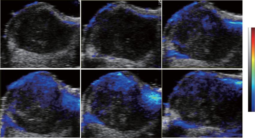
Natl Sci Rev, 2021, Vol. 8, nwaa156

is determined by three factors: molar extinction                (PA)/positron emission tomography (PET)/CT
coefficient, photothermal conversion efficiency and             imaging (Scheme 1).
allowable laser power density. Even though some
nanoagents have relatively high molar extinction co-
efficient or considerable photothermal conversion               RESULTS AND DISCUSSION
efficiency in the NIR-II window, their photothermal
performances in the NIR-II window are not as good               Synthesis and characterization of TeSex
as those in the NIR-I window because of lower                   nano-alloys
allowable laser power density [31]. However, it                 A series of rod-like TeSex nano-alloys with var-
is true that compared with NIR-I light, NIR-II                  ious ratios of Se/Te and length/diameter were
light possesses some intrinsic advantages in lesser             synthesized by a facile co-precipitation method.
photo-scattering and higher maximum permissible                 Tellurite and selenite were reduced simultaneously

                                                                                                                         Downloaded from https://academic.oup.com/nsr/article/8/6/nwaa156/5867802 by guest on 10 October 2021
exposure (MPE), consequently exhibiting higher                  by hydrazine to form TeSex nano-alloys, and the Se
tissue penetration depth with less background                   contents were adjusted by tuning the molar ratio
interference and higher spatial resolution, and                 of tellurite to selenite (Supplementary Table S1).
allowing tissue illustration at a relatively higher             TeSex nano-alloys with Se/Te precursor molar
power density of laser (1.0 W cm−2 for NIR-II                   ratios of 1 : 3, 2 : 3, 1 : 1 and 3 : 2 were named
versus 0.3 W cm−2 for NIR-I) [29]. Therefore,                   as TS1, TS2, TS3 and TS4, respectively. To as-
to develop biocompatible NIR-II-photothermal                    certain the crystal structure of the as-prepared
nanoplatforms with versatile imaging functions                  TeSex nanomaterials, X-ray diffraction (XRD)
is significant to precision cancer theranostics but             characterization was conducted (Fig. 1a and Sup-
challenging.                                                    plementary Fig. S1). XRD patterns were further
    Both selenium (Se) and tellurium (Te) belong                refined using the total pattern solution (TOPAS)
to the chalcogen elements, and their nanomaterials              Rietveld crystal-structure refinement software
exhibit some unique semi-conductive features                    (Fig. 1a and Supplementary Fig. S2). The refine-
[32–36]. Te nanoneedles and nanosheets have an                  ment results suggested the formation of Te-Se alloys,
extremely narrow band gap (about 0.35 eV) and a                 which were crystallized in a rhombohedral structure
strong absorbance in the NIR-II region in support of            with P31 21 space group. The refined structure of
NIR-II-photothermal therapy and imaging, but also               typical TS3 nano-alloy was further investigated by
demonstrate high cytotoxicity and poor biocompat-               high-resolution transmission electron microscope
ibility owing to their strong reducibility, restricting         (TEM). In a high-angle annular dark field (HAADF)
their biomedical applications [37–41]. By compari-              image and the corresponding elemental mapping in
son, Se is an essential element for human beings and            Fig. 1b–e, Te and Se were dispersed throughout the
the selenizing can eliminate the cytotoxicity of many           whole rod-like TeSex nano-alloys, and no core-shell
metals such as Cd and Cu [42,43]. Therefore, we                 structure can be obviously observed. To study the
hypothesize that controllable incorporation of bio-             radial elemental distribution of the as-prepared
compatible Se into the lattice of Te nanostructures             TeSex nano-alloys, depth profiling X-ray photoelec-
for construction of TeSex nano-alloys could intrin-             tron spectroscopy (XPS) analysis was conducted on
sically tune the inherent cytotoxicity of Te nano-              sample TS3 which was exposed to Ar+ for 0, 1 and
materials, enhance the biocompatibility of Te nano-             2 min. Te 3d and Se 3d XPS spectra of TS3 in Fig. 1f
materials and extend their functions for biomedical             revealed that there were only Te (0) and Se (0) in
applications. In this work, we synthesize a series of           the TeSex nano-alloy, and the binding energy of Te
TeSex nano-alloys with different Se incorporating               3d and Se 3d gradually decreased with the increase
proportions, and investigate their biocompatibility             of Ar+ etching time. Accordingly, the atomic ratio of
and develop their theranostic functions. We have                Se to Te obtained from XPS (Supplementary Table
discovered that the toxicity of Te nanomaterials                S2) decreased gradually, suggesting the gradient
mainly comes from irreversible oxidation stress and             increase of Se content from inside to outside in sup-
intracellular imbalance of organization and energy,             port of the formation of TeSex nano-alloy [44]. To
which is exterminated by the nano-alloying by in-               confirm the atomic structure of TeSex nano-alloy,
corporating a moderate proportion of Se (x = 0.43)              atomically resolved HAADF-scanning transmission
(Scheme 1). The synthesized TeSex nano-alloy                    electron microscopy (STEM) was conducted.
(x = 0.43) exhibits extraordinarily high NIR-                   Figure 1g–i and Fig. 1j–l showed the simultaneously
II-photothermal conversion efficiency (77.2%),                  recorded HAADF and bright-field (BF) STEM
64
   Cu coordination and computed tomography                      images acquired along a-axis and c-axis, respectively.
(CT) contrast capabilities, enabling high-efficacy              The observed atomic crystal structure from STEM
photothermal therapy of cancer under the guidance               was in high accordance with the simulated one from
of multimodal photothermal (PT)/photoacoustic                   XRD refinements (yellow spheres in Fig. 1i and l).
                                         Page 2 of 12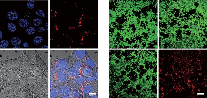
Natl Sci Rev, 2021, Vol. 8, nwaa156

                                                                                       PET imaging
                                             GAPD
                        Energy
                      production
                                                 RPL7A
                                                                                            64
                                                                                                 Cu
                         Subunit                   GSTs
                       organization                                                                              NIR-II light

                   Apoptosis
                                                                  Alloying

                                                                         Se                                              PT therapy

                                                                                  Te
                                                                                           Photothermal effect

                                                                                                                                       Downloaded from https://academic.oup.com/nsr/article/8/6/nwaa156/5867802 by guest on 10 October 2021
                                           Te nano-rods

                                                                              CT imaging                               PA/PT imaging
                                                                                       Multi-functionalization

Scheme 1. Schematic illustration of TeSex alloying strategy and mechanisms for detoxification and theranostic multi-
functionalization. Several main advances are achieved: (i) advanced TeSex nano-alloys are facilely constructed to intrinsi-
cally eliminate the inherent toxicity of Te nanomaterials by the moderate incorporation of biocompatible Se; (ii) advanced
mechanisms for Te nanomaterial toxification and TeSex alloying detoxification are uncovered; (iii) advanced theranostic per-
formances with extraordinarily high NIR-II-photothermal efficiency and multimodal PT/PA/CT/PET imaging capability are
achieved by nano-alloying.

Besides, no megascopic difference between Te and                     nano-alloys shifted towards lower binding energy,
Se can be observed, pinpointing that Se and Te                       which was also induced by the alloying formation
were thoroughly miscible in each other and formed                    between Te and Se. TEM images in Supplementary
homogenous trigonal-system TeSex nano-alloy.                         Fig. S4 indicated that the diameter and length
    The compositions of a series of TeSex nano-                      of rod-like TeSex nano-alloys decreased with the
alloys were measured by the inductively coupled                      increase of Se incorporation amount, while their
plasma optical emission spectrometry (ICP-                           morphologies remained nearly unchanged, suggest-
OES). As shown in Supplementary Table S1, the                        ing that the incorporation of Se inhibited the growth
chemical structures of TS1, TS2, TS3 and TS4 were                    of Te nano-rods. The particle size of synthesized
Te0.82 Se0.18 , Te0.75 Se0.25 , Te0.7 Se0.3 and Te0.67 Se0.33 ,      TeSex nano-alloys was less than 100 nm in favor
respectively. By varying the amounts of Se and Te                    of passive targeting accumulation in tumor by the
precursors, the molar fraction of Se in TeSex nano-                  enhanced permeability and retention (EPR) effect.
alloys could be controllably tailored. XRD patterns
of TS1, TS2, TS3 and TS4 in Supplementary Fig. S1
showed that the diffraction peaks were well matched                  Evolution of biocompatibility and
with the standard ones of Te (JCPDS card number                      photothermal properties of TeSex
36–1452) in the absence of impurity. In contrast                     nano-alloys
to the reported core−shell Te@Se nanowires [45]                      The biocompatibility of nanomedicines is vitally
and Se-coated Te nanoheterojunctions [46], no                        important to their biomedical application. Here we
characteristic XRD peaks of Se were observed in                      evaluated the biocompatibility of TeSex nano-alloys
TeSex nano-alloys in this work. It is worth carefully                and checked the effect of the Se incorporation
noticing that the finely identified TeSex nano-                      amount. Two cell lines (breast 4T1 cells and liver
alloying structure is easily mistaken for core-shell                 L-O2 cells) were employed for in vitro cytotoxicity
structure and heterojunction, possibly attributed to                 assay. In Fig. 2a and b and Supplementary Fig. S5,
improper sampling, characterization and analysis.                    when incubated with different samples at varied
As the content of Se in TeSex nano-alloys increased,                 concentrations for 24 h, Te nano-rods showed
all the diffraction peaks shifted slightly towards                   obvious inhibition effect to the growth and prolif-
high-angle direction, suggesting the decrease of the                 eration of both 4T1 and L-O2 cells, even at the low
interlayer distances which agreed with the variation                 concentration of 25 μg mL−1 . In comparison, all
of c/a obtained from the Rietveld refinement results                 the investigated TeSex nano-alloys did not exhibit
(Supplementary Table S3). XPS analysis (Supple-                      significant cytotoxicity at the concentration of
mentary Fig. S3) displayed that with the increase of                 25−100 μg mL−1 . At the high concentration of
the amount of Se, Te 3d and Se 3d peaks of TeSex                     200 μg mL−1 , TS1 and TS2 with relatively lower Se
                                              Page 3 of 12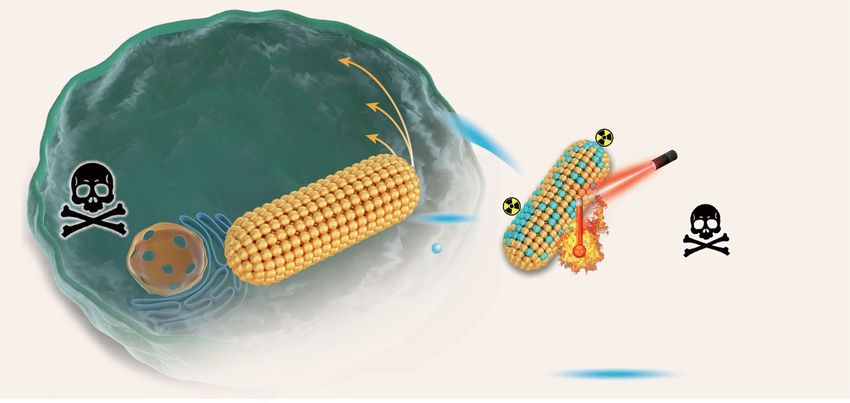
Natl Sci Rev, 2021, Vol. 8, nwaa156

a                                                                                        b                          c                         d                       e
                                          Ymea
                                          Ysim
                                          Ydiff
                                          hkl                                                            HAADF                          Te                       Se         Overlap
    Intensity (a.u.)

                                                                                                           30 nm

                                                                                         g                                   h                               i
                                                                                                                                    3.2 Å
                                                                                                                                    (101)

                       20         30     40        50     60            70          80
                                              2θ (degree)

                                                                                                                                                                                        Downloaded from https://academic.oup.com/nsr/article/8/6/nwaa156/5867802 by guest on 10 October 2021
                                                                                                                                                     2.3 Å
f                       Te 3d           573.2 eV          Se 3d
                                                                     55.1 eV                                                                         (102)
                                                                                             5 nm
    Intensity (a.u.)

                                                                               2 min
                                                                    54.9 eV              j                                   k                               l
                                        572.9 eV

                                                                                                                                             3.9 Å
                                                                                                                                             (010)
                                                                               1 min
                                                                    54.2 eV

                                          572.2 eV
                                                                               0 min

                            585   580   575   570       65     60      55      50            5 nm
                                         Binding energy (eV)

Figure 1. The structure, morphology and chemical composition of TeSex nano-alloys (TS3). (a) Measured (red line) and simulated (blue line) XRD patterns,
and differentiated profiles (green line) between them obtained from the Rietveld refinement of TS3 using P31 21 space group with hexagonal unit cell,
where the inset is the perspective view of the simulated crystal structure of TeSex nano-alloys (the atoms represent either Te or Se). HAADF-STEM
(b) and energy dispersive spectrum (EDS) elemental-mapping images (c−e) of TeSex nano-alloys. (f) Depth profiling XPS spectra of TeSex nano-alloys
exposed to Ar+ for 0, 1 and 2 min. Atomically resolved HAADF-STEM images acquired along the a-axis direction (g) and c-axis direction (j), with
more detailed views (h, k) and corresponding BF-STEM images (i, l). The scale bars in (h), (i), (k) and (l) correspond to 2 nm, 1 nm, 2 nm and 1 nm,
respectively.

                                                     incorporation amounts still can inhibit cell growth                         degradation of Te nano-rods remarkably (Supple-
                                                     to a certain extent, while TS3 and TS4 did not                              mentary Table S4) in support of depressed toxicity.
                                                     (Fig. 2b). In vivo toxicity of TeSex nano-alloys was                            To understand the mechanisms of the toxicity
                                                     further investigated. After intravenous injection                           of Te nano-rods and alloying detoxification, gene
                                                     with TeSex nano-alloys at the higher dose of                                expression studies were performed by RNA se-
                                                     50 mg kg−1 for one week, all the treated mice                               quencing (RNA-Seq) which allowed quantitative
                                                     were alive and well, and their blood samples were                           measurement of expression levels of genes in 4T1
                                                     taken from the orbital sinus to investigate the                             cells from six groups with different treatments
                                                     toxicity of TeSex nano-alloys. From the standard                            (blank control, Te, TS1, TS2, TS3 and TS4 at
                                                     blood biochemical indexes in Fig. 2c and d, the                             200 μg mL−1 ). We first screened the differen-
                                                     concentrations of aspartate transaminase (AST)                              tially expressed genes (DEGs) between TeSex
                                                     and creatinine (CREA) in the Te-treated group                               nano-alloys and blank control to characterize
                                                     were remarkably higher and lower than that of the                           the functional consequences of gene expression
                                                     blank control group, respectively, suggesting that                          changes induced by TeSex nano-alloys. As shown
                                                     Te nano-rods caused distinct damage to liver and                            in Supplementary Figs S6 and S7, there were 201
                                                     kidney functions. By comparison, all the investi-                           (Te), 172 (TS1), 199 (TS2), 198 (TS3) and
                                                     gated TeSex nano-alloys did not demonstrate visible                         189 (TS4) DEGs between Te/TeSex and blank
                                                     toxicity to liver and kidney. These in vitro and in                         control, respectively, where 110 (Te), 71 (TS1),
                                                     vivo toxicity results therefore suggested that the                          101 (TS2), 80 (TS3) and 114 (TS4) genes were
                                                     incorporation of Se into Te nano-rods at a relatively                       up-regulated by Te/TeSex , respectively. The heat
                                                     high amount can effectively reduce their toxicity. In                       map in Supplementary Fig. S8 clearly shows that
                                                     addition, different from TeSe nanoheterojunctions                           the negative effect of Te on gene expression was
                                                     [46], Te nano-rods synthesized in this work can                             significantly attenuated by the incorporation of Se.
                                                     degrade by only 1.4% in water after immersion for                           To further identify the functions of these DEGs,
                                                     8 months, and nano-alloying of TeSex inhibited the                          we performed gene ontology (GO) analysis. In
                                                                                                        Page 4 of 12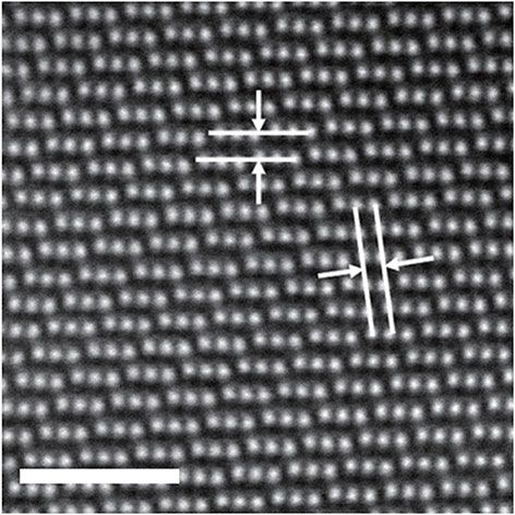
Natl Sci Rev, 2021, Vol. 8, nwaa156
a Te TS1 TS2 b Te TS1 TS2 c Blank Te TS1
d
TS3 TS4 TS3 TS4 400 11k Control
TS2 TS3 TS4
Te
*
Concentration (unit/L)
10k
Concentration (μM)
TS1
Cell viability (a.u.)
Cell viability (a.u.)
1.0 1.0
300 TS2
9k
TS3
8k TS4
200
0.5 0.5 7k
40.0
100
20.0
0.0 0.0 0 0.0
25 50 100 200 25 50 100 200 ALT ALP AST BUN CREA
Concentration (μg/mL) Concentration (μg/mL)
e Te TS1 TS2 TS3 TS4 f
GO:1902115 regulation of organelle assem bly
GO:0032732 positive regulation of interleukin−1 production
Subunit
Downloaded from https://academic.oup.com/nsr/article/8/6/nwaa156/5867802 by guest on 10 October 2021
GO:0071840 cellular component organization or biogenesis
GO:0043933 macromolecular complex subunit organization GSTs GSH organization
GO:0071822 protein complex subunit organization
GO:0007010 cytoskeleton organization
GO:2000379 positive regulation of reactive oxygen species metabolic process
GO:0006955 immune response
GO:0006952 defense response
GO:0098542 defense response to other organism
GO:0002252 immune effector process
GO:0071345 cellular response to cytokine stimulus
GO:0034097 response to cytokine
GO:0045087 innate immune response Te TeSex Apoptosis
GO:0043901 negative regulation of multi−organism process
GO:0034340 response to type I inter feron
GO:0035458 cellular response to inter feron−beta Increase
GO:0035456 response to interferon−beta
GO:0019221 cytokine−mediated signaling path way 8
GO:0051607 defense response to virus 6
GO:0009615 response to virus
GO:0051707 response to other organism 4
GO:0043207 response to external biotic stimulus 2
-log10p
GO:0009607 response to biotic stimulus Ribosome Subunit
GO:0051704 multi−organism process 0 RPL7A
-2
disorders organization
GO:0002376 immune system process
GO:0009605 response to external stimulus -4
GO:0006950 response to stress
GO:1903901 negative regulation of viral life cycle -6
GO:0048525 negative regulation of viral process -8
GO:0045069 regulation of viral genome replication Decrease Energy
GO:0019079 viral genome replication GAPD Glycolysis
GO:0045071 negative regulation of viral genome replication production
g 5 Te
h i H 2O 100 μg/mL j 60 2
0.2 W/cm NIR1060
80 50 μg/mL 200 μg/mL 2
0.5 W/cm NIR1060
TS1 70 2
1.0 W/cm NIR1060
Absorbance (a.u.)
4
Temperature (°C)
TS2
Temperature (°C)
TS3 60 60 50
η (%)
3 TS4
40 50
2 40
40
1 20
30
30
0 0
200 400 600 800 1000 1200 Te TS1 TS2 TS3 TS4 0 2 4 6 8 10 0 2 4 6 8 10
Wavelength (nm) Irradiation time (min) Irradiation time (min)
Figure 2. Biocompatibility and photothermal properties of TeSex nano-alloys. Cytotoxicity (a) and cell proliferation (b) of 4T1 cells incubated with
TeSex nano-alloys at varied particle concentrations. Haematological indexes of liver (c) and kidney (d) functions of the mice with intravenous ad-
ministration of TeSex nano-alloys. (e) Heat map showing scaled expression values of the differentially expressed genes for TeSex nano-alloys vs.
blank control comparison. (f) Schematic illustration of significant pathways of Te toxification and TeSex detoxification. (g) Absorption spectra of TeSex
nano-alloys (200 μg mL−1 ). (h) Calculated NIR-II-photothermal conversion efficiencies of Te nano-rods and TeSex nano-alloys with 1060 nm laser.
NIR-II-photothermal curves of TeSex solutions under the irradiation of 1060 nm laser at varied particle concentrations (50, 100 and 200 μg mL−1 )
at the same laser power density of 1 W cm−2 (i), and at different power densities (0.2, 0.5 and 1 W cm−2 ) at the same particle concentration of
100 μg mL−1 (j). P values in c were calculated by two-tailed Student’s t-test (∗ P < 0.05) by comparing with the blank control group.
Fig. 2e, several genes, involving subunit organi- incorporation of Se into Te nano-rods (such as
zation and positive regulation of reactive oxygen TS3) can recover the normal metabolic process of
species (ROS) metabolic process, were remarkably ROS and avoid damage to subunits, thereby greatly
down-regulated by Te nano-rods, but very slightly reducing the toxicity of Te nano-rods. But the
affected by TeSex nano-alloys, especially TS3 defense response from overhigh incorporation of
and TS4 with higher Se incorporation amounts. Se (such as TS4) would possibly cause toxicity. To
Furthermore, defense response was provoked by further identify the pathways of Te toxification and
Te nano-rods and can also be avoided to a certain TeSex detoxification, we performed the pathway
extent by TS1, TS2 and TS3 with relatively lower analysis of DEGs based on the KEGG (Kyoto
Se incorporation amounts. However, excessive Encyclopedia of Genes and Genomes) database.
Se incorporation for TS4 caused the strongest As summarized in Fig. 2f, and Supplementary
defense response. These GO results indicated that Tables S5 and S6, Te nano-rods positively stim-
Te nano-rods disturbed the normal metabolic ulated the drug metabolism pathway, which was
process of ROS and thus caused oxidative stress similar to the response of cells to many toxic
and damage to subunit organization. The moderate substances (Supplementary Fig. S9a). Furthermore,
Page 5 of 12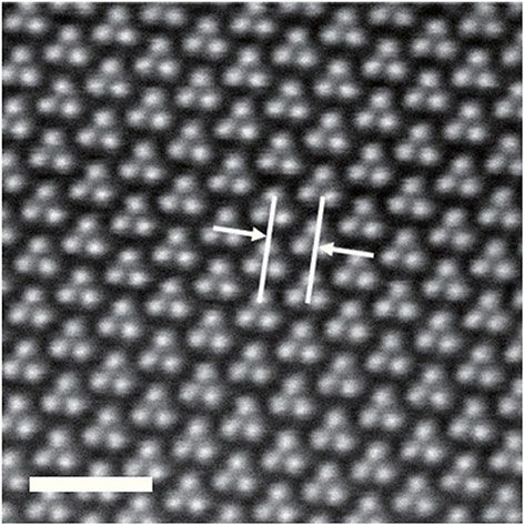
Natl Sci Rev, 2021, Vol. 8, nwaa156

Te nano-rods significantly promoted the                        laser at varied laser power densities (0.2, 0.5 and
metabolism of glutathione (GSH) by up-regulating               1.0 W cm–2 ) and at different particle concentrations
glutathione S-transferase (GSTs), resulting in                 (50, 100 and 200 μg mL–1 ) to investigate its NIR-
the decrease of intracellular GSH level and thus               II-photothermal effect. As shown in Fig. 2i and j,
oxidative stress to impair subunit organization,               the NIR-II-photothermal effect of TeSex nano-alloy
as illustrated in Fig. 2f. Moreover, Te nano-rods              was positively related to both the power density of
also significantly caused ribosome disorders and               the laser and the concentration of TeSex nano-alloy.
inhibited glycolysis by suppressing RPL7A and                  Typically, the temperature of the TeSex solution
GAPD, consequentially causing subunit organiza-                containing 200 μg mL–1 TS3 rose by 36.2◦ C after
tion dysfunction and reduced energy production,                7 min of 1060 nm laser irradiation at 1.0 W cm–2
as illustrated in Fig. 2f. The increased levels of             in great support of thermal therapy of cancer. The
GSTs protein expression and decreased levels of                NIR-II-photothermal stability of TS3 was further

                                                                                                                       Downloaded from https://academic.oup.com/nsr/article/8/6/nwaa156/5867802 by guest on 10 October 2021
RPL7A and GAPD protein expression in Te evi-                   investigated for five laser on/off cycles. As shown
dently confirmed the associated GSH metabolism                 in Supplementary Fig. S13, a temperature change
and energy production. In contrast, the TS3 got                of 36.6◦ C was achieved and did not show signif-
rid of the negative effects on GSTs, RPL7A and                 icant deterioration during five-cycle irradiation,
GAPD (Supplementary Fig. S10). Therefore, the                  suggesting that TeSex nano-alloy had high NIR-
Se incorporation into Te nano-rods to form TeSex               II-photothermal stability. From the above results,
nano-alloys got rid of the negative effects on GSTs,           TeSex nano-alloys with moderate Se incorporation
RPL7A and GAPD (Supplementary Tables S5 and                    demonstrated highest comprehensive performances
S6 and Fig. S10), suppressing the cytotoxicity of Te           including good biocompatibility and high NIR-II-
(Fig. 2f). The GSH depletion of Te nanomaterials by            photothermal efficiency, and was therefore chosen
surface coordination between Te and hydrosulfide               as a theranostic platform to execute the following
group is generally thought to be the main reason               evaluation of theranostic performances. In addition,
for their toxicity [47]. Indeed, this work also found          we further measured photothermal performances
that Te nano-rods could adsorb GSH but TeSex                   of TeSex nano-alloys using a 808 nm laser and com-
nano-alloys almost not (Supplementary Fig. S11).               pared them with the use of a 1060 nm laser. As in
Additionally, this work discovered that Te can                 Fig. 2 and Supplementary Fig. S14, it could be found
also reduce GSH by up-regulating GSTs, and also                that NIR-II-photothermal conversion efficiency of
uncovered other pathways involving ribosome and                TS3 nano-alloys (77.2% for 1060 nm) was higher
glycolysis for the first time. The identified mecha-           than that in the NIR-I window (62.3% for 808 nm,
nisms for Te toxification and TeSex detoxification in          Supplementary Fig. S14b). Although TeSex nano-
this work would greatly favor deep understanding of            alloys had higher extinction coefficient at 808 nm
the origination of Te nanomaterials toxicity and also          than at 1060 nm (Supplementary Fig. S15), they
provide a strategy for developing biocompatible Te-            still exhibited higher photothermal performance at
based nanomaterials for biomedical applications.               1060 nm (Fig. 2, Supplementary Figs S14 and S16)
    After detoxification, the NIR-II-photothermal              owing to higher photothermal efficiencies. There-
effect of TeSex nano-alloys was evaluated. As shown            fore, at the same laser power density, a 1060 nm
by UV–VIS–NIR spectra in Fig. 2g, all the investi-             laser should have a higher tissue penetration depth
gated TeSex nano-alloys (TS1–TS4) had distinct                 than a 808 nm laser, and PAI performance of TS3
NIR-II light absorption. The Se incorporation led to           nano-alloys in the NIR-II window could be better
the blue shift of absorption spectra and the reduc-            than that in the NIR-I window [27], implying the
tion in the NIR-II absorbance of Te nano-rods, but             possibility of using TS3 nano-alloys for photoa-
remarkably enhanced their NIR-II-photothermal                  coustic imaging in both NIR-I and NIR-II windows.
conversion efficiencies. As shown in Fig. 2h and               In addition, we have executed the measurement
Supplementary Fig. S12, the NIR-II-photothermal                of singlet oxygen yield under NIR irradiation, and
conversion efficiency of TeSex nano-alloys gradually           found that no distinct singlet oxygen was generated
increased and then decreased with the increase of              by TeSex nano-alloys (Supplementary Fig. S17),
Se incorporation amount. TS3 exhibited the highest             possibly because most of the photo energy had been
NIR-II-photothermal conversion efficiency (η) of               converted to heat.
77.2% under the irradiation of a 1060 nm laser,
which is much higher than that of Te nano-rods
(53.3%) and other reported NIR-II-photothermal
nanomaterials such as Au nanostar@MOF (48.5%)                  In vivo multimodal imaging performances
[48], Nb2 C nanosheet (46.7%) [19] and Pt spiral               of TeSex nano-alloys
(52.5%) [49]. Moreover, the aqueous solution                   Inherent imaging functions of theranostic nanoplat-
of TS3 nano-alloy was exposed to the 1060 nm                   forms are very helpful for precision medicine.
                                        Page 6 of 12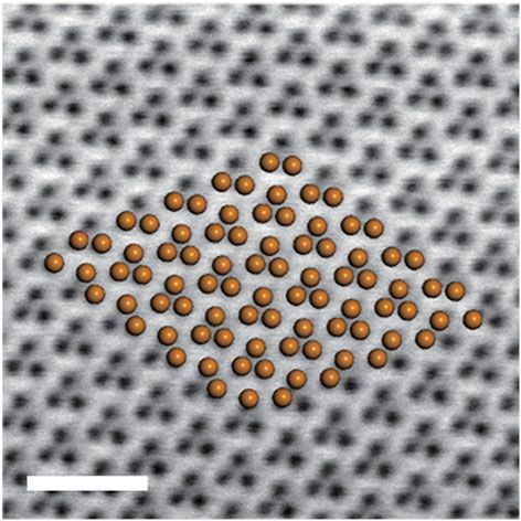
Natl Sci Rev, 2021, Vol. 8, nwaa156
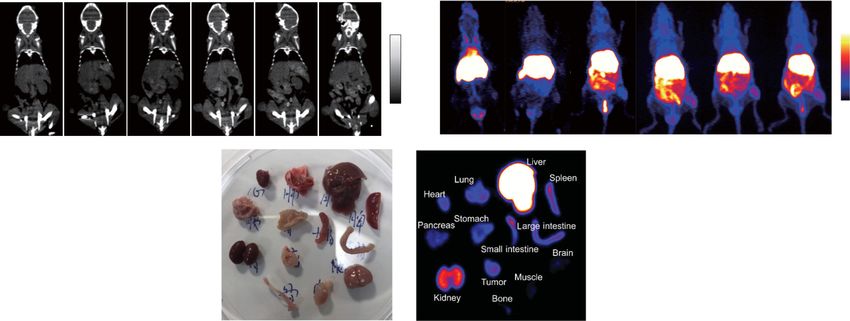
Especially, multimodal imaging with complemen- have no visible kidney/heart/lung toxicities and
tary advantages can be used to accurately guide longer circulation time, as well as tumor-targeted
cancer therapy. Based on the unique properties of ability in favor of tumor-targeted therapy. As in
TeSex nano-alloys in photothermal conversion, sur- Fig. 3e and f, intratumoral CT signal (yellow circles)
face incorporation/coordination and high density gradually increased and achieved the maximum at
(high atomic number), we tried to uncover mul- about 8 h post injection, indicating gradual intratu-
timodal PT/PA/PET/CT imaging performances moral accumulation process of TeSex nano-alloys in
of TeSex nano-alloys with the 4T1 tumor-bearing accordance with the above-mentioned PA imaging
mice model. As to PT imaging, TeSex nano-alloys results. Interestingly, we also observed that CT
(100 μL of TS3 at 2 mg mL–1 ) were intravenously signals in the kidney (red arrows, Supplementary
injected into mice, when tumors grew up to Fig. S21) and bladder (red circles, Supplementary
100 mm3 , followed by 1060 nm laser irradiation Fig. S21) enhanced with time, indicating that TeSex
Downloaded from https://academic.oup.com/nsr/article/8/6/nwaa156/5867802 by guest on 10 October 2021
(1 W cm–2 , 5 min) after 8 h post injection. The TeSex nano-alloys could be excreted through the urinary
group had a remarkably higher increase of temper- system, possibly owing to their small particle size
ature in the irradiated tumor site compared with (about 43 nm, Supplementary Fig. S4). Further-
the phosphate buffer saline (PBS) control group more, the radionuclide 64 Cu was facilely labeled
(Fig. 3a and b). After 1 min NIR-II light irradiation, to TeSex nano-alloys (TS3) by a surface coordi-
the increases of temperature in the TeSex and PBS nation method for PET imaging. Supplementary
groups were 17.6◦ C and 4.2◦ C, respectively, sug- Fig. S22 showed about 90.6% 64 Cu-labeled effi-
gesting that the irradiated tissue itself had low NIR- ciency of TeSex nano-alloys which was measured by
II-photothermal effect but TeSex nano-alloys effec- instant thin layer chromatography (iTLC). Then,
tively accumulated in the tumor in a passive targeting we employed PET to evaluate the in vivo delivery
way and exhibited high NIR-II-photothermal effect and biodistribution of TeSex nano-alloys. The
owing to high NIR-II-photothermal conversion decay-corrected PET images (Fig. 3g) displayed a
efficiency (Fig. 2h). Based on NIR-II-photothermal high tumor-to-background contrast in the TeSex -
64
effect, the PA imaging (PAI) performance of TeSex Cu treated 4T1 tumor-bearing nude mice. The
nano-alloys was further evaluated in vitro and in vivo tumor uptake efficiency of TeSex nano-alloys was
on the 4T1 tumor-bearing mice model. TeSex nano- measured by using a quantitative three-dimensional
alloys exhibited a high photoacoustic coefficient volume-of-interest analysis method. As shown in
of 0.109 mL mg−1 at 810 nm (Supplementary Fig. Fig. 3g–i, the intratumoral accumulation of TeSex
S18). As shown in Fig. 3c, the tumor itself displayed nano-alloys reached the maximum after about 12 h
relatively low PA signal before injection. After intra- post injection, and TeSex nano-alloys which were
venous injection of TeSex nano-alloys (TS3), the in- taken up by liver, spleen and kidney were gradually
tratumoral PA signal intensity gradually augmented eliminated with time. In Fig. 3g, the metabolic
over time and reached the maximum value at about process of TeSex nano-alloys was also clearly visible
8 h post injection (Fig. 3d), suggesting efficient (yellow arrows). At 24 h post injection, the mice
intratumoral accumulation of TeSex nano-alloys in were sacrificed and major organs were collected for
accordance with the above-mentioned PT results. biodistribution study. As shown in Fig. 3j–l, 6.42%
Leveraging the virtue of large X-ray absorption ID g−1 tumor uptake of TeSex nano-alloys was
coefficient of high atomic number elements for achieved at 24 h post injection, and other particles
CT contrast and the strong affinity of chalcogen to mainly distributed on liver, spleen, kidney, etc. Even
transitional metal ions for PET imaging [50,51], though TeSex nano-alloys widely distributed in the
we anticipated that TeSex nano-alloys could impart body, their continuous excretion could reduce the
the quantitative measure of their biodistributions potential risk of toxicity. In addition, the biodistri-
and metabolic processes by PET/CT imaging, be- butions of TeSex nano-alloys in major organs were
yond the localization photoacoustic/photothermal also determined by ICP-OES at the different time
(PT) imaging. We first investigated CT contrast points (2, 4, 8, 12 and 24 h) after injection (n =
performances of TeSex nano-alloys in vitro and in 3). As shown in Supplementary Figs S20 and S23,
vivo with 4T1 tumor-bearing mice with intravenous the ICP-OES results more accurately reflect the
injection of TS3 (100 μL, 10 mg mL–1 ). TeSex biodistribution of TeSex nano-alloys in basic accor-
nano-alloys exhibited a considerable X-ray absorp- dance with PET results, and the blood circulation
tion coefficient of 2.3 HU/mM equal to that of half-time of TS3 was calculated to be about 1.21 h.
an aqueous iodine standard (iopamidol) which is Nevertheless, TeSex nano-alloys were confirmed
popularly used clinically at 140 kV of X-ray tube to be an excellent theranostic platform with multi-
voltage [52] (Supplementary Fig. S19). In addition, modal PT/PA/CT/PET imaging functions in favor
superior to the iodine standard, TeSex nano-alloys of guiding and monitoring cancer treatment.
Page 7 of 12
Natl Sci Rev, 2021, Vol. 8, nwaa156
a 0s 20 s 40 s 1 min 2 min 3 min c 0h 2h 4h
High
PBS
60 °C
8h 12 h 24 h
TeSex
25 °C
Low
b d f h
Downloaded from https://academic.oup.com/nsr/article/8/6/nwaa156/5867802 by guest on 10 October 2021
TeSex nano-alloys 100
0.4 6
PBS
60
Temperature (°C)
PA intensity (a.u.)
75
CT value (HU)
4
ID%/g
50
0.2 50 Tumor
Muscle
2
40 25
30 0.0 0 0
0 60 120 180 0 2 4 8 12 24 0 2 4 8 12 24 0 5 10 15 20 25
Time (s) Time (h) Time (h) Time (h)
e 0h 2h 4h 8h 12 h 24 h g 0.5 h 1h 3h 6h 12 h 24 h
High 15%
Low 0%
ID/g
i 80 Liver j Lu Li k l
Spleen He 30
Liver
Sp
Intestine
Large intestine
60 Heart St
Small intestine
Pa
ID%/g
Kidney
ID%/g
Spleen
40 LIn
Tu
Stomach
SIn 15
Tumor
Pancreas
Kidney
Heart
Ki Br
Lung
20
Muscle
Brain
Bone
Bo
Mu
0
0 5 10 15 20 25 0
Time (h) Tissue
Figure 3. In vivo multimodal imaging performances of TeSex nano-alloys (TS3). (a) In vivo photothermal imaging tracking of one 4T1 tumor-bearing
mouse under the 1060 nm laser irradiation after intravenous injection with PBS or TeSex nano-alloys, and (b) the temperature change at the tumor site.
(c) In vivo PA images and (d) PA signal intensity change at the tumor site of one 4T1 tumor-bearing mouse before and after intravenous injection of TeSex
nano-alloys. (e) In vivo CT images and (f) CT signal intensity change at the tumor site of one 4T1 tumor-bearing mouse before and after intravenous
injection of TeSex nano-alloys. (g) In vivo PET images of one 4T1 tumor-bearing mouse obtained at different time points post injection of 64 Cu-labeled
TeSex nano-alloys. (h, i) Quantitative biodistribution obtained from ROI (region of interest) analysis of PET images. Ex vivo digital (j) and PET (k) images
and the corresponding biodistribution (l) of main organs after injection for 24 h.
fluorescence gradually increased inside cells (Fig. 4b
In vitro and in vivo NIR-II-photothermal
and Supplementary Fig. S24), indicating that TeSex
therapy performances of TeSex nano-alloys were efficiently internalized into 4T1
nano-alloys cells due to their small size of 43 nm. Thereafter,
Cellular uptake of TeSex nano-alloys was firstly NIR-II-photothermal cytotoxicity of TeSex nano-
investigated in vitro. TeSex nano-alloys were facilely alloys against varied cancer cell lines (4T1, B16,
labeled with red fluorescent dye 5,10,15,20-tetra(4- HeLa cells) at different particle concentrations and
pyridyl)-21H,23H-porphine (TPyP) by virtue of at different laser power densities were investigated
its coordination capability. Confocal fluorescence using the standard CCK-8 assay. From Fig. 4a and
images of 4T1 cells incubated with TPyP-labeled Supplementary Fig. S25, TeSex nano-alloys without
TeSex nano-alloys for 1 and 2 h showed that red 1060 nm laser irradiation did not show observable
Page 8 of 12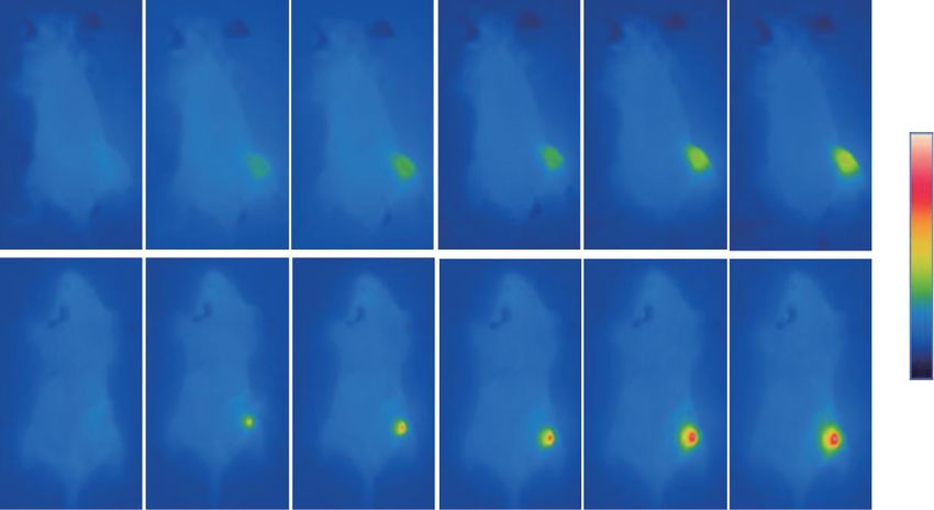
Natl Sci Rev, 2021, Vol. 8, nwaa156
a Without NIR
2
0.2 W/cm NIR1060
b DAPI TPyP-TeSex
c Control NIR1060
2 2
0.5 W/cm NIR1060 1.0 W/cm NIR1060
1.0
Cell viability (a.u.)
0.5 Bright Merge TeSex TeSex+NIR1060
0.0
0 12.5 25 50 100 200
Concentration (μg/mL)
d e PBS
Downloaded from https://academic.oup.com/nsr/article/8/6/nwaa156/5867802 by guest on 10 October 2021
PBS
2.0 PBS+NIR808
PBS+NIR1060
Tumor volume (cm3)
PBS+NIR808
TeSex nano-alloys
1.5 TeSex nano-alloys+NIR808
TeSex nano-alloys+NIR1060 PBS+NIR1060
1.0
TeSex
***
0.5 TeSex+NIR808
0.0 TeSex+NIR1060
0 5 10 15 20
0.0 0.2 0.4 0.6 0.8 1.0 1.2
Time (day)
Tumor weight (g)
f g h
Without NIR With NIR808 With NIR1060 Control Control
12k
300 -1
10 mg kg TeSex nano-alloys 10 mg kg-1 TeSex nano-alloys
Concentration (unit/L)
-1
50 mg kg TeSex nano-alloys 11k 50 mg kg-1 TeSex nano-alloys
Concentration (μM)
PBS
250 10k
200 9k
8k
150
7k
100 40.0
TeSex
50 20.0
0 0.0
ALT ALP AST BUN CREA
Figure 4. In vitro and in vivo NIR-II-photothermal therapy performances of TeSex nano-alloys (TS3). (a) Viability of 4T1 cells
after treatment with various concentrations of TeSex nano-alloys followed by 1060 nm laser irradiation for 5 min at different
power densities (0, 0.2, 0.5 and 1.0 W cm−2 ). (b) Cell internalization of TPyP-labeled TeSex nano-alloys in 4T1 cells (scale
bar: 10 μm). (c) Confocal fluorescence images of TeSex nano-alloys induced photothermal ablation (1.0 W cm−2 , 5 min,
100 μg mL−1 ) after the 1060 nm laser irradiation (scale bar, 100 μm). (d) Tumor growth of 4T1 tumor-bearing mice with or
without laser irradiation after intravenous injection with PBS or 100 μL TeSex nano-alloys. (e) 4T1 tumor weight comparison
after 22-day treatment and the corresponding digital images of extracted tumors (n = 5). (f) H&E staining images of tumor
tissues (scale bar, 100 μm). Haematological indexes of liver (g) and kidney (h) functions of the mice with intravenous admin-
istration of TeSex nano-alloys. P values in (d) and (e) were calculated by two-tailed Student’s t-test (∗∗∗ P < 0.005, ∗∗ P < 0.01)
by comparing with the PBS control group.
cytotoxicity to all the investigated cancer cells at a sion of TeSex nano-alloys in the NIR-I window, we
concentration up to 200 μg mL−1 . Under 1060 nm also found that TeSex nano-alloys had remarkable
laser irradiation, TeSex nano-alloys exhibited NIR-I-photothermal cytotoxicity against various
remarkable concentration- and power-dependent cancer cells (Supplementary Fig. S27), which can
cytotoxicity against various cancer cells (Fig. 4a and also be an alternative candidate for photothermal
Supplementary Fig. S25). Typically, 0.5 W cm−2 therapy of cancer.
1060 nm laser irradiation for 5 min on 100 μg mL−1 Encouraged with the above-confirmed NIR-
TeSex nano-alloys treated cancer cells killed 83.5% II-photothermal effects of TeSex nano-alloys, the
4T1 cells, 98.2% HeLa cells and 81.6% B16 cells. NIR-I-/NIR-II-photothermal ablation of solid
Additionally, green (calcein-AM) and red (propid- tumors with TeSex nano-alloys was further eval-
ium iodide) fluorescence staining results also clearly uated in vivo. Firstly, mice bearing breast 4T1
demonstrated high NIR-II-photothermal cytotoxic- tumor were randomly divided into six groups
ity of TeSex nano-alloys (Fig. 4c). In addition, based (n = 5 per group) with approximately the same
on obvious absorption and photothermal conver- tumor volume (ca. 100 mm3 ): (i) the PBS group
Page 9 of 12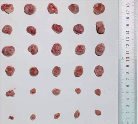
Natl Sci Rev, 2021, Vol. 8, nwaa156

(blank control), (ii) the PBS+NIR808 group,                     and had been proved to be a kind of versatile
(iii) the PBS+NIR1060 group, (iv) the TeSex                     nanotheranostic platform with multiple functions
group, (v) the TeSex +NIR808 group, and (vi) the                of NIR-II-photothermal therapy and multimodal
TeSex +NIR1060 group. The NIR treatment group                   PT/PA/PET/CT imaging, enabling multimodal
referred to the mice intravenously injected with PBS            imaging-guided NIR-II-photothermal therapy of
or TeSex nano-alloys (100 μL TS3, 10 mg kg−1 ,                  cancer with high theranostic performances.
three times at Day 1, Day 3 and Day 5) and irra-
diated with 808 nm or 1060 nm laser irradiation
(1.0 W cm−2 for 5 min) at fixed time points (Day
                                                                METHODS
2, Day 4 and Day 6) after 8 h post injection to                 The controlled preparation of rod-like TeSex
ensure sufficient thermal damage to tumor cells.                nano-alloys with different ratios of Te/Se and
From Fig. 4d, tumor growth was not affected by                  length/diameter were realized by a facile hy-

                                                                                                                                       Downloaded from https://academic.oup.com/nsr/article/8/6/nwaa156/5867802 by guest on 10 October 2021
TeSex nano-alloys, but significantly suppressed by              drothermal method, where the Se contents were
combination of TeSex nano-alloys with 1060 nm                   adjusted by tuning the molar ratio of tellurite to
laser irradiation. The 808 nm laser irradiation                 selenite. The comprehensive details, chemicals and
plus TeSex nano-alloys also generated remarkable                characterizations are in the Supplementary data.
inhibition effect on tumor growth, but the inhibition
efficacy was not as efficient as that of the 1060 nm
                                                                SUPPLEMENTARY DATA
laser irradiation. After 21-day treatment, tumors
were dissected to photograph and weigh as shown                 Supplementary data are available at NSR online.
in Fig. 4e. The results further demonstrated remark-
able inhibition effect of NIR-photothermal TeSex                ACKNOWLEDGEMENTS
nano-alloys on tumor growth. Although tumors
                                                                We thank the Instrumental Analysis Center of Shenzhen Univer-
had not been completely eradicated which possibly
                                                                sity (Xili campus) for the assistance in material characterizations.
resulted from short irradiation time of only 5 min,
we felt optimistic because in vivo therapeutic efficacy
could become better if we extended NIR irradiation              FUNDING
time. Furthermore, the hematoxylin-eosin (H&E)                  This work was supported by the National Natural Science Foun-
staining of major organs and tumor tissues was                  dation of China (51701127 and 51872188), the Special Funds for
conducted (Fig. 4f and Supplementary Fig. S28).                 the Development of Strategic Emerging Industries in Shenzhen
It was found that NIR-II-photothermal therapy                   (20180309154519685), the Shenzhen Basic Research Program
caused significant tumor cell damage. But there                 (JCYJ20170302151858466 and JCYJ20170818093808351),
was no obvious damage in all major organs after                 the Shenzhen Peacock Plan (KQTD2016053112051497
treatment (Supplementary Fig. S28) and no loss                  and KQTD2016053112042971), the High-Level University
in body weight during treatment (Supplementary                  Construction Project of Health Science Center, Shenzhen
Fig. S29), suggesting no obvious systematic toxicity            University (860-00000210), the Educational Commission of
                                                                Guangdong Province (2016KTSCX126), and the Guangdong
of TeSex nano-alloys. Even at the high injection dose
                                                                Special Support Program and Pengcheng Scholar Program.
of 50 mg kg−1 which was five-fold higher than treat-
                                                                This work was also supported, in part, by the University of
ment dose, no damage to liver and kidney functions              Wisconsin—Madison and the National Institutes of Health
was visible (Fig. 4g and h). These results indicated            (P30CA014520).
that TeSex nano-alloys were a biocompatible and
high-efficacy NIR-photothermal platform.
                                                                AUTHOR CONTRIBUTIONS
                                                                Q.J.H., C.L.S., X.L. and Z.K.J. conceived the study. Q.J.H. and
CONCLUSION                                                      C.L.S. supervised this project. X.L., Q.J. and Z.K.J. planned and
In conclusion, a series of TeSex nano-alloys with               performed the experiments, and collected and analyzed the data.
different ratios of Se/Te and length/diameter                   Q.J.H., X.T.W., B.W., Z.C.W., Y.S.X., T.Y.C., J.W.E. and W.B.C.
were controllably synthesized by the facile co-                 assisted with the experiments and characterization. Q.J.H., C.L.S.,
precipitation method. Incorporating a moderate                  X.L. and Z.K.J. co-wrote the manuscript.
content of Se (x = 0.43) into the lattice of Te nanos-
                                                                Conflict of interest statement. None declared.
tructure effectively eradicated the toxicity of Te,
which mainly originated from GSTs up-regulation,
and RPL7A/GAPD down-regulation caused
subunit organization dysfunction and energy pro-                REFERENCES
duction loss. TeSex nano-alloys exhibited high NIR-                 1. Chen HM, Zhang WZ and Zhu GZ et al. Rethinking cancer nan-
II-photothermal conversion efficiency (77.2%),                        otheranostics. Nat Rev Mater 2017; 2: 17024–41.

                                        Page 10 of 12
Natl Sci Rev, 2021, Vol. 8, nwaa156
2. Lim EK, Kim T and Paik S et al. Nanomaterials for theranostics: recent advances 21. Li SL, Chen YH and Liu HB et al. Graphdiyne materials as nanotransducer for in
and future challenges. Chem Rev 2015; 115: 327–94. vivo photoacoustic imaging and photothermal therapy of tumor. Chem Mater
3. Chen S, Weitemier AZ and Zeng X et al. Near-infrared deep brain stimula- 2017; 29: 6087–94.
tion via upconversion nanoparticle-mediated optogenetics. Science 2018; 359: 22. Tao W, Ji X and Zhu X et al. Two-dimensional antimonene-based photonic
679–84. nanomedicine for cancer theranostics. Adv Mater 2018; 30: 1802061.
4. Lukianova-Hleb EY, Kim YS and Belatsarkouski I et al. Intraoperative diagnos- 23. Ji XY, Kang Y and Ouyang J et al. Synthesis of ultrathin biotite nanosheets
tics and elimination of residual microtumours with plasmonic nanobubbles. Nat as an intelligent theranostic platform for combination cancer therapy. Adv Sci
Nanotechnol 2016; 11: 525–32. 2019; 6: 1901211.
5. Xue XD, Huang Y and Bo RN et al. Trojan horse nanotheranostics with dual 24. Tao W, Kong N and Ji XY et al. Emerging two-dimensional monoelemen-
transformability and multifunctionality for highly effective cancer treatment. tal materials (Xenes) for biomedical applications. Chem Soc Rev 2019; 48:
Nat Commun 2018; 9: 3653. 2891–912.
6. Yu GC, Yang Z and Fu X et al. Polyrotaxane-based supramolecular theranostics. 25. Tang ZM, Kong N and Ouyang J et al. Phosphorus science-oriented design and
Downloaded from https://academic.oup.com/nsr/article/8/6/nwaa156/5867802 by guest on 10 October 2021
Nat Commun 2018; 9: 766. synthesis of multifunctional nanomaterials for biomedical applications. Matter
7. Lovell JF, Jin CS and Huynh E et al. Porphysome nanovesicles generated by por- 2020; 2: 297–322.
phyrin bilayers for use as multimodal biophotonic contrast agents. Nat Mater 26. Kong N, Ji XY and Wan JQ et al. ROS-mediated selective killing effect of black
2011; 10: 324–32. phosphorus: mechanistic understanding and its guidance for safe biomedical
8. Kim JW, Galanzha EI and Shashkov EV et al. Golden carbon nanotubes as mul- applications. Nano Lett 2020; 20: 3943–55.
timodal photoacoustic and photothermal high-contrast molecular agents. Nat 27. Jiang YY, Li JC and Zhen X et al. Dual-peak absorbing semiconducting copoly-
Nanotechnol 2009; 4: 688–94. mer nanoparticles for first and second near-infrared window photothermal ther-
9. Seo WS, Lee JH and Sun XM et al. FeCo/graphitic-shell nanocrystals as apy: a comparative study. Adv Mater 2018; 30: 1705980.
advanced magnetic-resonance-imaging and near-infrared agents. Nat Mater 28. Sun TT, Dou JH and Liu S et al. Second near-infrared conjugated polymer
2006; 5: 971–6. nanoparticles for photoacoustic imaging and photothermal therapy. ACS Appl
10. Cheng L, Wang C and Feng L et al. Functional nanomaterials for phototherapies Mater Inter 2018; 10: 7919–26.
of cancer. Chem Rev 2014; 114: 10869–939. 29. Men XJ, Wang F and Chen HB et al. Ultrasmall semiconducting polymer dots
11. Liu YJ, Bhattarai P and Dai ZF et al. Photothermal therapy and photoacous- with rapid clearance for second near-infrared photoacoustic imaging and pho-
tic imaging via nanotheranostics in fighting cancer. Chem Soc Rev 2019; 48: tothermal cancer therapy. Adv Funct Mater 2020; 30: 1909673.
2053–108. 30. Wang Q, Xu JZ and Geng RY et al. High performance one-for-all phototheranos-
12. MacLeod MJ, Goodman AJ and Ye HZ et al. Robust gold nanorods stabilized by tics: NIR-II fluorescence imaging guided mitochondria-targeting phototherapy
bidentate N-heterocyclic-carbene–thiolate ligands. Nat Chem 2019; 11: 57–63. with a single-dose injection and 808 nm laser irradiation. Biomaterials 2020;
13. Ding XG, Liow CH and Zhang MX et al. Surface plasmon resonance enhanced 231: 119671.
light absorption and photothermal therapy in the second near-infrared window. 31. Zhu R, Su LC and Dai JY et al. Biologically responsive plasmonic assem-
J Am Chem Soc 2014; 136: 15684–93. blies for second near-infrared window photoacoustic imaging-guided concur-
14. Chen M, Tang SH and Guo ZD et al. Core-shell Pd@Au nanoplates as thera- rent chemo-immunotherapy. ACS Nano 2020; 14: 3991–4006.
nostic agents for in-vivo photoacoustic imaging, CT imaging, and photothermal 32. He Z, Yang Y and Liu JW et al. Emerging tellurium nanostructures: controllable
therapy. Adv Mater 2014; 26: 8210–6. synthesis and their applications. Chem Soc Rev 2017; 46: 2732–53.
15. Chang Y, Feng YL and Cheng Y et al. Anisotropic plasmonic metal heterostruc- 33. Liu YY, Wu WZ and Goddard WA. Tellurium: fast electrical and atomic transport
tures as theranostic nanosystems for near infrared light-activated fluorescence along the weak interaction direction. J Am Chem Soc 2018; 140: 550–3.
amplification and phototherapy. Adv Sci 2019; 6: 1900158. 34. Wu WZ, Qiu G and Wang YX et al. Tellurene: its physical properties, scal-
16. Cheng Y, Chang Y and Feng YL et al. Deep-level defect enhanced photothermal able nanomanufacturing, and device applications. Chem Soc Rev 2018; 47:
performance of bismuth sulfide-gold heterojunction nanorods for photothermal 7203–12.
therapy of cancer guided by computed tomography imaging. Angew Chem Int 35. Amani M, Tan CL and Zhang G et al. Solution-synthesized high-mobility tel-
Ed 2018; 57: 246–51. lurium nanoflakes for short-wave infrared photodetectors. ACS Nano 2018; 12:
17. Chen WS, Ouyang J and Liu H et al. Black phosphorus nanosheet-based drug 7253–63.
delivery system for synergistic photodynamic/photothermal/chemotherapy of 36. Chaudhary S, Umar A and Mehta SK. Selenium nanomaterials: an overview of
cancer. Adv Mater 2017; 29: 1603864. recent developments in synthesis, properties and potential applications. Prog
18. Yang K, Feng L and Hong H et al. Preparation and functionalization of graphene Mater Sci 2016; 83: 270–329.
nanocomposites for biomedical applications. Nat Protoc 2013; 8: 2392– 37. Yu N, Li JN and Wang ZJ et al. Blue Te nanoneedles with strong NIR photother-
403. mal and laser-enhanced anticancer effects as ‘all-in-one’ nanoagents for syn-
19. Lin H, Gao SS and Dai C et al. A two-dimensional biodegradable niobium car- ergistic thermo-chemotherapy of tumors. Adv Healthc Mater 2018; 7: 1800643.
bide (MXene) for photothermal tumor eradication in NIR-I and NIR-II biowin- 38. Huang W, Huang YY and You YY et al. High-yield synthesis of multifunctional
dows. J Am Chem Soc 2017; 139: 16235–47. tellurium nanorods to achieve simultaneous chemo-photothermal combination
20. Zhu HJ, Lai ZC and Fang Y et al. Ternary chalcogenide nanosheets with ultrahigh cancer therapy. Adv Funct Mater 2017; 27: 1701388.
photothermal conversion efficiency for photoacoustic theranostics. Small 2019; 39. Yang T, Ke HT and Wang QL et al. Bifunctional tellurium nanodots for photo-
13: 1604139. induced synergistic cancer therapy. ACS Nano 2017; 11: 10012–24.
Page 11 of 12Natl Sci Rev, 2021, Vol. 8, nwaa156
40. Xiong LH, Cui R and Zhang ZL et al. Harnessing intracellular biochemical path- 47. Deuticke B, Lütkemeier P and Poser B. Tellurite-induced damage of the ery-
ways for in vitro synthesis of designer tellurium nanorods. Small 2015; 11: throcyte membrane-manifestations and mechanisms. Biochim Biophys Acta
5416–22. Biomembr 1992; 1109: 97–107.
41. Lin Y, Wu Y and Wang R et al. Two-dimensional tellurium nanosheets for pho- 48. Deng XR, Liang S and Cai XH et al. Yolk-shell structured Au nanostar@metal-
toacoustic imaging-guided photodynamic therapy. Chem Commun 2018; 54: organic framework for synergistic chemo-photothermal therapy in the second
8579–82. near-infrared window. Nano Lett 2019; 19: 6772–80.
42. Durgadas CV, Sreenivasan K and Sharma CP. Bright blue emitting 49. Wang QS, Wang H and Yang Y et al. Plasmonic Pt superstructures
CuSe/ZnS/silica core/shell/shell quantum dots and their biocompatibil- with boosted near-infrared absorption and photothermal conversion effi-
ity. Biomaterials 2012; 33: 6420–9. ciency in the second bio-window for cancer therapy. Adv Mater 2019; 31:
43. Liu WH, Choi HS and Zimmer JP et al. Compact cysteine-coated CdSe(ZnCdS) 1904836.
quantum dots for in vivo applications. J Am Chem Soc 2007; 129: 14530–1. 50. Sun XL, Cai WB and Chen XY. Positron emission tomography imag-
44. Ferrando R, Jellinek J and Johnston RL. Nanoalloys: from theory to applications ing using radiolabeled inorganic nanomaterials. Acc Chem Res 2015; 48:
Downloaded from https://academic.oup.com/nsr/article/8/6/nwaa156/5867802 by guest on 10 October 2021
of alloy clusters and nanoparticles. Chem Rev 2008; 108: 845–910. 286–94.
45. Yang Y, Wang K and Liang HW et al. A new generation of alloyed/multimetal 51. Zhao YF, Sultan D and Detering L et al. Copper-64-alloyed gold nanoparticles for
chalcogenide nanowires by chemical transformation. Sci Adv 2015; 1: cancer imaging: improved radiolabel stability and diagnostic accuracy. Angew
e1500714. Chem Int Ed 2014; 53: 156–9.
46. Chen SY, Xing CY and Huang DZ et al. Eradication of tumor growth by delivering 52. Brown AL, Naha PC and Montes VB et al. Synthesis, X-ray opacity, and biolog-
novel photothermal selenium-coated tellurium nanoheterojunctions. Sci Adv ical compatibility of ultra-high payload elemental bismuth nanoparticle X-ray
2020; 6: eaay6825. contrast agents. Chem Mater 2014; 26: 2266–74.
Page 12 of 12You can also read