Optical Detection of Distal Lung Enzyme Activity in Human Inflammatory Lung Disease - Science
←
→
Page content transcription
If your browser does not render page correctly, please read the page content below
AAAS
BME Frontiers
Volume 2021, Article ID 9834163, 11 pages
https://doi.org/10.34133/2021/9834163

Research Article
Optical Detection of Distal Lung Enzyme Activity in Human
Inflammatory Lung Disease

 Alicia Megia-Fernandez ,1 Adam Marshall ,2 Ahsan R. Akram ,2 Bethany Mills ,2
 Sunay V. Chankeshwara ,1 Emma Scholefield ,2 Amy Miele,2 Bruce C. McGorum ,3
 Chesney Michaels,2 Nathan Knighton ,4 Tom Vercauteren ,5 Francois Lacombe,6
 Veronique Dentan,6 Annya M. Bruce,2 Joanne Mair,2 Robert Hitchcock,4 Nik Hirani,2
 Chris Haslett,2 Mark Bradley ,1 and Kevin Dhaliwal 2
 1
 EaStCHEM, The University of Edinburgh School of Chemistry, Joseph Black Building, West Mains Road, Edinburgh, UK EH9 3FJ
 2
 Translational Healthcare Technologies Group, Centre for Inflammation Research, Queen’s Medical Research Institute,
 University of Edinburgh, 47 Little France Crescent, Edinburgh BioQuarter, Edinburgh, UK EH16 4TJ
 3
 The Roslin Institute and Royal (Dick) School of Veterinary Studies, University of Edinburgh, Easter Bush, Midlothian, UK EH25 9RG
 4
 Department of Biomedical Engineering, University of Utah, 36 S Wasatch Dr, Salt Lake City, UT 84112, USA
 5
 School of Biomedical Engineering & Imaging Sciences, King’s College London, London, UK SE1 7EH
 6
 Mauna Kea Technologies, 9, Rue d’Enghien, Paris, France 75010

 Correspondence should be addressed to Mark Bradley; mark.bradley@ed.ac.uk and Kevin Dhaliwal; kev.dhaliwal@ed.ac.uk

 Alicia Megia-Fernandez, Adam Marshall, Ahsan R. Akram, and Bethany Mills contributed equally to this work.

 Received 10 August 2020; Accepted 10 March 2021; Published 7 April 2021

 Copyright © 2021 Alicia Megia-Fernandez et al. Exclusive Licensee Suzhou Institute of Biomedical Engineering and Technology,
 CAS. Distributed under a Creative Commons Attribution License (CC BY 4.0).

 Objective and Impact Statement. There is a need to develop platforms delineating inflammatory biology of the distal human lung.
 We describe a platform technology approach to detect in situ enzyme activity and observe drug inhibition in the distal human lung
 using a combination of matrix metalloproteinase (MMP) optical reporters, fibered confocal fluorescence microscopy (FCFM), and
 a bespoke delivery device. Introduction. The development of new therapeutic agents is hindered by the lack of in vivo in situ
 experimental methodologies that can rapidly evaluate the biological activity or drug-target engagement in patients. Methods. We
 optimised a novel highly quenched optical molecular reporter of enzyme activity (FIB One) and developed a translational
 pathway for in-human assessment. Results. We demonstrate the specificity for matrix metalloproteases (MMPs) 2, 9, and 13 and
 probe dequenching within physiological levels of MMPs and feasibility of imaging within whole lung models in preclinical
 settings. Subsequently, in a first-in-human exploratory experimental medicine study of patients with fibroproliferative lung
 disease, we demonstrate, through FCFM, the MMP activity in the alveolar space measured through FIB One fluorescence
 increase (with pharmacological inhibition). Conclusion. This translational in situ approach enables a new methodology to
 demonstrate active drug target effects of the distal lung and consequently may inform therapeutic drug development pathways.

1. Introduction rapid approaches to assess drugs that cause respiratory
 morbidity and mortality [3].
Respiratory diseases are one of the leading causes of mor- To date, the evaluation of early-phase clinical trials in
bidity and mortality worldwide [1]. Despite the significant human pulmonary disease has relied upon measurements,
burden of disease, there is a paucity of new pharmacologi- which include pulmonary function measurements, six-
cal moieties reaching clinical phases of development and minute walk tests, and imaging such as computerized
even fewer reaching clinical approval with spiraling costs tomography (CT) [4]. These investigations can inform on
of drug development [2]. Furthermore, the recent functional and structural abnormalities but are delayed
COVID-19 global pandemic has highlighted the need for surrogates of active disease and offer little insight into
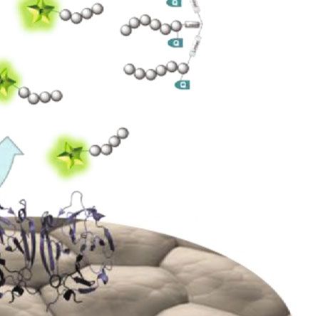
2 BME Frontiers

dynamic molecular processes where therapeutics may be the quenching effect of three quenchers [28]. The molecular
beneficial. Optical imaging modalities provide the potential probe generated (called FIB One (Figure 1)) consisted of a tri-
for rapid readouts of disease activity at the site of pathogen- branched peptide (Pro-Phe-Gly-Nle-Lys-βAla)3, containing
esis, such as the distal lung [5]. Therefore, we developed a three fluorescein units and three methyl red groups
novel optical imaging approach using a combination of an (Figure 1). FIB One was prepared using Fmoc-based solid-
existing miniaturized fibred confocal fluorescent micros- phase peptide synthesis (Scheme S1 and ESI), and after
copy (FCFM) system, a bronchoscope compatible delivery purification, was fully characterized by reverse-phase high-
device, a new molecular probe, and a drug inhibitor to pressure liquid chromatography (RP-HPLC) and matrix-
demonstrate real-time drug-target engagement in the distal assisted laser desorption ionising time-of-flight mass spec-
lung of patients. trometry (MALDI-ToF MS) (Figure 1(b) and ESI).
 The matrix metalloproteinase (MMP) pathway was Selective hydrolysis of the peptidic sequence by the
selected for detection and inhibition within a small patient active enzyme results in an increase in fluorescence due
population (eight patients with predicted aberrant lung to the release of the fragments containing fluorescein
fibrogenesis) to demonstrate the clinical tractability of our (Figure 1(c)), with excitation and emission maxima at
approach. MMPs are mediators of injury and repair, per- 490/530 nm, respectively (Figure 1(d)). FIB One demon-
forming pivotal roles in the turnover of the extracellular strated rapid and specific fluorescent amplification when
matrix (ECM), with dysregulation leading to an aberrant exposed to recombinant enzymes (MMPs 2, 9, and 13),
response in inflammation and tissue repair, which ultimately which was blocked by the pan-MMP inhibitor marimastat
leads to an impairment of organ function [6–9]. MMP dys- (Figure 1(e) and Figure S1), with MALDI-ToF MS showing
regulation has been associated with many acute and chronic confirmation of selective cleavage (Figure S1). In addition,
inflammatory respiratory diseases such as acute respiratory dis- comparison of FIB One with its analogous monomeric
tress syndrome (ARDS) [10], idiopathic pulmonary fibrosis linear structure confirmed improved signal amplification
(IPF) [11], chronic obstructive pulmonary disease (COPD) obtained with the multimeric approach (Figure S2).
[12], and lung cancer [13], with upregulation of the gelatinases
(MMP-2 and MMP-9) often found in IPF [9, 14] and associated 2.2. FIB One Is Cleaved by MMPs in Ex Vivo Primary Human
with a more aggressive disease phenotype [15]. The collagenase Models and in an Ex Vivo Lung Model. To determine the clin-
MMP-13 has also been shown to be abundant in the IPF tissue, ical tractability of this approach and for the compound to be
[16] and as such, there has been considerable interest in the cleaved at physiologically relevant/disease levels of MMPs,
assessment of pulmonary MMP activity as a biomarker of we evaluated the propensity for FIB One to be specifically
fibrotic lung disease [17, 18] and in the therapeutic targeting cleaved by MMPs 2, 9, and 13 in primary human lung tis-
of MMP activity in acute and chronic lung disease [19, 20] sue and also by stimulated human neutrophils. FIB One
and the malignant matrix [21, 22]. was cleaved in both these scenarios with cleavage inhibi-
 The aim of this exploratory translational study was to tion observed following coincubation with marimastat
develop and assess a novel optical reporter that was able to (Figures 2(a) and 2(b)). MALDI-ToF MS confirmed that
image MMP activity (measured though fluorescence increase the cleavage in these models was MMP specific (Figure S3).
of FIB One) within the alveolar space and demonstrate drug- The translational utility required evidence that the MMP
target engagement using a microdosing approach of a code- activity could be rapidly detected in the distal lung in real time
livered MMP inhibitor (moieties delivered being less than (within seconds to minutes) using an endoscopic approach.
100 μg of imaging agent or drug). This methodology and Therefore, we utilised an ex vivo ovine lung model
the demonstration of real-time drug-target engagement in (Figure 2(d)). Here, to demonstrate MMP upregulation, seg-
situ allowed the bedside assessment of pharmacological ments instilled with supernatant from spontaneously occurring
action in human disease using optical imaging. ovine pulmonary adenocarcinoma (OPA) enabled evaluation of
 FIB One (Figure 2(c)). FCFM-based imaging of segments with
2. Results high MMP activity (Figure S3) confirmed a diffuse pattern of
 increased fluorescence across the field of view, which was a
2.1. A Tribranched Scaffold Is Efficiently Cleaved by MMPs characteristic of cleaved FIB One (Figure 2(d)). In contrast,
with Amplification of Fluorescence Signal within Minutes. We vehicle control segments and marimastat (pan-MMP
recently described a MMP peptide substrate [23, 24] that was inhibitor)-dosed segments did not show an increase in FIB
selectively and specifically cleaved by secreted MMPs 2, 9, and One signal (Figure 2(d)). Together, these data supported that
13 resulting in fluorescent signal amplification and designed pulmonary delivery of FIB One could detect MMP activity in
to be resistant to plasmin and other bystander enzymes. How- situ when coupled with FCFM within a size relevant model
ever, although highly selective, this linear optimised probe dem- within physiological concentrations.
onstrated low signal-to-noise following cleavage.
 To maximise the fluorescence amplification, we modified 2.3. GMP-Manufactured FIB One Demonstrated Stability,
the linear probe by using a multiple Förster resonance energy Lack of Toxicity, and Favourable Alveolar Imaging in
transfer (FRET) system with insertion of the substrate pep- Fibrotic Lung Disease in Human Patients. The translational
tide sequence into a tribranched scaffold. This tribranched pathway required toxicological and functional evaluation
structure takes advantage of both the self-quenching effect of FIB One. FIB One was synthesised in accordance with
observed in multifluorophore constructs [25–27] as well as active pharmaceutical ingredient (API) development to
BME Frontiers 3

 HO

 Intens. 0_N13\1: +MS
 O O O
 O
 H
 O
 H
 O
 H
 O
 H
 O
 H
 O
 H
 O
 H
 O O H O
 6465.2218
 H O C N CHC N CHC N CHC N CHC N H2C CHC N CHC N O CHC O N
 CHC
 6465.2218
 CH2 H CH2 CH2 H CH2 O N N O
 HO N O H CH2 H
 O N O CH2
 CH2 CH2 CH2
 O CH2 CH2 CH CH2 CH2 6000 Experimental
 CH3 CH2 CH2
 2
 CH2
 CH2
 CH2
 CH2
 [M+H]+ = 6465
 NH2 NH2
 NH2 NH2
 O
 N
 N
 NH
 N
 O H NH2
 N N
 FAM-PEG2-Pro-Phe-Gly-Nle-Lys- Ala-Lys(MethylRed)-PEG2-(D)Lys-PEG2-(D)Lys H O

 O
 O
 5000
 NH
 FAM-PEG2-Pro-Phe-Gly-Nle-Lys- Ala-Lys(MethylRed)-PEG2-(D)Lys-PEG2-(D)Lys
 1+
 Theoretical 6465.3578
 (a) C324 H453N62O78
 4000

 Fib One

 3000
 Off On
 6440 6460 6480 6500

 2000

 MMPs
 1000

Extracellular
matrix

 0
 1000 2000 3000 4000 5000 6000 7000 8000 9000 m/z
 MMP cleavable sequence Quencher
 Fluorophore on

 (c) (b)

 100 50
 40
 Fold change (FAM) 10 min

 30
 10
 Intensity

 8
 50
 6

 4

 2
 0 0
 350 400 450 500 550 600 650 700
 MMP-2
 MMP-1
 MMP-3
 MMP-7
 MMP-8
 MMP-10
 MMP-11
 MMP-12

 MMP-9

 MMP-13

 Thrombin
 Plasmin
 FXa
 Wavelength (nm) Elastase

 Absorbance
 Emission
 Off-target enzyme
 Target enzyme
 Enzyme + Inhibitor
 (d) (e)

Figure 1: Structure, characterisation, mode of action, and in vitro validation of FIB One. (a) Chemical structure of FIB One. (b) FIB One
characterisation by MALDI-ToF MS analysis: m/z 6465 [M+H]+). (c) The FIB One structure showing how fluorescence switches from ‘off’
to ‘on’ following cleavage by the target MMP’s. (d) Absorbance and emission spectra of FIB One. (e) Fold-change in fluorescent signal of
FIB One (1 μM) following 10 min incubation with target (green) and off-target (black) enzymes, compared to enzyme-free control.
Activation of FIB One by target enzymes was prevented by the addition of the MMP inhibitor marimastat (20 μM, white). Data shows
mean and s.e.m. n = 3 performed in duplicate, MMPs 30 nM, thrombin 5 U/mL, plasmin 30 nM, factor Xa 500 nM, and
excitation/emission 485/528 nm.

good manufacturing practice (GMP) principles as stated in absence of erythrocyte hemolysis, and no preclinical in vivo
Eudralex [29]. It was prepared on a 100 mg scale and toxicity, supported by a rodent intratracheal instillation
demonstrated no degradation as an aqueous formulation model of high FIB One concentrations (500 μg/mL,
over 12 months (Figure S4 and Table S8). FIB One compared to 20 μg/mL for intended human pulmonary
demonstrated no overt biological toxicity, as evidenced by dosing), which resulted in no pulmonary inflammatory cell
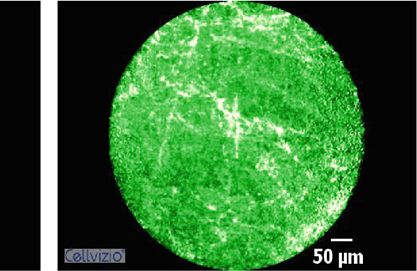
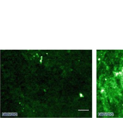
4 BME Frontiers

 ⁎⁎⁎⁎
 ⁎
 5 4
 ⁎⁎⁎⁎

 4 ⁎⁎⁎⁎ 3

 Fold-change (10 min)

 Fold-change
 supernatant
 3
 2
 2

 1
 1

 0 0

 Nonstimulated + M
 Stimulated

 Nonstimulated

 MMP-9

 MMP-9 + M
 Stimulated + M

 Tissue + M
 Tissue
 (a) (b)

 Lungs (+/– Heart) Ventilated OPA supernatant FIB One
 retrieved +/– inhibitor instillation &
 instillation imaging
 > 30 mins

 (c)
 5 ⁎

 OPA supernatant 4
 Normal lung OPA supernatant
 with marimastat
 Fold-change

 3

 2

 1

 50 m 50 m 50 m 0
 OPA OPA + M

 (d)

Figure 2: Validation of FIB One in physiologically relevant biological models. (a) Fold-change in FIB One (1 μM) signal following 10 min
incubation with supernatant from stimulated or nonstimulated neutrophils in vitro with and without a pan-MMP inhibitor, marimastat
(M). Recombinant human MMP-9 (30 nM) shown as reference. Bars show mean (+/-SEM), n ≥ 4 for each condition, analysis by one-way
ANOVA ∗∗∗∗ P < 0:0001. MALDI-ToF MS demonstrating site-specific probe cleavage in ESI Figure S3a. (b) Quantification from ex vivo
fibre-based imaging of aged human lung tissue (open circles of pulmonary fibrosis patients and black circles of aged nonfibrotic lung
tissue) with an FCFM imaging system (488 nm excitation). The fluorescence from samples with FIB One (1 μM) or FIB One plus
marimastat (100 μM) was imaged (10 min) and quantified. Graph shows fold-change compared to baseline autofluorescence of the same
sample. Data show mean (+/-SEM), n = 9, ∗ P = 0:0215, student’s paired t-test. (c) Experimental set-up of size-relevant ex vivo ovine
pulmonary adenocarcinoma (OPA) sheep lung model. (d) Representative images of ex vivo ovine lung and OPA segments following
administration of FIB One with and without inhibitor. Scale bar represents 50 μm. Graph demonstrates quantification of all experiments
relative to intrinsic autofluorescence. n = 3, bars show mean (+/-SEM), analysis by paired t-test for each normalised pre-post-FIB One
segment, ∗ P = 0:0476.
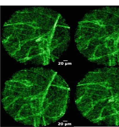
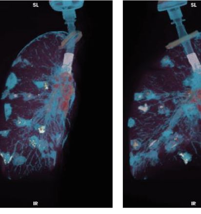
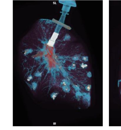
BME Frontiers 5

 Parenchymal
 fibrosis
 (a)
 60

 Median fluorescence (RFU)
 40

 20

 0
 Baseline

 Post-FIB

 (b)

Figure 3: Imaging of parenchymal fibrosis following delivery of FIB One endobronchially. (a) Representative CT images (axial image from a
patient with rheumatoid arthritis associated interstitial lung disease) with baseline and post-FIB One images acquired by confocal
endomicroscopy imaging. (b) Quantification of in vivo human lung fluorescence measured using confocal endomicroscopy before and
after bronchial delivery of the FIB One (n = 4 for each group, P > 0:05). Statistical analysis using a Mann–Whitney test. Scale bar
represents 50 μm.

recruitment, pulmonary toxicity, or systemic toxicity at with active fibrotic lung disease, our approach was hampered
either early or late time points (Table S7). by requiring the delivery of a proximal bolus of FIB One
 Using a conventional catheter in the working channel of a down a conventional catheter in a step immediately preced-
bronchoscope, we delivered FIB One endobronchially as a ing insertion of the imaging fibre. To enable concurrent FIB
bolus to the lungs of eight patients with fibroproliferative lung One delivery and imaging at the exact same location, we
disease in which we expected high MMP level (endobronchi- developed and deployed a triple lumen bronchoscopy cathe-
ally visible tumours and diffuse pulmonary fibrosis). Alveolar ter (TLBC) [30] which allows delivery of compounds and
FCFM imaging following endobronchial delivery of the probe simultaneous FCFM imaging in the field of view.
resulted in increased fluorescent signal in all patients with To assess the distribution of agents delivered to the lung
parenchymal fibrosis (Figure 3). For cancer imaging, ex vivo parenchyma, we undertook preclinical testing of the TLBC
data suggested that MMP activity utilising FIB One could be in an ex vivo ventilated human lung model. 500 μL aliquots
imaged but in vivo imaging of bronchial tumours was compli- of iodinated contrast agent were delivered into the alveolar
cated by FIB One washing off the bronchial mucosa rapidly space and distribution assessed by CT scan, demonstrating
(Figure S5). There were no significant adverse events in the a dispersal volume of 0.5 cm3 (Figure 4(a)). The TLBC
eight patients (Supplementary tables S1-S6). offered the capability to codeliver microdoses of a drug inhib-
 itor. Thus, we reformulated an orally bioavailable small
2.4. A Triple Lumen Bronchoscopy Catheter (TLBC) Enabled molecule inhibitor of MMPs (AZD1236) which had been
Direct Imaging of MMP Activity in the Alveolar Space in previously evaluated in Phase-II trials into a GMP aqueous
Patients which Was Inhibited by Codelivery of a MMP drug product and evaluated its toxicological and biological
Inhibitor. Although we detected MMP activity in patients activity (Figure S6 Table S9).
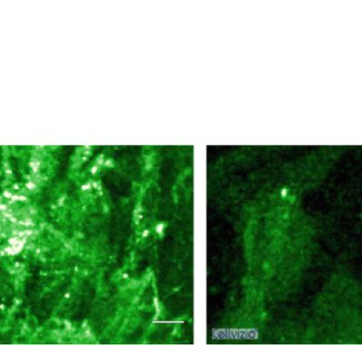
6 BME Frontiers

 Anterior Right Posterior

 (a)
 ⁎⁎
 (i) (ii)
 100 200
 (iv)
 80
 Fluorescence (RFU)

 150
 ns
 20 m 20 m 60
 (iii) (iv) (iii) 100
 40
 (i)
 50
 (ii)
 20
 20 m 20 m 0
 0

 Baseline

 PBS

 PBS + FIB
 0 500 1000 1500 2000 2500
 Frames

 Baseline
 PBS
 PBS + FIB One

 (b) (c) (d)

 (i) (ii)
 100 200

 80 ns
 Fluorescence (RFU)

 150
 20 m 20 m 60
 (iii) (iv) 100 ns
 40
 (i) (ii) (iii) (iv) 50
 20
 20 m 20 m 0
 0
 Baseline

 AZ

 AZ + FIB
 0 500 1000 1500 2000 2500 3000
 Frames

 Baseline
 AZ
 AZ + FIB One

 (e) (f) (g)

Figure 4: In vivo evidence of MMP activity with pharmacological inhibition in patients with pulmonary fibrosis. (a) CT images of an inflated
ex vivo human right lung with 500 μL aliquots of iodinated contrast delivered transbronchially. (b) Representative in vivo images from a
patient with IPF: (i) alveolar space before administering any agents, (ii) after administering PBS, and (iii, iv) after the administration of
FIB One. Dynamic range is identical for all images. Scale bar represents 20 μm. (c) Mean fluorescence of field of view over time during
administration of PBS and FIB One. Corresponding time points to the images in (b) are indicated with arrows. (d) Median fluorescence of
each video from 14 pulmonary segments (nsP = 0:36, ∗∗ P = 0:002). Error bars represent mean + / − s:d and statistical values from
Wilcoxon matched pairs. (e) Representative images from a different pulmonary segment in the same patient as in (b): (i) alveolar space
before administering any agents, (ii) after administering MMP inhibitor (AZD1236), and (iii, iv) after the administration of FIB One.
Dynamic range is identical for all images. (f) Mean fluorescence of field of view over time during administration of AZD1236 and FIB
One. Corresponding time points to the images in (e) are indicated with arrows. (g) Median fluorescence of each video from 12 pulmonary
segments dosed with AZD1236 (P = 0:51) or AZD1236 plus FIB One (P = 0:05). Error bars represent mean + / − s:d and statistical values
from Wilcoxon matched pairs. RFU: relative fluorescence unit; PBS: phosphate-buffered saline; AZ: AZD1236 MMP inhibitor.
BME Frontiers 7

 Using this approach, we obtained real-time imaging inate the possibility that FCFM fibre placement would miss a
sequences in nine patients with inflammatory fibroprolifera- predosed alveolar region. Our preclinical data supports the
tive disease before and after FIB One delivery, with and use of this approach in lung cancer, although there were tech-
without localised predelivery of AZD1236 (Figure 4, nical difficulties with imaging endobronchial tumours with
Table S5 and Figure S6), demonstrating in situ in vivo rapid dissipation of probe. These may be mitigated in the
drug-target engagement within the alveolar space. future with further adaptation to the technical approach or
 modifications to the probe to allow cancer cell labelling [24].
3. Discussion MMP inhibition has been identified as a target for drug
 interventions in inflammatory diseases [44, 45]. MMP inhib-
We report the first in-human pulmonary optical molecular itors used in trials as cancer therapies were disappointing due
imaging of enzyme activity, through fluorescence increase to lack of efficacy and significant adverse effects [46]. Studies
of an MMP reporter, and dynamic elucidation of drug- have also been limited by a lack of quantifiable biomarkers to
target engagement in the distal alveolar regions of the human reflect on their in vivo and functional activity [20]. In this
lung. study, we reformulated and toxicologically qualified
 The FIB One probe was specific to MMPs 2, 9, and 13 AZD1236, an orally available MMP-9/12 inhibitor which
which degrade gelatin, type IV collagen, and elastin within also demonstrated an ability to inhibit MMPs 2 and 13. It
the ECM, which are known to be abundant in the context had previously been used in a phase 2A safety and tolerability
of acute and chronic inflammation [31] where they are study in 74 emphysema patients randomised to placebo or
secreted by infiltrating inflammatory cells as well as bron- AZD1236 75 mg twice daily for 6 weeks [19]. The study dem-
chial and alveolar epithelial cells and fibroblasts [32]. To onstrated no effect on the secondary lung physiology end-
ensure future validity of our work, there would need to be points, and the investigators were unable to show that
inclusion of a noninflammatory control group to determine AZD1236 actually inhibited its intended targets in situ.
the detection of upregulated vs. homeostatic MMP activity. Indeed, a placebo-controlled, AZD1236 biomarker study of
However, if confirmed, MMPs represent an attractive target 55 COPD patients again failed to demonstrate MMP-9 or
as they serve as a marker in several human inflammatory dis- MMP-12 inhibition in induced sputum [20], whereas we
eases including IPF, COPD, ARDS, and lung cancer [10–14]. have demonstrated drug-target engagement of AZD1236 in
With the advent of increasingly available multiplexing humans.
approaches for microscopic interrogation, our approach of The pathway of drug validation utilises cell-based assays
using fluorescent reporters with FCFM and drug inhibition and animal models of disease followed by phased studies in
does represent a wider applicability and is readily extendable humans. At each stage of development, costs increase and
to multiple target pathways in several organ systems. there is a high attrition rate often leading to the failure or
 Current approaches to identifying enzyme activity in the deescalation of drug candidates. Although there are many
lung has been challenging and has typically been performed reasons for this, one major hurdle is demonstrating that
ex vivo using zymography on patient tissue or lavage samples enzymatic activity is present in the patients being studied.
[33]. However, this technique has several limitations, includ- Therefore, there is a rationale for performing translational
ing the requirement for an invasive biopsy and tissue proof of concept (experimental medicine) studies directly
retrieval and the utilisation of lavage fluid, which is prone and rapidly in humans, using small numbers of patients.
to sampling inconsistencies. Furthermore, zymography Consequently, here, we have described a tractable strategy
assessment may not reflect free enzyme activity in the native for rapidly assessing distal alveolar biology using a sensitive
environment, which in the case of MMPs, is dependent on and specific optical probe and direct visualisation of MMP
inhibitory antagonists such as tissue inhibitors of MMPs pathway activity/inhibition in situ in real time with FCFM.
(TIMPs). In the future, this could be assessed in a broader group of
 Molecular imaging of enzyme activity and pharmacolog- patients with inflammatory and noninflammatory lung dis-
ical perturbation in vivo has focused on either nuclear imag- ease correlated with the MMP activity and may present the
ing of radiolabelled probes or optical imaging with ability to offer a mechanism-based method of stratifying
fluorogenic probes. These studies have used animal models patients for drug studies and to interrogate biological path-
to image MMP activity in vivo in cancer [34–39], stroke ways in situ in vivo with molecular microscopy.
[40], and joint disease [41–43]; however, they have all relied
on systemic intravenous delivery of the probes and whole- 4. Materials and Methods
body optical imaging, which is to date, technically not possi-
ble in humans with sufficient resolution or sensitivity. Thus, 4.1. Ethics Statement. All experiments using human samples
we adopted an alternate approach to measure enzyme activ- in vitro were performed following approval of the appropri-
ity in the distal lung human lung, made possible by using ate regional ethics committee (REC) and with informed
FCFM to allow us to access alveolar regions and deliver consent of the patients. Blood sample collection and use
microdoses (
8 BME Frontiers

were deemed unsuitable for transplant and authorised for tubes (VWR) using a Pecellys 24 homogeniser. Homogenised
research as part of the Enlighten study (London-Central tissue was adjusted to 4 mg mL-1 protein (determined by a
Research Ethics Committee, REC Reference: 16/LO/1883). Pierce BCA Total Protein Assay Kit (Thermo Fisher Scien-
The in-human assessments were undertaken with informed tific), following the manufacturer’s instructions). Multiwell
consent of the participants and approved by the South East plate reader assays were performed as previously described,
Scotland Regional Ethics Committee 02, REC Number: replacing the MMP buffer and MMP enzymes for the homo-
15/SS/0235. The trial was registered with ClinicalTrials.gov genised and protein-adjusted lung tissue.
(Identifier: NCT02604862). For zymography, the samples were mixed 1 : 1 with 2x
 SDS sample buffer and 20 μL was loaded onto precast
4.2. In Vitro Enzymatic Validation of FIB One. To determine Novex® 10% Zymogram (Gelatin) Protein Gels (Thermo
the enzymatic specificity of FIB One, the probe was incubated Fisher Scientific). The gels were placed in an electrophoresis
at a concentration of 1 μM (unless otherwise stated) with the chamber with pre-chilled Novex diluted running buffer and
recombinant MMPs (Enzo Life Sciences) (active domains of electrophoresed at 150 V for approximately 90 min at 4°C.
MMP-1, MMP-2, MMP-3, MMP-7, MMP-8, MMP-9, Gels were removed and incubated with Novex renaturing
MMP-10, MMP-11, MMP-12, and MMP-13, all at 30 nM), buffer for 90 min at 4°C. They were washed in distilled water
thrombin (Sigma Aldrich) (5 U/mL), factor Xa (Sigma and incubated with Novex developing buffer for 30 min at
Aldrich) (500 nM), plasmin (Sigma Aldrich) (30 nM), and room temperature prior to overnight incubation at 37°C.
human neutrophil elastase (30 nM). Where appropriate, Control gels had marimastat (50 μM) added to the develop-
enzymes were incubated with specific inhibitors for 30 min ing buffer. Gels were then rinsed with distilled water prior
at 37°C prior to addition of FIB One. The pan-MMP inhibi- to staining with a colloidal blue staining kit and imaging
tor marimastat (Tocris Biosciences) was used at 20 μM, and using a transilluminator (UVItec BXT-20 M, UVItec Ltd,
the MMP inhibitor AZD1236 was used at 0-14 μM. Enzyme Cambridge, UK).
free reactions served as a control. Enzymatic reactions were For ex vivo imaging, human lung tissue was sliced into
performed in MMP buffer (10 mM CaCl2, 6.1 g Tris-HCl, 1 mm × 4 mm sections and placed in the wells of a 96-well
8.6 g NaCl per litre, pH 7.5) in a final volume of 20 μL. Reac- plate with 100 μL of MMP buffer and 30 nM MMP-13.
tions were performed in duplicate in 384-well plates (Life Where appropriate, AZD1236 (0-14 μM as indicated in the
Technologies) with optically clear plate seals (Thermo Scien- text) was added to the tissue and incubated at 37°C for
tific) and repeated thrice (independently). Fluorescence sig- 30 min prior to the addition FIB One (5 μM). The tissue
nal was measured for up to 60 min, ex/em 485/528 nm was imaged over 10 min, 12 frames sec-1 using a preclinical
using microplate reader (BioTek Synergy H1 multimode confocal laser scanning endomicroscopy device (Cellvizio®,
reader). Data were normalised to buffer alone and are pre- Mauna Kea Technologies, excitation 488 nm/detection band-
sented as fold-change in signal (relative fluorescent units, width 505 to 700 nm) with a compatible imaging fibre
RFU) compared to enzyme-free controls. Data were plotted (Alveoflex™, Mauna Kea Technologies). At each time point
using Prism 7 (GraphPad Software Inc., La Jolla, CA, USA). 60 s, images of captured with same laser power. The fluores-
 cence for each sample was calculated by determining the
4.3. Neutrophil Extraction and Evaluation of FIB One. average fluorescence per frame for each time point. All
Neutrophils were isolated from the blood of healthy human images were brightness and contrast enhanced with the same
volunteers as previously described [47]. The number of parameters. Five independent patient specimens were col-
retrieved neutrophils was determined with NucleoCounter lected and analysed. RFUs collected from off-target frames
NC-1000 (Chemo Metec). Neutrophils were resuspended at (showing motion artefact or absence of any fluorescence)
a concentration of 20 × 106 mL-1 in 0.9% NaCl with 0.9 mM were omitted during analysis.
CaCl2. Cells were incubated for 30 min at 37°C. Cells to be
stimulated were subsequently activated with 5 μM calcium 4.5. Ex Vivo Lung Experimental Procedure. Ovine pulmonary
ionophore A23187 (Tocris Bioscience) for 30 min at 37°C. adenocarcinoma (OPA) tissue was homogenised using a drill
Neutrophils were harvested by centrifugation at 400 x g for homogeniser, followed by lysis in cell lysis buffer. The super-
5 min. Supernatants were removed and stored. Plate reader natant was collected following centrifugation at 12,000 rpm
assays were performed as described above, with the MMP for 20 min. The samples were adjusted to 1 mg mL-1 total
buffer replaced with neutrophil supernatant. All experiments protein (determined by a Pierce BCA Total Protein Assay
were carried out in duplicate using three independent donors Kit (Thermo Fisher Scientific)). Zymography (as described
(one donor per independent repeat). Data were normalised above) was performed to confirm MMP activity across mul-
by background subtraction of intrinsic fluorescence. tiple OPA samples. Recombinant MMP-9 (2 nM) and recom-
 binant pro-MMP-9 (0.5 nM) were included in the assay as
4.4. Ex Vivo Human Lung Tissue Processing and Evaluation positive controls.
of FIB One. Human lung tissue samples (lung tumour and OPA supernatant (1 mL) and marimastat (100 μM) or
noncancerous adjacent tissue) were obtained following surgi- 1 mL 0.9% NaCl (control) was instilled into anatomically dis-
cal resection and stored at -80° C until use. Processing for tinct segments of ex vivo ventilated non-OPA ovine lungs.
multiwell plate reader assay or zymography analysis was con- Following >30 min, the segments were bronchoscopically
ducted as follows: 1 mm × 4 mm sections were homogenised identified and microdosed with 5 μM FIB One in 1 mL.
in 400 μL MMP buffer in Precellys 2.8 mm ceramic bead FCFM imaging was undertaken pre- and postinstillation
BME Frontiers 9

(Cellvizio®, Mauna Kea Technologies). This was performed 4.9. In Vivo Assessment of FIB One and AZD1236 with TLBC.
by passing a FCFM fibre into the disparate bronchopulmon- In subsequent testing the Miniaturized Alveoflex™-TLBC
ary segments (up to 5 passes per bronchopulmonary segment combination replaced the Alveoflex™ fibre. The entire appa-
to capture a regional representation of fluorescence), record- ratus was passed transbronchially to allow direct alveolar
ing images at 12 frames per second and imaging for up to 5 imaging before localised delivery of agents. Once transbron-
minutes. Videos were analysed as described above. Each chial pass was accomplished, baseline lung imaging was
experimental set was normalised to the control segment recorded for at least 30 sec. A 500 μL microdose of either
(segment with 1 mL 0.9% NaCl instilled) which represents AZD1236 (14 μg/mL) or sterile PBS (control) was adminis-
intrinsic autofluorescence. tered under direct observation, and further imaging was
 obtained for >60 sec. 500 μL of FIB One (20 μg/mL) (deliv-
4.6. Human Ex Vivo Experimental Procedure. The develop- ered only after at least 60s to allow MMP inhibition) was then
ment of the TLBC has been described previously [30]. This dosed to the same area with a further 90-120 sec of imaging
device was compatible with a slimmer (diameter 1 mm) captured. If considerable movement due to breathing or
imaging fibre approved for clinical investigation (Miniatur- coughing was encountered, imaging was halted and a new
ized AlveoFlex™ Confocal Miniprobe, Cellvizio®, Mauna segment identified. Up to 6 segments of interest were investi-
Kea Technologies) and had two hollow lumens to allow FIB gated per patient determined by patient tolerance.
One and/or AZD1236 drug delivery directly to tissue within
the field of view during image acquisition. The TLBC device 4.10. Image Analysis. All video sequences were assessed by
was validated preclinically within ventilated ex vivo human two clinicians with clinical experience of pulmonary endomi-
lungs to assess both fluid delivery capability and to determine croscopy. Frames with excessive movement or lack of recog-
which segments of the lung the device was able to reach. nizable alveolar structure were removed from analysis.
Iodinated contrast (Omnipaque 300, GE healthcare) was Videos were assessed to ensure stability throughout the imag-
diluted with PBS in a ratio of 1 : 5 and delivered transbronchi- ing procedure. To ensure the region imaged was the same as
ally in 500 μL doses through the device lumens to the distal region dosed, imaging from a segment was excluded if there
lung, repeated for multiple pulmonary segments. Following was obvious alveolar movement witnessed during image cap-
delivery, the lung was inflated, the trachea clamped, and ture. All video files were analysed using the Cellvizio® Viewer
computerized tomography (Biograph mCT, Siemens) images image processing software from Mauna Kea Technologies,
were taken to visualise the extent of alveolar contrast spread. Paris. A region of interest was drawn around the fibre field
Processing was performed with Horos DICOM viewer of view and processed to determine the mean RFU for each
(HOROS). frame. The videos were categorized as either baseline, post-
 PBS, post-AZD1236, post-FIB One, and PBS or post-FIB
4.7. Clinical Study Patient Selection. All participants were One and AZD1236 for each patient. The median RFU was
scheduled for routine, clinical diagnostic flexible bronchos- calculated for each category and used as a comparative value
copy in our institution with a known or suspected diagnosis between groups.
of fibrosis or lung cancer. All eight patients had computer- 4.11. Statistical Analysis. Statistical analyses were performed
ized tomography performed prior to bronchoscopy and using Prism 7 (GraphPad Software Inc., La Jolla, CA, USA).
regions of active disease identified for targeted investigation. Where appropriate, analyses were performed using the stu-
 dent’s t-test or one-way ANOVA. Unless otherwise stated
4.8. Bronchoscopy and FCFM Procedure. Bronchoscopy was error bars show standard error of the mean (s.e.m).
undertaken in an outpatient suite using local anaesthetic
and conscious sedation or in theatre under general anaes-
thetic. FCFM was performed using a clinically approved
 Conflicts of Interest
confocal laser scanning endomicroscopy device (Cellvizio®, KD, CH, and MB are shareholders of Edinburgh Molecular
Mauna Kea Technologies) with a compatible imaging fibre Imaging. MB, SVC, and AMF are inventors on a patent
(Alveoflex™ or Miniaturized Alveoflex™, Mauna Kea (WO 2016/151299 A1) held by the University Court of the
Technologies). University of Edinburgh that covers the probe and method
 In initial exploratory testing, 8 patients with either endo- of use. FL and VD are employees of Mauna Kea Technolo-
bronchial tumour or parenchymal fibrosis underwent FIB gies. TV owns stock from Mauna Kea Technologies. The
One microdosing (
10 BME Frontiers

developed the TLBC. CH, MB, and KD supervised the [6] D. C. Rockey, P. D. Bell, and J. A. Hill, “Fibrosis — a common
project. AMF, AA, AMa, BM, and KD wrote the manu- pathway to organ injury and failure,” New England Journal of
script. All authors approved the manuscript. Alicia Megia- Medicine, vol. 372, no. 12, pp. 1138–1149, 2015.
Fernandez, Adam Marshall, Ahsan R. Akram, and Bethany [7] S. D. Shapiro, “Matrix metalloproteinase degradation of extra-
Mills contributed equally to this work. cellular matrix: biological consequences,” Current Opinion in
 Cell Biology, vol. 10, no. 5, pp. 602–608, 1998.
 [8] M. G. Rohani and W. C. Parks, “Matrix remodeling by MMPs
Acknowledgments during wound repair,” Matrix Biology, vol. 44-46, pp. 113–121,
 2015.
The authors would like to thank the NHS Lothian SAHSC
Bioresource for facilitating the studies using ex vivo human [9] M. Corbel, C. Belleguic, E. Boichot, and V. Lagente, “Involve-
lung tissue, Dr Chris Cousins for the samples of OPA lung, ment of gelatinases (MMP-2 and MMP-9) in the development
 of airway inflammation and pulmonary fibrosis,” Cell Biology
AstraZeneca UK for providing AZD1236 Drug Product as
 and Toxicology, vol. 18, no. 1, pp. 51–61, 2002.
an in-kind contribution through their Open Innovation Pro-
 [10] A. Davey, D. F. McAuley, and C. M. O'Kane, “Matrix metallo-
gram, Warren’s Wish (Registered Scottish Charity: SC
 proteinases in acute lung injury: mediators of injury and
045290), and the human lung donor and their families. This drivers of repair,” European Respiratory Journal, vol. 38,
study was supported by the Medical Research Council (under no. 4, pp. 959–970, 2011.
the Developmental Pathway Funding Scheme grant number
 [11] A. Pardo and M. Selman, “Role of matrix metaloproteases in
MR/J014702), the Engineering and Physical Sciences idiopathic pulmonary fibrosis,” Fibrogenesis & Tissue Repair,
Research Council (EP/K03197X/1, EP/R005257/1, and vol. 5, Supplement 1, p. S9, 2012.
NS/A000049/1), and the Wellcome Trust (203148/Z/16/Z). [12] L. Segura-Valdez, A. Pardo, M. Gaxiola, B. D. Uhal, C. Becerril,
AA is supported by a Cancer Research UK Clinician Scientist and M. Selman, “Upregulation of gelatinases A and B, collage-
Fellowship (A24867). TV is supported by a Medtronic/Royal nases 1 and 2, and increased parenchymal cell death in
Academy of Engineering Research Chair (RCSRF1819\7\34). COPD,” Chest, vol. 117, no. 3, pp. 684–694, 2000.
The research leading to these results has received funding [13] S. A. Shah, F. G. Spinale, J. S. Ikonomidis, R. E. Stroud, E. I.
from the European Union Seventh Framework Programme Chang, and C. E. Reed, “Differential matrix metalloproteinase
FP7 2012 under grant agreement No. 326465 (AMF). Mauna levels in adenocarcinoma and squamous cell carcinoma of the
Kea Technologies provided support for the clinical develop- lung,” The Journal of Thoracic and Cardiovascular Surgery,
ment of the TLBC. vol. 139, no. 4, pp. 984–990, 2010.
 [14] A. Pardo, S. Cabrera, M. Maldonado, and M. Selman, “Role of
 matrix metalloproteinases in the pathogenesis of idiopathic
Supplementary Materials pulmonary fibrosis,” Respiratory Research, vol. 17, no. 1,
Supplementary Information containing supplementary p. 23, 2016.
Figures S1-6: chemistry materials and methods, patient data, [15] M. Selman and A. Pardo, “Revealing the pathogenic and aging-
toxicology studies, and stability studies. (Supplementary related mechanisms of the enigmatic idiopathic pulmonary
Materials) fibrosis. An integral model,” American Journal of Respiratory
 and Critical Care Medicine, vol. 189, no. 10, pp. 1161–1172,
 2014.
References
 [16] T. Nkyimbeng, C. Ruppert, T. Shiomi et al., “Pivotal role of
 [1] H. Wang, M. Naghavi, C. Allen et al., “Global, regional, and matrix metalloproteinase 13 in extracellular matrix turnover
 national life expectancy, all-cause mortality, and cause- in idiopathic pulmonary fibrosis,” PLoS One, vol. 8, no. 9, arti-
 specific mortality for 249 causes of death, 1980-2015: a system- cle e73279, 2013.
 atic analysis for the Global Burden of Disease Study 2015,” The [17] R. G. Jenkins, J. K. Simpson, G. Saini et al., “Longitudinal
 Lancet, vol. 388, no. 10053, pp. 1459–1544, 2016. change in collagen degradation biomarkers in idiopathic pul-
 [2] G. Khurana, A. Rohilla, and A. Deep, “Drug development pro- monary fibrosis: an analysis from the prospective, multicentre
 cess and novel drugs approved by FDA for 2017-18,” Applied PROFILE study,” The Lancet Respiratory Medicine, vol. 3,
 Clinical Research, Clinical Trials and Regulatory Affairs, no. 6, pp. 462–472, 2015.
 vol. 5, no. 2, pp. 80–98, 2018. [18] B. Ley, K. K. Brown, and H. R. Collard, “Molecular biomarkers
 [3] B. N. Rome and J. Avorn, “Drug evaluation during the Covid- in idiopathic pulmonary fibrosis,” American Journal of
 19 pandemic,” New England Journal of Medicine, vol. 382, Physiology-Lung Cellular and Molecular Physiology, vol. 307,
 no. 24, pp. 2282–2284, 2020. no. 9, pp. L681–L691, 2014.
 [4] P. Spagnolo, E. Cocconcelli, and V. Cottin, “Clinical trials in [19] H. Magnussen, H. Watz, A. Kirsten et al., “Safety and tolerabil-
 IPF: what are the best endpoints?,” in Idiopathic Pulmonary ity of an oral MMP-9 and -12 inhibitor, AZD1236, in patients
 Fibrosis: A Comprehensive Clinical Guide, K. C. Meyer and S. with moderate-to-severe COPD: A randomised controlled 6-
 D. Nathan, Eds., pp. 433–453, Springer International Publish- week trial,” Pulmonary Pharmacology & Therapeutics,
 ing, Cham, 2019. vol. 24, no. 5, pp. 563–570, 2011.
 [5] A. R. Akram, S. V. Chankeshwara, E. Scholefield et al., “In [20] R. Dahl, I. Titlestad, A. Lindqvist et al., “Effects of an oral
 situ identification of Gram-negative bacteria in human MMP-9 and -12 inhibitor, AZD1236, on biomarkers in mod-
 lungs using a topical fluorescent peptide targeting lipid A,” erate/severe COPD: a randomised controlled trial,” Pulmonary
 Science Translational Medicine, vol. 10, no. 464, article Pharmacology & Therapeutics, vol. 25, no. 2, pp. 169–177,
 eaal0033, 2018. 2012.
BME Frontiers 11

[21] K.-H. Shen, J. H. Hung, C. W. Chang, Y. T. Weng, M. J. Wu, [36] J. O. McIntyre, R. L. Scherer, and L. M. Matrisian, “Near-infra-
 and P. S. Chen, “Solasodine inhibits invasion of human lung red optical proteolytic beacons for in vivo imaging of matrix
 cancer cell through downregulation of miR-21 and MMPs metalloproteinase activity,” Methods in Molecular Biology
 expression,” Chemico-Biological Interactions, vol. 268, (Clifton, N.J.), vol. 622, pp. 279–304, 2010.
 pp. 129–135, 2017. [37] B.-W. Xie, I. M. Mol, S. Keereweer et al., “Dual-wavelength
[22] P. D. Brown, “Matrix metalloproteinase inhibitors in the treat- imaging of tumor progression by activatable and targeting
 ment of cancer,” Medical Oncology, vol. 14, no. 1, pp. 1–10, 1997. near-infrared fluorescent probes in a bioluminescent breast
[23] A. Megia-Fernandez, B. Mills, C. Michels et al., “Bimodal cancer model,” PLoS One, vol. 7, no. 2, article e31875, 2012.
 fluorogenic sensing of matrix proteolytic signatures in lung [38] L. Zhu, Y. Ma, D. O. Kiesewetter et al., “Rational design of matrix
 cancer,” Organic & Biomolecular Chemistry, vol. 16, no. 43, metalloproteinase-13 activatable probes for enhanced specificity,”
 pp. 8056–8063, 2018. ACS Chemical Biology, vol. 9, no. 2, pp. 510–516, 2013.
[24] B. Mills, D. Norberg, K. Dhaliwal, A. R. Akram, M. Bradley, [39] T. Ma, Y. Hou, J. Zeng et al., “Dual-ratiometric target-triggered
 and A. Megia-Fernandez, “A matrix metalloproteinase activa- fluorescent probe for simultaneous quantitative visualization
 tion probe for painting human tumours,” Chemical Communi- of tumor microenvironment protease activity and pH
 cations, vol. 56, no. 69, pp. 9962–9965, 2020. in vivo,” Journal of the American Chemical Society, vol. 140,
[25] N. Avlonitis, M. Debunne, T. Aslam et al., “Highly specific, no. 1, pp. 211–218, 2017.
 multi-branched fluorescent reporters for analysis of human [40] P. A. Barber, D. Rushforth, S. Agrawal, and U. I. Tuor, “Infra-
 neutrophil elastase,” Organic & Biomolecular Chemistry, red optical imaging of matrix metalloproteinases (MMPs) up
 vol. 11, no. 26, pp. 4414–4418, 2013. regulation following ischemia reperfusion is ameliorated by
[26] A. K. Galande, S. A. Hilderbrand, R. Weissleder, and C. H. hypothermia,” BMC Neuroscience, vol. 13, no. 1, p. 76, 2012.
 Tung, “Enzyme-targeted fluorescent imaging probes on a mul- [41] P. B. Satkunananthan, M. J. Anderson, N. M. de Jesus, D. R.
 tiple antigenic peptide core,” Journal of Medicinal Chemistry, Haudenschild, C. M. Ripplinger, and B. A. Christiansen, “In
 vol. 49, no. 15, pp. 4715–4720, 2006. vivo fluorescence reflectance imaging of protease activity in a
[27] J. M. Ellard, T. Zollitsch, W. J. Cummins, A. L. Hamilton, and mouse model of post-traumatic osteoarthritis,” Osteoarthritis
 M. Bradley, “Fluorescence enhancement through enzymatic and Cartilage, vol. 22, no. 10, pp. 1461–1469, 2014.
 cleavage of internally quenched dendritic peptides: a sensitive [42] T. Fukui, E. Tenborg, J. H. N. Yik, and D. R. Haudenschild,
 assay for the AspN endoproteinase,” Angewandte Chemie (Inter- “In-vitro and in-vivo imaging of MMP activity in cartilage
 national Ed. in English), vol. 41, no. 17, pp. 3233–3236, 2002. and joint injury,” Biochemical and Biophysical Research Com-
[28] T. H. Craven, N. Avlonitis, N. McDonald et al., “Super-silent munications, vol. 460, no. 3, pp. 741–746, 2015.
 FRET sensor enables live cell imaging and flow cytometric [43] H. Cho, F.-U.-R. Bhatti, T. W. Yoon, K. A. Hasty, J. M. Stuart,
 stratification of intracellular serine protease activity in neutro- and A.-K. Yi, “Non-invasive dual fluorescence in vivo imaging
 phils,” Scientific Reports, vol. 8, no. 1, article 13490, 2018. for detection of macrophage infiltration and matrix metallo-
[29] EU, “EU guidelines for good manufacturing practice for proteinase (MMP) activity in inflammatory arthritic joints,”
 medicinal products for human and veterinary use,” vol. 4, Biomedical Optics Express, vol. 7, no. 5, pp. 1842–1852, 2016.
 Heath and Consumers Directorate-General, European Comis- [44] H. Järveläinen, A. Sainio, M. Koulu, T. N. Wight, and
 sion, 2014, Part II. R. Penttinen, “Extracellular matrix molecules: potential targets
[30] N. Knighton, B. Cottle, V. Dentan et al., “Development of an in pharmacotherapy,” Pharmacological Reviews, vol. 61, no. 2,
 alveolar transbronchial catheter for concurrent fiber optics- pp. 198–223, 2009.
 based imaging and fluid delivery,” Journal of Medical Devices, [45] R. Ramachandran, C. Altier, K. Oikonomopoulou, and M. D.
 vol. 12, no. 3, article 035003, 2018. Hollenberg, “Proteinases, their extracellular targets, and
[31] W. C. Parks and S. D. Shapiro, “Matrix metalloproteinases in inflammatory signaling,” Pharmacological Reviews, vol. 68,
 lung biology,” Respiratory Research, vol. 2, no. 1, pp. 10–19, no. 4, pp. 1110–1142, 2016.
 2000. [46] N. B. Leighl, L. Paz-Ares, J. Y. Douillard et al., “Randomized
[32] M. M. Gueders, J.-M. Foidart, A. Noel, and D. D. Cataldo, phase III study of matrix metalloproteinase inhibitor BMS-
 “Matrix metalloproteinases (MMPs) and tissue inhibitors of 275291 in combination with paclitaxel and carboplatin in
 MMPs in the respiratory tract: potential implications in advanced non-small-cell lung cancer: National Cancer Insti-
 asthma and other lung diseases,” European Journal of Pharma- tute of Canada-Clinical Trials Group Study BR.18,” Journal
 cology, vol. 533, no. 1-3, pp. 133–144, 2006. of Clinical Oncology, vol. 23, no. 12, pp. 2831–2839, 2005.
[33] M. Toth and R. Fridman, “Assessment of gelatinases (MMP-2 [47] C. Haslett, L. A. Guthrie, M. M. Kopaniak, Johnston RB Jr, and
 and MMP-9 by gelatin zymography),” in Metastasis Research P. M. Henson, “Modulation of multiple neutrophil functions
 Protocols: Volume I: Analysis of Cells and Tissues, S. A. Brooks by preparative methods or trace concentrations of bacterial
 and U. Schumacher, Eds., pp. 163–174, Humana Press, lipopolysaccharide,” The American Journal of Pathology,
 Totowa, NJ, 2001. vol. 119, no. 1, pp. 101–110, 1985.
[34] M. Salaün, J. Peng, H. H. Hensley et al., “MMP-13 in-vivo
 molecular imaging reveals early expression in lung adenocarci-
 noma,” PLoS One, vol. 10, no. 7, article e0132960, 2015.
[35] B. Waschkau, A. Faust, M. Schäfers, and C. Bremer, “Perfor-
 mance of a new fluorescence-labeled MMP inhibitor to image
 tumor MMP activity in vivo in comparison to an MMP-
 activatable probe,” Contrast Media & Molecular Imaging,
 vol. 8, no. 1, pp. 1–11, 2013.
You can also read