Affordable mobile microfluidic diagnostics: minimum requirements for smartphones and digital imaging for colorimetric and fluorometric anti-dengue ...
←
→
Page content transcription
If your browser does not render page correctly, please read the page content below
Wellcome Open Research 2021, 6:57 Last updated: 06 APR 2021

RESEARCH ARTICLE

Affordable mobile microfluidic diagnostics: minimum
requirements for smartphones and digital imaging for
colorimetric and fluorometric anti-dengue and anti-SARS-CoV-
2 antibody detection [version 1; peer review: awaiting peer
review]
Sophie M. Jégouic1,2, Ian M. Jones                      2,   Alexander D. Edwards                  1

1Reading School of Pharmacy, University of Reading, Reading, RG6 1EE, UK
2School of Biological Sciences, University of Reading, Reading, UK

v1   First published: 11 Mar 2021, 6:57                                         Open Peer Review
     https://doi.org/10.12688/wellcomeopenres.16628.1
     Latest published: 11 Mar 2021, 6:57
     https://doi.org/10.12688/wellcomeopenres.16628.1                           Reviewer Status AWAITING PEER REVIEW

                                                                                Any reports and responses or comments on the

Abstract                                                                        article can be found at the end of the article.
Background: Miniaturised bioassays permit diagnostic testing near
the patient, and the results can be recorded digitally using
inexpensive cameras including smartphone and mobile phone
cameras. Although digital cameras are now inexpensive and portable,
the minimum performance required for microfluidic diagnostic
bioassays has not been defined. We present a systematic comparison
of a wide range of different digital cameras for capturing and
measuring results of microfluidic bioassays and describe a framework
to specify performance requirements to quantify immunoassays.
Methods: A set of 200 µm diameter microchannels was filled with a
range of concentrations of dyes used in colorimetric and fluorometric
enzyme immunoassays. These were imaged in parallel using cameras
of varying cost and performance ranging from £500.
Results: Higher resolution imaging allowed larger numbers of
microdevices to be resolved and analysed in a single image. In
contrast, low quality cameras were still able to quantify results but for
fewer samples. In some cases, an additional macro lens was added to
focus closely. If image resolution was sufficient to identify individual
microfluidic channels as separate lines, all cameras were able to
quantify a similar range of concentrations of both colorimetric and
fluorometric dyes. However, the mid-range cameras performed
better, with the lowest cost cameras only allowing one or two samples
to be quantified per image. Consistent with these findings, we
demonstrate that quantitation (to determine endpoint titre) of
antibodies against dengue and severe acute respiratory syndrome
coronavirus 2 (SARS-CoV-2) viruses is possible using a wide range of

                                                                                                                          Page 1 of 15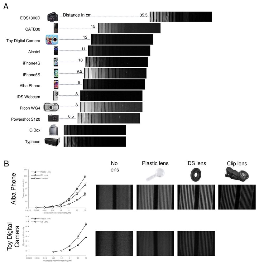
Wellcome Open Research 2021, 6:57 Last updated: 06 APR 2021

digital imaging devices including the mid-range smartphone iPhone
6S and a budget Android smartphone costing
Wellcome Open Research 2021, 6:57 Last updated: 06 APR 2021

Introduction                                                        with tightly regulated and standardised requirements for
The use of digital cameras in consumer devices, such as             regulatory approved diagnostics. An alternative to using
smartphones, to record the results of miniaturised bioassays,       consumer products is to exploit the underlying hardware
offers many potential advantages in clinical diagnostics.           (e.g. optoelectronic components) to build bespoke, regulatory
Smartphones can digitally record, interpret and quantify many       approved, camera-based readers/analysers. The CMOS sensors
measurements that have previously relied on a laboratory            and lenses found within smartphones are increasingly available
instrument to analyse, or needed a human operator to interpret      as camera modules. The most readily available are bundled
lines or colour change by eye and then manually record              with single-board computers such as the Raspberry Pi, or with
results. A wide spectrum of devices and bioassays have been         microcontrollers. Industrial use of digital cameras for example
read using smartphones1, including pH in clinical samples2          in manufacturing for machine vision has likewise grown
immunoassays3,4, nucleic acid detection5,6, microbiology7,8, and    accelerated by the fall in cost of cameras and network/process-
paper devices including urine analysis dipsticks9 or cholesterol    ing power16. The cameras used in industrial applications
test strips10. Smartphones offer a combination of three features    have one major advantage over consumer products, which
in an accessible package: high performance camera, on-board         is greater robustness and manufacturing specifications for
computing power, and networking. Firstly, optimised miniatur-       longer-term use coupled with more software control, compared
ised optics combined with high-performance complementary            to the short product life for consumer products where profit is
metal oxide semiconductor (CMOS) image sensors deliver              driven by frequent replacements. Furthermore, smartphone
increasingly high-performance digital cameras. Secondly, con-       camera software optimises images for consumer preference- not
stantly advancing processors deliver local fast and powerful        diagnostic accuracy. However, this comes at a cost, with machine
computing capacity, plus onboard random-access memory               vision cameras typically costing far more for equivalent sensors.
(RAM) and secure digital (SD) card data storage allow image
acquisition, storage, processing and analysis. Thirdly, mobile      As many studies17–19 have reported proof-of-concept demonstrat-
network bandwidth and expansion of wireless networking              ing that smartphone imaging can be used to read microfluidic
permits integration to connected health systems and – if            assays a systematic comparison of different digital camera
handset computing power is insufficient- cloud-based analysis       hardware is warranted to understand the key parameters for
of results. The latter two features can also be used without        digital imaging of bioassays. We developed a simple and low-
the camera to power external sensor modules11. Bioassays read       cost microfluidic platform that exploits the optical transparency
by smartphone can aid diagnosis either as stand-alone point-        of melt-extruded fluoropolymer microcapillary film (MCF).
of-care clinical measurement12, or results can be combined with     We previously showed both smartphone and consumer digital
other clinical data where smartphones have been explored for        cameras, as well as the Raspberry Pi camera, can quantify
providing clinical guidance in the field13,14.                      immunoassay and analytical microbiology assays within
                                                                    affordable microfluidic devices made from MCF3,20–24. The low
The large consumer markets and massive sales volumes have           cost of the devices and the optical transparency achieved through
driven up hardware performance and cut optoelectronic               refractive index matching makes this simple platform ideally
component price. However, the latest high-end products with         suited to systematic comparison of imaging performance.
highest hardware performance and most handset features
comes at increased price, with cost of many high-end smart-         Here, we compared the analytical performance of a range of
phone handsets exceeding £1000. Alternative, lower price and        digital cameras and tested if lower cost feature phones and
performance options have followed. Two classes of low-budget        budget smartphone handsets were capable of recording colori-
mobile phone handset have emerged. Firstly, in many regions         metric and fluorometric dyes within microfluidic devices. Using
of the world (e.g. India, many African states) mobile phones        MCF as an example of microfluidic bioassays, we systemati-
without the networking and microprocessor power of smart-           cally compared the optical sensitivity, resolution and therefore
phones, termed “feature phones” have been the most widely           analytical performance of these low-cost units against higher-
owned handsets15. These offer longer battery life and are better    specified smartphones and higher performance digital cameras.
suited to users and regions where network capacity is limited       We also compared these consumer products with an industrial
and communication by phone/SMS remains the most important           camera module developed for machine vision applications and a
driver for mobile phone ownership. Secondly, budget smart-          Raspberry Pi camera. Finally, we assessed if these digital
phones have proliferated, especially those with android operating   cameras could measure simulated antibody responses against
system, with reduced features (e.g. older operating systems) and    two viral infections of global health significance- dengue fever
less highly specified hardware (less memory, simpler screen,        and coronavirus disease 2019 (COVID-19).
lower resolution camera, cheap casework). Touchscreen, fully
networked android handsets are available under £50. Whilst          Methods
many feature phones do have cameras, the limited screen size        Experimental approach
and memory ensures these are hard to use and they are typically     We established a simple imaging rig to systematically determine
equipped with very simple low-resolution optoelectronics.           the performance of a range of digital cameras for imaging
                                                                    colorimetric and fluorometric assays within microfluidic devices
Consumer products such as smartphones can be hard to adopt          (Figure 1A). To allow us to control the microfluidic device
into diagnostics because their closed and rapidly changing          characteristics and directly compare camera performances, we
software, and variable hardware specification, is not compatible    used colorimetric and fluorescent dye solutions- rather than

                                                                                                                           Page 3 of 15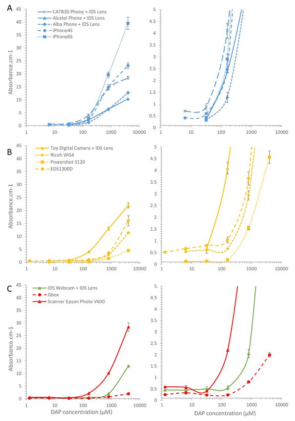
Wellcome Open Research 2021, 6:57 Last updated: 06 APR 2021
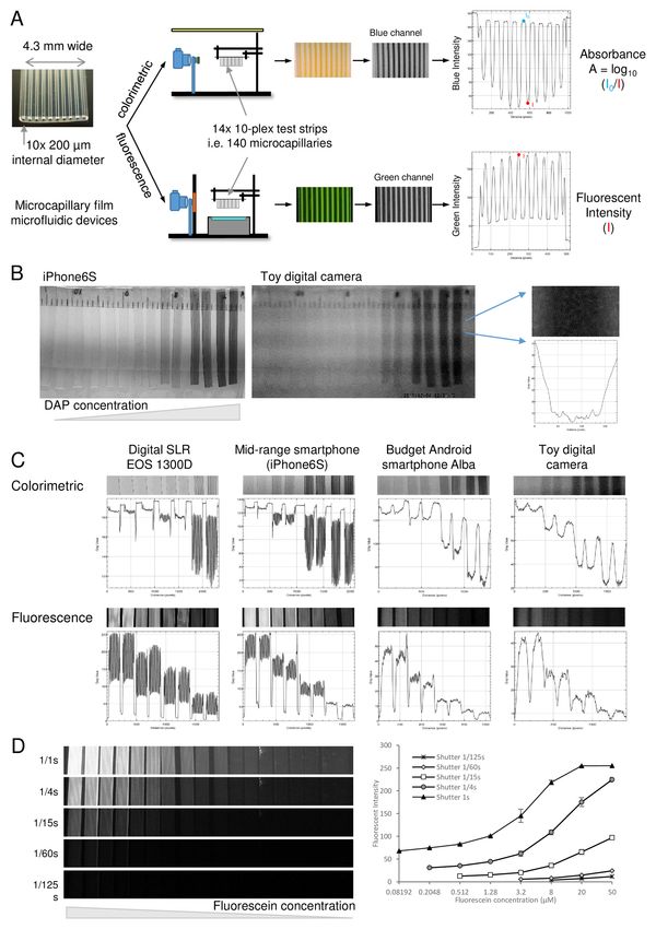
Figure 1. Experimental setup to systematically compare the digital image quantitation of microfluidic bioassays by different
cameras and smartphones. A) Microcapillary film strips were used as examples of microfluidic devices capable of performing immunoassays.
Sets of colorimetric or fluorescent dye solutions were loaded in a panel of strips and imaged either on a white light background for
colorimetric (absorbance) measurement, or with blue light emitting diode (LED) excitation imaged through an amber emission filter for
fluorescent. The absorbance or fluorescence intensity for each capillary was analysed for the appropriate red-green-blue (RGB) colour
channel using ImageJ as indicated. B) Comparison of image quality for mid-range smartphone vs the cheapest camera, illustrating how
lower quality images cannot resolve individual capillaries but variation in intensity is still clear. C) Illustration of systematic comparison of
cameras for imaging microfluidic bioassays. D) effect of camera settings on fluorescence detection, showing that varying shutter speed can
dramatically alter dynamic range of fluorophore measurement. The same set of serial fluorescein dilutions were imaged with the indicated
range of shutter speeds. Mean fluorescence intensity for 20 replicate capillaries in duplicate microcapillary film (MCF) test strips were
plotted, with error bars indicating standard deviation.
Page 4 of 15Wellcome Open Research 2021, 6:57 Last updated: 06 APR 2021
full immunoassays where substrate conversion is dynamic and supplemented with 10% foetal bovine serum (Sigma, UK) and
target dye concentrations are uncontrolled. Di-amino phenazine 5% goat serum (Sigma, UK). Positive control antiviral antibodies
(DAP) is the yellow dye product produced by the horseradish were recombinant human IgG and IgM anti-Flavivirus group
peroxidase enzyme, commonly used in colorimetric immu- antigen [D1-4G2-4-15 (4G2)] and human IgM Anti-COVID-19
noassays, acting upon the o-Phenylenediamine dihydrochloride & SARS-CoV S glycoprotein [CR3022], obtained from Absolute
substrate (OPD) substrate often used in colorimetric immu- Antibody (Oxford, UK). Anti-human IgG AP and anti-human
noassays. Fluorescein is used both directly for fluorescent IgM Ap secondary antibodies were purchased from ThermoFisher
detection and also as the product of alkaline phosphatase Scientific (UK). For imaging both dengue virus (DENV) and
conversion of the substrate fluorescein di-phosphate (FDP) and SARS-CoV-2 immunoassay strips, AttoPhos® AP Fluorescent
is spectrally similar to other alkaline phosphatase substrates Substrate System (Promega, UK) was added as the final sub-
such as Attophos™. Following this systematic comparison using strate. Baculovirus expression of viral proteins used Spodoptera
dyes, selected cameras were used to image full immunoassays that frugiperda (Sf9) and T.nao38 cells which were maintained in
simulated the measurement of antibodies against important viral EX-CELL 420 medium (Sigma, UK) supplemented with 2%
antigens, to confirm the findings could be applied to clinically fetal bovine serum (Sigma, UK), at 27°C with shaking. Virus
relevant diagnostics bioassays. For all the cameras but the growth used exclusively Sf9 cells while protein expression used
G:Box, the images were taken at about 10cm from the subject T.nao38 cells.
and using a digital setting of 1 (equivalent to no zoom). The
distance of the camera to the sample was recorded and this Digital cameras, mobile phones, and imaging devices
working distance shown for different cameras below alongside For the detection of fluorescent and colorimetric signals, we
example images. Automatic settings were used for the Raspberry used a wide range of cameras and phones, and professional
Pi 3B+, the toy camera, the Alba Phone and the iPhone 6S. imaging systems (Table 1). We used a DSLR camera EOS 1300D
Manual settings were used to take images with the Powershot with a Canon EF-S 60mm f/2.8 Macro USM Lens (Canon), a
S120, with a fixed ISO of 3200 and variable F-Stop and exposure. compact camera Powershot S120 (Canon), a compact waterproof
and shock-resistant WG4 camera (Ricoh) and a toy camera
Target microfluidic devices for fluorometric and colorimetric (Sakar). We used smartphones including an iPhones 6S and 4S
signal detection. Whilst the characteristics of the microchan- (Apple) and Alba SIM Free 5’’ Android and mobile phones
nels within MCF are typical of many microfluidic devices, such as CAT B30 Phone (Caterpillar) and Alcatel 2008G phone
with assay channels of 200 µm internal diameter, they have the (Alcatel) purchased from Argos (UK). The industrial machine
advantage of low cost allowing large numbers of devices to vision camera USB 3 uEye® XC with a Macro Lens (AE00126;
be filled with differing dye concentrations and simultaneously IDS Imaging Development Systems, Obersulm, Germany), a
imaged. They have some unique features including a cylindrical Raspberry Pi camera module v2 powered by a Raspberry Pi 3
cross-section that results in an elliptical intensity plot when B+ computer (Raspberry Pi Foundation, UK) and the labora-
projected onto a flat image, and refractive index matching tory imaging systems G:BOX (Syngene, UK) and Typhoon
between fluoropolymer device and water, avoiding distortion (Amersham, UK) were also used to detect fluorescent and
at the interface between aqueous sample and microdevice. colorimetric signals. Images were taken at a resolution of
Previous studies have shown these permit a wide range of 3280x2464 pixels with the Raspberry Pi camera.
bioassays to be conducted including immunoassays and analytical
microbiology. To improve the image quality and resolution of some imaging
systems by allowing closer focussing on the microfluidic
Materials and reagents channels, three type of lenses were used: a simple injection-
The MCF was manufactured by Lamina Dielectrics Ltd moulded plastic magnifying lens, a smartphone clip-on macro
(Billingshurst, West Sussex, UK) and consisted of an array of lens (Amazon, UK) and an industrial machine vision macro lens
10 micro-capillaries produced from Teflon®-FEP FEP (fluori- (IDS). These were held directly onto the front of the digital
nated ethylene propylene) (Dow, USA) using a continuous camera lens during imaging.
melt-extrusion process22,23. The MCF had a width of 4.3mm
and the mean internal diameter of the microcapillaries was Evaluation of digital imaging performance for
206µm. Fluorescein NIST-Traceable Standard (ThermoFisher colorimetric and fluorescent microfluidic assay
Scientific, UK) was used to assess the cameras/phones measurement
performance to detect fluorescence and was used as a MCF strips were prepared by firstly giving an internal
reference for the immunoassays. A stock solution of 100mM hydrophilic coating with PVOH incubated at room temperature
2,3-Diaminophenazine (DAP) (Sigma, UK) was prepared in (RT) overnight, followed by washing and cutting into individual
DMSO. Dilutions of DAP were prepared in SIGMA-FAST 75mm long test strips22. For the colorimetric signal detection,
OPD Buffer (Sigma, UK). DAP was used to assess the cameras/ a 4mM solution of DAP and 5-fold dilutions in SIGMA-FAST
phones performance to detect colorimetric signals. A 2 mg/mL OPD Buffer were added to the MCF strips, in duplicate, using
solution of high molecular weight polyvinyl alcohol (PVOH, a 10ml syringe. The images were taken under a white light. The
Sigma, UK) in PBS was used to coat the inner surface absorbance was calculated from the drop in blue light intensities
of the micro-capillaries to make them hydrophilic22,23. Human measured using ImageJ software25 to plot intensity across the
negative control serum was bought from Sigma. The blocking microdevice, with the lowest point being taken as maximal
buffer consisted of Superblock (ThermoFisher Scientific, UK) absorbance value (Figure 1A)3,20,21. For the fluorescent signal
Page 5 of 15Table 1. Cost and properties of different classes of digital cameras.
Market Product Type Class Price Brand/Camera Price Sensor Size Image Stated Sensor Aperture Focal length
Range when dimensions Resolution (35mm
purchased (pixels) (Megapixels) equivalent)
Consumer Mobile Smartphone Flagship > £700 Samsung/Galaxy 1/2.55” 12 f/1.5-2.4 26 mm
phone S9+ *
camera
Mid-range £400 Apple/iPhone 6S * 1/3” 12 f/2.2 29 mm
- £700
Budget £50 Apple/iPhone 4S * 1/3.2” 8 f/2.4 33 mm
- £400
Alba/SIM Free 5’’ £50 unknown 1920 x 2560 5 f/2.8 unknown
AndroidTM *
Phone Caterpillar/CAT B30 £80 unknown 1200 x 1600 2 unknown unknown
Phone
Feature < £50 Alcatel/2008G £35 unknown 2 unknown unknown
phone
Digital DSLR Flagship £250 Canon/EOS 1300D £570 22.3 x 14.9 mm 18 f/2.8-32 96 mm
Camera - £1000 + Canon EF-S 60mm including
f/2.8 Macro USM Lens macro lens
Compact Mid-range £100 Canon/Powershot £250 1/1.7” (~ 7.53 x 12.1 f/1.8-5.7 24 - 120 mm
- £250 S120 5.64 mm) 5.2 - 26 mm
Ricoh/Compact WG4 £280 1/2.3” (~ 6.16 x 16 f/2-4.9 25 - 100 mm
4.62 mm)
Kids camera Budget/ < £50 Toy camera (CA2- £25 unknown 3648 x 2736 10.1 f/2.8 4.1mm
Toy 10027 Frozen)
Camera powered by Budget < £50 Raspberry Pi Ltd/v2 £33 for 1/4” 8 f/2.0 (3.60 mm
single-board computer camera Raspberry Pi computer – not 35mm
3 B+ plus £22 equivalent)
for camera
Industrial Machine Webcam ≥ £150 IDS/USB 3 uEye® XC + £330 plus 1/2.45 4192 x 3104 13 f/2.8mm 5.3 mm
vision Macro Lens (AE00126) £30 for lens
Laboratory Gel Doc system > £5000 Syngene/G:Box unknown 1391 x 1039 4 unknown unknown
Amersham/Typhoon unknown N/A N/A N/A
Page 6 of 15
Wellcome Open Research 2021, 6:57 Last updated: 06 APR 2021Wellcome Open Research 2021, 6:57 Last updated: 06 APR 2021
detection, 50 uM Fluorescein and 5-fold dilutions in H2O were 3h at RT before adding blocking buffer. The MCF strips were
added to the MCF strips, in duplicate, by aspiration with a 10ml stored overnight at 4°C in blocking buffer. For the DENV2-E
syringe. The images were taken using a light-emitting diode assays, high (100 µg/ml) and low (5 µg/ml) amount of mono-
(LED) Transilluminator (IO Rodeo, USA) to provide blue light clonal IgG or IgM anti-Flavivirus was spiked into human serum
excitation in a dark room with the amber emission filter held and compared with human serum with no added anti-Flavivirus
between the camera and the test strips. The peak fluorescence antibody. For the SARS-CoV-2-S1, a high (50 µg/ml) and low
intensity was determined for each microcapillary in the green (5 µg/ml) amount of monoclonal IgM anti-COVID-19 was
image channel using ImageJ software (Figure 1A). spiked into human serum and compared with human serum with
no added anti-COVID-19 antibody. In both cases, seroreactivity
Image data publication against the antigen was measured using a conventional end-
All image files are published in the associated dataset for this point titre protocol, whereby the serum samples were serially
paper26, and can be accessed to assess the relative image quality diluted in blocking buffer. The serum samples were incubated for
of different camera types. The image examples used in the 10–20 min, followed by two washed with PBS/T. Then, the
figures are constructed from cropped images of individual MCF secondary antibody anti-human IgG or IgM conjugated with
test strips as outlined in the examples shown in Figure 1, but all alkaline phosphatase was added to the strips at a dilution of
original digital images recorded by the cameras are available 1 µg/ml in blocking buffer. The secondary antibody was incu-
as underlying data26. A CSV data sheet summarises the image bated for 10–20 min before being washed three times with PBS/T.
file names in this data set of over 170 original image files, and Finally, AttoPhos® AP Fluorescent Substrate System (Promega)
this lists the conditions and camera in each image file. was added and the fluorescent signal was captured using various
imaging systems and measured using ImageJ software. For every
Baculovirus expression and purification of DENV2-E and assay, a reference MCF strip containing 2uM of fluorescein was
SARS-CoV-2-S1 added and was used to normalise the fluorescent signals from
The sequence of DENV2-E containing domains I and II (EI/II) the assays. The relative fluorescent intensity was determined by
(nt 1 to 891) (accession number NC_001474) was codon dividing the fluorescent intensity of the assay by the fluorescent
optimised for Spodoptera frugiperda cells and the honeybee intensity of the reference.
melittin signal peptide was added upstream of the sequence. The
sequence was flanked by 18bps at the 5’ and 3’ ends homolo- Results and discussion
gous to the intended expression vector, pTriEx1.1 before being A wide range of digital cameras are capable
ordered (IDT Europe, Belgium). The 3’ flanking nucle- of recording and quantifying colorimetric and
otides were also designed to fuse the EI/II ORF in frame to the fluorometric microfluidic test results
vector’s 6xHis tag encoding sequence. The gene and the We established a simple testing rig to systematically compare
vector were assembled by recombination using the In-Fusion digital images of microfluidic devices with a wide range of
HD Cloning kit (Takara, USA). The assembly reaction was then different cameras of different costs and formats (Figure 1A).
used to transform NovaBlue Singles Competent Cells (EMD Overall, we found that all digital cameras tested – even the
Millipore, UK). The sequence of SARS-CoV-2 S1 was obtained lowest quality and cost – were capable of recording and
from the cloned full-length S sequence and was cloned into distinguishing signal intensity of microfluidic test results to
the expression vector pTriEx1.1 (EMD Millipore, UK) and some extent. However, the optics and optoelectronics affected
characterised as described previously27. sharpness of image and sensitivity of detection for both
fluorescent and coloured dyes. For example, the iPhone 6S gave
Sf9 cells were transfected with the baculovirus expression sharp images with high enough resolution to clearly distinguish
vector FlashBAC Gold (Oxford Expression Technologies, UK) all capillaries, in contrast to the cheapest toy camera where all
and with either DENV2-EI/II or SARS-CoV2-S1 constructs to capillaries were blurred together (Figure 1B). This indistinct
produce recombinant baculovirusesr28. Large-scale protein image is likely a consequence of both poor optics unable to
expression was performed by infecting 1L of T.nao38 cells resolve the individual capillaries, and limitations of the image
with a high titre stock of the recombinant baculovirus and sensor showing noise and with a pixel size too large to distin-
incubated for 3-5 days at 27°C. After incubation the superna- guish capillaries. In spite of this poor image, a clear concen-
tant containing the secreted protein was harvested, clarified by tration-intensity relationship was still very clearly measurable
centrifugation at 4,300xg for 20min and filtered through a for cameras across the range of cost and quality, from the
0.45um filter. The clear supernatant was supplemented with digital SLR with largest image sensor and highest quality
0.5nM nickel sulphate before being loaded onto the Bio-Scale optics, through both mid-range and budget smartphones, to the
Mini Profinity IMAC Cartridge (Bio-Rad, UK). The elution was cheapest toy camera (Figure 1C). A concentration-intensity
carried out at a flow rate of 2.5 ml/min with a gradient elution relationship could be clearly quantified for both colorimetric
of 0.05–0.5M imidazole or 0.05-0.25M imidazole over 60 min and fluorescent dyes. This indicates that the most important
for DENV2-EI/II or SARS-CoV-2-S1 respectively. requirement for digital quantitation may be obtaining an image of
sufficient quality to resolve the microchannels, but that even the
Indirect ELISA for the detection of IgG and IgM simplest optical sensors can quantify intensity levels.
MCF strips were coated with 5 ug/ml of DENV2-EI/II or with
15 ug/ml SARS-CoV2-S1 and incubated at RT for minimum Whilst the imaging setup was standardised as far as possible,
1h. The MCF strips were then coated with 0.1mg/ml PVOH for it was clear that camera settings significantly affect signal and
Page 7 of 15Wellcome Open Research 2021, 6:57 Last updated: 06 APR 2021
made a big impact on both analytical sensitivity and dynamic and quantify colorimetric assays (Figure 2B), there was a
range of detection. It was only possible to control camera significant trade-off as closer focusing gave a smaller field of
settings such as exposure, aperture and focus for a subset of view and significantly reduced the number of microfluidic devices
cameras. By increasing the exposure time (but with fixed aper- that could be simultaneously recorded.
ture and ISO sensitivity), the analytical sensitivity of fluorescence
detection increased significantly, with lower concentrations In conclusion, the camera type significantly influenced the
of fluorescein becoming detectable, alongside an increase in number of microfluidic devices that could be recorded
background (Figure 1D). At the longest exposure, the highest simultaneously, with the lower the quality of optics and
concentrations of fluorescein became saturated, with the digital sensor, the fewer the microdevice measurements that can
8-bit image intensity scale providing a limit to the measurable be captured in one single image. However, as long as the camera
dynamic range. For those cameras with automatically controlled was able to resolve individual microchannels, dye quantitation
image settings, we could still compare intensity between devices was possible. All original image files are shared through the
within the same image, but if different devices were imaged experimental dataset accompanying this paper.
independently, a reference sample would become essential to nor-
malise sample intensity between images taken at different times3. Performance differences and quantitation. Having estab-
lished that multiple microcapillary devices could be imaged,
Quantitative comparison of camera performance for with some resolution limitations for the lower quality cam-
digital imaging of colorimetric microfluidic tests eras, colorimetric absorbance was quantified and plotted
For each camera tested representative images of colorimetric against concentration to determine the relative analytical per-
dye filled microcapillary devices on a white light background formance of different cameras (Figure 3). With the lower
are shown, listed in order of the working distance required to resolution images, every effort was made to record the intensity
image the full set of microcapillary devices (Figure 2A). The for each capillary where the test strip was clearly visible.
working distance is also indicated in the figure, and relates to However, in many strips capillaries with lower concentrations
the field of view for each camera setup. When imaging this could not be distinguished and no intensity could be measured,
full set of samples, all cameras were clearly able to quantify the and so data points were only recorded when the intensity value
variation in signal intensity between strips to some degree, but for individual capillaries could be clearly identified after
individual capillaries could only be resolved with a subset of plotting intensity profiles on ImageJ. Comparing camera
cameras. The iPhone 6s, the DSLR Camera EOS1300D and the phones and smartphones showed that the whole range of phone
Powershot S120 were the best cameras to image a full of set of cameras tested were capable of recording and quantifying
samples at high resolution. In contrast, the low cost phones colorimetric dye in microfluidic devices (Figure 3A). However,
(CATB30, Alcatel and Alba phones) and the toy camera provided the entry-level smartphone plus two feature phones – mobile
poor resolution images which could not be analysed. phones with simple cameras but without touchscreens and with
simple button interface – required the macro lens to resolve
For the lower performance cameras with images where individual capillaries and permit quantitation. This reduced
the resolution was too low to resolve individual capillaries the number of test strips that could be imaged simultaneously.
multiple devices, we added an additional lens to permit closer With both entry-level and mid-range smartphones 14 devices
focus. With the budget smartphone (Alba Phone) the capillaries each containing 10 microcapillaries were imaged (i.e. 140 data
were clearly distinguishable at higher dye concentrations but points), in contrast after adding the macro lens to the three
not as sharp as the higher performance cameras, and with the lower-quality camera phones for closer focus reduced this to
cheapest toy camera it was impossible to resolve any different recording 4 or fewer devices measuring only 40 capillaries
capillaries in spite of the clear difference in intensity visible i.e. 3.5x less data per image.
between strips of different concentration. Nevertheless, simply
by adding additional plastic convex lens – sold as “macro Surprisingly, the better optics and larger sensor in the three
lens” for addition to small cameras and as clip-on lenses for digital cameras did not appear to improve colorimetric
smartphones – it was possible to clearly resolve the individual quantitation, and both compact digital cameras (Powershot
capillaries when only a few strips were imaged (Figure 2B). S120 and Ricoh WG4) and the DSLR showed very limited
Three additional lenses were compared: the cheapest was an difference in absorbance between the 140 µM and 32 µM
injection moulded magnifying lens that allowed focus on 6 test concentrations, whereas all phone cameras showed a large
strips (around 35mm field of view); whereas both the profes- absorbance change over this range. When equipped with a
sional macro lens for machine vision imaging and the consumer macro lens to focus closely, the toy camera also showed a
smartphone clip-on macro lens allowed focus on 3 test strips clear difference in absorbance between these concentrations
(around 15–20mm field of view). Although the clip-on lens (Figure 3A, B). The largest sensor and best optics found in
could not be used with the toy camera, the benefits of macro the DSLR with macro lens did allow capillaries to be clearly
lens addition for closer focus was clear for both the budget identified with dye concentration as low as 1.3 µM and
smartphone and toy camera, and it was possible to quantify there was a clear increase in absorbance across the 1.3 µM to
absorbance within 200 µm diameter microfluidic channels 140 µM range with this camera, but given the absorbance only
with both of these very low cost cameras- in spite of their low increased 2-fold over this >100-fold increase in concentration,
quality image sensor and optics. Whilst the addition of macro the gradient is barely steep enough for accurate quantitation.
lens made it possible to resolve individual microcapillaries Thus, although colorimetric detection is clearly feasible with a
Page 8 of 15Wellcome Open Research 2021, 6:57 Last updated: 06 APR 2021
Figure 2. Example images of colorimetric microfluidic assays taken with range of digital cameras and phones. A series of five
5-fold concentrations of diaminophenazine dye (DAP) dye was made, representing the colorimetric product of horseradish peroxidase
enzyme substrate conversion used in conventional enzyme immunoassays. These simulated immunoassay samples, plus a negative control,
were loaded into pairs microcapillary film test strips. The full set of 12 samples were imaged using a range of different digital cameras, plus a
laboratory gel scanner and consumer A4 flatbed scanner. A) example images illustrating the image quality are shown, with the blue channel
where maximal absorbance of DAP presented. The distance of the camera to the sample to achieve an appropriate field of view is indicated
in cm. B) Two examples of lower quality cameras, where individual capillaries could not be resolved when imaging the full set of 12 samples,
so an additional macro lens was added allowing closer focus that permitted dye concentration to be quantified.
wide range of digital cameras, with high resolution optics and cameras (Figure 3C). The industrial machine vision camera
larger sensors permitting capture of many devices, or lower required the macro lens for closer focus and was only able to
quality digital imaging able to capture fewer devices, there capture 4 devices per image, in contrast the flatbed scanner was
may be limits to both analytical sensitivity and to measurable capable of capturing very large numbers of devices, with at least
dynamic range. However, the dynamic range can be increased 150 devices (each with 10 capillaries, i.e. 1500 microchannels)
by varying camera settings such as exposure times and sensor fitting on a single scan. It is possible with further optimisation
sensitivity (Figure 1D). of imaging or scanning conditions, these digital imaging devices
would meet the performance of the digital cameras, however
Finally, an industrial machine vision camera, a flatbed scanner, this demonstrates that consumer digital cameras are more than
and a laboratory gel scanner were compared, none of which capable of recording quantitative bioassay readouts alongside
offered improved quantitation over the phone cameras or digital industrial/laboratory devices.
Page 9 of 15Wellcome Open Research 2021, 6:57 Last updated: 06 APR 2021
Figure 3. Quantitation of colorimetric bioassay signal by different digital cameras. Absorbance was calculated for the images in
Figure 2 from the reduction in blue channel intensity for each individual capillary. The mean absorbance for 10 individual capillaries was
then plotted against concentration, to illustrate the relative analytical performance for microfluidic colorimetric bioassay measurement by
a range of digital cameras. A) Comparison of feature phones vs smartphones; where indicated a macro lens was used to be able to resolve
individual capillaries. B) Comparison of consumer digital cameras. C) Comparison of industrial camera vs laboratory scanners vs consumer
flatbed scanner. Error bars indicate the standard deviation of 10 capillaries. Lower absorbance values are plotted (Right) with smaller y-axis
range to permit comparison of detection of limiting concentrations of fluorophore.
Comparison of camera performance for digital imaging and low-cost fluorescence imaging setup comprising an open
of fluorometric microfluidic tests source blue LED array transillumination device plus a simple
Fluorescent readouts can offer higher analytical sensitivity amber acrylic emission screen, significantly lower concentra-
than colorimetric detection, although sensitivity of fluorescent tions of fluorescein were measurable – with some cameras clearly
detection depends significantly on excitation intensity and detecting 0.5 µM and lower – than with the colorimetric dye
wavelength, quality of emission filters, in addition to the where the lowest measurable concentration was 10 µM or higher
optical detector used. We found that even with the very simple (Figure 1D, Figure 4).
Page 10 of 15Wellcome Open Research 2021, 6:57 Last updated: 06 APR 2021
As with the colorimetric measurements, images with a wide original image files are shared through the experimental dataset
range of cameras are compared in Figure 4, with the working accompanying this paper26.
distance needed to capture the full set of devices indicated.
Although all cameras clearly recorded the changes in concentration The least effective fluorescence detection within the group of
between test strips, for many images the individual capillaries phone cameras was with the two feature phones (Alcatel and
could not be resolved. As with colorimetric measurements, the CAT B30), which although equipping with the macro lens for
macro lens allowed closer focussing and thereby facilitated closer focussing permitted individual capillaries to be clearly
quantitation of fluorescence within individual capillaries resolved, very limited difference in intensity was measured
for the lower quality cameras, with even the cheapest toy across a wide range of fluorescein dye (Figure 5A). In contrast,
camera capable of recording individual capillary intensity. All the budget smartphone (Alba Phone) gave adequate quantitation
Figure 4. Example images of fluorescent microfluidic readout taken with a range of digital cameras and phones. A series of 5-
fold dilutions of fluorescein were made to represent a range of results of fluorescent bioassays. A panel of microcapillary film strips filled
with these fluorescein samples were imaged in parallel using the indicated range of cameras and lab scanners. A) example images for
these cameras, with the distance from camera to sample to image the full sample set indicated in cm. B) For two cameras unable to resolve
individual microcapillaries when imaging the full set of samples, macro lenses were added to allow closer focusing, with example images
illustrating the improved resolution of macro images, and mean fluorescence of 10 replicate capillaries plotted to show how quantification
of fluorescein remains possible even with these lowest performance cameras.
Page 11 of 15Wellcome Open Research 2021, 6:57 Last updated: 06 APR 2021
Figure 5. Quantitation of fluorescent microfluidic readout with a range of digital cameras and phones. The signal intensity
for the images in Figure 4 were determined and mean fluorescence intensity for 10 individual capillaries plotted against concentration.
A) Comparison of feature phones vs smartphones; where indicated a macro lens was used to be able to resolve individual capillaries.
B) Comparison of consumer digital cameras. C) Comparison of industrial camera vs two laboratory fluorescent scanners. Error bars indicate
the standard deviation of 10 capillaries. Lower intensity values are plotted (Right) with smaller y-axis range to permit comparison of detection
of limiting concentrations of fluorophore.
of fluorescence with the macro lens. This difference could be digital cameras performing similarly to the DSLR and all
influenced not only by the sensor and lens, but also possibly by capable of measuring 0.2 µM fluorescein (Figure 5B). However,
the image acquisition software. The budget smartphone has a far the higher concentrations of fluorescein were saturated for
bigger screen and far better processing power onboard than two of the digital cameras, suggesting that careful utilisation
the two feature phones, that may permit improved imaging of of what are typically 8-bit intensities (i.e. 256 shades) is needed
fluorescence within microfluidic devices. However, the higher to maximise assay dynamic range. Even the cheapest toy
performance smartphones (iPhone 4S and iPhone 6S) were camera was capable of adequate quantitation, although at far
clearly superior at quantitation of fluorescent microfluidic higher concentrations – with the first steep rise in intensity
devices, showing clear quantitation below 0.2 µM and 0.5 µM visible from 2 µM up to 8 µM fluorescein – and requiring the
respectively. The digital cameras were all far better at quantitation macro lens for close focussing. Again, the industrial machine
of fluorescence than colorimetric dye, with the two compact vision camera gave less sensitive detection of fluorescence than
Page 12 of 15Wellcome Open Research 2021, 6:57 Last updated: 06 APR 2021
many of the consumer digital cameras and phone cameras, Thus although the lowest performance (and cheapest) camera
and the macro lens was essential for close focussing to resolve was still capable of quantifying overall fluorescent intensity
individual capillaries. The two laboratory scanners, designed and thus capably of quantifying microfluidic immunoassay
for scanning fluorescence, proved no better at quantitation of results, the reduced analytical performance combined with an
fluorescence within microfluidic devices than the consumer inability to capture as many test strips in a single photo makes it
digital cameras (Figure 5C). less suitable to quantitative clinical measurements. We conclude
that a very wide range of digital imaging devices including
Antiviral antibody levels can be measured using a wide smartphones, feature phones, consumer digital camera, and
range of smartphones, cameras or laboratory imaging camera modules are all effective at recording clinically impor-
systems tant immunoassays in microfluidic devices. Higher resolu-
Anti-viral antibody measurement assays were selected to tion digital imaging can offer higher density of data capture. All
illustrate fluorometric bioassays where microfluidic point-of-care original image files are shared through the experimental dataset
and testing outside a lab could be valuable, especially in accompanying this paper.
resource limited regions where use of low-cost consumer
digital cameras is of greatest benefit. We wanted to demonstrate Critical parameters for successful digital imaging of
the quantitation of bioassays was largely unaffected by camera microfluidic devices
type, and we therefore made use of recombinant positive We suggest the following framework for selecting a camera for
control antibodies spiked into negative control plasma to digital imaging of microdevices and bioassays. Firstly, establish
provide uniform assays. Indirect immunoassays with multiple the clinical need and analytical requirements (i.e. measurement
patient sample dilution are typically used to quantify levels of range, limit of detection) and thereby define the number and
antibody against viral antigens, for example for serosurveil- size of devices that need to be recorded. Alongside this, it is
lance programs. Although we published proof-of-concept of important to understand any regulatory constraints such as any
indirect immunoassays using mouse antibodies, here we used need to have complete control over software or camera settings.
recombinant humanised monoclonal antibodies spiked into Whilst separate from the technical requirements evaluated in this
human plasma. Microcapillary film strips internally coated study, such regulatory or software requirements may rule-out
with DENV2 E protein or SARS-CoV-2 S protein were used to use of consumer products. Once the number and size of devices
measure reactivity at multiple dilutions of simulated plasma are defined, both the overall image size needed (camera field of
samples, and after assay was completed were imaged using view) and the resolution required to quantify individual test
the indicated cameras. With all cameras, all response curves areas can be established. As far as we could establish in this
showed the expected dilution-intensity relationship (Figure 6), study, as long as the image acquired can resolve individual
with both IgM assays only showing weak signal at the lower microdevice channels i.e. each analytical chamber can be
dilutions (1:20 and 1:60) vs the far higher signal that is clearly identified on the final image, the camera will be able to
maintained after multiple serial dilutions for the spiked quantify signal. We do suggest real-world images are taken to
positive control samples. For anti-DENV IgG the background establish image resolution however, as the observed resolution
was somewhat higher at these lower dilutions for control may not match manufacturers claimed performance. The
samples, as expected given the higher levels of IgG found in working distance from camera to devices may also influence
plasma, but the spiked positive control still showed far higher setup. Finally, the dynamic range of quantitation of signal, and
signals when more highly diluted. Both the stand-alone camera analytical sensitivity required must both be considered. It may
module and a laboratory scanner showed similar performance be necessary to modify camera settings to expand dynamic range
to the consumer digital cameras, with a Raspberry Pi 3 B + or to match the required analytical sensitivity.
connected to the 8MP camera module v2, representing an open
source and lower cost version of the IDS industrial machine Conclusion
vision camera. We found that a wide range of digital cameras – including the
lowest cost consumer products – were capable of recording
Each set of dilutions represented 8 test strips, each with 10 rep- and quantifying fluorescence and colorimetric signal in 200 µm
licate capillaries, hence each individual image captured 80 data diameter channels within microfluidic devices. Nevertheless,
points. For this comparison of cameras with immunoassays, there are clear benefits of higher performance optoelectronics
the full set of test strips were imaged in one single image, but modules, both in terms of sensitivity and quantitation of lower
for the lowest resolution camera (the toy camera) it was not concentrations of target signal, and in number of microfluidic
possible to distinguish clearly distinct capillaries (see Figure 1B channels that can be simultaneously captured. Likewise, camera
for examples), yet clear differences in intensity were measured optics had some influence on detection, but even addition of a
for the whole strip. very simple macro lens was sufficient to allow low resolution
digital cameras to capture and quantify microfluidic device
All the imaging systems gave similar response curves with signal. With the higher performance cameras, a higher resolu-
exception of the toy camera, which showed reduced signal for tion image made it easier to resolve individual microfluidic
the strong positive control in the IgG assay. Furthermore, the channels, and allowed simultaneous capture of larger numbers
low-quality image with the toy camera made it hard to distin- of microdevices. Finally, we found that that most digital
guish individual capillaries, so multiplex analysis would not cameras including budget smartphones are very capable of
be feasible with distinct bioassays in each of the 10 capillaries. capturing and quantifying fluorescent microfluidic immunoassays
Page 13 of 15Wellcome Open Research 2021, 6:57 Last updated: 06 APR 2021
Figure 6. Comparison of a wide range of digital cameras for recording microfluidic immunoassays to measure antiviral antibody.
Three simulated patient samples were made by spiking two different levels of recombinant antiviral antibodies into human serum, plus a
negative control. Simulated IgM anti-dengue (Left), recombinant IgG anti-dengue (Middle) and recombinant IgM anti-SARS-CoV2 samples
were serially diluted and 7 dilutions plus one blank (no serum) tested in microcapillary film (MCF) test strips coated with the respective
viral protein, followed measuring human antibody level by enzyme fluorescence. The same set of 8 samples were imaged by the indicated
cameras or scanner, and mean fluorescence intensity for 10 individual capillaries plotted; error bars indicated the standard deviation of 10
capillaries. These data are representative of three or more independent assays and camera comparisons.
Page 14 of 15Wellcome Open Research 2021, 6:57 Last updated: 06 APR 2021
to measure antibody responses to two viral infections of global This project contains the following underlying data:
health significance: dengue fever and COVID-19. Overall, this - Images and list of imaging systems and conditions.
study highlights the range of performance requirements for
digital capture of miniature diagnostic tests, and provides a
Data are available under the terms of the Creative Commons
framework to develop a clear specification combining the
Attribution 4.0 International license (CC-BY 4.0).
clinical diagnostic application with the device type, to permit
use of the lowest cost digital camera for result capture, analysis
and quantitation.
Author contributions
Data availability SJ: Data Curation, Formal analysis, Investigation, Methodology,
Underlying data Writing- review and editing
Figshare: Dataset associated with the article “Affordable
IJ: Resources, Supervision, Writing- review and editing
mobile microfluidic diagnostics: minimum requirements for
smartphones and digital imaging for colorimetric and fluoro- ADE: Conceptualisation, Formal analysis, Funding acquisition,
metric viral antibody detection”. https://doi.org/10.6084/ Methodology, Project administration, Supervision, Writing-
m9.figshare.1310341426. original draft
References
1. Zhu H, Isikman SO, Mudanyali O, et al.: Optical imaging techniques for point- global challenge. Lancet Glob Health. 2015; 3(7): e356–7.
of-care diagnostics. Lab Chip. 2013; 13(1): 51–67. PubMed Abstract | Publisher Full Text
PubMed Abstract | Publisher Full Text | Free Full Text
16. Pérez L, Rodríguez I, Rodríguez N, et al.: Robot guidance using machine
2. Shen L, Hagen JA, Papautsky I: Point-of-care colorimetric detection with a vision techniques in industrial environments: A comparative review.
smartphone. Lab Chip. 2012; 12(21): 4240–4243. Sensors (Basel). 2016; 16(3): 335.
PubMed Abstract | Publisher Full Text PubMed Abstract | Publisher Full Text | Free Full Text
3. Barbosa AI, Gehlot P, Sidapra K, et al.: Portable smartphone quantitation 17. Erickson D, O'Dell D, Jiang L, et al.: Smartphone technology can be
of prostate specific antigen (PSA) in a fluoropolymer microfluidic device. transformative to the deployment of lab-on-chip diagnostics. Lab Chip.
Biosens Bioelectron. 2015; 70: 5–14. 2014; 14(17): 3159–64.
PubMed Abstract | Publisher Full Text PubMed Abstract | Publisher Full Text | Free Full Text
4. Berg B, Cortazar B, Tseng D, et al.: Cellphone-Based Hand-Held Microplate 18. Hernández-Neuta I, Neumann F, Brightmeyer J, et al.: Smartphone-based
Reader for Point-of-Care Testing of Enzyme-Linked Immunosorbent Assays. clinical diagnostics: towards democratization of evidence-based health
ACS Nano. 2015; 9(8): 7857–7866. care. J Intern Med. 2019; 285(1): 19–39.
PubMed Abstract | Publisher Full Text PubMed Abstract | Publisher Full Text | Free Full Text
5. Priye A, Bird SW, Light YK, et al.: A smartphone-based diagnostic platform for 19. Huang X, Xu D, Chen J, et al.: Smartphone-based analytical biosensors.
rapid detection of Zika, chikungunya, and dengue viruses. Sci Rep. 2017; 7: Analyst. 2018; 143(22): 5339–5351.
44778. PubMed Abstract | Publisher Full Text
PubMed Abstract | Publisher Full Text | Free Full Text
20. Edwards AD, Reis NM, Slater NKH, et al.: A simple device for multiplex ELISA
6. Rajendran VK, Bakthavathsalam P, Bergquist PL, et al.: Smartphone detection made from melt-extruded plastic microcapillary film. Lab Chip. 2011; 11(24):
of antibiotic resistance using convective PCR and a lateral flow assay. 4267–73.
Sensors and Actuators B: Chemical. 2019; 298: 126849. PubMed Abstract | Publisher Full Text
Publisher Full Text
21. Barbosa AI, Castanheira AP, Edwards AD, et al.: A lab-in-a-briefcase for rapid
7. Ding X, Mauk MG, Yin K, et al.: Interfacing Pathogen Detection with prostate specific antigen (PSA) screening from whole blood. Lab Chip. 2014;
Smartphones for Point-of-Care Applications. Anal Chem. 2019; 91(1): 655–672. 14(16): 2918–28.
PubMed Abstract | Publisher Full Text | Free Full Text PubMed Abstract | Publisher Full Text
8. Drancourt M, Michel-Lepage A, Boyer S, et al.: The Point-of-Care Laboratory in 22. Reis NM, Pivetal J, Loo-Zazueta AL, et al.: Lab on a stick: multi-analyte cellular
Clinical Microbiology. Clin Microbiol Rev. 2016; 29(3): 429–47. assays in a microfluidic dipstick. Lab Chip. 2016; 16(15): 2891–9.
PubMed Abstract | Publisher Full Text | Free Full Text PubMed Abstract | Publisher Full Text
9. Leddy J, Green JA, Yule C, et al.: Improving proteinuria screening with mailed 23. Needs SH, Diep TT, Bull SP, et al.: Exploiting open source 3D printer
smartphone urinalysis testing in previously unscreened patients with architecture for laboratory robotics to automate high-throughput time-
hypertension: a randomized controlled trial. BMC Nephrol. 2019; 20(1): 132. lapse imaging for analytical microbiology. PLoS One. 2019; 14(11): e0224878.
PubMed Abstract | Publisher Full Text | Free Full Text PubMed Abstract | Publisher Full Text | Free Full Text
10. Oncescu V, Mancuso M, Erickson D: Cholesterol testing on a smartphone. Lab 24. Dönmez Sİ, Needs SH, Osborn HMI, et al.: Label-free smartphone
Chip. 2014; 14(4): 759–63. quantitation of bacteria by darkfield imaging of light scattering
PubMed Abstract | Publisher Full Text in fluoropolymer micro capillary film allows portable detection of
11. Kwon L, Long KD, Wan Y, et al.: Medical diagnostics with mobile devices: bacteriophage lysis. Sens Actuators B Chem. 2020; 323: 128645.
Comparison of intrinsic and extrinsic sensing. Biotechnol Adv. 2016; 34(3): Publisher Full Text
291–304. 25. Abràmoff MD, Magalhães PJ, Ram SJ: Image processing with ImageJ.
PubMed Abstract | Publisher Full Text Biophotonics International. 2004; 11(7): 36–42.
12. Kozel TR, Burnham-Marusich AR: Point-of-Care Testing for Infectious Reference Source
Diseases: Past, Present, and Future. J Clin Microbiol. 2017; 55(8): 2313–2320. 26. Jegouic S: Dataset associated with the article “Affordable mobile
PubMed Abstract | Publisher Full Text | Free Full Text microfluidic diagnostics: minimum requirements for smartphones and
13. Haque F, Ball RL, Khatun S, et al.: Evaluation of a Smartphone Decision- digital imaging for colorimetric and fluorometric viral antibody detection”.
Support Tool for Diarrheal Disease Management in a Resource-Limited figshare. Dataset. 2021.
Setting. PLoS Negl Trop Dis. 2017; 11(1): e0005290. http://www.doi.org/10.6084/m9.figshare.13103414.v1
PubMed Abstract | Publisher Full Text | Free Full Text 27. Jegouic SM, Loureiro S, Thom M, et al.: Recombinant SARS-CoV-2 spike
14. Cohen-Bacrie S, Ninove L, Nougairède A, et al.: Revolutionizing clinical proteins for sero-surveillance and epitope mapping. bioRxiv. 2020.
microbiology laboratory organization in hospitals with in situ point-of- Publisher Full Text
care. PLoS One. 2011; 6(7): e22403. 28. Zhao Y, Chapman DAG, Jones IM: Improving baculovirus recombination.
PubMed Abstract | Publisher Full Text | Free Full Text Nucleic Acids Res. 2003; 31(2): E6–6.
15. Royston G, Hagar C, Long LA, et al.: Mobile health-care information for all: a PubMed Abstract | Publisher Full Text | Free Full Text
Page 15 of 15You can also read