Properties of a bovine collagen type I membrane for guided bone regeneration applications
←
→
Page content transcription
If your browser does not render page correctly, please read the page content below

e-Polymers 2021; 21: 210–221
Research Article
Igor S. Brum, Carlos N. Elias*, Jorge J. de Carvalho, Jorge L. S. Pires, Mario J. S. Pereira, and
Ronaldo S. de Biasi
Properties of a bovine collagen type I membrane
for guided bone regeneration applications
https://doi.org/10.1515/epoly-2021-0021 was homogeneous, with collagen fiber webs and open
received December 12, 2020; accepted January 22, 2021 pores. It had no sign of cytotoxicity and the cells at the
Abstract: Dental implant treatment requires an avail- insertion site showed no bone morphological changes.
able bone volume in the implantation site to ensure the There was no tissue reaction and no statistical difference
implant’s mechanical stability. When the bone volume is between Blue Bone® and Bio Oss® groups. The proposed
insufficient, one must resort to surgical means such as membrane has no cytotoxicity and displays a biocompat-
guided bone regeneration (GBR). In GBR surgery, bone ibility profile that makes it suitable for GBR.
grafts and membranes are used. The objective of this Keywords: membrane, collagen, bone regeneration,
work is to manufacture and characterize the in vitro and biomaterials
in vivo properties of resorbable collagen type I mem-
branes (Green Membrane®) for GBR. Membrane surface
morphology was characterized by SEM and roughness
was measured using an interferometric noncontact 3D 1 Introduction
system. In vivo skin sensitization and toxicity tests have
been performed on Wistar rats. Bone defects were pre- In dentistry, membranes are used for guided bone regen-
pared in 24 adult male rats, filled with biomaterials (Blue eration (GBR) and guided tissue regeneration (GTR). The
Bone® and Bio Oss®) and covered with collagen mem- purpose of GBR is to restore large bone defects in ortho-
branes to maintain the mechanical stability of the site for pedic and maxillofacial surgery. In these surgery proce-
bone regeneration. The incisions were closed with simple dures, membranes play an important role. The membrane
stitches; and 60 days after the surgery, the animals were used together with a bone graft provides mechanical sta-
euthanized. Results showed that the analyzed membrane bility to a biomaterial inside the defect site. The mem-
brane prevents epithelial and connective cells from
invading the site during the postsurgical wound healing
phase while allowing periodontal cells to selectively
* Corresponding author: Carlos N. Elias, Materials Science migrate into the defect (1,2).
Department, Instituto Militar de Engenharia, Rio de Janeiro, RJ, The success of GBR depends on the mechanisms
Brazil, e-mail: elias@ime.eb.br
involved in the proliferation and differentiation of
Igor S. Brum: Dentistry Department, Universidade do Estado do Rio
de Janeiro, Rio de Janeiro, RJ, Brazil
mesenchymal cells (MSCs) in the surgery site. When the
Jorge J. de Carvalho: Dentistry Department, Universidade do Estado interactions of proteins with the biomaterial are rapid
do Rio de Janeiro, Rio de Janeiro, RJ, Brazil, and the differentiation of cells into fibroblasts or pre-
e-mail: jjcarv@gmail.com odontoblasts occurs in large numbers, cell differentiation
Jorge L. S. Pires: Dentistry Department, Universidade do Estado do occurs in immature odontoblasts and finally in mature
Rio de Janeiro, Rio de Janeiro, RJ, Brazil,
odontoblasts in less time (3). The main function of odon-
e-mail: jorgepires45@gmail.com
Mario J. S. Pereira: Dentistry Department, Universidade do Estado toblasts is to produce an extracellular matrix, which at
do Rio de Janeiro, Rio de Janeiro, RJ, Brazil, the beginning of the reactions is formed by collagen type
e-mail: mariojps@gmail.com I. Collagen type I has the function of improving cell
Ronaldo S. de Biasi: Materials Science Department, Instituto Militar growth and migration (4). The use of collagen type I
de Engenharia, Rio de Janeiro, RJ, Brazil, e-mail: rsbiasi@ime.eb.br
membranes in GBR helps to create the most surgery
ORCID: Igor S. Brum 0000-0002-7522-205x; Carlos N. Elias 0000-
0002-7560-6926; Jorge J. de Carvalho 0000-0002-9426-6381;
favorable conditions so that the cells involved in the pro-
Jorge L. S. Pires 0000-0002-0572-2577; Mario J. S. Pereira 0000- cess can perform their function without unwanted tissue
0002-3950-3954; Ronaldo S. de Biasi 0000-0001-8897-5969 invagination or the action of an external agent that
Open Access. © 2021 Igor S. Brum et al., published by De Gruyter. This work is licensed under the Creative Commons Attribution 4.0
International License.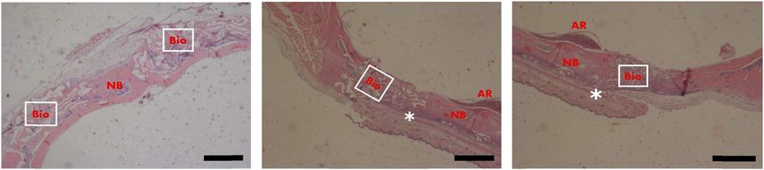
Bovine collagen membrane 211
impairs the natural progress of the process (5). The type I the most common in the human body. Type I collagen
collagen used in the membrane is formed by a polysac- consists of 90% total collagen and is found in the main
charide protein, which contains a small amount of galac- connective tissues such as tendons, ligaments, skin,
tose and glucose (4). Collagen is the main component bone, periodontal connective tissue, and cornea. Type
that forms the extracellular matrix and allows for better II collagen is mainly found in cartilage and intervertebral
cell growth and migration during healing. discs. Type III collagen is found in the cardiovascular
Several natural and synthetic membranes have been system and granulation tissues, and type IV collagen is
commercialized for biomedical applications (6,7), includ- mostly found in the basal membrane (15).
ing polytetrafluoroethylene (PTFE), expanded PTFE, nat- In the market, a variety of natural membranes are
ural collagen, freeze-dried fascia lata, freeze-dried dura available, which possess different times of resorption,
mater allografts, polylactic acid, polyglycolic acid, poly- i.e., from 8 to 38 weeks. Each membrane has advantages
orthoester, polyurethane, polyhydroxybutyrate, calcium and disadvantages (9,16,17). The collagen type I mem-
sulfate, titanium mesh, and titanium foils; but natural branes can be derived from different sources such as
type-I collagen is the preferred choice (8,9). The mem- bovine tendon, porcine pericardium, porcine submucosa,
brane helps to create a space for fibroblasts and osteo- equine tendon, and equine pericardium. In the present
blasts to remodel the damaged tissues (7). Collagen work, a new bovine tendon membrane was developed.
membranes are preferred due to biocompatibility, the The advantages of collagen membranes are biocompat-
capability of promoting wound healing, and the fact ibility, lack of rigidity, good malleability during sur-
that a second surgery is not necessary to remove it. Fibro- gery, manipulation, and the capacity to absorb blood
blasts are responsible for the synthesis of collagen fibrils clots. The purpose of this work was to develop and
that provide physical support for the cellular matrix (10), characterize the in vitro and in vivo properties and the
while osteoblasts, thanks to the barrier created by the potential of resorbable collagen type I membranes for
collagen membrane, do not have to compete with con- GBR procedures.
nective tissues and improve the deposition of bone matrix
(8). Regarding clinical results, the use of resorbable col-
lagen membranes in GBR is comparable with nonresorb-
able membranes (9). 2 Materials and methods
Chia-Lai et al. (11) studied in vivo cellular reactions
for several types of collagen membranes. The data In the present work, an experimental bovine collagen
showed two kinds of cellular reactions that depend on membrane was prepared for GBR. This membrane is a
the physicochemical properties and processing techni- natural type I collagen made from a bovine tendon.
ques. The membranes that induce a physiological reac- The membrane biocompatibility was analyzed in vitro
tion using mononuclear cells undergo an integration and in vivo tests. Scanning electron microscopy (SEM)
process and maintain their structure for 60 days. The and 3D interferometry microscopy analyzed the surface
reaction of collagen-based materials is dominated by morphology. The surface roughness parameters and
mononuclear cells, which lead to their integration into wettability were measured. In vivo tests have been per-
the host tissue (12). formed in rats.
The advantages of collagen membranes over other
natural and synthetic materials are the hemostatic func-
tions that allow early wound stabilization; capable of
promoting cell attachment and proliferation; capable of 2.1 Membrane preparation
attracting fibroblasts, fibroblasts, and osteoblast, which
can attach to its surface; capable of integrating with soft The experimental collagen membranes with a long
tissues; and permeability that facilitates the diffusion of resorption time (60 days) were prepared in the laboratory
nutrients (12). Another important advantage is that nat- of Regener Biomateriais (Curitiba, Brazil) based on highly
ural resorbable membranes do not require a second sur- purified collagen type I fibers derived from bovine Achilles
gery for removal. When the collagen membrane is used, tendons. The collagen underwent purification and proces-
the cell activity starts 3–5 days after the surgery (13,14). sing procedures including the use of sodium hydroxide to
Collagen is of more than 20 different types, and they inactivate pathogens. The purification and processing pro-
change their structure and composition based on the cedures followed the Brazilian and international standards
location and function. Collagen from types I to IV are for handling and supplying animal tissues.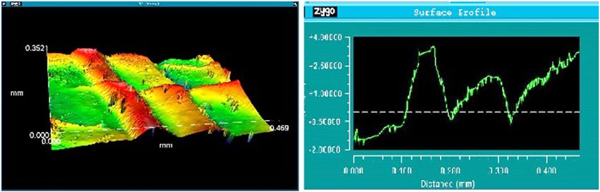
212 Igor S. Brum et al.
2.2 Surface morphology, roughness, and positive control, Biocure® (Pele; Nova Biotecnologia,
wettability Co., Brazil) latex fragment with 5 × 5 mm of proven toxic
nature was used. The samples were tested in triplicate on
The surface structure morphology was characterized by separate plates. The plates were analyzed microscopi-
SEM using a Field Emission Gun FEI QUANTA FEG 250® cally for cell integrity and macroscopically for the pre-
(FEI Corporate, Hillsboro, OR, USA). sence of a halo.
The membrane surface roughness was investigated The cytotoxicity was measured by the diameter of the
using an interferometry noncontact 3D surface measure- light halo, which is classified according to the reactivity
ment system (New View 7100 Profilometer; Zygo Co., scale shown in Table 1.
Middlefield, CT, USA). The parameters for numerically
characterized roughness were the arithmetic mean of
the absolute values of roughness (Ra), the peak-to-valley
roughness (Rz), the mean of the third maximum peak-to- 2.4 Tests for irritation and skin sensitization
valley height (R3z), the root square value of average
roughness (Rq), the highest peak to valley (PV), root Intracutaneous tests on rats were carried out to evaluate
mean square (Rms), area above (Aa), and area below (Ab). the irritation and skin sensitization of the membrane.
The surface wettability was determined by measuring Two incisions were made in the skin of each animal’s
the contact angle with a goniometer First Ten Angstroms calvaria. The skin was raised and sutured. The first inci-
model FTA-100 (First Ten Angstroms Co., Portsmouth, sion was used to introduce a sample of the membrane
VA, USA). The contact angles were determined by aver- under the skin. The second incision was left as a control.
aging the values obtained at five different areas on the The degree of irritation and skin sensitization are quan-
two sample surfaces using distilled water. tified by the index shown in Table 2.
The biocompatibility classification was based on a
comparison of the observed erythema, edema, and
necrosis effects with those of the control incision. The
2.3 In vitro cytotoxicity testing tissue reaction index on the side with the membrane was
subtracted from the reaction index on the control side.
NCTC Clone 929 cell lines and mouse connective tissue cells Table 2 shows the tissue reaction index classification.
(ATCC CCL 1), at a concentration of 3.0 to 105 cells/mL, were
seeded in Petri dishes. Membrane fragments (10 × 10 mm)
were incubated for 48 h at 37°C in a humidified incubator
with an atmosphere of 5% CO2 to form a cell monolayer. The 2.5 Tissue reaction in vivo testing
liquid culture medium was replaced with a solid covering
medium composed of equal parts of concentrate and agar at The in vivo study was carried out at the Instituto de
1.8% with 0.01% neutral red. Biologia Roberto Alcântara of Universidade do Estado
Membrane fragments (10 × 10 mm) were placed on do Rio de Janeiro, Rio de Janeiro, Brazil. Twelve male
this covering medium before its complete solidification. adult Wistar rats weighing approximately 350 g were
The plates were stored at 37°C in an atmosphere of 5% selected. The research was approved by the ethics com-
CO2 for 24 h. For the negative control, nontoxic filter mittee (Process 001/2019). The rats were trichotomized
paper discs with 5 mm in diameter were used. As a and underwent a triangular incision in the calvaria
Table 1: Cytotoxicity classification
Description of the reactivity zone Cytotoxicity Classification
No detectable zones around or under the sample None 0
Some malformed or degenerate cells under the sample Light 1
Zone limited to the area under the sample Mild 2
Zone extends from 0.5 to 1.0 cm beyond the sample Moderate 3
Zone extends more than 1.0 cm beyond the sample Strong 4
Source: BS EN ISO 10993-10 – Biological evaluation of medical devices.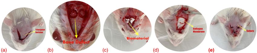
Bovine collagen membrane 213
Table 2: Tissue reaction index in formalin 10% and sent for histological and ultrastruc-
tural analyses.
Reaction Classification The authors confirm that they have complied with
Formation of erythema and bedsores the World Medical Association Declaration of Helsinki
Without erythema 0 regarding the ethical conduct of research involving
Very mild erythema 1 animals.
Well-defined erythema 2 After euthanasia, the animal heads were decalcified
Moderate erythema 3
using 7% EDTA and 0.1 M phosphate buffer (pH 7.4) for
Severe erythema 4
Edema formation
approximately 40 days. The samples were washed in dis-
Without edema 0 tilled water, dehydrated with ethanol (70%, 95%, and
Very mild edema 1 100%), clarified with dimethylbenzene, and embedded
Well-defined edema 2 in paraffin blocks (Paraplast®) at 65°C.
Moderate edema 3 Ten serial coronal sections of 7 µm thick were made
Severe edema 4
using a microtome (LEICA Co., Ltd., Nussloch, Germany).
Maximum possible irritation score
Other adverse changes must be recorded and reported The sections were mounted on glass slides and subjected
to the hematoxylin–eosin (HE) staining technique and
Source: BS EN ISO 10993-10 – Biological evaluation of medical Masson’s trichrome staining. The images were obtained
devices. Tests for irritation and skin sensitization.
with 25× and 40× magnifications using a Carl Zeiss
Axiolab optical microscope.
To examine the surface morphology, the membrane
region (Figure 1a). Two surgical sites were prepared samples were cut into 8 × 8 mm pieces, mounted on a
in each animal using a sterilized punch (cutting edge support with the aid of adhesive tape, and coated with
Ø 3 mm). The bone fragments were carefully removed to gold using a sputtering coating machine.
avoid damage to the dura mater. In each animal, one
surgical site was filled with Bio Oss® (Geistlich, Switzer-
land) and the other was filled with Blue Bone® (Regener,
Brazil). After the biomaterial was inserted in the bone 2.6 In vivo intracutaneous biological
defect (Figure 1b and c), a membrane was placed in the toxicity/reactivity testing
region (Figure 1d) to cover the entire surgical wound. The
animal skin was carefully placed and a suture was made The intracutaneous biological toxicity/reactivity test aims
with 5.0 silk to close the surgical region (Figure 1e). A to determine the biological response of substances
control group with 12 animals was subjected to the same inserted through intracutaneous injections. This test
procedure but without placing the membrane over the was conducted to study the possible harmful effects
biomaterial. of the test substance on the calvaria of animals.
The rats were placed in separate cages after surgery, Samples of the membrane with a concentration of
and an analgesic was administered (sodium dipyrone, 0.2 g/mL of polar extraction solution (0.9% saline solu-
0.1 mL/100 g orally) for 7 consecutive days. After 60 days, tion) were used. The samples were placed in sterile boro-
the animals were euthanized according to the animal silicate containers with a capacity of 25 mL and heated
welfare protocol. The collected fragments were conditioned in an autoclave at 121°C for 1 h. The same process was
Figure 1: In vivo testing: (a) triangular incision in the calvaria region; (b) bone defect prepared; (c) insertion of biomaterial; (d) collagen
membrane in the subcutaneous region; (e) suture with 5.0 silk to close the surgical region.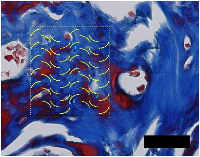
214 Igor S. Brum et al.
performed for controls containing only the polar solution. identify the number of interceptions of the tissue image
Three female rats were used for each test group. with the stereological grid. This procedure was adopted
The animals were kept individually in galvanized due to the complexity of the image. The double-blind
steel cages and acclimated to the conditions of the principle was used to circumvent the possible biased
laboratory for at least 5 days before the test. The diet assessments. In rehearsals, one person marked the
consisted of commercial feed and filtered water, both main points in the images and the second person
provided at will. counted. The two people who did the counting did not
Subjects and controls were inoculated with 0.2 mL by know the sample groups. In the counts, Buffon’s recom-
intracutaneous injection at 5 points on one side of the mendations were observed, a similar procedure has been
spine. The animals were kept for 72 h after application adopted in previous studies (18–20).
and observed for the presence of erythema, edema, and Data on osteocytes in bone defects were analyzed
necrosis as well as other changes. using one-way ANOVA, followed by a Wilcoxon matched-
During this period, the environmental conditions pair test (p < 0.05). All analyses were performed by
were the following: ventilation with 10–15 air changes specific software (GraphPad Prism Version 8.0 and
per hour, temperature between 19°C and 23°C, relative BioEstat 5.0).
humidity between 30% and 70%, controlled illumination
with 12 h of light and 12 h of darkness, the light intensity
between 150 and 325 lux, and noise between 50 and
70 dB. 3 Results
3.1 Membrane surface morphology
2.7 Number of osteocytes in the regenerated
defect Figure 2 shows a scanning electron surface image of the
representative membrane structure at increasing magni-
Stereological analysis was used to determine the number fications. SEM images showed that the outer membrane
of osteocytes. Samples were analyzed using a microscope surface was homogeneous and not compact (Figure 2),
(Carl Zeiss – JVC TK-1270) with a color video camera and with a tightly woven and fibrous structure.
400× magnification. In this analysis, a stereological grid
composed of cycloid segments was placed over the image
of the defect after healing. All points that the grid cut the
osteocytes were counted. The osteocytes were quantified 3.2 Surface morphology and roughness
using Image-Pro Plus for Window, version 7.0.1 (Media analysis with interferometry
Cybernetics).
The number of osteocytes was counted in a semiau- The most characteristic structural morphology of mem-
tomatic way. The researcher decided to define and brane for bone regeneration is the cellular structure.
Figure 2: Samples of the outer surface collagen membrane at different magnifications (from left to right: 500×, 2,500×, and 10,000×). A
tightly woven and fibrous morphology is observed. At high magnification, one sees the typical periodic pattern of collagen fibrils.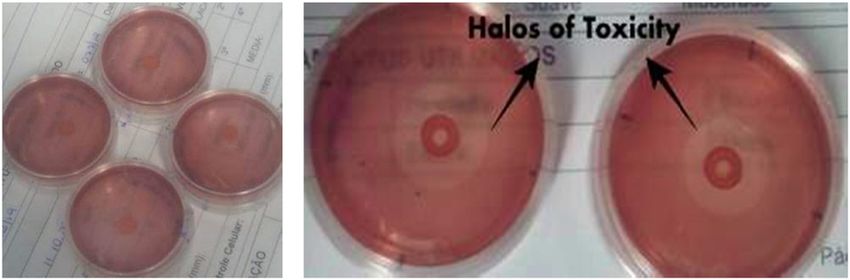
Bovine collagen membrane 215
Typically, collagen membrane morphology characteriza- Table 3: Cytotoxicity test results
tion is performed by SEM, interferometry, MRI, small-
angle XRD, and AFM. In the present work, the mem- Material Halo diameter (mm)
branes were characterized by SEM and interferometry. Sample 1 Sample 2 Sample 3
The membrane characterization using interferometry is
Membrane 0 0 0
not found in the literature. Figure 3 shows a representa-
Negative control 0 0 0
tive membrane surface morphology observed by inter- Positive control 1.2 1.0 1.1
ferometry. The membrane surface morphology has an
irregular shape with valleys and peaks.
Several techniques can be used to measure the mate- PV = 5.246 ± 0.768 µm, Rms = 1.441 ± 0.114 µm, area
rial surface roughness, some of which involve contact above = 579.49 ± 68.795 µm 2 , and area below =
with the material. In the contact-type instruments, a 121.49 ± 20.825 µm 2 .
stylus tip scans the surface of the sample. Direct contact
has many disadvantages, among them the fact that the
measuring pressure may scratch the surface of a soft 3.3 Surface wettability
sample. Moreover, the stylus cannot measure the rough-
ness properly if the scratch width is smaller than the Wettability is an important property for resorbable bio-
diameter of the stylus tip. This is the reason a noncontact materials. The larger the wettability, the greater are the
instrument was used in this work. Another advantage contact with body fluids and the resorption rate. In the
of the laser noncontact equipment is that it provides present work, the wettability of the proposed membrane
simultaneous observation of the surface images during was observed to be very high. A drop of water penetrates
a roughness measurement. quickly into the membrane pores, making it difficult to
Figure 3 shows details of the membrane surface measure the contact angle. The contact angle of each
structure; no sample surface damage can be observed sample was measured several times and it was found to
due to roughness measurement. Figure 3 shows a surface be smaller than 10°. This result shows that the membrane
roughness profile, which is an image like a contact surface is very hydrophilic.
instrument across one direction.
The interferometric analysis showed the 3D structure
of the collagen and elastic fibers (Figure 3). It was pos-
sible to observe the collagen fibers arranged in stacked 3.4 In vitro cytotoxicity results
layers parallel to the membrane surface. A pattern of
curled fibrils with a certain degree of orientation was Table 3 shows the results of the cytotoxicity tests. The
seen. Some regions displayed a network of long, straight, membrane showed no signs of cytotoxicity. In the tests,
and uniformly oriented fibrils. Similar morphology was no halos of toxicity were observed around or under the
observed by Mostaço-Guidolin et al. (21). samples (Figure 4). The cells showed characteristics
The membrane roughness parameters were Ra = 1.235 ± without any morphological changes identical to those
0.125 µm, Rz = 2.759 ± 0.337 µm, R3z = 3.065 ± 0.412 µm, of the negative control.
Figure 3: Interferometric image of the proposed collagen membrane, showing 3D bundle fibrils and the roughness surface profile.216 Igor S. Brum et al.
membrane. Cellular action can be observed 60 days after
the surgery. The histological sections stained for HE was
marked with a square showing the area where the mem-
brane acted during tissue regeneration. The microscopic
examination showed newly formed bone in the prepared
defects with Masson’s trichrome staining (Figure 6). The
graft particles were surrounded by newly formed bone
Figure 4: In vitro cytotoxicity testing results. Samples of negative and improved bone regeneration. Few osteocytes were
control (filter paper) are marked *. Samples of the membrane are
observed in the marrow bone.
marked +. The tin samples did not show halos of toxicity. The
samples from the positive control group (latex) showed halos of
toxicity.
3.6 Osteocyte counting
In the positive control, toxicity was revealed by the
presence of a clear halo with an average diameter of Figure 7 shows a representative image cut from the Blue
1.1 mm (Figure 4). This halo is observed when the red Bone® group obtained from stereological analysis for
dye that was incorporated in the cells is released fol- counting the number of osteocytes. Ninety-six cycloids
lowing cell death. were counted in the. Among the cycloids, six intercept
The sample had a cytotoxic action index of 0, pre- some osteocytes.
senting any effect for the cell line NCTC clone 929. A statistical difference was observed in the number of
osteocytes between the groups covered with the mem-
brane and without a membrane (Table 5). The control
3.5 Tissue reaction in vivo testing results group (without membrane) induced a lower number of
osteocytes than the group with membranes. Table 5
The membrane irritation and skin sensitization classifica- shows that using Blue Bone® biomaterial in the bone
tion were given according to the comparison of the defect, it forms a greater number of osteocytes than
observed erythema, edema, and/or necrosis values with that in the side with Bio Oss®.
those of the control side. Table 4 shows the results of the Table 5 shows the statistical analysis of the number
intracutaneous tests. The tissue reaction index was null. of infiltrated osteocytes in the new bone inside the bone
In the present work, the tissue reaction index (0) on the defect observed 60 days after the surgery.
side with the membrane was subtracted from the irrita-
tion index (Table 3) on the control side (0). The result was
that the tissue reaction had an index equal to 0 and thus
was being considered a nonreactive substance when 4 Discussion
intracutaneously applied in the proportion of 0.2 g/mL
of polar extracting solution (0.9% physiological solution) The purpose of the present work was to develop and
in rats. investigate the properties of a collagen type I bovine
Figure 5 shows the micrography from the control membrane for GBR and guided tissue regeneration
group without a membrane and sample from the region (GTG). Collagen is a component of the bone matrix. It is
of the bone defect filled with Bio Oss® and the membrane the most abundant protein in the human body and has
covering the region. Figure 5a was from the region above a significant influence on the adhesion of fibroblasts.
the defect. Figure 5c shows a slice of the defect filled with The collagen-based membrane has also been reported to
Blue Bone® biomaterial and covered with the proposed influence the cellular functions of fibroblasts, including
cell shape, differentiation, and migration, due to the pre-
Table 4: Intracutaneous test results sence of Arginine-glycine-aspartic acid - Arg-Gly-Asp (RGD)
and GFOGER (integrin-specific glycine–phenylalanine–
Animal 1, 2, and 3 hydroxyproline–glycine–glutamate–arginine) sequences
Side Erythema Edema Bedsores (22). Wound healing, which is a complex and multicel-
lular process that aims to restore the structural and func-
Membrane 0 0 0
tional integrity of bone tissues, follows GBR and GTG
No membrane 0 0 0
procedures. The body can regenerate and repair itselfBovine collagen membrane 217
Figure 5: Photomicrographs of slides stained with hematoxylin and eosin (HE). (left side picture) Sample cut from the region above the
defect without membrane. (middle picture) Samples cut from the defect filled with Bio Oss® biomaterial and covered with an experimental
membrane. Sixty days after the surgery. (*) membrane. (right side picture) Samples cut from the defect filled with Blue Bone® biomaterial
and covered with an experimental membrane. Sixty days after the surgery. (*) membrane. NB = native bone; Bio = biomaterial; AR = artery.
Scale bar = 100 µm, 25× magnification.
Figure 6: Enhanced photomicrograph of slides stained with Masson’s trichrome. (left side picture) Removed from the defect filled with Blue
Bone® biomaterial and covered with the membrane. Many blood vessels can be observed. (right side picture) Removed from the defect filled
with Bio Oss® biomaterial and covered with the membrane. Sixty days after surgery. Blue shows the mineralized new bone. Scale bar =
100 µm, 40× magnification.
Table 5: Statistical analysis of osteocytes numbers in new bone
No membrane With membrane
Bio Oss® Blue Bone® Bio Oss® Blue Bone®
Mean 0.94 1.45 2.68 3.99
SD 0.27 0.42 0.77 1.15
P values P = 0.0001
SD, standard deviation.
Figure 7: Representative images from the stereological analysis. soft tissues. Large bone defects can only be repaired with
Ninety-six cycloids were counted from the image and only six the help of biomaterials such as scaffolds, graft, and
intercepted some osteocytes (scale bar = 15 µm, 40× magnification).
membranes (23,24).
Image cut from the Blue Bone® group.
Type I collagen membranes are used in GBR surgeries
to guarantee the maintenance of the fully elaborated
but only in the case of relatively small defects. Sponta- human intestinal epithelium (25). It is also used in normal
neous bone regeneration involves the migration, prolif- corneal endothelial cells producing mainly type I collagen
eration, and differentiation of several kinds of cells, from the basement membrane (26). It helps in the regen-
including osteoblasts, fibroblasts, macrophages, and pla- eration of the basement membrane of the liver (27), among
telets. The process starts with an inflammatory response other indications (28,29). All applications adopt proce-
to the injury and ends with the growth of new bone and dures similar to the present study.218 Igor S. Brum et al.
Maurer et al. characterized four naturally porcine The results of the present work show that the contact
membranes. Their membranes displayed a smooth, com- angle of this bovine collagen type I membrane is very low
pact, and irregularly crumpled surface morphology (30). and the surface energy is high. This is consistent with the
They observed a finely formed collagen fibrils, loosely histological results; materials with a high wettability and
arranged, and undulating collagen bundles. The surface high surface energy have greater cellular activity (34).
showed a single fibril or small bundles interconnect- The histological results of the present study corrobo-
ing larger bundles. In some area, a compact collagen rate studies in the literature (35). Collagen membranes
arrangement was observed, which was interrupted by provide organized cell growth, in addition to presenting
clusters of circular discontinuities. Their analyzed mem- the potential for the production of tissue substitutes.
brane surface morphologies were different than the pre- The results of cytotoxicity tests showed that the
sent work showed in Figure 2. The difference is possibly developed type I collagen membrane is a nontoxic mate-
due to the manufacturing membrane processing. rial. This result indicates that this kind of membrane,
In the present work, the analyzed collagen type I when used for wound covering, helps tissue regenera-
membrane showed a porous and open-cell structure tion. The membrane can also improve the healing of
(Figure 2). This surface morphology is important to burns and ulcers (36). In the cytotoxicity tests, no inflam-
satisfy critical criteria for membrane, including osteocon- matory reactions were found, showing that the material is
ductive. Open pores are essential for bone healing and nontoxic and does not promote an inflammatory response.
regeneration by allowing adhesion, attachment, migra- Borges et al. carried out a study of the use of collagen
tion, a proliferation of protein, and cells. The fibril bun- membranes with and nonresorbable membranes in the
dles are interconnected and form larger bundles. Some same surgical procedure in the femur of Wistar rats
fibrils showed helical grooves, which are indicative of (37). The results showed that the simultaneous use of
twisted microfibrils. This mesh is permeable to the collagen membranes and nonresorbable membranes in
macromolecules necessary for providing nutrition for GBR procedures do not improve the bone quality of the
tissue repair of the underlying membrane and is a very rat femur. Tanaskovic et al. analyzed the effectiveness of
retentive surface. Similar morphology was observed by the simultaneous use of collagen membranes and tita-
Zenóbio et al. (31). nium mesh nonresorbable membranes and compared
SEM results of the present work (Figure 2) show that them with the separate use of membranes (38). The
the membrane has a three-dimensional structure made of results showed that only using the nonresorbable mem-
collagen fibrils forming bundles in random orientation. brane promotes an intense inflammatory reaction in the
There are no data in the literature about the influence of host tissues, which can cause fibrosis. When the nonre-
the surface morphology of a collagen membrane on cell sorbable membrane was covered with a collagen mem-
behavior such as differentiation, migration, proliferation, brane, the inflammatory response reduced (39). The best
and gene expression. Li et al. studied the effects of micro- result was obtained only using the collagen membrane.
grooved collagen membrane on the behavior of mesench- Toledano et al. analyzed the degradation of three
ymal stem cells (32). They observed that microgrooves types of non-cross-linked resorbable membranes (40).
such as the ones observed in this work (Figures 2 and 3) They concluded that some membranes start to degrade
have a significant effect on the morphology, alignment, in the first 8 h and this early degradation decreases GBR.
and collagen synthesis of the cells. Fadel et al. studied the influence of using collagen mem-
The membrane roughness (Ra = 1.235 µm) and the brane in the regeneration of defects in the calvaria of
contact angle (10°) of the membrane explain the good Wistar rats (41). They concluded that defects that were
in vivo test results. The membrane surface wettability, covered with resorbable collagen membranes showed
energy, and roughness are important parameters that better bone regeneration than defects without a mem-
influence its performance during bone regeneration. brane. The present work corroborates the results of the
Open porous, rough and chemically activated surfaces literature (39–42).
provide ideal conditions for direct protein adsorption Previous work compared the performance of non-
and facilitate the adsorption of fibronectin and albumin cross-linked type I collagen membranes with the nonab-
due to modifications in their ionic state. These mechan- sorbable PTFE membrane (43). It has been observed that
isms are important for bone regeneration. The basic pre- membranes of non-cross-linked collagens promote a high
mise is that the driving force for protein adsorption is the rate of vascularization 60 days after the surgery. This
free energy of the membrane, which is higher if the sur- result was also observed in the in histological analyses
face is rough (33). of the present study.Bovine collagen membrane 219
The in vivo testing shows that the defects that were References
covered with a membrane had a larger number of blood
vessels (Figure 6) and osteocytes (Figures 6 and 7, (1) Kwon KJ, Seok H. Silk protein-based membrane for guided
Table 5) than the defects without a membrane. Similar bone regeneration. Appl Sci. 2018;8:1214. doi: 10.3390/
app8081214.
results were obtained by other researchers who observed
(2) Korzinskas T, Jung O, Smeets R, Stojanovic S, Najman S,
that the use of resorbable membrane improves bone
Glenske K, et al. In vivo analysis of the biocompatibility and
repair (44,45). macrophage response of a non-resorbable PTFE membrane for
guided bone regeneration. Int J Mol Sci. 2018;19:2952. doi:
10.3390/ijms19102952.
(3) da Silva Brum I, Frigo L, Lana Devita R, da Silva Pires JL, Hugo
5 Conclusions Vieira de Oliveira V, Rosa Nascimento AL, et al.
Histomorphometric, immunohistochemical, ultrastructural
characterization of a nano-hydroxyapatite/beta-tricalcium
The results of this work showed that: phosphate composite and a bone xenograft in sub-critical size
(a) The structure of the developed collagen type I mem- bone defect in rat calvaria. Materials (Basel). 2020 Oct
brane exhibited wide cross-linked fibrils of various 15;13(20):4598. doi: 10.3390/ma13204598. PMID: 33076561;
thicknesses forming collagen bundles. PMCID: PMC7602735.
(b) The fibrils have various thicknesses arranged in sev- (4) Zhou J, Zhang K, Ma S, Liu T, Yao M, Li J, et al. Preparing an
injectable hydrogel with sodium alginate and type I collagen to
eral directions and forming highly interconnected
create better MSCs growth microenvironment. E-Polymers.
cross-links. 2019;19:87–91.
(c) The proposed membrane does not have cytotoxicity (5) Makuszewska M, Bonda T, Cieślińska M, Bialuk I,
and does not promote inflammatory reactions. Winnicka MM, Skotnicka B, et al. Expression of collagens type
(d) The use of the developed membrane to keep the graft I and V in healing rat’s tympanic membrane. Int J Pediatr
Otorhinolaryngol. 2019 Mar;118:79–83. doi: 10.1016/
stable significantly increases the number of osteo-
j.ijporl.2018.12.020. Epub 2018 Dec 18. PMID: 30590281.
cytes during bone regeneration. (6) Jo Y-Y, Oh JH. New resorbable membrane materials for guided
(e) The developed membrane morphology is adequate bone regeneration. Appl Sci. 2018;8:2157. doi: 10.3390/
for GBR surgery and has characteristics suitable for app8112157.
use in GBR. (7) Sbricoli L, Guazzo R, Annunziata M, Gobbato L, Bressan E,
Nastri L. Selection of collagen membranes for bone regen-
eration: a literature review. Materials. 2020;13:786. doi:
10.3390/ma13030786.
Research funding: The authors thank the Brazilian Agencies (8) da Silva IB, de Carvalho JJ, da Silva PJL, de Carvalho MAA, Dos
CNPq and FAPERJ for the financial support, and Regener Co Santos LBF, Elias CN. Nanosized hydroxyapatite and β-trical-
for using their facilities. cium phosphate composite: physico-chemical, cytotoxicity,
morphological properties and in vivo trial. Sci Rep.
2019;9(1):19602. Published 2019 Dec 20. doi: 10.1038/
Author contributions: Igor S. Brum was involved in
s41598-019-56124-4.
experimental testing and original draft preparation; (9) Sheikh Z, Hamdan N, Ikeda Y, Grynpas M, Ganss B,
Carlos N. Elias was in charge of conceptualization, the Glogauer M. Natural graft tissues and synthetic biomaterials
surface structure morphology analysis, roughness mea- for periodontal and alveolar bone reconstructive applications:
surements, results analysis, and writing original draft a review. Biomater Res. 2017;21:9. doi: 10.1186/s40824-017-
preparation; Jorge J. de Carvalho contributed to the intra- 0095-5.
(10) Tonelli P, Duvina M, Barbato L, Biondi E, Nuti N, Brancato L,
cutaneous biological toxicity/reactivity test; Jorge L. S.
et al. Bone regeneration in dentistry. Clin Cases Min Bone
Pires was involved in vivo testing; Mario J. S. Pereira per- Metab. 2011;8(3):24–8.
formed the in vivo study and cytotoxicity testing; and (11) Chia-Lai PJ, Orlowska A, Al-Maawi S, Dias A, Zhang Y, Wang X,
Ronaldo S. de Biasi contributed to revision and concepts. et al. Sugar-based collagen membrane cross-linking increases
barrier capacity of membranes. Clin Oral Investig.
2018;22(4):1851–63. doi: 10.1007/s00784-017-2281-1.
Conflict of interest: Authors state no conflict of interest
(12) Kirpatovskii VI, Efimenko AY, Sysoeva VY, Mudraya IS,
and declare any personal circumstances or interests Kamalov DM, Akopyan ZA, et al. Collagen-1 membrane for
that may be perceived as inappropriately influencing replacing the bladder wall. Bull Exp Biol Med. 2016
the representation or interpretation of reported research Nov;162(1):102–6. doi: 10.1007/s10517-016-3556-2. Epub
results. 2016 Nov 23. PMID: 27878492.220 Igor S. Brum et al.
(13) Pokrywczynska M, Jundzill A, Rasmus M, Adamowicz J, Sep 15;9(9):e107814. doi: 10.1371/journal.pone.0107814.
Balcerczyk D, Buhl M, et al. Understanding the role of PMID: 25222024; PMCID: PMC4164635.
mesenchymal stem cells in urinary bladder regeneration-a (26) Kay ED, Cheung CC, Jester JV, Nimni ME, Smith RE. Type I
preclinical study on a porcine model. Stem Cell Res Ther. 2018 collagen and fibronectin synthesis by retrocorneal fibrous
Nov 28;9(1):328. doi: 10.1186/s13287-018-1070-3. PMID: membrane. Invest Ophthalmol Vis Sci. 1982
30486856; PMCID: PMC6260700. Feb;22(2):200–12. PMID: 7035394.
(14) Kamalov AA, Mksimov VA, Kirpatovskiĭ VI, Kudriavtsev IuV, (27) Konomi H, Sano J, Nagai Y. Immunohistochemical localization
Karpov VK, Tokarev FK, et al. Building of defects of the bladder of type I, III and IV (basement membrane) collagens in the
wall by membrane, created on the basis of type I collagen (an liver. Acta Pathol Jpn. 1981 Nov;31(6):973–8. doi: 10.1111/
experimental study). Urologiia. 2012 Sep–Oct;6:33–6. j.1440-1827.1981.tb02011.x. PMID: 7032198.
Russian. PMID: 23379236. (28) David G, Nusgens B, van der Schueren B, van Cauwenberge D,
(15) Leonhäuser D, Stollenwerk K, Seifarth V, Zraik IM, Vogt M, van den Berghe H, Lapière C. Collagen metabolism and base-
Srinivasan PK, et al. Two differentially structured collagen ment membrane formation in cultures of mouse mammary
scaffolds for potential urinary bladder augmentation: proof of epithelial cells. Induction of ‘assembly’ on fibrillar type I col-
concept study in a Göttingen minipig model. J Transl Med. 2017 lagen substrata. Exp Cell Res. 1987 Jun;170(2):402–16.
Jan 4;15(1):3. doi: 10.1186/s12967-016-1112-5. PMID: doi: 10.1016/0014-4827(87)90316-8. PMID: 3297741.
28049497; PMCID: PMC5209890. (29) Tateya T, Tateya I, Bless DM. Immuno-scanning electron
(16) Roca-Millan E, Jané-Salas E, Estrugo-Devesa A, López-López J. microscopy of collagen types I and III in human vocal fold lamina
Evaluation of bone gain and complication rates after guided propria. Ann Otol Rhinol Laryngol. 2007 Feb;116(2):156–9. doi:
bone regeneration with titanium foils: a systematic review. 10.1177/000348940711600212. PMID: 17388240.
Materials. 2020;13(23):5346. doi: 10.3390/ma13235346. (30) Maurer T, Stoffel MH, Belyaev Y, Stiefel NG, Vidondo B,
(17) Raz P, Brosh T, Ronen G, Tal H. Tensile properties of three KuÈker S, et al. Structural characterization of four different
selected collagen membranes. Biomed Res Int. 2019 Dec naturally occurring porcine collagen membranes suitable for
5;2019:5163603. doi: 10.1155/2019/5163603. medical applications. PLoS One. 2018 Oct 3;13(10):e0205027.
(18) García-Fiñana M, Cruz-Orive LM, Mackay CE, Pakkenberg B, doi: 10.1371/journal.pone.0205027.
Roberts N. Comparison of MR imaging against physical sec- (31) Zenóbio EG, Zenóbio MAF, Martins AGV, de Olivieira LJ, de
tioning to estimate the volume of human cerebral compart- Abreu FAM, de Souza PEA. Morphological analysis of resorb-
ments. Neuroimage. 2003 Feb;18(2):505–16. doi: 10.1016/ able collagen membranes by scanning electron microscopy.
s1053-8119(02)00021-6. PMID: 12595203. J Int Acad Periodontol. 2018;20/1:19–24.
(19) Glaser E. Comments on the shortcomings of predicting the pre- (32) Li L, Li X, Chen L, Sun P, Hao N, Jiang B. Morphology, prolif-
cision of Cavalieri volume estimates based upon assumed mea- eration, alignment, and new collagen synthesis of mesench-
surement functions. J Microsc. 2005;218(Pt 1):1–5. doi: 10.1111/ ymal stem cells on a microgrooved collagen membrane.
j.1365-2818.2005.01465.x. PMID: 15817057 discussion 6–8. J Biomater Sci Polym Ed. 2016;27(7):581–98. doi: 10.1080/
(20) Schmitz C, Hof PR. Recommendations for straightforward and 09205063.2015.1136919.
rigorous methods of counting neurons based on a computer (33) Elias CN. Factors affecting the success of dental implants,
simulation approach. J Chem Neuroanat. 2000 implant dentistry – a rapidly evolving practice. Ilser
Oct;20(1):93–114. doi: 10.1016/s0891-0618(00)00066-1. Turkyilmaz: IntechOpen; 2011. doi: 10.5772/18746.
PMID: 11074347. (34) Bao L, Yang W, Mao X, Mou S, Tang S. Agar/collagen mem-
(21) Mostaço-Guidolin LB, Ko ACT, Wang F, Xiang B, Hewko M, brane as skin dressing for wounds. Biomed Mater.
Tan G, et al. Collagen morphology and texture analysis: from 2008;3(4):044108. doi: 10.1088/1748-6041/3/4/044108.
statistics to classification. Sci Rep. 2013;3:2190. doi: 10.1038/ (35) Duan X, Sheardown H. Dendrimer crosslinked collagen as a
srep02190. corneal tissue engineering scaffold: mechanical properties
(22) Sun D, Song B, Sun D. Cytocompatibility of collagen mem- and corneal epithelial cell interactions. Biomaterials.
branes with bladder transitional cells of rabbit in vitro). 2006;27(26):4608–17. doi: 10.1016/
Zhongguo Xiu Fu Chong Jian Wai Ke Za Zhi. 2004 j.biomaterials.2006.04.022.
May;18(3):217–9. Chinese. PMID: 15211840. (36) Sohn DS, Moon JW, Lee WH, Kim SS, Kim CW, Kim KT, et al.
(23) Pizzicannella J, Marconi GD, Pierdomenico SD, Comparison of new bone formation in the maxillary sinus with
Cavalcanti MFXB, Diomede F, Trubiani O. Bovine pericardium and without bone grafts: immunochemical rabbit study. Int J
membrane, gingival stem cells, and ascorbic acid: a novel Oral Maxillofac Implant. 2011 Sep–Oct;26(5):1033–42. PMID:
team in regenerative medicine. Eur J Histochem. 2019 Sep 22010087.
25;63(3):3064. doi: 10.4081/ejh.2019.3064. PMID: 31696691; (37) Borges CD, Faria PE, Pessôa de Oliveira PG, Sales de Melo
PMCID: PMC6767323. Soares M, Ricoldi MS, Costa MS, et al. Influence of collagen
(24) Silva Júnior ZS, Botta SB, Ana PA, França CM, Fernandes KP, membrane on bone quality in titanium meshes reconstructions
Mesquita-Ferrari RA, et al. Effect of papain-based gel on type I – study in rats (published online ahead of print. J Periodontol.
collagen–spectroscopy applied for microstructural analysis. 2020;91:1673. doi: 10.1002/JPER.19-0399.
Sci Rep. 2015 Jun 23;5:11448. doi: 10.1038/srep11448. PMID: (38) Tanaskovic N, Trajkovski B, Kačarević ŽP, Rider PM,
26101184; PMCID: PMC4477230. Houshmand A, Xiong X, et al. Periorbital reconstruction by
(25) Jabaji Z, Brinkley GJ, Khalil HA, Sears CM, Lei NY, Lewis M, “Periorbital Patch” technique using a pericardium-based col-
et al. Type I collagen as an extracellular matrix for the in vitro lagen membrane and titanium mesh. Materials (Basel).
growth of human small intestinal epithelium. PLoS One. 2014 2019;12(15):2343. doi: 10.3390/ma12152343.Bovine collagen membrane 221
(39) Pitaluga LH, Souza MT, Zanotto ED, Romero MES, Hatton PV. (42) Lyu C, Shao Z, Zou D, Lu J. Ridge alterations following socket
Electrospun F18 bioactive glass/PCL-poly (ε-caprolactone)- preservation using a collagen membrane in dogs. Biomed Res
membrane for guided tissue regeneration. Materials. Int. 2020;2020:1487681. Published 2020 Mar 3. doi: 10.1155/
2018;11:400. doi: 10.3390/ma11030400. 2020/1487681.
(40) Toledano M, Asady S, Toledano-Osorio M, García-Godoy F, (43) Ghanaati S. Non-cross-linked porcine-based collagen I–III
Serrera-Figallo MA, Benítez-García JA, et al. Differential membranes do not require high vascularization rates for their
Biodegradation Kinetics of Collagen Membranes for Bone integration within the implantation bed: a paradigm shift. Acta
Regeneration. Polymers (Basel). 2020 Jun 4;12(6):1290. Biomater. 2012 Aug;8(8):3061–72. doi: 10.1016/
doi: 10.3390/polym12061290. PMID: 32512861; PMCID: j.actbio.2012.04.041. Epub 2012 May 3. PMID: 22561669.
PMC7362079. (44) Das A, Abas M, Biswas N, Banerjee P, Ghosh N, Rawat A, et al.
(41) Fadel RA, Samarani R, Chakar C. Guided bone regeneration in A modified collagen dressing induces transition of inflamma-
calvarial critical size bony defect using a double-layer tory to reparative phenotype of wound macrophages. Sci Rep.
resorbable collagen membrane covering a xenograft: a histo- 2019;9:14293.
logical and histomorphometric study in rats. Oral Maxillofac (45) Choi NR, Sándor GK, Kim Y-D. Efficacy of collagen-based
Surg. 2018 Jun;22(2):203–13. doi: 10.1007/s10006-018-0694- membranes in alveolar bone augmentation. Appl Sci.
x. Epub 2018 Apr 14. PMID: 29654386. 2018;8:2048. doi: 10.3390/app8112048.You can also read