Evaluation of Prophylactic Corticosteroid Eye Drop Use in the Management of Corneal Abnormalities Induced by the Antibody-Drug Conjugate ...
←
→
Page content transcription
If your browser does not render page correctly, please read the page content below
Author Manuscript Published OnlineFirst on November 9, 2018; DOI: 10.1158/1078-0432.CCR-18-2474
     Author manuscripts have been peer reviewed and accepted for publication but have not yet been edited.

    Evaluation of Prophylactic Corticosteroid Eye Drop Use in
    the Management of Corneal Abnormalities Induced by the
      Antibody-Drug Conjugate Mirvetuximab Soravtansine
Ursula A. Matulonis1*, Michael J. Birrer2*†, David M. O’Malley3, Kathleen N. Moore4,
Jason A. Konner5, Lucy Gilbert6, Lainie P. Martin7, Todd M. Bauer8, Amit M. Oza9,
Karim Malek10, Jan Pinkas10, and Stella K. Kim11

*These authors contributed equally to this work.
1
 Dana Farber Cancer Institute, Boston MA; 2Massachusetts General Hospital, Boston,
MA; 3The Ohio State University – James CCC, Columbus, OH; 4University of Oklahoma
Health Sciences Center, Oklahoma City, OK; 5Memorial Sloan Kettering Cancer Center,
New York, NY; 6McGill University Health Centre, Montreal, Canada; 7Fox Chase Cancer
Center, Philadelphia, PA; 8Sarah Cannon Research Institute/Tennessee Oncology,
PLLC., Nashville, TN; 10Princess Margaret Cancer Centre, Toronto, Canada;
10
   ImmunoGen, Inc., Waltham, MA; 11University of Texas McGovern Medical School,
Houston, TX.
†
 Present address: Comprehensive Cancer Center, University of Alabama at Birmingham,
Birmingham, AL

Running Title: Pathogenesis and management of ADC-induced ocular toxicity

Keywords: antibody-drug conjugate; folate receptor alpha; ocular toxicity, ovarian
cancer; phase I trial

Corresponding author: Stella K. Kim, MD, The University of Texas McGovern Medical
School, Department of Ophthalmology and Visual Sciences, Robert Cizik Eye Clinic,
6400 Fannin Street Suite 1800, Houston, TX 77030 e-mail: stella.k.kim@uth.tmc.edu

Potential Conflicts of Interest: K.M and J.P. are employees of ImmunoGen, Inc;
U.A.M., M.J.B., D.M.O’M., K.N.M., L.P.M., and A.M.O. have served on an ImmunoGen
advisory board. There are no other competing interests to declare.

Word Count: 4639                          Tables/Figures: 3 Tables/3 Figures

                                                       1

    Downloaded from clincancerres.aacrjournals.org on March 1, 2021. © 2018 American Association for Cancer
                                                   Research.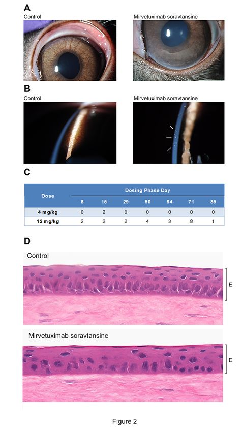
Author Manuscript Published OnlineFirst on November 9, 2018; DOI: 10.1158/1078-0432.CCR-18-2474
Author manuscripts have been peer reviewed and accepted for publication but have not yet been edited.
Translational Relevance
Ocular toxicities associated with antibody-drug conjugate (ADC) administration have
been observed clinically but underlying mechanisms and strategies for symptomatic
mitigation in patients remain to be elucidated. Mirvetuximab soravtansine is a folate-
receptor alpha (FR)-targeting ADC in late-stage clinical development showing promise
for the treatment of recurrent ovarian cancer. The primary ocular abnormalities with
mirvetuximab soravtansine include low-grade, reversible blurred vision and keratopathy.
This report describes non receptor-mediated effects on the corneal epithelium that
account for this ocular toxicity profile, evidenced by a lack of FR expression in human
corneal tissues and preclinical modeling studies in rabbits. The addition of primary
prophylaxis with corticosteroid eye drops as a means to alleviate ocular adverse events
as part of a phase I study in patients receiving mirvetuximab soravtansine is
summarized. These results may have broad implications for the application of this and
other ADCs for which ocular side effects are an important clinical consideration.
2
Downloaded from clincancerres.aacrjournals.org on March 1, 2021. © 2018 American Association for Cancer
Research.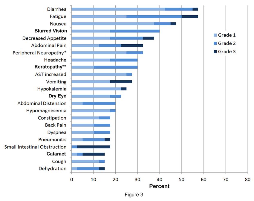
Author Manuscript Published OnlineFirst on November 9, 2018; DOI: 10.1158/1078-0432.CCR-18-2474
Author manuscripts have been peer reviewed and accepted for publication but have not yet been edited.
Abstract
Purpose: Reversible, low-grade ocular adverse events (AEs) are associated with
administration of mirvetuximab soravtansine, a folate receptor alpha (FR)-targeted
antibody-drug conjugate undergoing Phase III clinical evaluation in platinum-resistant
ovarian cancer. This study investigated the underlying mechanisms of ocular toxicity
and evaluated primary prophylactic use of corticosteroid eye drops in patients receiving
mirvetuximab soravtansine.
Experimental Design: Target expression in the human eye was determined by
immunohistochemistry. The ocular toxicity profile of mirvetuximab soravtansine was
assessed preclinically using Dutch-Belted rabbits. In a Phase I clinical study, ovarian
cancer patients were treated with 6 mg/kg mirvetuximab soravtansine intravenously
once every 3 weeks, including one expansion cohort with corticosteroid eye drops
administered daily for the first 10 days of each treatment cycle.
Results: FR expression was absent from human corneal tissues. Ocular abnormalities
in the rabbit eye appeared phenotypically consistent with off-target effects on the
cornea. Forty patients were enrolled in the expansion cohort. Reversible grade 1 or 2
blurred vision and keratopathy occurred in 16 (40%) and 12 (30%) patients respectively;
no grade 3/4 ocular events were observed. Compared to those patients who did not
receive primary prophylaxis, corticosteroid eye drop use resulted in fewer dose
reductions (5% versus 15%) and none discontinued due to ocular AEs.
Conclusion: Preclinical modeling was predictive of the corneal-related symptoms seen
in some patients dosed with mirvetuximab soravtansine. Primary prophylactic use of
3
Downloaded from clincancerres.aacrjournals.org on March 1, 2021. © 2018 American Association for Cancer
Research.Author Manuscript Published OnlineFirst on November 9, 2018; DOI: 10.1158/1078-0432.CCR-18-2474
Author manuscripts have been peer reviewed and accepted for publication but have not yet been edited.
topical corticosteroid eye drops resulted in a trend towards symptomatic improvement
and a reduction in ocular AE-related dose modifications in patients treated with
mirvetuximab soravtansine.
4
Downloaded from clincancerres.aacrjournals.org on March 1, 2021. © 2018 American Association for Cancer
Research.Author Manuscript Published OnlineFirst on November 9, 2018; DOI: 10.1158/1078-0432.CCR-18-2474
Author manuscripts have been peer reviewed and accepted for publication but have not yet been edited.
INTRODUCTION
It is well established that the eye is susceptible to toxic insults that arise in response to
systemic chemotherapy, resulting in a variety of ophthalmic complications that range in
severity from mild irritation to visual loss, including conjunctivitis, blurred vision,
photophobia, keratitis, retinopathy, and optic neuropathy (1,2). This broad spectrum of
ocular toxicities reflects the unique anatomical, physiological, and biochemical features
of the eye (1) and varies with the class of cytotoxic drug used (e.g., alkylating agents,
antimetabolites, taxanes, or platinum agents). Ocular side effects have also emerged as
an important clinical concern for molecularly targeted therapies entering standard
oncology practice, despite these being more tumor selective than traditional cytotoxic
chemotherapy (3,4). In some cases they can be attributed to on-target effects due to
target antigen expression in the eye, as exemplified by the class effect visual
disturbances seen during the early development of MEK and HSP90 (heat shock
protein 90) inhibitors (5). Alternatively, toxicities may occur via off-target mechanisms
and the etiology of such events is less clearly defined.
Antibody-drug conjugates (ADCs) are designed for targeted delivery of potent cytotoxic
compounds through conjugation to monoclonal antibodies that recognize tumor-
associated antigens (6). Currently four ADCs are approved for use in a variety of solid
and hematological malignancies, and more than 60 others are under active clinical
evaluation (7). Importantly, this unique method of site-selective drug delivery affords a
means to reduce off-target toxicities in patients by limiting the exposure of normal
tissues to the payload (8). In this regard, the safety and tolerability profiles for this
5
Downloaded from clincancerres.aacrjournals.org on March 1, 2021. © 2018 American Association for Cancer
Research.Author Manuscript Published OnlineFirst on November 9, 2018; DOI: 10.1158/1078-0432.CCR-18-2474
Author manuscripts have been peer reviewed and accepted for publication but have not yet been edited.
rapidly growing class of anticancer therapeutics were expected to correlate with the
levels of target antigen found in normal tissues. However, for most ADCs, the clinical
experience has revealed that toxicities (including ocular) are driven by the payload
present and not antigen expression (9). Indeed, there appears to be a clear payload
association with ocular side effects, which have been most commonly reported for
ADCs that bear either the maytansinoid DM4 (derivative of maytansine-4) or the
auristatin metabolite MMAF (monomethyl auristatin-F) as their cytotoxic effector
molecules (10). In most cases, these toxicities are consistent with corneal changes
(causing symptoms of blurred vision due to keratitis, microcystic epithelial changes etc.)
and appear irrespective of the cellular targets of the individual ADC, which are typically
absent or only minimally expressed in the eye (9,10).
Mirvetuximab soravtansine is a folate receptor alpha (FR)-targeting ADC, comprised of
a humanized anti-FR monoclonal antibody linked to DM4 (11), currently undergoing
pivotal phase III evaluation in patients with platinum-resistant ovarian cancer (12). In the
first-in-human study of mirvetuximab soravtansine, ocular abnormalities emerged as
adverse events of interest during the escalation stage, prompting dose modifications
(13). These primarily manifested as blurred vision and/or keratopathy, which were
generally mild (≤ grade 2), reversible, and similar in nature to those reported for other
maytansinoid-conjugated antibodies (8). Early recognition of dose- and exposure-
dependent correlations with these ocular events resulted in modification of
mirvetuximab soravtansine dosing from total to adjusted ideal body weight, in order to
decrease the range of variance in interpatient drug exposures (13). Moreover,
implementation of daily lubricating eye drop use and other proactive measures (e.g.,
6
Downloaded from clincancerres.aacrjournals.org on March 1, 2021. © 2018 American Association for Cancer
Research.Author Manuscript Published OnlineFirst on November 9, 2018; DOI: 10.1158/1078-0432.CCR-18-2474
Author manuscripts have been peer reviewed and accepted for publication but have not yet been edited.
avoidance of contact lenses, application of compresses over the eyes etc.)
subsequently decreased both the incidence and grade of visual disturbances in patients
while on treatment (14). In addition to these mitigating strategies, steroid eye drops
were used as treatment or in the secondary prophylaxis setting to help manage ocular
symptoms.
To examine the pathogenesis of the ocular side effects observed in patients treated with
mirvetuximab soravtansine, the distribution pattern of FR expression in the human eye
was determined, followed by preclinical assessment of the ocular toxicity profile of
mirvetuximab soravtansine in rabbits, a commonly used species for modeling potential
drug-mediated ocular toxicities (15). We further report on the clinical findings of a
dedicated expansion cohort, opened as part of the initial phase I study, in individuals
with relapsed ovarian cancer to investigate the potential benefits of primary prophylactic
corticosteroid eye drop use with mirvetuximab soravtansine monotherapy.
7
Downloaded from clincancerres.aacrjournals.org on March 1, 2021. © 2018 American Association for Cancer
Research.Author Manuscript Published OnlineFirst on November 9, 2018; DOI: 10.1158/1078-0432.CCR-18-2474
Author manuscripts have been peer reviewed and accepted for publication but have not yet been edited.
MATERIALS AND METHODS
Immunohistochemistry
Immunohistochemical staining for FRα expression in formalin-fixed, paraffin-embedded
(FFPE) normal human eye whole-section samples (Cooperative Human Tissue
Network) was performed by automated immunohistochemistry (IHC) assay using the
anti-human FRα mouse monoclonal FOLR1-2.1 and OptiView Detection Kit. The
FOLR1-2.1 antibody was developed at ImmunoGen and recognizes FR in FFPE
tissues and the assay was optimized and validated with a broad dynamic range to
detect FR staining in FFPE tissue samples with weak receptor expression. Non-small
cell lung cancer FFPE samples with confirmed FRα expression levels were used as
positive controls. Staining was evaluated by a Board-certified pathologist. For positive
samples, staining intensity (weak, moderate, or strong) and localization (membranous,
cytoplasmic) were recorded.
Rabbit ocular toxicity model
Rabbit studies were performed by Covance Laboratories (Madison, WI), an organization
fully accredited by the Association for Assessment and Accreditation of Laboratory
Animal Care (AAALAC). All procedures were in compliance with applicable animal
welfare acts and approved by the local Institutional Animal Care and Use Committee
(IACUC). Male Dutch-Belted rabbits (n=5/group) were administered mirvetuximab
soravtansine once every 3 weeks for four consecutive doses (i.e. on days 1, 22, 43 and
64) by intravenous (i.v.) infusion via a marginal ear vein over approximately 15 minutes.
Two dose levels were tested, 4 and 12 mg/kg, and animals were monitored until study
8
Downloaded from clincancerres.aacrjournals.org on March 1, 2021. © 2018 American Association for Cancer
Research.Author Manuscript Published OnlineFirst on November 9, 2018; DOI: 10.1158/1078-0432.CCR-18-2474
Author manuscripts have been peer reviewed and accepted for publication but have not yet been edited.
day 107 (end of 3-week recovery phase). A control group was administered the
formulation buffer following the same dosing schedule. The antibody component of
mirvetuximab soravtansine is not cross-reactive with rabbit FR, therefore no
unconjugated antibody arm was included in the study. Assessment of toxicity was
based on mortality, clinical observations, qualitative food consumption, body weights,
ophthalmic examinations, and anatomic pathology (day 85 terminal sacrifice; day 107
recovery sacrifice). Ocular toxicity was assessed by external examination via slit lamp
biomicroscopy as well as microscopic analysis. The adnexa and anterior portion of both
eyes were examined using a slit-lamp biomicroscope and ocular fundus of both eyes
examined using an indirect ophthalmoscope. Prior to examination with the indirect
ophthalmoscope, pupils were dilated with a mydriatic agent (1% tropicamide). In
addition, corneal fluorescein staining was performed at each interval. Animals were
euthanized on days 85 (end of dosing phase) and 107 (end of 3-week recovery phase)
of the study and selected tissues placed in fixative according to established procedures
for IHC microscopic analysis by a Board-certified pathologist.
Patient selection and eligibility criteria
An expansion cohort was opened as part of the first-in-human, phase I study of
mirvetuximab soravtansine monotherapy in adults with FR-positive ovarian tumors to
evaluate primary prophylaxis with corticosteroid eye drops. Adults with histologically-
confirmed advanced epithelial ovarian, primary peritoneal, or fallopian tube cancer who
had received either 3 or 4 prior lines of systemic therapy were eligible to enroll. Patients
had to have met the minimum requirement of FR positivity on archival tumor samples
by IHC (≥ 25% of tumor staining at ≥ 2+ intensity). Tumor tissues were analyzed for
9
Downloaded from clincancerres.aacrjournals.org on March 1, 2021. © 2018 American Association for Cancer
Research.Author Manuscript Published OnlineFirst on November 9, 2018; DOI: 10.1158/1078-0432.CCR-18-2474
Author manuscripts have been peer reviewed and accepted for publication but have not yet been edited.
FR expression at Ventana Medical Systems, Inc. using a validated assay for
sensitivity, specificity and reproducibility. Patients had measurable or non-measureable
disease according to Response Evaluation Criteria in Solid Tumors (RECIST) version
1.1 (16). Patients were also required to be ≥ 18 years of age; have an Eastern
Cooperative Oncology Group (ECOG) performance status 0 or 1; have adequate
hematologic, renal, and hepatic function; and be willing and able to self-administer low-
dose corticosteroid eye drops for the first 10 days of each cycle during active study
treatment. Key exclusion criteria included neuropathy > grade 1; as well as any active or
chronic corneal disorder such as Sjogren’s syndrome, Fuchs corneal dystrophy, history
of corneal transplantation, active herpetic keratitis, active ocular conditions requiring on-
going treatment/monitoring like wet age-related macular degeneration requiring
intravitreal injections, active diabetic retinopathy with macular edema, presence of
papilledema, or acquired monocular vision. All patients provided written informed
consent in accordance with federal, local and institutional guidelines.
Study design and treatment administration
The primary objective of the expansion cohort was to evaluate the impact of primary
prophylactic use of corticosteroid eye drops on the incidence and/or severity of ocular
AEs observed in patients dosed with mirvetuximab soravtansine. Patients were
administered mirvetuximab soravtansine i.v. once every 3 weeks at 6 mg/kg (adjusted
ideal body weight), established as the recommended phase II dose during dose-finding
(13). In addition to daily use of lubricating eye drops, patients self-administered
corticosteroid eye drops (1% prednisolone acetate) 4-6 times daily for the first 10 days
of each treatment cycle. Patients continued on mirvetuximab soravtansine until
10
Downloaded from clincancerres.aacrjournals.org on March 1, 2021. © 2018 American Association for Cancer
Research.Author Manuscript Published OnlineFirst on November 9, 2018; DOI: 10.1158/1078-0432.CCR-18-2474
Author manuscripts have been peer reviewed and accepted for publication but have not yet been edited.
intolerable toxicity or adverse events (AEs), disease progression, or investigator/patient
decision. The trial was conducted in accordance with the US Food and Drug
Administration regulations, the International Conference on Harmonisation Guidelines
for Good Clinical Practice and the Declaration of Helsinki. The study was compliant with
all relevant Institutional Review Board and Independent Ethics Committee requirements
and is registered at ClinicalTrials.gov (NCT01609556).
Clinical Assessments
Baseline assessments included medical history and physical examination, ECOG
performance status, blood chemistry and hematology, and electrocardiogram. Toxicities
were graded according to the National Cancer Institute Common Terminology Criteria
for Adverse Events (NCI CTCAE) version 4.03 and monitored continuously throughout
the study from the time of first dose until 28 days after treatment cessation. For the
purposes of this study, keratopathy was used as a grouped term to capture the specific
corneal pathologies of the observed AEs, including occurrences of keratopathy,
keratitis, and corneal epithelial microcysts. Baseline ophthalmic exams were performed
by a Board-certified ophthalmologist and included slit-lamp examination under dilatation,
intraocular pressure measurement, corneal photography and dilated funduscopic
examination. A Schirmer test was performed at baseline for all patients and, for those
who experienced ocular symptoms, was repeated at the first on-study ophthalmic
examination (and subsequently, if clinically indicated). Ocular symptom assessment
was performed prior to the start of each cycle by the treating physician.
11
Downloaded from clincancerres.aacrjournals.org on March 1, 2021. © 2018 American Association for Cancer
Research.Author Manuscript Published OnlineFirst on November 9, 2018; DOI: 10.1158/1078-0432.CCR-18-2474
Author manuscripts have been peer reviewed and accepted for publication but have not yet been edited.
Patients who experienced ocular symptoms had a complete ophthalmologic exam
performed every other cycle from the point where the ocular AE was first reported,
including patients with blurred vision symptoms without any obvious clinical findings.
Dose modification guidelines were as follows. For patients who experienced a grade 1
ocular AE, mirvetuximab soravtansine dosing continued and the individual monitored for
worsening symptoms. For grade 2 events, mirvetuximab soravtansine dosing was held
and patients subjected to weekly symptomatic ocular assessments until symptoms
resolved to grade 1 or baseline. Patients were permitted to resume therapy at the same
dose level unless the dosing delay was >14 days, in which case treatment resumed at a
reduced dose level. All patients received a complete ophthalmologic exam at the end of
treatment or 28-day follow up visit.
Statistical Analysis
The eye drop expansion cohort enrolled 40 patients. Given this sample size, the power
to detect a difference of 20% against benchmark values is 68% using one-sided alpha
of 20% and Chi-Square test statistics. For comparative purposes in this paper, an
analysis of the ocular AE profiles from a pooled population of ovarian cancer patients
enrolled as part of the overall phase I trial was performed. For both groups, descriptive
statistics were used to summarize demographic and baseline characteristics and
additional analyses were performed using SAS statistical software (version 9.4). For the
safety evaluations, baseline was defined as the last available assessment prior to day 1,
cycle 1 and any AE with the same onset date as the start of study treatment or later was
reported as treatment-emergent. The safety population included all patients who
received at least one dose of mirvetuximab soravtansine.
12
Downloaded from clincancerres.aacrjournals.org on March 1, 2021. © 2018 American Association for Cancer
Research.Author Manuscript Published OnlineFirst on November 9, 2018; DOI: 10.1158/1078-0432.CCR-18-2474
Author manuscripts have been peer reviewed and accepted for publication but have not yet been edited.
RESULTS
FR expression in human ocular tissues
FR has previously been reported to be expressed in the mammalian retina, particularly
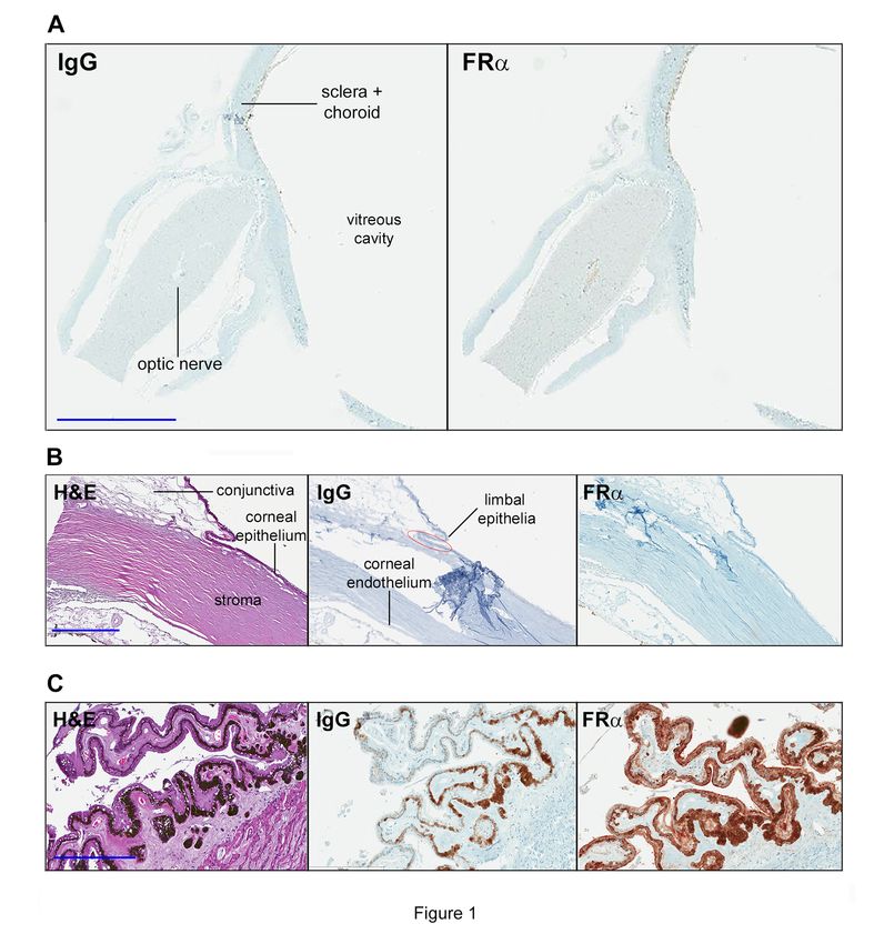
in the retinal pigmented epithelium (17,18). Representative images from
immunohistochemical assessment of FR expression in human ocular tissues are
shown in Figure 1. No FR staining was observed in the major non-retinal structures in
the human eye, including the optic nerve, sclera, and choroid (Fig.1A). Importantly, FR
expression was also notably absent throughout the entire cornea, including the corneal
epithelium, stroma, and endothelium, as well as the adjacent conjunctiva and limbal
region (n = 7 samples) (Fig. 1B). Positive receptor staining was only seen in ciliary body
epithelia (strong membranous and cytoplasmic staining in 4/4 samples) (Fig. 1C), a
result consistent with recent gene expression profiling in this tissue (19).
Preclinical modeling in rabbits predicts for potential corneal abnormalities
Mirvetuximab soravtansine was administered to Dutch-Belted rabbits at either 4 or 12
mg/kg (five/group) and animals monitored until study day 107 in order to assess the
reversibility, persistence, and/or delayed occurrence of any ocular effects. Gross
examination of the eyes revealed the development of corneal haze (Fig. 2A) in one
animal administered the high dose of mirvetuximab soravtansine, on days 71 and 85.
The key observation to arise from ophthalmic evaluations was the appearance of
punctate microcystic lesions within the corneal epithelium (associated with multifocal
fluorescein stain uptake on slit-lamp microscopy) in response to mirvetuximab
soravtansine exposure (Fig. 2B). This development of corneal microcystic lesions in the
13
Downloaded from clincancerres.aacrjournals.org on March 1, 2021. © 2018 American Association for Cancer
Research.Author Manuscript Published OnlineFirst on November 9, 2018; DOI: 10.1158/1078-0432.CCR-18-2474
Author manuscripts have been peer reviewed and accepted for publication but have not yet been edited.
rabbit was similar to that seen in patients receiving mirvetuximab soravtansine
monotherapy. The microcystic lesions were largely restricted to the perilimbal region of
the cornea and tended to be less frequent, later in onset, and faster to resolve in
animals given the lower dose of mirvetuximab soravtansine. Figure 2C summarizes the
temporal incidence of corneal microcystic epitheliopathy observed during the study
period for both dose levels. For animals treated at 4 mg/kg/dose, the maximal incidence
of corneal lesions was 20% (i.e. two of ten eyes affected) which occurred on day 15,
two weeks following the initial mirvetuximab soravtansine dose. Symptomatic onset was
more rapid in rabbits treated at 12 mg/kg/dose, with 20% of eyes affected within one
week of initial dosing (day 8) and peaking at 80% on day 71, one week after the last
dose. Despite the higher prevalence in rabbits dosed at 12 mg/kg/dose, the effects
showed signs of reversibility as evidenced by a marked reduction in numbers seen
during the recovery phase (80% to 10% between days 71 and 85), although complete
resolution was observed only in animals treated with 4 mg/kg/dose.
At terminal sacrifice on day 85, histologic examination of the eyes revealed a clear
attenuation of the corneal epithelium that persisted in animals dosed at the 12
mg/kg/dose level (5/6 eyes examined) (Fig. 2D), characterized by clear cellular
disorganization (including fewer and larger epithelial cells, discontinuous basal layer).
These microscopic findings showed signs of recovery with a lower incidence (1/4 eyes)
noted at the recovery sacrifice (day 107). Of note, no retinal effects were observed in
any of the ophthalmic or histologic examinations. Together with the absence of FR
expression in the cornea, the development of corneal abnormalities in response to
mirvetuximab soravtansine exposure appeared to be non-target-related.
14
Downloaded from clincancerres.aacrjournals.org on March 1, 2021. © 2018 American Association for Cancer
Research.Author Manuscript Published OnlineFirst on November 9, 2018; DOI: 10.1158/1078-0432.CCR-18-2474
Author manuscripts have been peer reviewed and accepted for publication but have not yet been edited.
Patient characteristics
Forty patients were enrolled in the expansion cohort evaluating primary prophylactic use
of corticosteroids, starting from cycle 1. Patients were required to record eye drop
administration in a diary format. Of those who provided diaries (87.5%), the median
compliance with self-administration was 90% (range, 45-100%). Patient demographics
and baseline characteristics are summarized in Table 1. The median age was 60 years
(range, 49-83). The distribution of tumor types was epithelial ovarian carcinoma (85%),
fallopian tube cancer (7.5%), and primary peritoneal cancer (7.5%), with a majority of
patients (90%) presenting with serous histology. All individuals were heavily pretreated,
with 100% having prior platinum and taxane exposure. Twenty-four patients (60%)
received 3 previous systemic therapies and 15 (37.5%) had undergone 4 prior lines.
Safety
All 40 patients were included in the safety analyses. Treatment-emergent adverse
events (TEAEs) occurring in ≥ 15% of patients are presented in Figure 3. The most
frequently reported TEAEs were diarrhea, fatigue (each 58%), and nausea (48%), the
majority of which were either grade 1 or 2. No grade 4 events were observed and no
deaths occurred in patients while on study. Related adverse events led to study
discontinuation for four individuals (10%), involving three cases of pneumonitis (grade
1/2) and one case of grade 2 thrombocytopenia.
Ocular AEs
The two major ocular adverse events of interest, blurred vision and keratopathy, were
seen in 16 (40%) and 12 (30%) patients respectively, with no grade 3 or 4 events
15
Downloaded from clincancerres.aacrjournals.org on March 1, 2021. © 2018 American Association for Cancer
Research.Author Manuscript Published OnlineFirst on November 9, 2018; DOI: 10.1158/1078-0432.CCR-18-2474
Author manuscripts have been peer reviewed and accepted for publication but have not yet been edited.
observed. A comparator pooled population of patients from the same phase I trial (n =
73, Table 1) received the same dosing regimen of mirvetuximab soravtansine,
underwent identical ocular management procedures, but did not receive primary
prophylactic corticosteroid eye drops. Although no significant differences in the
frequency or median time to onset of the two toxicities were observed between groups,
there was a trend towards a lower incidence, particularly of keratopathy, in those
patients who received corticosteroid prophylaxis (Table 2). Further, the percentage of
patients in each group requiring a dose delay due to ocular toxicity was similar, however
the number of dose reductions was lower (2 patients, 5%) and no discontinuations in
response to ocular events were seen in the corticosteroid cohort (Table 3). Consistent
with the lower frequency of dose modifications associated with primary prophylactic
steroid eye drop use, the median relative dose intensity (RDI) in the eye drop cohort
was 98.6% versus 95.6% in the comparator group. With respect to other ocular AEs, dry
eye and cataracts were reported in nine (23%) and six patients (15%) respectively
(Figure 3) in the eye drop cohort. Importantly, no retinal-related toxicities were observed
in patients who received mirvetuximab soravtansine.
16
Downloaded from clincancerres.aacrjournals.org on March 1, 2021. © 2018 American Association for Cancer
Research.Author Manuscript Published OnlineFirst on November 9, 2018; DOI: 10.1158/1078-0432.CCR-18-2474
Author manuscripts have been peer reviewed and accepted for publication but have not yet been edited.
DISCUSSION
Mirvetuximab soravtansine is a FR-targeting ADC currently undergoing late-stage
clinical development in ovarian cancer, and was granted Fast Track designation in June
2018 by the Food and Drug Administration following complete enrollment of a pivotal
Phase III study (FORWARD I; NCT02631876) (20). The reversible, low grade ocular
abnormalities seen with mirvetuximab soravtansine are similar to those reported for a
variety of other ADCs that bear tubulin-disrupting payloads (10,21). The exact
mechanisms of such ocular toxicities are still poorly defined. Beyond the apparent
payload association, the type of linker employed for drug attachment has also been
suggested as a potential contributory factor, with prolonged retention in the circulation
conferred by stable linkers (such as that present in mirvetuximab soravtansine)
proposed to enhance overall exposure in normal tissues, including the eye (22).
Understanding the etiology of the ocular disturbances of mirvetuximab soravtansine and
developing mitigation strategies remain important clinical considerations for the optimal
application of this promising investigational agent.
To better understand the pathophysiology of mirvetuximab soravtansine-induced ocular
adverse events, the distribution pattern of FR in the human eye was evaluated using
immunohistochemistry. FRhas previously been reported to be expressed in retinal
tissues, primarily localized to the basolateral surface of the retinal pigmented epithelium,
where it is believed to be involved in vectorial transfer of folate from the choroidal blood
supply into the retina (17,18). In this manuscript, we show that FR protein is also
expressed in the ciliary body, a multifunctional tissue whose principal roles include
17
Downloaded from clincancerres.aacrjournals.org on March 1, 2021. © 2018 American Association for Cancer
Research.Author Manuscript Published OnlineFirst on November 9, 2018; DOI: 10.1158/1078-0432.CCR-18-2474
Author manuscripts have been peer reviewed and accepted for publication but have not yet been edited.
production of aqueous humor and accommodation of the lens by the ciliary muscle (23).
The ciliary body, along with the iris and choroid, form the uvea, the pigmented middle
layer of the eye that is structurally and functionally distinct from the cornea. Of note, this
anatomical differentiation suggests that there is little likelihood of any potential on-target
effects of mirvetuximab soravtansine on the ciliary body manifesting as corneal
damage. The ciliary body has been linked to a number of pathologies, the most
important of which are glaucoma and anterior uveitis/iritis (24). Further, there are few
reports of specific drug-induced effects on this tissue, with the exception of certain
sulfamate-derived drugs such as the anti-epileptic topiramate, which can cause swelling
and lead to the development of angle-closure glaucoma (25). However, none of these
toxicities, nor any retinal abnormalities, were observed in the preclinical modelling or in
human subjects treated with mirvetuximab soravtansine. Together with the absence of
FR expression in corneal tissues, the ocular adverse event profile of mirvetuximab
soravtansine thus appears to be target independent.
Studies of ocular drug toxicities are best performed in species for which the information
can be applied to the clinical setting. Dutch-Belted rabbits, a strain with pigmented eyes
and non-cross reactivity to the mirvetuximab soravtansine antibody moiety, were used
to assess the preclinical ocular toxicity profile of mirvetuximab soravtansine. The major
ophthalmic observation to emerge from this animal model was the development of
punctate microcystic lesions in the cornea suggesting a close phenotypic match to the
keratopathy observed in human trial subjects. Histopathologic evaluation of the
keratopathy at terminal sacrifice showed marked attenuation and disorganization of the
corneal epithelium in mirvetuximab soravtansine-treated animals. These corneal effects
18
Downloaded from clincancerres.aacrjournals.org on March 1, 2021. © 2018 American Association for Cancer
Research.Author Manuscript Published OnlineFirst on November 9, 2018; DOI: 10.1158/1078-0432.CCR-18-2474
Author manuscripts have been peer reviewed and accepted for publication but have not yet been edited.
occurred in a dose-dependent manner with respect to onset, duration, and severity.
Importantly, the abnormalities also showed evidence of reversibility, with the degree of
resolution also correlating with the dose of mirvetuximab soravtansine. Taken together,
these results are consistent with damage occurring within the proliferative compartment
of the corneal epithelium, mediated by the antimitotic activity of the DM4 payload. The
mechanism of action underlying these off-target yet selective effects on the corneal
epithelium were not determined in this assay. However, a recent report by Zhao and
colleagues (26) revealed that macropinocytosis-mediated uptake by corneal epithelial
and other primary cells was responsible for ocular toxicity of AGS-16C3F, an ENPP3-
targeting ADC containing a MMAF payload. Further, they showed that the biophysical
properties of the ADC itself (overall hydrophobicity and/or presence of positive charges
on the antibody) were important determinants for this non-receptor mediated process
and could affect the ocular toxicity profile in animal models. The ophthalmic
observations of our clinical study are also in agreement with the current model proposed
to explain the observed changes in the ocular surface, in which corneal damage begins
peripherally after ADCs reach the cornea via the vascularized limbal region, followed by
internalization and consequent accumulation of the cytotoxic payload into transient
amplifying cells. These damaged progenitor cells then migrate centripetally, sufficient to
account for the development of microcystic deposits seen in patients (26,27). Overall,
the findings validate the use of this rabbit model to assess the pathogenesis of ocular
abnormalities, as well as the risks of visual disturbances, induced by mirvetuximab
soravtansine exposure in human subjects.
19
Downloaded from clincancerres.aacrjournals.org on March 1, 2021. © 2018 American Association for Cancer
Research.Author Manuscript Published OnlineFirst on November 9, 2018; DOI: 10.1158/1078-0432.CCR-18-2474
Author manuscripts have been peer reviewed and accepted for publication but have not yet been edited.
An important consideration related to ocular toxicity relates to prior treatment history.
While a cumulative effect of chemotherapy on the corneal epithelium cannot be ruled
out, it is unlikely that this represents a major contributing factor to the corneal damage
seen in patients in this study. Systemic chemotherapy can induce ocular side effects,
affecting multiple sites within the eye including the optic nerve, retina, and anterior
chamber (1). All patients in the present study had received prior platinum compound
and taxane exposure; however, these agents are not associated with corneal
abnormalities but instead can induce optic neuropathy and retinopathy (2), neither of
which were reported as adverse events. Keratitis/keratopathy is more commonly seen
with chemotherapeutics such as 5-fluorouracil, tamoxifen, and cytarabine (1,2), but
none of these drugs are used in ovarian cancer therapy and no patients in the present
study were exposed to them as part of their treatment history.
A number of mitigation strategies have been implemented clinically to help reduce the
incidence and severity of the corneal and visual disorders induced by mirvetuximab
soravtansine exposure. During early dose-escalation, in which doses were determined
using total body weight, pharmacokinetic analysis suggested an association between
the degree of reversible ocular toxicity and high early exposure levels of the ADC (13).
A change in the weight-based dosing strategy to adjusted ideal body weight was
therefore undertaken in order to reduce peak plasma concentrations to levels below the
threshold for ocular toxicity. This approach is now standard in all clinical evaluations of
mirvetuximab soravtansine. Further, daily use of lubricating eye drops in conjunction
with other proactive ocular management procedures reduced the incidence of blurred
vision and corneal keratopathy to levels seen in the pooled population of ovarian cancer
20
Downloaded from clincancerres.aacrjournals.org on March 1, 2021. © 2018 American Association for Cancer
Research.Author Manuscript Published OnlineFirst on November 9, 2018; DOI: 10.1158/1078-0432.CCR-18-2474
Author manuscripts have been peer reviewed and accepted for publication but have not yet been edited.
patients used for comparative purposes in this study. Prophylactic use of steroid eye
drops is another approach that has been reported to be successful in reducing the
frequency and severity of ocular events in trials of other ADCs, such as ABT-414 and
SGN-CD19A (both of which utilize MMAF as their cytotoxic payload) (28-30). The actual
mechanism(s) by which steroids can reduce ADC-induced keratopathy remain poorly
defined and, as supported by our preclinical modeling, there appears to be no
inflammatory component underlying the etiology of the corneal toxicity observed with
mirvetuximab soravtansine. However, it has been hypothesized that ocular steroids can
slow down the proliferation of limbal stem cells, potentially leading to a lower sensitivity
to the damaging effects of chemotherapeutics, including cell cycle-dependent agents
like the DM4 payload present in mirvetuximab soravtansine. Further, ocular steroids
may contribute to a thinning of the corneal epithelium, thereby facilitating shedding of
corneal microcysts induced by exposure to the ADC.
In the clinical expansion cohort, primary prophylaxis with corticosteroid eye drops
starting with the first cycle of mirvetuximab soravtansine infusion resulted in a reduced,
albeit not significant, incidence of keratopathy that is suggestive of potential clinical
benefit. The more modest effects on blurred vision suggests that additional
mechanisms, less influenced by steroid prophylaxis, are likely contributing to this
symptom in patients. Of interest, in the ADC studies where steroid prophylaxis has been
shown to be effective, the ocular AEs were more severe at onset (grades 3 or 4) and
were subsequently reduced to grade 1/2 events (27-30), comparable to the baseline
levels seen with mirvetuximab soravtansine. Prophylactic steroid eye drop use as a
mitigation strategy has not eliminated ADC-induced keratopathy, and it is reasonable to
21
Downloaded from clincancerres.aacrjournals.org on March 1, 2021. © 2018 American Association for Cancer
Research.Author Manuscript Published OnlineFirst on November 9, 2018; DOI: 10.1158/1078-0432.CCR-18-2474
Author manuscripts have been peer reviewed and accepted for publication but have not yet been edited.
suggest that there is a role for additional strategies to further optimize ADC-related
ocular AE profiles. Also, any potential side effects of topical ophthalmic steroid use (e.g.
rise in intraocular pressure, accelerated cataract formation) are both easily treatable
and outweighed by the prospective therapeutic benefit for a patient staying on treatment
longer with an effective ADC. There was no apparent influence on the development of
cataracts in patients who received corticosteroids in this study since the observed
incidence was 15%, and the prevalence of cataracts in women of this age group
(median, 60 years) in the general population is approximately 17-20% and increases
sharply with age (31). In this regard, the most significant observation of the clinical study
was the requirement for fewer dose reductions and lack of discontinuations due to
ocular adverse events in patients receiving primary steroid prophylaxis compared to
those individuals on study without it. This has important therapeutic implications, since
better compliance with the treatment schedule would be expected to maintain
mirvetuximab soravtansine dose intensity. Indeed, the improved median RDI seen in the
eye drop cohort is consistent with this premise.
In summary, the principal ocular AEs associated with mirvetuximab soravtansine can be
attributed to off-target effects on the cornea, characterized by primary involvement of
the corneal epithelium and manifesting with blurred vision that is often associated with
microcystic keratopathy. Primary corticosteroid eye drop prophylaxis provides clinical
benefit to patients while on study and, based on these findings, prophylactic steroid eye
drop use is now mandated along with lubricating eye drops in ongoing trials of this
agent in patients with advanced ovarian cancer. Given that the ocular AEs are not
eliminated by prophylactic topical steroid measures, explorations of additional mitigating
22
Downloaded from clincancerres.aacrjournals.org on March 1, 2021. © 2018 American Association for Cancer
Research.Author Manuscript Published OnlineFirst on November 9, 2018; DOI: 10.1158/1078-0432.CCR-18-2474
Author manuscripts have been peer reviewed and accepted for publication but have not yet been edited.
strategies are ongoing. The findings of this study underscore the need for patients to
continue to be appropriately screened for ocular adverse events and highlight the
importance of close collaboration between treating physicians and ophthalmologists to
tailor treatment options for patients experiencing ocular AEs from ADC-directed therapy.
23
Downloaded from clincancerres.aacrjournals.org on March 1, 2021. © 2018 American Association for Cancer
Research.Author Manuscript Published OnlineFirst on November 9, 2018; DOI: 10.1158/1078-0432.CCR-18-2474
Author manuscripts have been peer reviewed and accepted for publication but have not yet been edited.
ACKNOWLEDGMENTS
All work was funded by ImmunoGen, Inc. The authors wish to thank all the patients who
participated and their families, as well as co-investigators, nurses, study coordinators,
and operations staff at each of the clinical sites. We also thank Jianhua Zhao for the
immunohistochemical analyses and Richard Bates, Assoc. Director of Medical Affairs at
Immunogen, who provided drafts and editorial assistance during the production of this
manuscript.
24
Downloaded from clincancerres.aacrjournals.org on March 1, 2021. © 2018 American Association for Cancer
Research.Author Manuscript Published OnlineFirst on November 9, 2018; DOI: 10.1158/1078-0432.CCR-18-2474
Author manuscripts have been peer reviewed and accepted for publication but have not yet been edited.
REFERENCES
1. al-Tweigeri T, Nabholtz JM, Mackey JR. Ocular toxicity and cancer chemotherapy. A
review. Cancer 1996;78(7):1359-73.
2. Singh P, Singh A. Ocular adverse effects of anti-cancer chemotherapy and targeted
therapy. Journal of Cancer Therapeutics & Research 2012:1-5.
3. Renouf DJ, Velazquez-Martin JP, Simpson R, Siu LL, Bedard PL. Ocular toxicity of
targeted therapies. J Clin Oncol 2012;30:3277-86.
4. Fu C, Gombos DS, Lee J, George GC, Hess K, Whyte A, et al. Ocular toxicities
associated with targeted anticancer agents: an analysis of clinical data with
management suggestions. Oncotarget 2017;8:58709-27.
5. Dy GK, Adjei AA. Understanding, recognizing, and managing toxicities of targeted
anticancer therapies. CA Cancer J Clin 2013;63:249-79.
6. Chari RV, Miller ML, Widdison WC. Antibody-drug conjugates: an emerging concept in
cancer therapy. Angew Chem Int Ed Engl 2014;53:3796-827.
7. Beck A, Goetsch L, Dumontet C, Corvaia N. Strategies and challenges for the next
generation of antibody-drug conjugates. Nat Rev Drug Discov 2017;16:315-37.
8. Parslow AC, Parakh S, Lee FT, Gan HK, Scott AM. Antibody-Drug Conjugates for
Cancer Therapy. Biomedicines 2016;4:14
9. Donaghy H. Effects of antibody, drug and linker on the preclinical and clinical toxicities of
antibody-drug conjugates. MAbs 2016;8:659-71.
10. Eaton JS, Miller PE, Mannis MJ, Murphy CJ. Ocular Adverse Events Associated with
Antibody-Drug Conjugates in Human Clinical Trials. J Ocul Pharmacol Ther
2015;31:589-604.
11. Gunderson CC, Moore KN. Mirvetuximab soravtansine. FRa-targeting ADC; treatment of
epithelial ovarian cancer. Drugs of the Future 2016;41:539-45.
25
Downloaded from clincancerres.aacrjournals.org on March 1, 2021. © 2018 American Association for Cancer
Research.Author Manuscript Published OnlineFirst on November 9, 2018; DOI: 10.1158/1078-0432.CCR-18-2474
Author manuscripts have been peer reviewed and accepted for publication but have not yet been edited.
12. Moore KN, Martin LP, O'Malley DM, Matulonis UA, Konner JA, Vergote I, et al. A review
of mirvetuximab soravtansine in the treatment of platinum-resistant ovarian cancer.
Future Oncol 2018;14:123-36.
13. Moore KN, Borghaei H, O'Malley DM, Jeong W, Seward SM, Bauer TM, et al. Phase 1
dose-escalation study of mirvetuximab soravtansine (IMGN853), a folate receptor alpha-
targeting antibody-drug conjugate, in patients with solid tumors. Cancer 2017;123:3080-
87.
14. Moore KN, Martin LP, O'Malley DM, Matulonis UA, Konner JA, Perez RP, et al. Safety
and Activity of Mirvetuximab Soravtansine (IMGN853), a Folate Receptor Alpha-
Targeting Antibody-Drug Conjugate, in Platinum-Resistant Ovarian, Fallopian Tube, or
Primary Peritoneal Cancer: A Phase I Expansion Study. J Clin Oncol 2017;35:1112-18.
15. Wilson SL, Ahearne M, Hopkinson A. An overview of current techniques for ocular
toxicity testing. Toxicology 2015;327:32-46.
16. Eisenhauer EA, Therasse P, Bogaerts J, Schwartz LH, Sargent D, Ford R, et al. New
response evaluation criteria in solid tumours: revised RECIST guideline (version 1.1).
Eur J Cancer 2009;45:228-47.
17. Smith SB, Kekuda R, Gu X, Chancy C, Conway SJ, Ganapathy V. Expression of folate
receptor alpha in the mammalian retinol pigmented epithelium and retina. Invest
Ophthalmol Vis Sci 1999;40:840-8.
18. Chancy CD, Kekuda R, Huang W, Prasad PD, Kuhnel JM, Sirotnak FM, et al.
Expression and differential polarization of the reduced-folate transporter-1 and the folate
receptor alpha in mammalian retinal pigment epithelium. J Biol Chem 2000;275:20676-
84.
19. Janssen SF, Gorgels TG, Bossers K, Ten Brink JB, Essing AH, Nagtegaal M, et al.
Gene expression and functional annotation of the human ciliary body epithelia. PLoS
One 2012;7:e44973.
26
Downloaded from clincancerres.aacrjournals.org on March 1, 2021. © 2018 American Association for Cancer
Research.Author Manuscript Published OnlineFirst on November 9, 2018; DOI: 10.1158/1078-0432.CCR-18-2474
Author manuscripts have been peer reviewed and accepted for publication but have not yet been edited.
20. Moore KN, Vergote I, Oaknin A, Colombo N, Banerjee S, Oza A, et al. FORWARD I: a
Phase III study of mirvetuximab soravtansine versus chemotherapy in platinum-resistant
ovarian cancer. Future Oncol 2018;14:1669-1678.
21. Younes A, Kim S, Romaguera J, Copeland A, Farial Sde C, Kwak LW, et al. Phase I
multidose-escalation study of the anti-CD19 maytansinoid immunoconjugate SAR3419
administered by intravenous infusion every 3 weeks to patients with relapsed/refractory
B-cell lymphoma. J Clin Oncol 2012;30:2776-82.
22. Polakis P. Antibody Drug Conjugates for Cancer Therapy. Pharmacol Rev 2016;68:3-19.
23. Rehman I, Ali T. Anatomy, Head, Eye, Muscles, Ciliary. In: StatPearls. Treasure Island
(FL); 2018; available at https://www.ncbi.nlm.nih.gov/books/NBK482301/
24. Coca-Prados M, Escribano J. New perspectives in aqueous humor secretion and in
glaucoma: the ciliary body as a multifunctional neuroendocrine gland. Prog Retin Eye
Res 2007;26:239-62.
25. Li J, Tripathi RC, Tripathi BJ. Drug-induced ocular disorders. Drug Saf 2008;31:127-41.
26. Zhao H, Atkinson J, Gulesserian S, Zeng Z, Nater J, Ou J, et al. Modulation of
Macropinocytosis-Mediated Internalization Decreases Ocular Toxicity of Antibody-Drug
Conjugates. Cancer Res 2018;78:2115-26.
27. Gan HK, Reardon DA, Lassman AB, Merrell R, van den Bent M, Butowski N, et al.
Safety, pharmacokinetics, and antitumor response of depatuxizumab mafodotin as
monotherapy or in combination with temozolomide in patients with glioblastoma. Neuro
Oncol 2018;20:838-47.
28. Goss GD, Vokes EE, Gordon MS, Gandhi L, Papadopoulos KP, Rasco DW, et al.
Efficacy and safety results of depatuxizumab mafodotin (ABT-414) in patients with
advanced solid tumors likely to overexpress epidermal growth factor receptor. Cancer
2018;124:2174-83.
27
Downloaded from clincancerres.aacrjournals.org on March 1, 2021. © 2018 American Association for Cancer
Research.Author Manuscript Published OnlineFirst on November 9, 2018; DOI: 10.1158/1078-0432.CCR-18-2474
Author manuscripts have been peer reviewed and accepted for publication but have not yet been edited.
29. Fathi AT, Chen R, Trippett TM, O'Brien MM, DeAngelo DJ, Shah BD, et al. Interim
analysis of a Phase I study of the antibody-drug conjugate SGN-CD19A in relapsed or
refractory B-lineage acute leukemia and highly aggressive lymphoma. Blood
2014;124:963.
30. Moskowitz CH, Forero-Torres A, Shah BD, Advani R, Hamlin P, Kim S, et al. Interim
analysis of a Phase I study of the antibody-drug conjugate SGN-CD19A in relapsed or
refractory B-lineage non-Hodgkin lymphoma. Blood 2014;124:1471.
31. Klein R, Klein BE. The prevalence of age-related eye diseases and visual impairment in
aging: current estimates. Invest Ophthalmol Vis Sci 2013;54:ORSF5-ORSF13.
28
Downloaded from clincancerres.aacrjournals.org on March 1, 2021. © 2018 American Association for Cancer
Research.Author Manuscript Published OnlineFirst on November 9, 2018; DOI: 10.1158/1078-0432.CCR-18-2474
Author manuscripts have been peer reviewed and accepted for publication but have not yet been edited.
Table 1. Patient demographics and baseline characteristics
Corticosteroid No corticosteroid
Characteristic prophylaxis prophylaxis
(n = 40) (n = 73)
Age in years, median (range) 60 (49-83) 62 (38-81)
Race, n (%)
White 34 (85.0) 66 (90.4)
Black or African American 1 (2.5) 2 (2.7)
Asian 3 (7.5) 2 (2.7)
American Indian or Alaskan native 0 (0.0) 2 (2.7)
Not reported 2 (5.0) 1 (1.4)
Primary cancer diagnosis, n (%)
Epithelial ovarian cancer 34 (85.0) 67 (91.8)
Fallopian tube cancer 3 (7.5) 5 (6.8)
Primary peritoneal cancer 3 (7.5) 1 (1.4)
Histology, n (%)
Serous 36 (90.0) 67 (91.8)
Endometrioid 2 (5.0) 1 (1.4)
Mixed 2 (5.0) 2 (2.7)
Carcinosarcoma 0 (0.0) 2 (2.7)
Mullerian carcinoma 0 (0.0) 1 (1.4)
ECOG PS, n (%)
0 20 (50.0) 32 (43.8)
1 20 (50.0) 41 (56.2)
Platinum resistance, n (%)
Yes 31 (77.5) 65 (89.0)
No 9 (22.5) 8 (11.0)
Number of prior systemic therapies, n (%)
1-2 1* (2.5) 16 (22)
3-4 39 (97.5) 30 (41)
5+ 0 (0) 27 (37)
Prior compound exposure, n (%)
Platinum 40 (100.0) 73 (100.0)
Taxane 40 (100.0) 73 (100.0)
Bevacizumab 24 (60.0) 53 (72.6)
PARP inhibitor 8 (20.0) 15 (20.5)
Abbreviations: ECOG PS, Eastern Cooperative Oncology Group performance status; PARP, poly ADP
ribose polymerase
*1 patient enrolled with 2 prior lines of therapy, although 3 or 4 prior lines were defined in the protocol
29
Downloaded from clincancerres.aacrjournals.org on March 1, 2021. © 2018 American Association for Cancer
Research.Author Manuscript Published OnlineFirst on November 9, 2018; DOI: 10.1158/1078-0432.CCR-18-2474
Author manuscripts have been peer reviewed and accepted for publication but have not yet been edited.
Table 2. Summary of ocular TEAEs
Corticosteroid No corticosteroid
Ocular AE prophylaxis prophylaxis
(n = 40) (n = 73)
Blurred Vision
Total, n (%) 16 (40.0) 34 (46.6)
Grade 1 7 (17.5) 16 (21.9)
Grade 2 9 (22.5) 18 (24.7)
Time to onset (days)
Median 33 36
Keratopathy*
Total, n (%) 12 (30.0) 30 (41.1)
Grade 1 4 (10.0) 18 (24.7)
Grade 2 8 (20.0) 11 (15.1)
Grade 3 0 1 (1.4)
Time to onset (days)
Median 42 43
* grouped term which includes keratopathy, keratitis, and corneal epithelial microcysts.
30
Downloaded from clincancerres.aacrjournals.org on March 1, 2021. © 2018 American Association for Cancer
Research.Author Manuscript Published OnlineFirst on November 9, 2018; DOI: 10.1158/1078-0432.CCR-18-2474
Author manuscripts have been peer reviewed and accepted for publication but have not yet been edited.
Table 3. Action taken due to ocular adverse events
Corticosteroid No corticosteroid
prophylaxis Prophylaxis
(n= 40) (n = 73)
No. of patients with ocular TEAEs n = 18 n = 38
Action taken due to ocular events, n (%)
Dose interruption 0 (0) 0 (0)
Dose delay 9 (22.5) 17 (23.3)
Dose reduction 2 (5.0) 11 (15.1)
Dose discontinuation 0 (0) 1 (1.4)
31
Downloaded from clincancerres.aacrjournals.org on March 1, 2021. © 2018 American Association for Cancer
Research.Author Manuscript Published OnlineFirst on November 9, 2018; DOI: 10.1158/1078-0432.CCR-18-2474
Author manuscripts have been peer reviewed and accepted for publication but have not yet been edited.
FIGURE LEGENDS
Fig 1. FR expression in human ocular tissues. A, Low magnification images from the
rear sagittal section of a normal human eye incubated with control IgG or anti-FR
antibodies, respectively. Scale bar, 5 mm. B, Hematoxylin and eosin (H&E), IgG, and
anti-FR immunohistochemical staining of the cornea, limbus, and conjunctiva regions.
Scale bar, 500 m. C, Representative images showing FR immunoreactivity in the
ciliary body (non-specific pigmentation seen in IgG control). Scale bar, 300 m.
Fig 2. Mirvetuximab soravtansine exposure induces corneal abnormalities in rabbits. A,
images of rabbit eyes from control (left panel) or mirvetuximab soravtansine-treated (12
mg/kg/dose; right panel) animals that developed corneal haze. B, Slit-lamp images
showing the appearance of diffuse, perilimbal microcysts within the corneal epithelium
of mirvetuximab soravtansine-treated rabbits. Arrows depict multifocal punctate
perlimbal corneal microcysts in affected animals. C, Incidence of corneal microcysts
identified during ophthalmic examinations. Values represent numbers of eyes affected.
D, H&E staining of corneal tissue sections taken from control (upper panel) or
mirvetuximab soravtansine-treated animals (12 mg/kg/dose; lower panel) following
terminal sacrifice on day 85. Attenuation of the epithelium (E) in response to
mirvetuximab soravtansine exposure is characterized by fewer and larger epithelial cells
and disorganization of the basal epithelial layer. Original magnification, 40X.
Fig 3. Treatment-emergent adverse events (TEAEs) reported in ≥ 15% of patients. All
40 patients enrolled in the corticosteroid expansion cohort were included in the safety
analysis. Ocular events are highlighted in bold text. *Peripheral neuropathy is a grouped
32
Downloaded from clincancerres.aacrjournals.org on March 1, 2021. © 2018 American Association for Cancer
Research.You can also read