The Changing Face of Mastectomy: An Oncologic and Cosmetic Perspective
←
→
Page content transcription
If your browser does not render page correctly, please read the page content below

Preservation of the nipple is the

                                                                                   ultimate aesthetic outcome for a

                                                                                   woman undergoing mastectomy.

Photo courtesy of Lisa Scholder. Victory Martini, 24ʺ × 32ʺ.

                               The Changing Face of Mastectomy:
                             An Oncologic and Cosmetic Perspective
                           Christine Laronga, MD, FACS, Jaime D. Lewis, MD, and Paul D. Smith, MD

Background: The history of surgical treatment of breast cancer is rich with contributions from many surgeons
over the centuries. Among the recent advances in technique is the nipple-sparing mastectomy, which reflects
the emerging focus on cosmetic outcomes.
Methods: We took a backward glance at the literature illustrating the evolution of surgical management of
breast cancer, culminating with nipple-sparing mastectomy. The growing clinical data with nipple-sparing
mastectomy are explored.
Results: The demand for nipple-sparing mastectomy has been steadily increasing at many institutions. Based
on the clinical data reported, nipple-sparing mastectomy is an oncologically safe procedure for selected women
who have or are at high risk for breast cancer.
Conclusions: For women facing mastectomy and their surgeons, the optimal aesthetic result centers on
preservation of the nipple. However, nipple-sparing mastectomy is technically challenging, with long-term safety
not yet confirmed. Evidence-based data are needed to document local tumor recurrence, distant metastasis,
cosmetic outcomes, patient satisfaction, and procedural complications.

Introduction                                                          following mastectomy with reconstruction includes
Surgical management of breast cancer has evolved                      preservation of the nipple-areola complex. This ar-
dramatically over the years. The guiding principles                   ticle reviews the “changing face of mastectomy” from
are based on patient safety followed by oncologic                     a historic perspective, culminating in an in-depth look
safety. Recent advances have also taken into account                  at the current status of nipple-sparing mastectomy
cosmetic outcome. The optimal cosmetic outcome                        from an oncologic and cosmetic perspective. The
                                                                      focus is solely on mastectomy rather than breast-
                                                                      preserving surgery.
From the Comprehensive Breast Program, Department of Women’s
Oncology at the H. Lee Moffitt Cancer Center & Research Institute,
Tampa, Florida (CL, JDL), and the Department of Surgery at the Uni-
                                                                      A Historic Perspective: The Early Years
versity of South Florida, Tampa, Florida (PS).                        Over the years, dramatic changes have occurred in
Submitted November 4, 2011; accepted February 22, 2012.               the surgical management of breast cancer. In fact, the
Address correspondence to Christine Laronga, MD, FACS, Compre-        oldest recorded history comes from ancient Egypt in
hensive Breast Program, Moffitt Cancer Center, 12902 Magnolia         1600 bc, where a scroll titled “Instructions concerning
Drive, MCC-BR PROG, Tampa, FL 33612. E-mail: Christine.Laronga        tumors of his breast” stated that there was no treat-
@moffitt.org
No significant relationship exists between the authors and the com-
                                                                      ment for breast cancer.1 Twelve hundred years later,
panies/organizations whose products or services may be referenced     Hippocrates described a woman with bloody nipple
in this article.                                                      discharge and breast cancer and also recommended

286 Cancer Control                                                                                    October 2012, Vol. 19, No. 4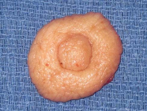
no surgical management as it would certainly only          Later in his career, he recommended the additional
hasten her death. For the next 500 years, women with       removal of the latissimus dorsi, subscapularis, teres
breast cancer were offered no treatment.                   minor, and serratus. With the Halsted methodology
     It was not until the first century AD that a Greek    plus anesthesia and antisepsis, the operative mortal-
physician, Leonides, performed the first operation         ity declined tremendously, and the tides turned to
for breast cancer. The technique, which came to be         reconsidering surgical management of breast cancer.
known as the “Escharotomy” method, consisted of            Thus, the Halsted radical mastectomy became the
using a hot poker to make repeated incisions into          standard of practice, the primary treatment of breast
the breast until the breast was completely burned          cancer, for the next 70 years.
off the chest wall. Women then applied a homemade
poultice for wound care. As expected, most of these        From the Halsted Revolution to the Present
women died of infection. The first surgical cure for       During the Halsted revolution, Cushman Haagensen,
breast cancer using this method did not take place for     a surgeon and pathologist at Columbia University in
another 100 years and is credited to Galen.                New York, noted that although the operation became
     With limited success and high mortality, this tech-   more “radical,” the survival rates from breast cancer
nique did not gain much favor, and by the Renais-          did not likewise increase over those achieved with the
sance era, physicians were searching for new ways          less “radical” mastectomy.3 This observation was also
to remove the breast. Surgical instruments were be-        noted by European colleagues (Veronesi) and opened
ing created at a rapid pace; however, without the          the door to consideration of a less radical operation.4
advent of anesthesia and antisepsis, the goal was to            In 1948, Patey and Dyson5 introduced the concept
remove the breast as swiftly and completely as pos-        of the modified radical mastectomy — removal of the
sible. The new technique was thus nicknamed the            entire breast, levels I, II, and III lymph nodes, and
“Guillotine” method because surgeons literally am-         the necessary skin (including the nipple-areola com-
putated the breast with a large sharp knife. In fact,      plex) — to allow primary closure flat against the chest
stories are told of the surgeon arriving unannounced       wall with minimal redundancy of skin. No muscles
at a woman’s house to perform the operation, with          would be removed during the operation, including
a few male helpers to hold the woman down on her           the pectoralis major and minor.
own kitchen table during the procedure. The skin                Shortly thereafter, in the 1950s, silicone gel im-
was not reapproximated, and the woman was bound            plants came on the scene. However, women with
to stop bleeding. Not surprisingly, many of these          breast cancer were not offered immediate reconstruc-
women died of exsanguination. Those who survived           tion but rather delayed reconstruction for two reasons.
the acute postoperative period succumbed to infection      First, it was well known that if a woman was going to
or endured significant morbidity.                          develop a recurrence or distant disease, these events
     This practice continued into the 18th century. In-    would most likely happen within 3 years of her cancer
terestingly, during that time, a surgeon, Jean Louis       diagnosis. Thus, they wanted to declare the woman a
Petit, advocated leaving all the skin not involved with    survivor before performing any breast reconstruction.
the tumor, including the nipple-areola complex, and in     Second, the techniques of immediate reconstruction
essence described the concept of nipple-sparing mas-       after mastectomy were still in their infancy. If the
tectomy.2 Unfortunately, he was considered a heretic,      woman lived flat-chested, with all the imperfections of
and his beliefs and methods were not adopted. In           a mastectomy without reconstruction for a significant
fact, the number of “mastectomies” being performed         period, she would be much more appreciative of any
dropped off precipitously in the 18th century and early    reconstructive outcome she had.
19th century due to poor results and “indiscriminant            As such, early experience with immediate breast
mutilation.” Even the development of primary skin clo-     reconstruction came from women having subcutane-
sure of wounds by Joseph Pancoast and Samuel Gross         ous mastectomies for benign disease.6,7 Without the
did not result in a return of “mastectomy.” However, in    benefit of mammography, breast ultrasonography,
the late 19th century, a revolution was about to begin.    and core needle biopsies, many women had multiple
William Halsted is credited with describing the exact      open surgical biopsies in the 1960s and 1970s for be-
technique to safely perform a radical mastectomy, an       nign disease (fibrocystic changes and fibroadenomas).
operation that bears his name to this day. Halsted had     Repetitive biopsies led to scarring, pain, anxiety, and
two advantages over his predecessors: the advent of        deformity, and thus these women opted to have their
anesthesia and the concept of antisepsis. With these       breasts removed.
advances, he advocated a meticulous dissection with             A subcutaneous mastectomy was performed, most
avoidance of a hematoma or hemorrhage.                     commonly via an inframammary incision, with re-
     Early in his career, his operation entailed com-      moval of most of the breast tissue, leaving a rim of
plete removal of the breast with all the overlying skin    normal breast tissue on the undersurface of the na-
(thus requiring a skin graft for coverage of the chest     tive breast skin (especially subareolar). No muscles
wall), removal of levels I, II, and III axillary lymph     or lymph nodes were removed, nor was any of the
nodes, and removal of the pectoralis major and minor.      skin (including the nipple-areola complex). Thick

October 2012, Vol. 19, No. 4                                                                     Cancer Control 287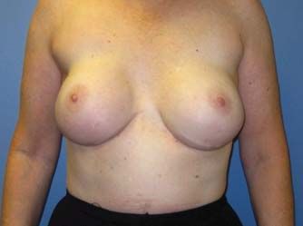
mastectomy flaps (> 10 mm) were raised, leaving a                  Improving nipple reconstruction techniques be-
cushion of tissue anterior to the implant and beneath         came paramount in the 1980s, as new methods of
the skin. This cushion maintained skin viability and          breast reconstruction were being developed, namely
created a more natural “feel” to the reconstruction.          autologous tissue transfers. These modalities (latis-
As this technique was for benign disease, oncologic           simus dorsi myocutaneous flaps, transverse rectus
safety was not questioned, and it actually provided           abdominis myocutaneous flaps [TRAMs]) offered an
an excellent risk-reduction benefit to these women.           alternative to tissue expander/implant reconstruc-
However, most women in the 1960s were still treated           tion and provided the ability to create a larger breast
with the Halsted radical mastectomy. Without any              mound with ptosis if needed to match the contralat-
available muscles or native breast skin, implant re-          eral natural breast.
construction was not possible.                                     In concert with these improved reconstruction
     Fortunately, by the 1970s, the modified radical          options, Toth and Lappert12 introduced the concept of
mastectomy was gaining traction. Now plastic sur-             the skin-sparing mastectomy in 1984. A skin-sparing
geons had some native breast skin, albeit thinner             mastectomy still removes the entire breast and pro-
than a subcutaneous mastectomy, to provide coverage           vides access to the axilla for removal of lymph nodes
over the implant. However, the high risk of implant           (level I/II axillary node dissection at this time) but
exposure from wound dehiscence was quickly dis-               preserves more of the native breast skin than would a
covered. The implant was too heavy for the delicate           traditional modified radical mastectomy with immedi-
mastectomy skin, but women were demanding im-                 ate reconstruction. In fact, by definition, a skin-sparing
mediate reconstruction. This led to the development           mastectomy removes < 20% of the native breast skin
of tissue expanders.                                          but always removes the nipple-areola complex. By
     Tissue expanders are essentially deflated balloons       preserving the maximal amount of breast skin, the
placed beneath the pectoralis major muscles at the            breast/general surgeon can provide the plastic surgeon
time of mastectomy. They are then slowly inflated             with an envelope that is the same size, color, contour,
with saline over time (weekly or bimonthly injec-             and ptosis as the original breast. Toth and Lappert12
tions), stretching the pectoralis major until the in-         also recommended avoiding incisions in the upper
tended breast size is achieved. The rate of expansion         portion of the breast, which could impact cosmesis.
is adjusted per patient based on skin integrity and                Undeniably, this approach would improve cosme-
patient tolerance. The expanders are subsequently             sis, but it was technically more challenging. Initial
exchanged for a permanent, more natural-appearing             concerns existed regarding the oncologic safety of
implant a few months later as an outpatient proce-            preserving additional skin (echoes of Halsted). Thus,
dure, without risk of skin compromise. As the nipple-         in the first several years after the introduction of the
areola complex is always removed with a modified              skin-sparing mastectomy, the literature focused on
radical mastectomy, the nipple can be reconstructed           technical feasibility and local recurrence rates.13-18
during a third operation at least 6 weeks later; the          Studies had shown that if the undersurface of the re-
areolar disc can later be tattooed.                           maining native breast skin and the anterior surface of
                                                              the pectoralis major muscles are scraped after a mas-
The Evolution of Nipple-Preservation Techniques               tectomy, approximately 1 g of normal breast epithelial
Plastic surgeons recognized early the importance of           cells could be identified.19 Despite the assumption
preserving the nipple. The nipple-areola complex              that leaving more skin behind would increase the
defines a breast as a breast and therefore likewise de-       number of remaining breast cells, the local recur-
fines a reconstructed breast mound. As such, women
were offered a choice of having nipple reconstruction
using skin from the local area or donated from the
groin area. Alternatively, the nipple-areola complex
could be harvested at the time of mastectomy, with
“banking” of the nipple in the patient’s groin as a
full-thickness skin graft and later replacing it on the
reconstructed breast (Fig 1). Attempts to maintain the
nipple in a tissue bank for future reimplantation re-
sulted in poor viability of the preserved nipple-areola
complex. However, whether it was “banked” in the
groin or the biorepository, concerns about transplant-
ing cancer contained within the preserved nipple
started to appear in the literature, and this practice fell
out of favor.8-10 Focus shifted back to improving the
techniques of nipple reconstruction and investigating
the psychological importance of re-creating a nipple          Fig 1. — Harvest of the nipple-areola complex for banking and reimplantation at a
on a reconstructed breast mound.11                            second surgery.

288 Cancer Control                                                                                            October 2012, Vol. 19, No. 4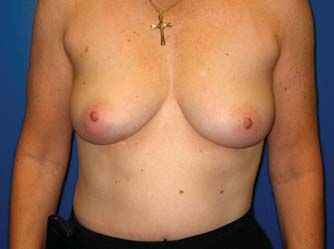
rence rates were no different between skin-sparing         Re-creating the Nipple
and non–skin-sparing mastectomies.13,14,17,18 Currently,   A multitude of techniques have been developed to
skin-sparing mastectomies with immediate breast re-        re-create the nipple. However, they all fall into one
construction are now the standard of practice.             of three groups. The first method is called composite
     In the 1980s, the Halsted dogma was challenged        grafting. This approach entails transfer of tissue from
and the concept of breast conservation (lumpectomy/        a distant source (contralateral normal nipple, cartilage
partial mastectomy plus whole breast irradiation) was      graft from the ear, skin from the groin area), use of
introduced. Clinical trials such as the National Sur-      various acellular dermal matrices, or use of fillers that
gical Adjuvant Breast and Bowel Project (NSABP)            can be injected/placed under the breast skin. The
B-06 trial were opening, demonstrating the oncologic       second approach involves local flaps, which employ
safety of preserving the breast20 if followed by irra-     the breast skin itself to re-create a projecting nipple
diation. Although local recurrence rates are higher        with primary closure of the donor site. The final op-
with lumpectomy/partial mastectomy than with any           tion is a similar re-creation of the nipple with local
mastectomy when matched for stage, they did not            breast skin, but rather than closing the donor site
translate into a survival disadvantage. As a result        primarily, a skin graft is used to cover the area to cre-
of similar trials, the 1990s and early 2000s saw the       ate the nipple. Typically, this option manifests itself
widespread adoption of breast-conserving surgery:          as a round graft mimicking an areolar disc.
70% to 80% of newly diagnosed breast cancer patients            Each technique has its own advantages and dis-
could be and were treated with lumpectomy. When            advantages. The greatest long-term projection of the
local recurrences occurred, 75% of them were in the        reconstructed nipple is attributed to the last of these
lumpectomy bed and were not de novo primary breast         methods, typically referred to as a “Skate” flap. The
cancers in another quadrant. This finding led to a         disadvantage is the need for an additional scar to
return to mastectomy and again raised the question         harvest the skin graft. This graft is usually taken
of sparing the nipple-areola complex, as it was not        from the groin, suprapubic area, upper inner thigh, or
the harbinger of tumor recurrence.                         even in some instances the labia. The second method
     Armed with this observation and documentation of      allows adequate projection initially but does tend to
years of performing skin-sparing mastectomy without        lose this projection over time. The first methods are
compromise in oncologic safety, nipple-sparing mas-        less reliable, and when using foreign material, can
tectomy was again considered. However, aside from          increase the risk of infection or exposure of the graft.
Petit in the 18th century, preserving the nipple-areola         For a time in the 1970s, “banking” the nipple was
complex goes against surgical dogma. In support of         a popular option. This option entailed removing the
this belief are several studies from the 1970s to 1990s    native nipple-areola complex and grafting it into the
demonstrating unsuspected nipple involvement in 6%         groin or upper thigh. After the reconstruction was
to 58% of mastectomy specimens.21 These studies var-       complete, this “banked” nipple would be harvested
ied in sampling technique and number of mastectomy         and placed back over the reconstructed breast mound.
specimens (patients) examined. However, all studies        This technique fell out of favor due to reports of trans-
took a cylinder of tissue encompassing the nipple-         posing breast cancer to the “banked” grafting sites.
areola complex and the tissue up to 20 mm deep. The             Despite numerous advances in nipple reconstruc-
nipple was considered positive for tumor if any part       tion, loss of projection as well as diminished color
of the cylinder contained tumor. Currently, 20 mm of       variation and asymmetry continued to plague plastic
tissue would not be left on the nipple flap during a       surgeons. Thus, in the 1990s, general surgeons and
nipple-sparing mastectomy, but rather it would be the      surgical oncologists began to explore the option of
same as the rest of the flap (ie, 3 mm to 5 mm).           areola-sparing mastectomy. Akin to skin-sparing mas-
     Considering only the 5 mm of tissue deep to the       tectomy, this technique needed to be defined and a
nipple-areola complex, the involvement of the nipple       decision made as to the use of the areolar disc. Two
with a true occult second cancer (not extension of the     options evolved: the areolar disc could be left in
primary tumor into this zone) would be much lower          situ and a nipple created using donor skin from an
(< 5%).21 Despite this knowledge, general surgeons         autologous tissue transfer, or alternatively, the areolar
and surgical oncologists still were not comfortable        disc could be used to create a nipple, and the sur-
with the prospect of sparing the nipple. However,          rounding skin could be tattooed later to re-create an
they recognized that the areolar disc was a skin ap-       areolar disc around the newly created nipple.
pendage and not breast tissue; the areolar disc can             We have learned that the areola makes a natural-
become involved by direct extension of a primary           appearing nipple and does a better job maintaining its
breast cancer but cannot generate a de novo breast         projection than a re-created nipple using donor skin,
cancer. This realization was especially important to       especially if the nipple creation is performed during
reconstructive surgeons because the nipple-areola          the initial reconstruction operation. On the negative
complex has a surface texture that presents multiple       side, the color tones and pigmentation within the
subtle surface qualities, which remain unattainable        areola are typically variegated and highly individual,
through reconstruction.                                    defying an exact match, even by the most skillful tat-

October 2012, Vol. 19, No. 4                                                                       Cancer Control 289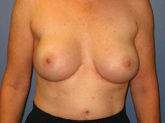
too artist. We have also discovered that areola-sparing      came from separate institutions, both in 2006.26,27 The
mastectomy is not only technically feasible but onco-        Petit group in Milan, Italy, described a subcutane-
logically safe. To date, there have been no reports of       ous mastectomy (a type of mastectomy intentionally
a de novo breast cancer arising from the preserved           leaving a 1-cm to 1.5-cm cuff of breast tissue on the
areolar disc regardless of whether it is maintained as       mastectomy flaps, especially in the area of the nipple
an areolar disc or used to create the nipple.22              base). Because of the thickness of the mastectomy
                                                             flaps and the inherent increased risk of occult or
The Birth of Nipple-Sparing Mastectomy                       secondary cancer, they coupled their operation with
The time had come to re-explore the option of nipple-        the use of 16 Gy of intraoperative radiation therapy
sparing mastectomy. To take the next leap to spare           with electrons (ELIOT) to the nipple-areola complex
the nipple, many surgeons searched for factors that          and remaining breast tissue.26 At 6 months of follow-
might predict occult nipple involvement. Common              up in 25 patients, no local recurrences in the nipple
themes from prior studies of occult tumor involvement        areolar complex were reported. There was one local
of the nipple included poorly differentiated primary         recurrence at 13 months of follow-up far away from
tumors > 2 cm and centrally located tumors.21,23,24 Mul-     the nipple in the infraclavicular region.
ticentric tumors, lymphovascular tumor invasion, and              Gerber et al27 studied 61 patients having nipple-
lymph node involvement were also common features,            sparing mastectomy and compared them with women
which formed the basis for initial eligibility criteria.     having skin-sparing or non–skin-sparing mastectomy.
Interestingly, these same criteria would also define an      At a mean follow-up of 4.9 years, the local recurrence
ideal candidate for breast conservation. Yet a por-          rate was the same (5%) in all three cohorts. The fol-
tion of these women were requesting nipple-sparing           lowing year, Crowe et al28 reported on 44 patients
mastectomy. Perhaps, they considered the local recur-        having nipple-sparing mastectomy for breast cancer
rence rate of approximately 10% after whole breast           treatment or prophylaxis, with no local recurrences
irradiation too high compared with 3% following              noted at a short mean follow-up of 6 weeks.
mastectomy without irradiation. Maybe the concept                 As surgeons worldwide learned of this “new”
of receiving radiation in and of itself was enough of        technique, they began offering the procedure to their
a deterrent, or perhaps this was a way for women to          patients. Initially, many nipple-sparing mastectomies
gain control and take charge by choosing the best            were performed prophylactically in high-risk women.
risk-reduction prevention strategy for breast cancer         In these women, many of whom were later confirmed
development. After all, women who have had breast            to be BRCA mutation carriers, the nipple-sparing mas-
cancer are at the highest risk to develop it again.          tectomy was performed as a subcutaneous mastec-
     The 1990s also saw the dawn of genetic testing for      tomy. However, the development of a primary breast
breast cancer risk assessment. With increased testing        tumor in the residual breast tissue was higher than
and public awareness via the Internet and advocacy           anticipated29,30 (1.9% at a median follow-up of 6.4 years
groups, the number of mastectomies being performed           in a study by Rebbeck et al30) and begs the question
was on the rise. Over the past 5 years, the trend is         of oncologic safety in this high-risk population. From
toward an equalization of lumpectomy vs mastectomy           2004 to 2008, the literature was bare on this topic, as
selected by patients. The true explanation of this           surgeons were grappling with learning the technique,
phenomenon is largely speculative.                           deciding whether to perform the cosmetically more ap-
     Whatever the reason, demand for nipple-sparing          pealing subcutaneous mastectomy or the true nipple-
mastectomy steadily increased, and the next step was         sparing mastectomy, and defining eligibility criteria
technical development of the procedure itself. The           from both oncologic safety and cosmetic standpoints.
design of the incision had to allow complete removal              In 2008 and 2009, a plethora of reports spout-
of the breast as well as access to the axilla for staging.   ing technical feasibility surfaced in the literature
Additionally, the ability to easily obtain tissue from the   but with small numbers of patients and short-term
base of the nipple for assessment of atypia or occult        follow-up.31-33 Some articles focused on generating
cancer was an absolute requirement. The base of the          an algorithm for eligibility without providing much
nipple could be examined by imprint cytology or fro-         information about their own institutional experience
zen section, depending on each institution’s expertise.      or outcomes from a cosmetic standpoint.34,35 For ex-
If any atypical or cancerous cells were identified, the      ample, in the Memorial Sloan-Kettering Cancer Center
nipple would be sacrificed intraoperatively. Similarly,      experience, 25 women had 42 nipple-sparing mastec-
if vascular viability was of concern to the plastic sur-     tomies, of which 81% were prophylactic.35 All of these
geon, the nipple would likewise be sacrificed.               women had tissue expander/implant reconstruction.
                                                             Partial nipple loss was seen in 5% of mastectomies,
The Growing Clinical Data                                    and complete nipple loss occurred in 2% of the wom-
Introduction of nipple-sparing mastectomy was first          en. Additionally, at 2 weeks following surgery, 48%
presented as a case report at the Southwestern Surgi-        had nipple discoloration or ischemia.
cal Congress in 1999, followed by an editorial in 2000            The choice of incision appears to affect cosmesis,
describing the technique.25 The first reported series        technical ease of performing the operation, and vascu-

290 Cancer Control                                                                            October 2012, Vol. 19, No. 4
lar viability of the nipple. Ischemia was most common (75%) have implant-alone reconstruction (“one-step”
with a lateral incision (curvilinear at the edge of the procedure). Other complications included partial
breast in the lower outer quadrant). In a recent update necrosis of the nipple in 6.5% of patients and full-
of the Memorial Sloan-Kettering Cancer Center experi- thickness necrosis of the nipple in 3.9%. Necrosis
ence, de Alcantara Filho et al36 reported that since 2005, was more common in larger-breasted women than
eligibility criteria have changed to encompass more in others. Depigmentation of the nipple occurred
women with breast cancer. Approximately 4% of all in 31% of women. Sixty-four percent of the nipples
mastectomies performed at this institution are nipple- were insensate or had minimal sensation, but 82%
sparing, demonstrating continued careful selection of and 84% of the patients and surgeons, respectively,
patients resulting in no local recurrences on short-term reported the cosmetic outcome as good to excellent.
follow-up and only 1 patient with distant metastasis. Complications specific to tissue expander reconstruc-
None of the nipple-sparing mastectomies per- tion was tilting (radiodystrophy) of the nipple toward
formed at Memorial Sloan-Kettering Cancer Center used the axilla and delayed capsular contracture, which
an inframammary approach. In that regard, the study occurred in 16.5% of patients. Overall, the women
by Kiluk et al37 bears further mention. An inframam- expressed a positive effect on body image, intimacy/
mary incision was made in all patients akin to that used sex, and satisfaction.
in breast augmentation or subcutaneous mastectomy, Several months later, at the 16th Annual Multi-
utilized for the performance of a true nipple-sparing disciplinary Symposium on Breast Disease in Amelia
mastectomy. No counter incision was made in the Island, Florida, Veronesi42 reiterated his associate’s
axilla for sentinel lymph node biopsy. Demonstrating presentation in San Antonio and included some fur-
technical feasibility of the inframammary incision to ac- ther updates. Their group has now performed over
complish all the goals (complete removal of the breast, 2,000 nipple-sparing mastectomies. The complication
access to the axilla for staging, and ease of obtaining rates are similar, but they now have 53.2 months of
pathological assessment of the base of the nipple) set follow-up in terms of oncologic outcomes. The local
the bar high for nipple-sparing mastectomy. The infra- recurrence rate was 3.9%, with 12 of 39 (31%) lo-
mammary incision has become the preferred incision cal recurrences being de novo cancers involving the
by women (patients) and plastic surgeons. preserved nipple (11 noninvasive cancers, 1 invasive
As others were reporting technical feasibility at cancer) despite intraoperative radiation. Interestingly,
short-term outcomes, the original pioneers were pub- 75 additional patients had a nipple base with cancer
lishing updates.38-40 Gerber et al39 presented longer (25 invasive cancers, 50 noninvasive cancers) found
follow-up on 60 of the original 61 nipple-sparing on final pathology review at the time of the nipple-
mastectomy patients. At a median follow-up of 101 sparing mastectomy. These nipples were observed in
months, the local recurrence rate was higher, at situ, and none developed local tumor recurrence, most
11.7%, but it was not statistically different from that likely due to intraoperative radiation. Yet that does
of their skin-sparing and non–skin-sparing cohorts. not explain the occurrence of the aforementioned de
This study was nonrandomized and retrospective, novo cancer (1.2%), as these patients also received
and although the local recurrences rates are higher the same intraoperative radiation dose. Eight percent
than those reported in the literature, they represent of women have developed distant disease, with a
a single institution/same surgeon clinical experience. mortality rate of 2.7%.
Also in 2009, Petit et al40 reported on 1,001 pa- Another group to report 5-year data is the John
tients having subcutaneous mastectomy with ELIOT; Wayne Cancer Center.43 In this series of 99 patients,
at 20 months, the local recurrence rate was 1.4%. 14% had their nipples removed due to atypical cells
Nipple-sparing mastectomy has become a prominent or cancer cells in the base of the nipple (apart from
entity at their institution. In fact, at the San Antonio the Italian group, intraoperative radiation is not rou-
Breast Cancer Symposium in 2010, Petit stated that tinely performed with nipple-sparing mastectomy).
37% of their mastectomies are nipple-sparing.41 Their Six percent of patients had the nipple sacrificed for
current eligibility criteria for nipple-sparing mastecto- vascular insufficiency. As a result, they have modi-
my include all clinical stage T1 and T2 invasive breast fied their technique to incorporate a “delay” proce-
cancers but exclude those with a history of breast dure. A delay procedure entails making an inferior
irradiation. They use a radial incision in the upper circumareolar incision to obtain a nipple base bi-
outer quadrant of the breast from the areolar border opsy and detaching the entire nipple-areola com-
to the axilla and always perform an intraoperative plex from the breast mound. Two weeks later, after
frozen section of the base of the nipple. All patients skin collateralization has occurred to the nipple and
receive 20 Gy of intraoperative radiation using ELIOT. pathological assessment of the base of the nipple
Immediate complications included infection shows no tumor, the nipple-sparing mastectomy is
(2.1%) and the need to remove the tissue expander/ performed. In their hands, this modified technique
implant (4.2%). In their hands, TRAM reconstruction has decreased vascular compromise to the nipple and
offers the best cosmetic outcome; when the recon- thus nipple loss. Additionally, this strategy should
struction is a tissue expander/implant, the majority lead to fewer local recurrences. Early in their se-
October 2012, Vol. 19, No. 4 Cancer Control 291A B C
Fig 2A-C. — (A) Preoperative, (B) 6-month postoperative, and (C) 2-year postoperative views of bilateral nipple-sparing mastectomy with tissue expander/silicone implant
reconstruction.
ries, patients with a positive nipple base for cancer Assessment of oncologic safety is limited by a
intraoperatively had a 14% local recurrence rate. By short mean follow-up of only 22 months (range, <
identifying these patients beforehand, they can de- 1 to 57 months). Of the patients intended for nip-
clare them ineligible for nipple-sparing mastectomy. ple-sparing mastectomy, 93% ultimately maintained
their nipples. Four nipples (2.1%) were sacrificed
The Moffitt Experience for atypia or cancer at the nipple base on pathology
Over the past several years, Moffitt Cancer Center review. Local regional recurrences occurred in 5 of
has performed nipple-sparing mastectomy for ge- 187 patients (2.7%). Distant metastases developed in
netic carriers and in select women with breast cancer 1 patient (0.5%). Overall survival was 97%. The cause
(Fig 2). About 5 years ago, we established stringent of death was unknown in 2 patients who were lost
eligibility criteria based on oncologic factors and tech- to follow-up in 2007; a third patient died 48 months
nical/cosmetic constraints (Table). Due to concerns after nipple-sparing mastectomy, secondary to widely
over the 1.2% de novo cancer rate evidenced by the metastatic ovarian cancer.
Milan group, our nipple-sparing mastectomy is not All of the studies to date offer the same message:
performed as a subcutaneous mastectomy. Our mas- nipple-sparing mastectomy is an oncologically safe
tectomy flaps are 3 mm to 5 mm thick and extend procedure for women who have or are at high risk
directly onto the base of the nipple. Initially, we cored for breast cancer. Knowing that the local recurrence
the nipple proper, but based on the data from Stolier rates for skin-sparing mastectomy are comparable to
et al,44 we no longer core the nipple. Rather, we those for non–skin-sparing mastectomy, logic pre-
perform intraoperative frozen sections of the nipple dicts that the addition of leaving the nipple in situ
base; if atypical or malignant cells are identified, the should have minimal risk of increased local tumor
nipple (not the areolar disc) is sacrificed. recurrence. In fact, a study by Stolier et al44 demon-
We recently reviewed our complications following strated that only 25% of nipples possess a terminal
187 nipple-sparing mastectomies performed on 111 duct lobular unit, which is the progenitor of all breast
patients (108 women, 3 men). The median age was cancers. More important, the terminal duct lobular
48.5 years (range, 18 to 82 years), and the median unit is always found at the base of the nipple, not
body mass index (BMI) was 23 kg/m2 (range, 17 kg/ within the nipple proper. This information has led
m2 to 34 kg/m2). The majority (68%) had bilateral to a change in our surgical technique, away from
nipple-sparing mastectomy, with 80% of patients hav- “coring out” the nipple proper to frozen sections at
ing nipple-sparing for an invasive cancer. The major- the base of the nipple alone. Coring of the nipple
ity of women (> 80%) experienced some amount of has been associated with a higher rate of vascular
epidermolysis to the nipple tip at 5 to 10 days after compromise to the nipple. Avoidance of this ma-
surgery. No intervention was warranted, and the area neuver has decreased our nipple loss rate (partial
healed without sequelae. Other complications dur- and full) as well as the amount of epidermolysis.
ing the immediate postoperative period (< 30 days) Coring of the nipple is one of the many lessons
included partial or complete nipple loss (5.6% or learned by surgeons during the infancy of imple-
0.6%, respectively), infection requiring removal of a menting nipple-sparing mastectomy into their prac-
tissue expander (1.2%), skin flap necrosis (6.8%), and tice to limit nipple ischemia. Additional lessons in-
malposition or leakage of the tissue expander (3.7%). clude selection of the appropriate incision, the type
Delayed complications consisted of asymmetry of the of nipple-sparing mastectomy technique used, and
nipples (8%), depigmentation of the nipples (1.2%), the often detrimental effects of several patient fac-
infection requiring intravenous antibiotics (5.6%), tors (heavy smoking, diabetes, large size, and ptotic
irrigation of the surgical site (4.2%), or removal of breasts).45 BMI > 25 kg/m2 is also associated with
the tissue expander/implant/acellular dermal matrix an increased risk for complications including flap
(12.9%). The median time to these delayed infections necrosis and wound dehiscence; this risk increases
was 7 months (range, 1 to 51 months). as BMI increases.46 An excised breast mass of > 750
292 Cancer Control October 2012, Vol. 19, No. 4g is associated with an overall increase in complica- This finding correlated with independent observers’
tions (especially wound dehiscence), and a sternal ratings. However, although preserved sensation was
notch-to-nipple distance of > 26 cm is associated with acknowledged by a majority of patients, it was re-
a general increase in complications.46 Thus, reporting ported to be only fair or poor.48
of all complications is paramount to make technical Recently, the MD Anderson Cancer Center report-
improvements and expand patient eligibility criteria. ed its experience with nipple-sparing mastectomy,
As such, we have modified our initial eligibility criteria including evaluation of nipple sensation in terms of
to a less stringent (oncologically and cosmetically) responsiveness to touch preoperatively and post-
checklist (Table). operatively at 6 months and at 1 year.49 Although
response time to “erection” slowed at 6 months (re-
Evaluating Patient Satisfaction mained constant at 1 year), data are limited to only 11
Additionally, we have launched a prospective clinical evaluable breasts at 1 year. The majority of women
trial evaluating body image, quality of life, and skin/ had displacement of the nipple position (75%) and
nipple sensation before surgery and after surgery at breast mound (58%), resulting in an excellent to very
6 months and at 1 year. We are comparing women good overall appearance of the nipple (23%) and
having skin-sparing vs nipple-sparing mastectomy to mound (35%), and most women were “not at all” self-
ascertain what impact the nipple itself has on these conscious in their clothes.
components. All women enrolled must meet our
eligibility criteria (Table) regardless of the cohort as- Conclusions
signed. Such a study is important, as a dearth of lit- Similar studies worldwide will help to assess the value
erature currently exists regarding patient satisfaction of nipple-sparing mastectomy to individual women.
and sensation of the preserved nipple. From the perspective of breast/general surgeons and
In one report by Yueh et al,47 two-thirds of pa- plastic surgeons, preservation of the nipple is the
tients were satisfied with their aesthetic result. Al- ultimate in aesthetic outcome for a patient facing
though 75% reported preservation of nipple sensation, mastectomy. However, it is technically challenging,
this sensation was rated poorly on a scale of 1 to 10, and long-term oncologic safety is unknown (albeit
with a mean of only approximately 3. In a similar predicted to be low risk). Demonstrating the impor-
study, patients assessed the appearance, symmetry, tance of preserving a nipple, whether sensate or not,
color, position, and texture of the breast mound, with will encourage surgeons to not only remain steadfast
a majority of the results rated as good or excellent.48 in their learning curve, but also acquire evidence-
based data documenting local tumor recurrence, dis-
Table. — Inclusion and Exclusion Criteria for Nipple-Sparing Mastectomy tant metastasis, cosmetic outcomes, patient satisfac-
tion (quality of life, body image, nipple sensation),
Inclusion Criteria
and procedural complications/risks.
Histologically proven diagnosis of breast cancer: unifocal invasive ductal, invasive
lobular, or a sarcoma
Invasive tumor 3 cm or smaller based on preoperative breast imaging
References
1. Wagner FB, Martin RG, Bland KI. History of the therapy of breast disease.
Tumor margin > 2 cm from the areolar edge based radially and 2 cm from the In: Bland KI, Copeland EM, eds. The Breast: Comprehensive Management of Benign
posterior margin of the nipple-areola base based on preoperative breast imaging and Malignant Diseases. 2nd ed. Philadelphia, PA: WB Saunders; 1991:1-18.
2. Olson JS, ed. Bathsheba’s Breast. Women, Cancer, & History. Baltimore, MD:
Lymph node-negative status and performance of a sentinel lymph node biopsy John Hopkins University Press; 2002:9-45.
at the time of the mastectomy on the cancer side (not required for a prophylactic 3. Haagensen CD, ed. Carcinoma of the Breast. New York, NY: American Can-
mastectomy) cer Society; 1950.
Prophylactic mastectomy (unilateral or bilateral) for risk reduction for a nipple- 4. Lacour J, Bucalossi P, Cacers E, et al. Radical mastectomy versus radical
mastectomy plus internal mammary dissection: five-year results of an interna-
sparing mastectomy of the breast without cancer tional cooperative study. Cancer. 1976;37(1):206-214.
Female, age 18 to 85 5. Patey DH, Dyson WH. The prognosis of carcinoma of the breast in relation
to the type of operation performed. Br J Cancer. 1948;2(1):7-13.
6. Freeman BS. Subcutaneous mastectomy for benign breast lesions with
Exclusion Criteria immediate or delayed prosthetic replacement. Plast Reconstr Surg Transplant Bull.
Age older than 85 years at the time of surgery 1962;30:676-682.
7. Freeman BS. Complications of subcutaneous mastectomy with prosthetic
Extensive ductal carcinoma in situ (area > 3 cm) replacement, immediate or delayed. South Med J. 1967;60(12):1277-1280.
8. Cucin RL, Guthrie RH Jr, Luterman A, et al. Transplantation of the cryo-
History of breast cancer (invasive or noninvasive) with radiation preserved nipple-areolar complex. Ann Plast Surg. 1980;4(5):391-395.
History of irradiation to the breast area (ie, mantle radiation for lymphoma) 9. Rose JH Jr. Carcinoma in a transplanted nipple. Arch Surg. 1980;115(9):
1131-1132.
Invasive cancer > 3 cm, multicentric, within 2 cm from the areolar margin or 10. Allison AB, Howorth MG Jr. Carcinoma in a nipple preserved by hetero-
within 2 cm from the posterior aspect of the nipple-areolar base topic auto-implantation. N Engl J Med. 1978;298(20):1132.
11. Wellisch DK, Schain WS, Noone RB, Little JW 3rd. The psychological con-
Clinically suspicious axillary lymph nodes on palpation or by fine-needle aspiration tribution of nipple addition in breast reconstruction. Plast Reconstr Surg. 1987;
History of smoking within 6 weeks of the intended surgery 80(5):699-704.
12. Toth BA, Lappert P. Modified skin incisions for mastectomy: the need for
Obesity (defined as a body mass index of > 30 kg/m2) plastic surgical input in preoperative planning. Plast Reconstr Surg. 1991;87(6):
Not a candidate for immediate breast reconstruction 1048-1053.
13. Newman LA, Kuerer HM, Hunt KK, et al. Presentation, treatment, and out-
Not a candidate for nipple-areola skin-sparing mastectomy due to the location come of local recurrence after skin-sparing mastectomy and immediate breast
of the nipple below the inframammary fold with the patient sitting, breast size is reconstruction. Ann Surg Oncol. 1998;5(7):620-626.
> 700 g or significant contour abnormalities of the nipple-areola complex itself 14. Carlson GW, Bostwick J 3rd, Styblo TM, et al. Skin-sparing mastectomy:
oncologic and reconstructive considerations. Ann Surg. 1997;225(5):570-578.
15. Fersis N, Hoenig A, Relakis K, et al. Skin-sparing mastectomy and immedi-
October 2012, Vol. 19, No. 4 Cancer Control 293ate breast reconstruction: incidence of recurrence in patients with invasive breast immediate implant reconstruction: cosmetic outcomes and technical refine-
cancer. Breast. 2004;13(6):488-493. ments. Plast Reconstr Surg. 2010;126(5):1460-1471.
16. Greenway RM, Schlossberg L, Dooley WC. Fifteen-year series of skin-spar- 46. Davies K, Allan L, Roblin P, et al. Factors affecting post-operative com-
ing mastectomy for stage 0 to 2 breast cancer. Am J Surg. 2005;190(6):918-922. plications following skin sparing mastectomy with immediate breast reconstruc-
17. Kroll SS, Khoo A, Singletary SE, et al. Local recurrence risk after skin- tion. Breast. 2011;20(1):21-25.
sparing and conventional mastectomy: a 6-year follow-up. Plast Reconstr Surg. 47. Yueh JH, Houlihan MJ, Slavin SA, et al. Nipple-sparing mastectomy:
1999;104(2):421-425. evaluation of patient satisfaction, aesthetic results, and sensation. Ann Plast Surg.
18. Kroll SS, Schusterman MA, Tadjalli HE, et al. Risk of recurrence after treat- 2009;62(5):586-590.
ment of early breast cancer with skin-sparing mastectomy. Ann Surg Oncol. 48. Djohan R, Gage E, Gatherwright J, et al. Patient satisfaction following
1997;4(3):193-197. nipple-sparing mastectomy and immediate breast reconstruction: an 8-year out-
19. Barton FE Jr, English JM, Kingsley WB, Fietz M. Glandular excision in total come study. Plast Reconstr Surg. 2010;125(3):818-829.
glandular mastectomy and modified radical mastectomy: a comparison. Plast 49. Wagner JL, Fearmonti R, Hunt KK, et al. Prospective evaluation of the
Reconstr Surg. 1991;88 (3):389-394. nipple-areola complex sparing mastectomy for risk reduction and for early-stage
20. Fisher B, Bauer M, Margolese R, et al. Five-year results of a randomized breast cancer. Ann Surg Oncol. 2012;19(4):1137-1144.
clinical trial comparing total mastectomy and segmental mastectomy with or
without radiation in the treatment of breast cancer. N Engl J Med. 1985;312(11):
665-673.
21. Laronga C, Kemp B, Johnston D, et al. The incidence of occult nipple-
areola complex involvement in breast cancer patients receiving a skin-sparing
mastectomy. Ann Surg Oncol. 1999;6(6):609-613.
22. Simmons RM, Brennan M, Christos P, et al. Analysis of nipple/areolar
involvement with mastectomy: can the areola be preserved? Ann Surg Oncol.
2002;9(2):165-168.
23. Lagios MD, Gates EA, Westdahl PR, et al. A guide to the frequency of
nipple involvement in breast cancer: a study of 149 consecutive mastecto-
mies using a serial subgross and correlated radiographic technique. Am J Surg.
1979;138(1):135-142.
24. Smith J, Payne WS, Carney JA. Involvement of the nipple and areola in
carcinoma of the breast. Surg Gynecol Obstet. 1976;143(4):546-548.
25. Laronga C, Robb GL, Singletary SE. Feasibility of skin-sparing mastec-
tomy with preservation of the nipple-areola complex. Breast Dis Yearb Q. 1998;
19(2):125-127.
26. Petit JY, Veronesi U, Orecchia R, et al. Nipple-sparing mastectomy in as-
sociation with intra operative radiotherapy (ELIOT): a new type of mastectomy
for breast cancer treatment. Breast Cancer Res Treat. 2006;96(1):47-51.
27. Gerber B, Krause A, Reimer T, et al. Skin-sparing mastectomy with conser-
vation of the nipple-areola complex and autologous reconstruction is an onco-
logically safe procedure. Ann Surg. 2003;238(1):120-127.
28. Crowe JP Jr, Kim JA, Yetman R, et al. Nipple-sparing mastectomy: tech-
nique and results of 54 procedures. Arch Surg. 2004;139(2):148-150.
29. Hartmann LC, Schaid DJ, Woods JE, et al. Efficacy of bilateral prophylac-
tic mastectomy in women with a family history of breast cancer. N Engl J Med.
1999;340(2):77-84.
30. Rebbeck TR, Friebel T, Lynch HT, et al. Bilateral prophylactic mastectomy
reduces breast cancer risk in BRCA1 and BRCA2 mutation carriers: the PROSE
Study Group. J Clin Oncol. 2004;22(6):1055-1062.
31. Sookhan N, Boughey JC, Walsh MF, Degnim AC. Nipple-sparing mastec-
tomy: initial experience at a tertiary center. Am J Surg. 2008;196(4):575-577.
32. Voltura AM, Tsangaris TN, Rosson GD, et al. Nipple-sparing mastectomy:
critical assessment of 51 procedures and implications for selection criteria. Ann
Surg Oncol. 2008;15(12):3396-3401.
33. Garwood ER, Moore D, Ewing C, et al. Total skin-sparing mastectomy:
complications and local recurrence rates in 2 cohorts of patients. Ann Surg.
2009;249(1):26-32.
34. Spear SL, Hannan CM, Willey SC, Cocilovo C. Nipple-sparing mastectomy.
Plast Reconstr Surg. 2009;123(6):1665-1673.
35. Garcia-Etienne CA, Borgen PI. Update on the indications for nipple-spar-
ing mastectomy. J Support Oncol. 2006;4(5):225-230.
36. de Alcantara Filho P, Capko D, Barry JM, et al. Nipple-sparing mastectomy
for breast cancer and risk-reducing surgery: the Memorial Sloan-Kettering Cancer
Center experience. Ann Surg Oncol. 2011;18(11):3117-3122.
37. Kiluk JV, Santillan AA, Kaur P, et al. Feasibility of sentinel lymph node bi-
opsy through an inframammary incision for a nipple-sparing mastectomy. Ann
Surg Oncol. 2009;15 (12):3402-3406.
38. Benediktsson KP, Perbeck L. Survival in breast cancer after nipple-sparing
subcutaneous mastectomy and immediate reconstruction with implants: a pro-
spective trial with 13 years median follow-up in 216 patients. Eur J Surg Oncol.
2008;34(2):143-148.
39. Gerber B, Krause A, Dieterich M, et al. The oncological safety of skin spar-
ing mastectomy with conservation of the nipple-areola complex and autologous
reconstruction: an extended follow-up study. Ann Surg. 2009;249(3):461-468.
40. Petit JY, Veronesi U, Orecchia R, et al. Nipple sparing mastectomy with
nipple areola intraoperative radiotherapy: one thousand and one cases of a five
years experience at the European Institute of Oncology of Milan (EIO). Breast Can-
cer Res Treat. 2009;117(2):333-338.
41. Petit JY. Nipple-sparing mastectomy with nipple areola intra-operative
radiotherapy: one thousand and one cases of a five years experience at the
European Institute of Oncology of Milan (EIO). Oral presentation at the 33rd
Annual San Antonio Breast Cancer Symposium; December 8-12, 2010; San An-
tonio, Texas.
42. Veronesi U. Progress in breast cancer management. Oral presentation at
the 16th Annual Multidisciplinary Symposium on Breast Disease; February 10-13,
2011; Amelia Island, FL.
43. Jensen JA, Orringer JS, Giuliano AE. Nipple-sparing mastectomy in 99 pa-
tients with a mean follow-up of 5 years. Ann Surg Oncol. 2011;18(6):1665-1670.
44. Stolier AJ, Sullivan SK, Dellacroce FJ. Technical considerations in nipple-
sparing mastectomy: 82 consecutive cases without necrosis. Ann Surg Oncol.
2008;15(5):1341-1347.
45. Salgarello M, Visconti G, Barone-Adesi L. Nipple-sparing mastecomy with
294 Cancer Control October 2012, Vol. 19, No. 4You can also read


