Original Article A peptide derived from D. melanogaster Hairless protein promotes the negative regulation of Notch Aberrant Constitutive Signaling ...
←
→
Page content transcription
If your browser does not render page correctly, please read the page content below
Int J Clin Exp Med 2021;14(1):62-75
www.ijcem.com /ISSN:1940-5901/IJCEM0108892

Original Article
A peptide derived from D. melanogaster Hairless
protein promotes the negative regulation of
Notch Aberrant Constitutive Signaling
on human breast cancer cells
Germán Saucedo-Correa, Rosa E Nuñez-Anita, Ulises Maciel-Ponce, Humberto Contreras-Cornejo, Alejandro
Bravo-Patiño

Facultad de Medicina Veterinaria y Zootecnia UMSNH, Michoacán, México
Received February 8, 2020; Accepted April 2, 2020; Epub January 15, 2021; Published January 30, 2021

Abstract: The Notch Signaling Pathway (NSP), by a Notch intracellular domain (NICD) constitutive overexpression,
has been related to many cancer types. In breast cancer, the constitutively activated NSP plays a principal role in
aberrant cell cycle progression, poor cellular differentiation and apoptosis inhibition. It is well known the high con-
servation of Notch proteins through the metazoarians. Hairless is a negative regulator of NSP in D. melanogaster,
but a homologous Hairless protein in mammals is unclear. The design of an expression plasmid, pReNegAID, which
encodes a peptide based on the CSL binding domain of the Hairless protein of D. melanogaster will be used for ana-
lyzing the ReNegAID peptide participation it the negative regulation of NSP in the mammary gland cancer context.
Both the pReNeg-AID plasmid and the mock plasmid were transfected into breast cancer cells in order to analyze
cell proliferation (MTT assay) and gene expression pattern related to the NSP common genes between Hedgehog
and Wingless pathways and genes related to apoptosis, cell differentiation and cell cycle. Moreover, control expres-
sion of Luciferase reported plasmid was performed and data showed that ReNeg-AID peptide induces a switch in
the gene expression pattern related to the NSP and induce G1/S cell cycle arrest by the negative regulation of the
Notch-1 receptor expression and it suggests cross talking between Hedgehog pathway (Hh) and NSP in mammary
cancer cells to avoid the molecular machinery of initial epithelial to mesenchymal transition (EMT).

Keywords: Breast cancer, MCF-7, negative regulation, cancer therapy, notch-1, notch signaling pathway

Introduction                                                    cancer cells appearing and maintaining the dis-
                                                                ease state [6, 7]. The role of the NSP in cancer
The Notch Signaling Pathway (NSP) (Figure S1)                   has been firstly described in acute lymphoblas-
is an ancestral cell communication circuit high-                tic leukemia. Today, the NSP is considered as a
ly conserved in all metazoan. Their transcrip-                  primordial target for the strategy against can-
tion activation complex evolution through time                  cer disease, especially for breast cancer [6, 8,
has been minimal, however, the transcription                    9].
repression complex presents more variability
and specific tissue behavior in different organ-                The NSP exerts control over different events
isms where it has been described [1, 2]. In the
                                                                essential for the proper function of the cell,
last years the NSP activation mechanism has
                                                                such as differentiation, apoptosis, signaling
been elucidated and in recent years the Notch
                                                                pathways cross talking with both Hedgehog
mechanism of negative regulation, mainly in
different cell populations and different cellular               (Hh) and Wingless (Wnt) pathways. It is an
context, as well as its implications in different               essential regulator of the cell cycle G1/S phas-
diseases such as cancer [3-5].                                  es transitions and it has been shown that if the
                                                                Notch signaling pathway is not regulated prop-
In the cancer context, it has been known that                   erly, aberrations arise in its information flow,
NSP has a principal role in both promoting the                  leading the cell to a cancer state [10, 11].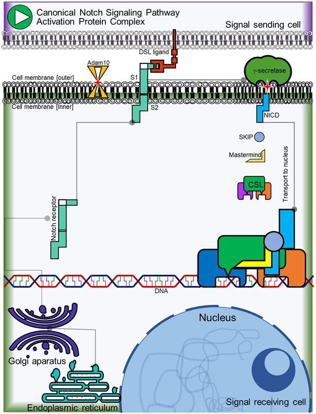
Negative regulation of Notch pathway on mammary cancer cells

It is known that in breast cancer the NSP pres-     gene in non-cancer mammary cells (MCF-12F)
ents a constitutive activation mainly through       has not shown changes, but it caused changes
Notch1 and Notch2 receptors and rarely              in MCF7 and MDA-MB-231 cells. In summary,
through Notch4 receptor, promoting a strike on      the result supported the hypothesis that
the mammary gland cell differentiation, an          pReNeg-AID could be employed as an adjuvant
epithelial to mesenchymal transition (EMT)          together with other anti-cancer therapies for
through deregulating the cross talking between      mammary cancer or other cancer related to
Hh and NSP, promoting high levels of cell migra-    aberrant expression of Notch-1 cancer which is
tion and more aggressive metastasis. Also, this     opening the opportunity to propose a new strat-
constitutive activation of the NSP promotes the     egy against breast cancer where the Notch
stop of the cell cycle checkpoints performed by     pathway is involved.
cyclin D1 and cyclin E1, as well as changes in
the transcriptional rates of Fos and FosL genes     Material and methods
promoting the apoptosis evasion [11-15].
                                                    For cell culture: Dulbecco’s Modified Eagle’s
The strategies focused against breast cancer        Medium (DMEM, Gibco, Cat. No. 12100046)
related to NSP distorted activity are based         for MCF-7 and MDA-MB-231 cells, DMEM-F12
mainly on the γ-secretase inhibitors (GSI’s) to     Ham (Gibco, Cat. No. 12500096) for MCF12-F
block the NICD release from the cellular mem-       cells, fetal bovine serum (FBS, Gibco, Cat. No.
brane avoiding its transport into the nucleus.      10437028), penicillin, streptomycin, and tryp-
Also, antibodies directed against ligands and       sin-EDTA (supplied by GIBCO-USA). For DMEM-
receptors interactions are used to prevent the      F12 Ham complete growth medium 20 ng/ml
NSP cascade. Antibodies against co-activator        of epidermal growth factor (Gibco, Cat. No.
Mastermind are also used, to prevent the tran-      PHG0315), 0.01 mg/ml of human insulin and
scriptional activation complex inhibiting the       500 ng/ml of hydrocortisone were added. The
CSL/Mastermind interaction [16, 17]. However,       plasmid used for cell transfection (pFN21K
these strategies still have collateral damage to    HaloTag® CMV Flexi® Vector) was supplied by
the patients since the total inhibition of the      Promega™.
NSP on healthy cells can have contradictory
and lethal effects in the long-term [18-20].        ReNeg-AID peptide

Due to the high conservation of the NSP             The ReNeg-AID peptide (patent pending) co-
through the metazoan kingdom, inter species         mes from the Hairless (H) protein of Drosophila
experiments have been conducted to corrobo-         melanogaster, specifically from the binding
rate the correct interaction between proteins       domain of the Hairless protein to the transcrip-
from D. melanogaster NSP and proteins from          tional factor Su(H) [CSL]. The amino acid
the Mus musculus and human NSP, mainly              sequence of pReNeg-AID was determined from
between proteins involved in the transcriptional    position 1987aa-2782aa sequence for the
negative regulation. It has been demonstrated       Hairless protein. The ReNeg-AID DNA sequence
that D. melanogaster Hairless protein, respon-      was cloned into the vector pFN21K HaloTag
sible for negative regulation of the NSP at the     CMV (pReNeg-AID, patent pending) following
fruit fly early embryo development, is capable      the manufacturer’s specifications (PROME-
of bounding, with high affinity, to mammal CSL      GA™) (Figure 1A).
transcription factor and downregulating the
NSP activity [21-23].                               Cell culture

The aim of this study was to demonstrate that       The non-cancer cell line of human epithelial
the ReNeg-AID (Regulation Negative-AID) pep-        breast MCF-12F (ATCC Cat# CRL-10783, RR-
tide (Figure S2), derived from the CSL binding      ID:CVCL_3745) (CRL-10783), tumorous estro-
domain of the D. melanogaster Hairless protein      gen receptor-positive MCF-7 (CLS Cat# 300-
(Figure 1A), is capable of modifying the tran-      273/p2720_MCF-7, RRID:CVCL_0031) (HTB-
scriptional pattern of genes related to the NSP     22) and tumorous triple negative MDA-MB-231
which are involved in both proliferation inhibi-    (ATCC® HTB-26™) cells were purchased from
tion and cell cycle arrest in mammary cancer        American Type Culture Collection (ATCC, Ma-
cells. Interestingly, the transcriptional pattern   nassas, VA, USA). The cells were maintained in

63                                                             Int J Clin Exp Med 2021;14(1):62-75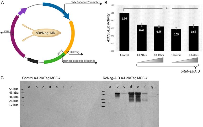
Negative regulation of Notch pathway on mammary cancer cells

Figure 1. A. Plasmid map of pReNeg-AID. The pFN21K HaloTag® CMV Flexi® Vector (Promega) was used to design
the pReNeg-AID. From Drosophila melanogaster larvae, the Hairless sequence that joins the CSL transcriptional
factor to form a protein repression complex of NSP was obtained and tagged with the HaloTag flag. B. The vector
pReNeg-AID decreases luciferase activity. MCF-7 cells were transfected in a molar ratio 1:1 and 1:3, respect to
pGL4xCSL and pReNeg-AID vectors at 24 and 48 hours. Relative activity of luciferase was calculated as the lucif-
erase activity of firefly against luciferase activity of Renilla. Each bar represents ± EST of average for triplicated
experiments. Values for P were determined by student t test (**P ≤ 0.01 vs control. n=4). C. pReNeg-AID Western.
The ReNeg-AID peptide is bound to a HaloTag flag (Promega) that uses monoclonal antibodies to be detected, the
peptide bound to the tag has a relative weight of 45 kDa, the molecular protein marker used was the 10 to 180
kDa PageRuler Prestained Protein Ladder (Thermo Scientific, # 26616). Control a-HaloTag MCF-7 panel shows the
controls without transfection of the ReNeg-AID peptide in MCF-7 cells. ReNeg-AID a-HaloTag MCF-7 panel shows
the following data: (a) Control without transfection; (b) Positive control with HaloTag mock plasmid; (c) MCF-7 cells
transfected with the pReNeg-AID, 12 hours after transfection; (d) MCF-7 cells transfected with the pReNeg-AID, 24
hours after transfection; (e, f and g) show the transfection at 48, 72 and 96 hours respectively.

DMEM supplemented with 10% FBS, 100 U/ml                       GIBCO-USA, Cat. No. 31985062). The nucleo-
penicillin, and 100 µg/ml streptomycin (basal                  fection solution was added to the cells and the
medium) at 5% CO2 and 37°C for 48 h before                     cells were placed in a 4 mm electroporation
transfections. The cell lines were obtained by                 cuvette in an electroporator system (supplied
the ATCC provider at the beginning of the exper-               by BTX® cuvettes & electroporation systems-
iments; therefore, these cell lines were new.                  Harvard Apparatus ECM 630 exponential decay
                                                               wave electroporation system, item 45-2051)
Transfection & electroporation                                 and the following conditions were applied: 140
                                                               V, 70 ms with one pulse; then the cuvette with
The cell culture was incubated with 5% of CO2                  the cell solution was incubated for five minutes
at 37°C until reaching 80% of confluent. The                   at room temperature between 18°C to 25°C
culture was then tripzinated and an 1 × 10⁶                    and the cells were cultured in a six-well plates
cells were harvested, centrifuged until the pill               with 1.5 ml of supplemented culture media in a
was formed. Then, a 100 μl nucleofection solu-                 humidified 37°C/5% CO2 incubator for 48 h.
tion (Cell Line Nucleofector® Kit V, protocol                  MTT assay
number T/C-28a2, supplied by AMAXA®) was
prepared with 2 ng of the plasmid pReNeg-AID                   Cell proliferation was determined by 3-(4,5-
and 400 μl of OptiMEM medium (supplied by                      dimethylthiazol-2-yl)-2,5-diphenyltetrazolium

64                                                                          Int J Clin Exp Med 2021;14(1):62-75
Negative regulation of Notch pathway on mammary cancer cells

bromide (MTT) assay. 48 hours before trans-          360 V. Analysis was performed with FlowJo
fection cells were cultured at 3 × 10³ cells/well    software V. 10 (Tree Star, Inc).
in DMEM medium and incubated at 37°C in
96-well plates with 5% CO2. Then, 20 µL of MTT       Statistical analysis
(5 mg/mL in PBS) was added to each well, and
cells were incubated for 4 hours at 37°C. The        MTT proliferation, luciferase assay and cyto-
supernatants were removed, 200 µL of dimeth-         metric analysis were analyzed by student’s
yl sulfoxide (DMSO) was added, and the absor-        t-test, and values with P < 0.05 were consid-
bance (595 nm) was determined by a micro-            ered statistically significant. All experiments
plate reader (Bio-Rad, Hercules, CA, USA). For       were performed at least three times with n=4
each cell line there were 3 trials.                  for each of them.

Luciferase assay                                     For gene expression Qiagen’s online web analy-
                                                     sis tool was utilized to produce comparative
One day before transfection, 450,000 cells           scatter plots and fold change was calculated by
were seeded on a petri dish. Later, the cells        determining the ratio of mRNA levels to control
were co-transfected with the pGL4xCSL and            values using the ΔCt method (2-ΔΔCt). All data
the pReNeg-AID vectors, following the protocol       were normalized to an average of two of this
of Xfect™ (Clontech). The luciferase activity was    housekeeping genes, ACTB and GAPDH. All
measured 24 and 48 hours post transfection           experiments were performed at least three
following the Dual-Luciferase® Reporter Assay        times with n=4 for each of them.
System (Promega) protocol.
                                                     Results
Quantitative real time PCR (RT-qPCR)
                                                     Luciferase assay and western blot
RT2 Profiler PCR (Qiagen, Cat. PAHS-059Z, No.
330231) Notch related gene arrays: Total             The reporter vector pGL4xCSL was designed
cell RNA was isolated from MCF12-F, MCF-7            for controlling the gene transcription of the
and MDA-MB-321, subsequently treated with            luciferase enzyme under the control of one pro-
DNase I and purified using RNeasy Mini Kit           moter that contains regulatory binding ele-
(Qiagen, Cat. No. 74034) according to manufac-       ments from the CSL transcription factor. The
turer’s instructions. 25 μg of high-quality total    co-transfection results showed that MCF-7
RNA was then reverse transcribed using the           cells with the pGL4xCSL and pReNeg-AID vec-
First Strand Synthesis Kit (Qiagen Cat. No.          tors decreased the enzymatic activity of lucifer-
330401) and subsequently loaded onto Human           ase to 41% (P ≤ 0.01).
Notch RT2 profiler array (PAHS-059Z) accord-
                                                     The molar relation between the transfection
ing to manufacturer’s instructions. The real
                                                     vector pGL4xCSL and pReNeg-AID was 1:1 and
time PCR was performed by using SYBR
                                                     1:3 respectively. The luciferase activity was
Green as a marker for DNA amplification on a
                                                     measured 24- and 48-hours post-transfection.
thermocycler StepOnePlus™ System (Applied
                                                     However, there was no significant difference
BioSystems, Thermo Fisher Scientific).
                                                     between the readings of luciferase enzyme
Flow cytometry                                       activity taken at 24 and 48 hours after trans-
                                                     fection. The decrease in the luciferase enzyme
For flow cytometry the control cells and ReNeg-      activity was not drastic, although H is the larg-
AID cells were harvested 48 hours post-trans-        est NSP antagonist in D. melanogaster, possi-
fection. A total of 1 × 10⁶ cells in PBS were dyed   bly because the polypeptide coded by the
with 400 μl of IP followed by adding 50 μl of        pReNeg-AID vector lacks the co-binding
RNAsa and incubated for 30 minutes to 1 hour.        domains for Groucho and CtBP co-repressors.
Data were collected on the Attune™ NXT Flow          However, the polypeptide coded by the pReNeg-
Cytometer (Thermo Fischer Scientific) using the      AID vector was able to decrease the luciferase
BL2-H (lin)/Histogram channel to obtain the cell     activity (Figure 1B). The Anti-HalgoTag mono-
cycler phases graphics with the following            clonal antibody (Promega) was used to detect
parameters: FCS-260 V, SSC-280 V and BL2-            the ReNeg-AID peptide expression (Figure 1C).

65                                                              Int J Clin Exp Med 2021;14(1):62-75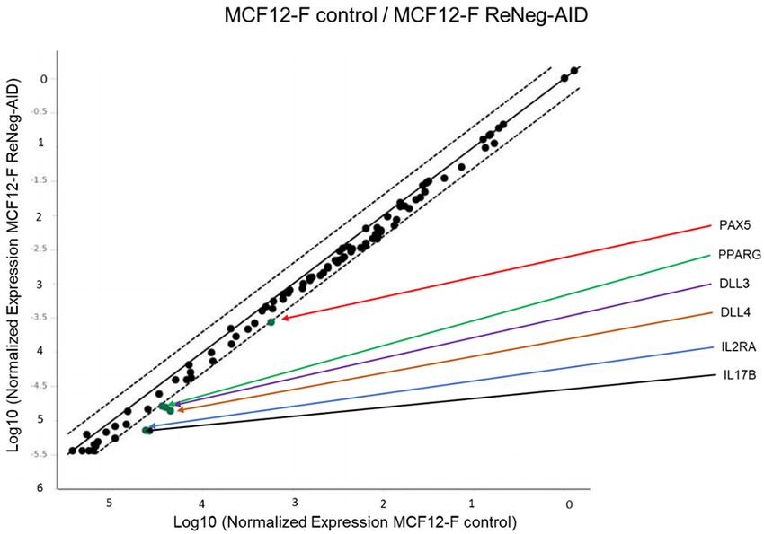
Negative regulation of Notch pathway on mammary cancer cells

                                                    Figure 2. Effect of ReNeg-AID peptide overexpression
                                                    on proliferation of MCF12-F, MCF-7 and MDA-MB-231
                                                    cells. A. Shows proliferation of MCF12-F cells after
                                                    transfection with pFN21K (Mock) or pReNeg-AID
                                                    for 12, 24, 36 or 48 h. B. Shows proliferation of
                                                    MCF-7 cells after transfection with pFN21K (Mock)
                                                    or pReNeg-AID for 12, 24, 36 or 48 h. C. Shows
                                                    proliferation of MDA-MB-231 cells after transfection
                                                    with pFN21K (Mock) or pReNeg-AID for 12, 24, 36 or
                                                    48 h. Both determined by the 3-(4, 5-dimethylthiazol-
                                                    2-yl)-2, 5-diphenyltetrazolium bromide (MTT) assay.
                                                    Significant differences in *, control cells vs transfected
                                                    cells (P < 0.05) between its respective control (one-
                                                    way ANOVA and Tukey’s test) (n=4 individual samples).

MTT proliferation assay                              Fold-change gene expression related to inhibi-
                                                     tion of the NSP and cell differentiation process
Figure 2A shows the proliferation behavior of        mediated by the NSP in mammary gland cells
MCF-12F cells after transfection with mock
vector pFN21K (black line) and pReNegAID             For gene related to inhibition of the NSP,
vector (blue line), at 12, 24, 36- and 48-hours      the comparative analysis of the transcription-
post-transfection (hpt). The statistical analysis    al rate fold-change of AES, CTNNB1, DTX1,
indicated that there are no significant differ-      GLI1, NCOR2, POFUT1 and SMO genes, MCF-7
ences between the pReNeg-AID transfected             control cells vs MCF-7 ReNeg-AID cells 48 hpt
and mock transfected MCF12-F cells. This             (Figure 3A) showed that the over expression of
result means that the ReNeg-AID peptide activ-       the ReNeg-AID peptide caused down regulation
ity has no effect on non-cancerous mammary           of the expression pattern for CTNNB1 (P ≤
gland cells. Figure 2B shows the behavior of         0.00004, -15.22 times), NCOR2 (P ≤ 0.00004,
MCF-7 cells proliferation after transfection with    -37.14 times), POFUT1 (P ≤ 0.000002, -24.54
both mock vector pFN21K (black line) or              times) and SMO (P ≤ 0.000104) genes. Also, it
pReNeg-AID vector (green line) at 12, 24, 36 or      caused an up-regulation for the expression
48 hpt. The statistical analysis indicated sig-      pattern of AES (P ≤ 0.000105, 7.05 times),
nificant differences (*) at 36 hpt (P ≤ 0.005)       and there were no changes in DTX1 and GLI1
and 48 hpt (P ≤ 0.005), causing a decrease in        genes.
cell proliferation when the ReNeg-AID peptide
was over expressed in this cancer cells. As          For gene related to differentiation a compara-
shown in Figure 2C, the MDA-MB-231 cell line         tive analysis of the transcriptional rate fold-
presented an irregular proliferation, due to its     change of DLL4, DTX1, HES1, HES5, HE-
genetic background with respect to the NSP           Y1, HEY2, HEYL, JAG1, NOTCH1 and NOTCH4
that is activated in its non-canonical way. For      genes, MCF-7 control cells vs MCF-7 ReNeg-AID
this reason, the peptide used is not able to con-    cells 48 hpt (Figure 3B) showed that the over
trol the constitutive activation of the Notch        expression of the ReNeg-AID peptide caused a
pathway in triple negative cancer cells.             down-regulations of the expression pattern for

66                                                                Int J Clin Exp Med 2021;14(1):62-75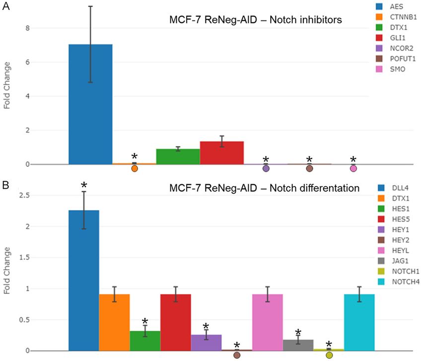
Negative regulation of Notch pathway on mammary cancer cells

                                                                                 HES1 (P ≤ 0.000553, -3.08
                                                                                 times), HEY1 (P ≤ 0.002341,
                                                                                 -3.86 times), HEY2 (P ≤ 0.00002,
                                                                                 -58.61 times), JAG1 (P ≤ 0.0012,
                                                                                 -5.53 times) and NOTCH1 (P ≤
                                                                                 0.00005, -30.41 times) genes.
                                                                                 Also, it caused an up-regulation
                                                                                 of the expression pattern for
                                                                                 DLL4 (P ≤ 0.000004, 2.26
                                                                                 times). For DTX1, HES5, HEYL
                                                                                 and NOTCH4 genes, there were
                                                                                 no changes.

                                                                                 Transcriptional rate fold-change
                                                                                 of genes related to the cross talk
                                                                                 between Hh and NSP pathways
                                                                                 in mammary gland cells

                                                                                 A comparative analysis (Figure 4)
                                                                                 of the transcriptional rate fold-
                                                                                 change of GLI1, GSK3B, HES5,
Figure 3. Profile of gene expression related to differentiation and Inhibi-      NOTCH4, SHH, SMO and SUFU
tion of Notch pathway after transfection with pReNeg-AID in MCF-7 cells          genes, MCF-7 control cells vs
in contrast with Mock transfected cells. A. Fold change of the expression        MCF-7 ReNeg-AID cells, 48 hpt,
of genes related to the inhibition of NSP. mRNA for AES, CTNNB1, DTX1,           showed that the over expression
GLI1, NCOR2, POFUT1 and SMO were quantified by qPCR method. B.                   of the ReNeg-AID peptide caus-
Fold change of the expression of genes related to cellular differentiation.
                                                                                 ed a down-regulation of the tran-
mRNA for DLL4, DTX1, HES1, HES5, HEY1, HEY2, HEYL, JAG1, NOTHC1
and NOTCH4 were quantified by qPCR. In all cases mRNA was quan-                  scriptional expression pattern for
tified using a real-time PCR method (RT2 ProfileTM PCR Array Human Notch         GSK3B (P ≤ 0.000069, -17.01
Signaling Pathway, Qiagen). ACTB and GAPDH genes served as internal              times), SHH (P ≤ 0.000017,
control and was used to normalize for differences in input RNA, and the          -21.82 times), SMO and SUFU (P
fold change threshold was cut-off in 2. Significant differences in *, MCF-       ≤ 0.000286, -7.47 times). Also, it
7 transfected cells (P < 0.05) between its respective control (one-way
                                                                                 caused an up-regulation of the
ANOVA and Tukey’s test) (n=4 individual samples).
                                                                                 transcriptional expression pat-
                                                                                 tern for GLI1, but there were no
                                                                                 changes for HES5 and NOTCH4
                                                                                 genes.

                                                                                 Transcriptional rate fold change
                                                                                 of genes involved in the cross
                                                                                 talk between Wnt and NSP path-
                                                                                 ways in mammary gland cells

                                                                                 A comparative analysis (Figure 5)
                                                                                 of the transcriptional rate fold-
                                                                                 change of AXIN1, CTNNB1, FZD2,
                                                                                 FZD3, FZD4, FZD7, GSK3B,
Figure 4. Profile of gene expression of genes interconnected between
Notch and Hh pathways, after transfection with pReNeg-AID in MCF-7               LRP5, WISP1 and WNT11 genes,
cells in contrast with Mock transfected cells. Fold change of the                MCF-7 control cells vs MCF-7
expression of genes interconnected between Notch and Hh signaling                ReNeg-AID cells at 48 hpt was
pathways. mRNA for GLI1, GSK3B, HES5, NOTCH4, SSH, SMO and SUFU                  performed. The over expression
were quantified by qPCR method. mRNA was quantified using a real-time            of the ReNeg-AID peptide caused
PCR method (RT2 ProfileTM PCR Array Human Notch Signaling Pathway,               a down-regulation of the gene
Qiagen). ACTB and GAPDH served as internal control used to normalize
for differences in input RNA, and the fold change threshold was cut-off          expression pattern for AXIN1
in 2. Significant differences in *, MCF-7 transfected cells (P < 0.05) be-       (P ≤ 0.000041), CTNNB1 (P ≤
tween its respective control (one-way ANOVA and Tukey’s test) (n=4 indi-         0.00004, -15.02 times), FZD2 (P
vidual samples).                                                                 ≤ 0.00247, -2.02 times), FZD3 (P

67                                                                            Int J Clin Exp Med 2021;14(1):62-75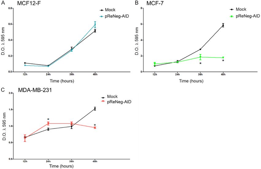
Negative regulation of Notch pathway on mammary cancer cells

                                                                               CTNNB1, FOS, FOSL1, IL2RA,
                                                                               NEURL1, NR4A2 and PTCRA
                                                                               genes, MCF-7 control cells vs
                                                                               MCF-7 ReNeg-AID cells 48 hpt
                                                                               was performed. The over expres-
                                                                               sion of the ReNeg-AID peptide
                                                                               caused a down-regulation of the
                                                                               expression pattern for AXIN1 and
                                                                               CFLAR (P ≤ 0.00004, -1348.80
                                                                               times), CTNNB1 and FOS (P ≤
                                                                               0.000176, -3.74 times), FOSL1 (P
Figure 5. Profile of gene expression of genes interconnected between
Notch and Wnt pathways, after transfection with pReNeg-AID in MCF-7            ≤ 0.00004, -3.83 times), IL2RA
cells in contrast to Mock transfected cells. Fold change of the expression     (P ≤ 0.000193), NEURL1 and
of genes interconnected between Notch and Hh signaling pathways.               PTCRA (no significant change).
mRNA for AXIN1, CTNNB1, FZD2, FZD3, FZD4, FZD7, GSK3B, LRP5,
WISP1 and WNT11 were quantified by qPCR method. mRNA was quan-                 Transcriptional rate fold change
tified using a real-time PCR method (RT2 ProfileTM PCR Array Human Notch       of genes related to cell cycle un-
Signaling Pathway, Qiagen). ACTB and GAPDH served as internal control
used to normalize for differences in input RNA, and the fold change
                                                                               der NSP control
threshold was cut-off in 2. Significant differences in *, MCF-7 transfect-
ed cells (P < 0.05) between its respective control (one-way ANOVA and          A comparative analysis (Figure
Tukey’s test) (n=4 individual samples).                                        7A) of the transcriptional rate
                                                                               fold change of AXIN1, CCND1,
                                                                               CCNE1, CDK1A, JAG2 and NOT-
                                                                               CH2 genes, MCF-7 control cells
                                                                               vs MCF-7 ReNeg-AID cells 48 hpt
                                                                               was performed. The ReNeg-AID
                                                                               peptide over expression caused a
                                                                               down-regulation of the expres-
                                                                               sion pattern for AXIN1, CCND1 (P
                                                                               ≤ 0.0005, -392.06 times), CCNE1
                                                                               (P ≤ 0.00005), CDK1A (P ≤
                                                                               0.00006, -50.59 times), JAG2 (P
                                                                               ≤ 0.000184, -12.77 times),
                                                                               NOTCH1 (P ≤ 0.00005, -30.41
Figure 6. Profile of gene expression of genes related to regulation of the
apoptosis by the control of Notch Signaling Pathway. Fold change of the        times), NOTCH2 (P ≤ 0.000007,
expression of genes related to the regulation of the apoptosis. mRNA           -34.62 times) genes.
for AXIN1, CFLAR, CTNNB1, FOS, FOSL1, IL2RA, NEURL1, NR4A2 and
PTCRA genes were quantified by qPCR method. mRNA was quantified                Flow cytometry and cell cycle
using a real-time PCR method (RT2 ProfileTM PCR Array Human Notch
Signaling Pathway, Qiagen). ACTB and GAPDH served as internal control         Figure 7B shows the cell cycle
used to normalize for differences in input RNA, and the fold change           histograms of MCF-7 control cells
threshold was cut-off in 2. Significant differences in *, MCF-7 transfect-
ed cells (P < 0.05) between its respective control (one-way ANOVA and         compared with MCF-7 ReNeg-AID
Tukey’s test) (n=4 individual samples).                                       cells, 3n experiments with 3
                                                                              repeats were performed for each
                                                                              condition. G1 phase peak on
≤ 0.00006, -10.61 times), FZD4 (P ≤ 0.00026,                   MCF-7 control showed an average percentage
-4.81 times), GSK3B and LRP5 (P ≤ 0.000073,                    of 39.9 ± 2.6 vs an average of 34.6 ± 2.5 for
-10.33 times). FZD7 showed no statistically sig-               MCF-7 ReNeg-AID without statistical signifi-
nificant change. For WISP1 and WNT11 genes,                    cance. S phase valley on MCF-7 control showed
there were no changes.                                         an average percentage of 26.4 ± 3.6 vs an
                                                               average of 37.7 ± 0.98 for MCF-7 ReNeg-AID
Transcriptional rate fold change of genes re-
                                                               with statistical significance (P ≤ 0.0382). The
lated to apoptosis regulation under the control
                                                               G2/M phases peak on MCF-7 control showed
of the NSP in mammary gland cells
                                                               an average percentage of 18.1 ± 3.6 vs an
A comparative analysis (Figure 6) of the tran-                 average of 6.91 ± 1.4 for MCF-7 ReNeg-AID
scriptional rate fold-change of AXIN1, CFLAR,                  with statistical significance (P ≤ 0.0183).

68                                                                           Int J Clin Exp Med 2021;14(1):62-75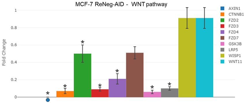
Negative regulation of Notch pathway on mammary cancer cells

                                                                              cer cell proliferation and, more-
                                                                              over, it seems that this peptide
                                                                              somehow is capable of affecting
                                                                              cell proliferation, and does not
                                                                              generate changes in gene expres-
                                                                              sion in the MCF-12F cell line
                                                                              (Figure S3).

                                                                                Gene related to the NSP inhibi-
                                                                                tion: Expression of the ReNeg-
                                                                                AID peptide in MCF-7 cells pro-
                                                                                moted that the AES gene, which
                                                                                participates in NSP inhibition, will
                                                                                show an increase in its transcrip-
                                                                                tion rate, which codifies for the
                                                                                Groucho protein. This protein is
                                                                                one of the main pleiotropic co-
                                                                                repressors of the NSP at nuclear
                                                                                level and has been related to the
                                                                                correct expression of the NSP
                                                                                target genes. The binding activa-
Figure 7. Profile of gene expression of genes related to cell cycle con-        tion complex structured by the
trol and analysis of cell cycle arrest. A. Fold change of the expression of     CSL transcriptional factor, Master-
genes related to the cell cycle control. mRNA for AXIN1, CCND1, CCNE1,          mind (MAML) and NICD proteins,
CDKN1A, JAG2 and NOTCH2 were quantified by qPCR method. mRNA
was quantified using a real-time PCR method (RT Profile PCR Array
                                                      2       TM                is inhibited by the Groucho pro-
Human Notch Signaling Pathway, Qiagen). ACTB and GAPDH served as                tein which recruits histone
internal control used to normalize for differences in input RNA, and the        deacetylases and inhibits the
fold change threshold was cut-off in 2. Significant differences in *, MCF-      NSP target genes [5, 21, 24]. No
7 transfected cells (P < 0.05) between its respective control (one-way          changes in DTX1 gene expres-
ANOVA and Tukey’s test). (n=4 individual samples). B. Cell cycle analysis.
After 48 h cell transfection with Mock and pReNeg-AID respectively, the         sion, the gene codifies to a pro-
cells were pooled, stained with Propidium Iodide (PI), and analyzed by          tein named Deltex, were reported
flow cytometry as described in the Materials and Methods section. Each           This protein has been reported to
histogram shows a flow cytometric plot of 10,000 cells per sample and            be able to physically interact with
is representative of three independent experiments. The percentage of           the Notch-1 receptor avoiding its
cells (mean ± S.D.) in G1, S, and G2/M phases is listed. Significant differ-
ences in *, MCF-7 control vs MCF-7 transfected cells (P < 0.05) between
                                                                                translocation to the nucleus, and,
its respective control (one-way ANOVA and Tukey’s test).                        as a consequence, the NSP tar-
                                                                                get genes will not be expressed
                                                                                (Figure 3A) [6, 25, 26]. The GLI1
Discussion                                                       gene, which codes for GLI1 protein, in the con-
                                                                 text of breast cells can negatively regulate NSP
Cellular proliferation. It has been demonstrated                 by direct physical interaction or cross-talk with
that the ReNeg-AID peptide has no any effect                     the HIF1α factor [27-29]. This prevents NICD1
on non-cancerous mammary gland cells at any                      from entering the nucleus and promotes vacu-
of the tested time, and cell proliferation remains               olar proteolysis when the SMO gene, which
unaltered if we compare transfected (blue line)                  encodes the Smoothness protein, is negatively
and not transfected (black line) MCF-12F cells                   regulated by not presenting changes of these
(Figure 2A). On the other hand, the ReNeg-AID                    proteins normally function in the context of NSP
peptide presents an effect over the MCF-7 cells                  [15, 30, 31].
proliferation (Figure 2B, green line). Cells prolif-
eration began to diminish at 36 hpt, and at 48                   Also, the ReNeg-AID peptide expression in
hpt it is clear that the cell proliferation is severe-           MCF-7 cells promotes that three genes experi-
ly compromised, compared to MCF-7 cells                          ence a reduction in their transcriptional rate.
transfected with the mock plasmid (Figure 2B,                    The first one, NCOR2 gene, which codes for the
black line). All these together suggest that the                 NCoR2 protein, shows a negative regulation;
ReNeg-AID peptide is capable of stopping can-                    and it has been known that this is a specific tis-

69                                                                        Int J Clin Exp Med 2021;14(1):62-75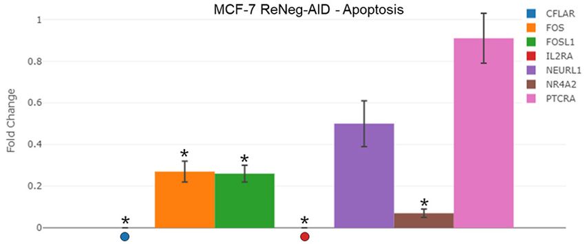
Negative regulation of Notch pathway on mammary cancer cells

sue repressor for NSP in mammary gland cells.        and it is necessary to complete the whole ves-
In contrast to its negative regulation, the AES      sel differentiation process. This change in the
gene positive regulation is capable of compen-       expression pattern of Notch4 and Notch1
sating the regulatory protein complexes mecha-       genes interrupts the differentiation of the Tip/
nisms involved in the inhibition of NSP (Figure      Stalk cells, where the expression of Notch-1
3A) [32-34]. The second gene, CTNNB1, that           gene is required to get a fully functional vessel
codes for the β-catenin protein, shows a nega-       cells (Figure 3B) [42, 44-46].
tive regulation in its transcriptional rate, which
could be indicating that the physical interaction    Cross talking between NSP and Hh pathways: It
reported between Notch-1 and β-catenin was           had been observed that ReNeg-AID peptide
happening when the ReNeg-AID peptide was             overexpression promotes a negative regulation
expressed; this interaction of Notch-1/β-catenin     of GSK3B, SHH, SMO and SUFU genes (Figure
promotes the half-live for Notch-1 and Notch-2       4), which encode for GSK-3β, Sonic Hedgehog,
in the mammary gland (Figure 3A) [35, 36]. The       Smoothened and SUFU proteins, respectively.
third is the POFUT1 gene, which encodes the          The negative regulation of Smoothened and
OFUT1 protein, is responsible for a correct          Sonic Hedgehog proteins has been related to
ligand-receptor interaction in the NSP trans-        the progenitor cancer cells population reduc-
duction mechanism; its negative regulation           tion [31, 47, 48]. This phenomena, together
promotes control at the membrane level to the        with the negative regulation of SUFU and GSK-
NSP in a negative way when an interaction            3β proteins, two negative regulators of the Hh
between the Jagged-1 ligand and the Notch-1          pathway, prevents the GLI1 and GLI2 proteins
receptor is established [37-39].                     degradation, which is essential for the correct
                                                     differentiation process of progenitor mammary
Gene related to NSP and cell differentiation:        gland cells [49-51]. As a consequence, the GLI1
A negative regulation was observed for the           protein cytoplasm accumulation increases the
JAG1 and NOTCH1 genes, which encode Jag-             amount of HIF1α protein in cytoplasm which
ged-1 and Notch-1 proteins respectively. Over-       avoids the NICD1 translocation to the nucleus
expression of ReNeg-AID promoted the de-             [15, 52]. All this together has been related to a
crease of ligand/receptor interaction, so that       diminished cell migration and EMT decrease on
the NSP target genes such as HES1, HEY1 and          mammary cancer cells led by Hh pathway and
HEY2 have a negative regulation. This phenom-        would suggest a cross talk with the γ-secretase
ena would suggest that the mammary gland             activity by the Notch-1 overexpression [53, 54].
cancer cells come to a dedifferentiated state in
order to recover the normal control of cell cycle    Cross talk between NSP and WNT pathways: A
and apoptosis [2, 40]. On the other hand, a          diminished transcriptional rate of those genes
positive regulation of DLL4 gene has been            encoding AXIN and β-catenin proteins has been
observed. DLL4 codes for Delta-4 ligand pro-         observed (Figure 5). It has been known that
teins; this positive regulation at mammary           the interaction between these proteins results
gland cancer cells seems to be promoting the         in a transcriptional complex that triggers the
establishment of Delta-4/Notch-4 (ligand/            expression of genes dependent on the WNT
receptor) interaction, in order to take control in   pathway activity, which is involved in cell cycle
the absence of Jagged-1/Notch-1 interaction,         and cell growth. When the ReNeg-AID peptide
although it has been shown that Notch-4/             is overexpressed, the complex AXIN/β-catenin
Delta-4 is not able to recover the whole cell pro-   is not formed. In normal cells, when the
cess mediated by Jagged-1/Notch-1 interac-           β-catenin protein is released from the AXIN/β-
tion [26, 41].                                       catenin complex, it interacts with the NICD
                                                     present at cell cytoplasm, avoiding its degrada-
When the ReNeg-AID peptide was over ex-              tion. In these cancer cells the β-catenin in cyto-
pressed in breast cancer cells, Hes5 gene did        plasm is also diminished by the ReNeg-AID pep-
not present changes, promoting the direct            tide overexpression, which shrinks its interac-
cross talking between vessel cells and epithe-       tion with NICD, causing its degradation which,
lial cells through Notch-4/Delta-4 interaction       instead, causes a negative transcriptional regu-
which, in consequence, promotes a normal de          lation of the NSP target genes [35, 55, 56].
novo angiogenesis process [42, 43] but the
new vessels will not be functional because the       It had been observed that the ReNeg-AID pep-
Notch-1/Jagged-1 interaction is not occurring        tide overexpression promotes a negative regu-

70                                                              Int J Clin Exp Med 2021;14(1):62-75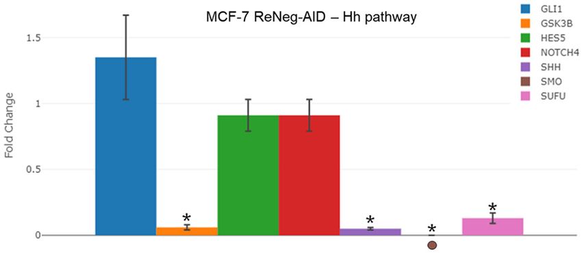
Negative regulation of Notch pathway on mammary cancer cells

lation of FZD2, FZD3, FZD4, and LRP5 genes.           D1, Cyclin E1, CDK1, Jagged-2, Notch-1 and
Therefore, a WNT signaling pathway inactiva-          Notch-2 proteins. It has been reported that in
tion occurs because the receptors Frizzled 2          MCF-7 cells the Jagged-2/Notch-2 interaction
(FZD2), Frizzled 3 (FZD3) and Frizzled 4 (FZD4)       promotes the cell cycle initiation mediated by
are diminished in their membrane concentra-           the activity of Cyclin D1 [11]. In breast cancer
tion. In contrast, the WNT11 and WISP1 genes          the Notch-1 constitutive overexpression pro-
did not show changes, which do not have any           motes the Notch-2 receptor overexpression
receptors to interact with. This effect is potenti-   and, as a consequence, the expression of the
ated by the lower LRP-5 protein concentration,        NSP target genes. One of these genes is CNND1
which has the important role in stabilizing           (Cyclin D1) which is responsible of the cell cycle
ligand/receptor interaction in the WNT pathway        G1/S phase transition. It is suggested that
context. All this together could be meaning that      Cyclin D1 overexpression could promote a
the WNT pathway is not activated by the defi-         G1/S checkpoint malfunction and, as a conse-
ciency of receptors. Nevertheless, at the same        quence, a loop cell cycle that will eventually
time the ReNeg-AID peptide overexpression on          lead the mammary gland cells to an uncon-
MCF-7 cell promotes a deficiency of the CTNNB1        trolled proliferation. The ReNeg-AID peptide
gene transcription, avoiding the presence of          overexpression in MCF-7 cells negatively regu-
β-catenin in cell cytoplasm, which instead pre-       lates the overexpression of both Notch-2 and
vents its translocation into the nucleus, pro-        Jagged-2 proteins, and therefore promotes the
moting the expression of WNT pathway target           negative regulation of Cyclin D1 and Cyclin E1
genes. The decrease of β-catenin in the cyto-         [60, 61]. This negative regulation of those pro-
plasm promotes a cytoplasmic NICD half-live           teins arrests the cell cycle at G1/S phase in
decrement, because NICDs are caught by pro-           MCF-7 cells (Figure 7B), where the cell popula-
teasomes [35, 36, 44, 57].                            tion is mainly arrested in the S phase, as a con-
                                                      sequence causing a diminished proliferation.
NSP and apoptosis: A negative regulation of           Finally, it had been observed that a down regu-
CFLAR, FOS, FOSL, NR4A2 and IL2RA genes               lation of CDK1A gene transcription, caused by
was observed (Figure 6) by the ReNeg-AID pep-         the ReNeg-AID peptide overexpression, in
tide overexpression in MCF-7 cells. Those             MCF-7 cells should cause an instability of the
genes encode for FADD-L1, C-Fos, Fra1, Nurr1          cell cycle associated with an early apoptotic
and CD25 proteins, respectively. The ReNeg-           activation process. This could mean that the
AID peptide overexpression suggests that              combined down regulation of both CDK1A and
FADD-L1/C-Fos/Fra1 interaction is able to posi-       CNND1 genes promotes the cell cycle arrest of
tively regulate the activation mechanism of           mammary gland cancer cells [12, 62, 63].
FAS/FADD apoptotic receptors by the extrinsic
way, together with the Nurr1 and CD25 regula-         Overexpression of ReNeg-AID peptide in MCF-7
tion on MCF-7 cells [54, 58]. However, it is          cells regulates negatively the constitutive
known that NSP is able to regulate the intrinsic      expression of Notch-1 receptor at different lev-
apoptotic pathway by the expression of NEURL1         els. On cellular membrane level it negatively
and PTCRA genes. It is also known that these          regulates the Jagged1/Notch-1 pathway by
proteins are involved in the differentiation pro-     negative regulation of POFUT1 and the normal
cesses and in the negative regulation of the          expression of DTX1. At cytoplasmic level, it neg-
NSP, promoting the regulation of the apoptotic        atively regulates the half-lives of the Notch
processes mediated by PUMA and Bcl-2 pro-             receptor by negative regulation of β-catenin
teins. Nevertheless, it is clear that more experi-    and Axin1 and by the normal expression of the
mental data is necessary to elucidate the par-        GLI1 protein. At nuclear level, the activation
ticipation of these proteins in the apoptotic         complex protein between CSL/MAML/NICD is
processes regulation in MCF-7 cells carried out       negatively regulated by the positive expression
by NSP [14, 59].                                      of Groucho protein and by the very nature of the
                                                      ReNeg-AID peptide. The most significant effect
Cell cycle and NSP: As shown in Figure 7A, a          of the ReNeg-AID overexpression occurs in the
negative regulation of AXIN1, CNND1, CNNE1,           cell cycle. The MCF-7 cells were arrested in the
CDK1A, JAG2, NOTCH1 and NOTCH2 genes,                 G1/S phase by the negative regulation of Cyclin
which are involved in cell cycle, has been            D1 and Cyclin E1, and by the effect of ReNeg-
observed. These genes encode for Axin1, Cyclin        AID peptide expression on normal cells (MCF-

71                                                               Int J Clin Exp Med 2021;14(1):62-75
Negative regulation of Notch pathway on mammary cancer cells
12F) that does not cause significant effects on centennial for notch signaling. Cancer Cell
the pattern genes related to the Notch path- 2014; 25: 318-334.
way. The effect of the ReNeg-AID peptide over- [2] Fior R and Henrique D. “Notch-Of”: a perspec-
tive on the termination of Notch signalling. Int
expression opens the doors for future research
J Dev Biol 2009; 53: 1379-1384.
based on the negative regulation at nuclear [3] Contreras-Cornejo H, Saucedo-Correa G, Ovie-
level in cancer cells that present a constitutive do-Boyso J, Valdez-Alarcón JJ, Baizabal-Aguirre
activation of the Notch signaling pathway and VM, Cajero-Juárez M and Bravo-Patiño A. The
that it can be used as an alternative adjuvant CSL proteins, versatile transcription factors
strategy against breast cancer. It remains to and context dependent corepressors of the
analyze the possible routes of administration notch signaling pathway. Cell Div 2016; 11:
and/or action, by which the peptide ReNeg-AID 12-12.
[4] Oswald F, Winkler M, Cao Y, Astrahantseff K,
can have an effect in vivo. The results of the
Bourteele S, Borggrefe T, Al OET and Iol
MDA-MB-231 cell line were performed as with MOLCELLB. RBP-J/SHARP recruits CtIP/CtBP
the MCF-7 and MCF-12F cell line, however, the corepressors to silence notch target genes.
data are not shown as conclusive results due to Society 2005; 25: 10379-10390.
the nature of expression of the Notch pathway [5] Yuan D, Yang X, Yuan Z, Zhao Y and Guo J.
since it presents a non-canonical signaling of TLE1 function and therapeutic potential in
the Notch pathway and merits more detailed cancer. Oncotarget 2017; 8: 15971-15976.
studies to answer that question. [6] Ling H, Sylvestre JR and Jolicoeur P. Notch1-
induced mammary tumor development is cy-
clin D1-dependent and correlates with expan-
Acknowledgements
sion of pre-malignant multipotent duct-limited
progenitors. Oncogene 2010; 29: 4543-4554.
GSC, ABP and RENA were responsible for the [7] Radtke F and Raj K. The role of Notch in tu-
conception, design and writing of the manu- morigenesis: oncogene or tumour suppressor?
script. All authors read and approved the final Nature reviews. Cancer 2003; 3: 756-767.
manuscript. GSC thanks to CONACYT for the [8] Li P, Barraclough R, Fernig DG, Smith JA and
financial support. ABP thanks for the manu- Rudland PS. Stem cells in breast epithelia. Int
script founding to SEP-CONACYT and CIC- J Exp Pathol 2008; 79: 193-206.
[9] Allenspach EJ, Maillard I, Aster JC and Pear
UMSNH. Consejo Nacional de Ciencia y Tecno-
WS. Notch Signaling in Cancer. Cancer Biol
logía (CONACYT) México, SEP-CONACYT (regis- Ther 2002; 1: 466-476.
tration number CB-2011-01 167947); and a [10] Arnold A and Papanikolaou A. Cyclin D1 in
CIC-UMSNH (registration number 521765 CIC breast cancer pathogenesis. J Clin Oncol
2015) funded this manuscript for Dr. Bravo 2005; 23: 4215-4224.
Patiño. We are grateful for the technical sup- [11] Wang Z, Sugano E, Isago H, Murayama N,
port to Proteomic and Cellular Bioengineering Tamai M and Tomita H. Notch signaling path-
Unit supported by CONACYT INFR-2015-01 way regulates proliferation and differentiation
of immortalized Müller cells under hypoxic
255010. Saucedo Correa would like to thank
conditions in vitro. Neuroscience 2012; 214:
CONACYT for her doctorate studies grant num- 171-180.
ber 27023. [12] Dash BC and El-Deiry WS. Cell cycle check-
point control mechanisms that can be disrupt-
Disclosure of conflict of interest ed in cancer. Drug Des Devel Ther 2004; 280:
99-161.
None. [13] Bravo A and Baizabal VM. L A VÍA DE S
EÑALIZACIÓN N OTCH Y EL D ESARROLLO E
Address correspondence to: Alejandro Bravo-Pa- MBRIONARIO A NIMAL. REB 2005; 24: 87-96.
tiño, Facultad de Medicina Veterinaria y Zootecnia [14] Gopalakrishnan N, Sivasithamparam ND and
Devaraj H. Synergistic association of Notch
UMSNH, Michoacán, México. Tel: (52) 443-2 95
and NFκB signaling and role of Notch signaling
8029; E-mail: brapal68@gmail.com; alejandro. in modulating epithelial to mesenchymal tran-
bravo@umich.mx sition in colorectal adenocarcinoma. Biochimie
2014; 107: 310-318.
References [15] Zheng X, Narayanan S, Zheng X, Luecke-
Johansson S, Gradin K, Catrina SB, Poellinger
[1] Ntziachristos P, Lim JS, Sage J and Aifantis I. L and Pereira TS. A Notch-independent mecha-
From fly wings to targeted cancer therapies: a nism contributes to the induction of Hes1 gene
72 Int J Clin Exp Med 2021;14(1):62-75Negative regulation of Notch pathway on mammary cancer cells
expression in response to hypoxia in P19 cells. transcription factor Hes1 via the PHB2 chaper-
Exp Cell Res 2017; 358: 129-139. one. J Biol Chem 2018; 293: 8285-8294.
[16] Wu Y, Cain-Hom C, Choy L, Hagenbeek TJ, De [27] Guen VJ, Chavarria TE, Kröger C, Ye X, Weinberg
Leon GP, Chen Y, Finkle D, Venook R, Wu X, RA and Lees JA. EMT programs promote basal
Ridgway J, Schahin-Reed D, Dow GJ, Shelton A, mammary stem cell and tumor-initiating cell
Stawicki S, Watts RJ, Zhang J, Choy R, Howard stemness by inducing primary ciliogenesis and
P, Kadyk L, Yan M, Zha J, Callahan CA, Hymowitz Hedgehog signaling. Proc Natl Acad Sci U S A
SG and Siebel CW. Therapeutic antibody tar- 2017; 114: E10532-E10539.
geting of individual Notch receptors. Nature [28] Liu Z, Tu K, Wang Y, Yao B, Li Q, Wang L, Dou C,
2010; 464: 1052-1057. Liu Q and Zheng X. Hypoxia accelerates ag-
[17] Previs RA, Coleman RL, Harris AL and Sood AK. gressiveness of hepatocellular carcinoma cells
Molecular pathways: translational and thera- involving oxidative stress, epithelial-mesenchy-
peutic implications of the Notch signaling mal transition and non-canonical hedgehog
pathway in cancer. Clin Cancer Res 2015; 21: signaling. Cell Physiol Biochem 2018; 44:
955-961. 1856-1866.
[18] Lamy M, Ferreira A, Dias JS, Braga S, Silva G [29] Cejudo-Martin P and Johnson RS. A new Notch
and Barbas A. Notch-out for breast cancer in the HIF belt: how hypoxia impacts differenti-
therapies. N Biotechnol 2017; 39: 215-221. ation. Dev Cell 2005; 9: 575-576.
[19] Rizzo P, Osipo C, Foreman K, Golde T, Osborne [30] Brechbiel J, Miller-Moslin K and Adjei AA.
B and Miele L. Rational targeting of Notch sig- Crosstalk between hedgehog and other signal-
naling in cancer. Oncogene 2008; 27: 5124- ing pathways as a basis for combination thera-
5131. pies in cancer. Cancer Treat Rev 2014; 40:
[20] Yuan X, Wu H, Xu H, Xiong H, Chu Q, Yu S, 750-759.
Wu GS and Wu K. Notch signaling: an emerg- [31] Hallahan AR, Pritchard JI, Hansen S, Benson
ing therapeutic target for cancer treatment. M, Stoeck J, Hatton BA, Russell TL, Ellenbogen
Cancer Lett 2015; 369: 20-27. RG, Bernstein ID, Beachy PA and Olson JM. The
[21] Maier D, Chen AX, Preiss A and Ketelhut SmoA1 mouse model reveals that notch sig-
M. The tiny Hairless protein from Apis mel- naling is critical for the growth and survival of
lifera: a potent antagonist of Notch signaling in sonic the SmoA1 mouse model reveals that
Drosophila melanogaster. BMC Evol Biol 2008; notch signaling is critical for the growth and
8: 175-175. survival of sonic hedgehog-induced medullo-
[22] Paz-Gómez D, Baizabal-Aguirre VM, Valdez- blastomas. Cancer Res 2004; 64: 7794-7800.
Alarcón JJ, Cajero-Juárez M, Nagel AC, Preiss A, [32] Giaimo BD, Oswald F and Borggrefe T. Dynamic
Maier D and Bravo-Patiño A. Structural analy- chromatin regulation at Notch target genes.
sis of point mutations in the Hairless gene and Transcription 2017; 8: 61-66.
their association with the activity of the [33] Schwanbeck R. The role of epigenetic mecha-
Hairless protein. Int J Biol Macromol 2008; 43: nisms in notch signaling during development. J
426-432. Cell Physiol 2015; 230: 969-981.
[23] Bray SJ, Takada S, Harrison E, Shen SC and [34] Oswald F, Kostezka U, Astrahantseff K, Bour-
Ferguson-Smith AC. The atypical mammalian teele S, Dillinger K, Zechner U, Ludwig L, Wilda
ligand Delta-like homologue 1 (Dlk1) can regu- M, Hameister H, Knöchel W, Liptay S and
late Notch signalling in Drosophila. BMC Dev Schmid RM. SHARP is a novel component of
Biol 2008; 8: 11. the Notch/RBP-Jkappa signalling pathway.
[24] Cheng YC, Huang YC, Yeh TH, Shih HY, Lin CY, EMBO J 2002; 21: 5417-5426.
Lin SJ, Chiu CC, Huang CW and Jiang YJ. [35] de Cássia Viu Carrara R, Fontes AM, Abraham
Deltex1 is inhibited by the Notch-Hairy/E(Spl) KJ, Orellana MD, Haddad SK, Palma PVB,
signaling pathway and induces neuronal and Panepucci RA, Zago MA and Covas DT.
glial differentiation. Neural Dev 2015; 10: Expression differences of genes in the PI3K/
1-15. AKT, WNT/b-catenin, SHH, NOTCH and MAPK
[25] Narayanappa R, Rout P, Aithal MGS and Chand signaling pathways in CD34+ hematopoietic
AK. Aberrant expression of Notch1, HES1, and cells obtained from chronic phase patients
DTX1 genes in glioblastoma formalin-fixed par- with chronic myeloid leukemia and from
affin-embedded tissues. Tumor Biol 2016; 37: healthy controls. Clin Transl Oncol 2018; 20:
6935-6942. 542-549.
[26] Perron A, Nishikawa Y, Iwata J, Shimojo H, [36] Gopalakrishnan N, Saravanakumar M, Madan-
Takaya J, Kobayashi K, Imayoshi I, Mbenza NM, kumar P, Thiyagu M and Devaraj H. Colo-
Takenoya M, Kageyama R, Kodama Y and calization of b-catenin with Notch intracellular
Uesugi M. Small-molecule screening yields a domain in colon cancer: a possible role of
compound that inhibits the cancer-associated Notch1 signaling in activation of CyclinD1-
73 Int J Clin Exp Med 2021;14(1):62-75Negative regulation of Notch pathway on mammary cancer cells
mediated cell proliferation. Mol Cell Biochem [48] Karamboulas C and Ailles L. Developmental
2014; 396: 281-293. signaling pathways in cancer stem cells of sol-
[37] Okajima T and Irvine KD. Regulation of Notch id tumors. Biochim Biophys Acta 2013; 1830:
signaling by O-linked fucose. Cell 2002; 111: 2481-2495.
893-904. [49] Xie G, Karaca G, Swiderska-Syn M, Michelotti
[38] Buono KD, Robinson GW, Martin C, Shi S, Stan- GA, Krüger L, Chen Y, Premont RT, Choi SS
ley P, Tanigaki K, Honjo T and Hennighausen L. and Diehl AM. Cross-talk between Notch and
The canonical Notch/RBP-J signaling pathway Hedgehog regulates hepatic stellate cell fate in
controls the balance of cell lineages in mam- mice. Hepatology 2013; 58: 1801-1813.
mary epithelium during pregnancy. Dev Biol [50] Merchant AA and Matsui W. Targeting He-
2006; 293: 565-580. dgehog - a cancer stem cell pathway. Clin
[39] Zavadil J, Cermak L, Soto-Nieves N and Böt- Cancer Res 2010; 16: 3130-3140.
tinger EP. Integration of TGF-β/Smad and [51] Correction: the notch target hes1 directly mod-
Jagged1/Notch signalling in epithelial-to-mes- ulates gli1 expression and hedgehog signaling:
enchymal transition. EMBO J 2004; 23: 1155- a potential mechanism of therapeutic resis-
1165. tance. Clin Cancer Res 2016; 22: 3700-3701.
[40] Murata K, Hattori M, Hirai N, Shinozuka Y, [52] Asnaghi L, Lin MH, Lim KS, Lim KJ, Tripathy A,
Hirata H, Kageyama R, Sakai T and Minato N. Wendeborn M, Merbs SL, Handa JT, Sodhi A,
Hes1 directly controls cell proliferation through Bar EE and Eberhart CG. Hypoxia promotes
the transcriptional repression of p27Kip1. Mol uveal melanoma invasion through enhanced
Cell Biol 2005; 25: 4262-4271. notch and MAPK activation. PLoS One 2014;
[41] Sun Y, Lowther W, Kato K, Bianco C, Kenney N, 9: e105372.
Strizzi L, Raafat D, Hirota M, Khan NI, Bargo S, [53] Salaritabar A, Berindan-neagoe I, Darvish
Jones B, Salomon D and Callahan R. Notch4 B, Hadjiakhoondi F, Manayi A, Pandima K,
intracellular domain binding to Smad3 and in- Barreca D, Erdogan I, Süntar I, Ahmad A, Gulei
hibition of the TGF-beta; signaling. Oncogene D, Fazel S, Sureda A and Daglia M. Targeting
Hedgehog signaling pathway: paving the road
2005; 24: 5365-5374.
for cancer therapy. Pharmacol Res 2019; 141:
[42] Patel NS, Li JL, Generali D, Poulsom R,
466-480.
Cranston DW and Harris AL. Up-regulation of
[54] Huang CC, Cheng SH, Wu CH, Li WY, Wang JS,
delta-like 4 ligand in human tumor vasculature
Kung ML, Chu TH, Huang ST, Feng CT, Huang
and the role of basal expression in endothelial
SC and Tai MH. Delta-like 1 homologue pro-
cell function. Cancer Res 2005; 65: 8690-
motes tumorigenesis and epithelial-mesenchy-
8697.
mal transition of ovarian high-grade serous
[43] Pedrosa AR, Trindade A, Carvalho C, Graça J,
carcinoma through activation of Notch signal-
Carvalho S, Peleteiro MC, Adams RH and
ing. Oncogene 2019; 38: 3201-3215.
Duarte A. Endothelial Jagged1 promotes solid
[55] Shao S, Zhao X, Zhang X, Luo M, Zuo X, Huang
tumor growth through both pro-angiogenic and
S, Wang Y, Gu S and Zhao X. Notch1 signaling
angiocrine functions. Oncotarget 2015; 6:
regulates the epithelial-mesenchymal transi-
24404-23. tion and invasion of breast cancer in a Slug-
[44] Guo S, Liu M and Gonzalez-perez RR. Bio- dependent manner. Mol Cancer 2015; 14: 28-
chimica et Biophysica Acta Role of Notch and 28.
its oncogenic signaling crosstalk in breast can- [56] Chen Y, Zheng S, Qi D, Zheng S, Guo J, Zhang S
cer. Biochim Biophys Acta 2011; 1815: 197- and Weng Z. Inhibition of notch signaling by a
213. γ-secretase inhibitor attenuates hepatic fibro-
[45] Jubb AM, Soilleux EJ, Turley H, Steers G, sis in rats. PLoS One 2012; 7: 1-11.
Parker A, Low I, Blades J, Li JL, Allen P, Leek R, [57] Guo S, Liu M and Gonzalez-Perez RR. Role of
Noguera-Troise I, Gatter KC, Thurston G and Notch and its oncogenic signaling crosstalk in
Harris AL. Expression of vascular Notch ligand breast cancer. Cancer Lett 2019; 461: 123-
delta-like 4 and inflammatory markers in 131.
breast cancer. Am J Pathol 2010; 176: 2019- [58] Zhang J, Tian XJ and Xing J. Signal transduc-
2028. tion pathways of EMT induced by TGF-β, SHH,
[46] Eilken HM and Adams RH. Dynamics of endo- and WNT and their crosstalks. J Clin Med
thelial cell behavior in sprouting angiogenesis. 2016; 5: 41-41.
Curr Opin Cell Biol 2010; 22: 617-625. [59] Corbin EA, Kong F, Lim CT, King WP and Bashir
[47] Liu, Zi Hao Dai, Xiao Meng Du, Bin. Hes1: a key R. Biophysical properties of human breast can-
role in stemness, metastasis and multidrug re- cer cells measured using silicon MEMS reso-
sistance. Cancer Biol Ther 2015; 16: 1538- nators and atomic force microscopy. Lab Chip
4047. 2015; 15: 839-847.
74 Int J Clin Exp Med 2021;14(1):62-75Negative regulation of Notch pathway on mammary cancer cells
[60] Ansardamavandi A, Tafazzoli-Shadpour M, [62] Cascione M, de Matteis V, Rinaldi R and
Omidvar R and Jahanzad I. Quantification of ef- Leporatti S. Atomic force microscopy combined
fects of cancer on elastic properties of breast with optical microscopy for cells investigation.
tissue by Atomic Force Microscopy. J Mech Microsc Res Tech 2017; 80: 109-123.
Behav Biomed Mater 2016; 60: 234-242. [63] Jamdade VS, Sethi N, Mundhe NA, Kumar P,
[61] Berquand A, Meunier M, Thevenard-Devy J, Lahkar M and Sinha N. Therapeutic targets of
Ivaldi C, Campion O, Dedieu S, Molinari M and triple-negative breast cancer: a review. Br J
Devy J. A gentle approach to investigate the Pharmacol 2015; 172: 4228-4237.
influence of LRP-1 silencing on the migratory
behavior of breast cancer cells by atomic
force microscopy and dynamic cell studies.
Nanomedicine 2019; 18: 359-370.
75 Int J Clin Exp Med 2021;14(1):62-75Negative regulation of Notch pathway on mammary cancer cells Figure S1. NSP canonical activation protein complex. The Notch receptor in mammals (Notch1-4) undergoes post- transcriptional modifications in the endoplasmic reticulum and in the Golgi apparatus where it is finally granted the specificity of binding by its ligands of the DSL family (Delta, Serrate and Lag-1). Once the Notch receptor is found on cell membrane it is recognized by the ADAM10/TACE metallopreotease that makes a proteolytic cut by separat- ing the extracellular domain of the Notch protein once it is bound to its ligand (S1). The Notch intracellular domain (NICD) is then released from the cell membrane by the action of γ-secretase (S2). Once NICD is released from the cell membrane it is directed to the nucleus where it forms an activation complex by recruiting the Mastermind and SKIP co-activators to bind to the CSL transcriptional factor and initiate the expression of genes dependent on the Notch pathway like Hes and Hey family genes, CCND1, CCND3, Notch receptor and Notch ligands. 1
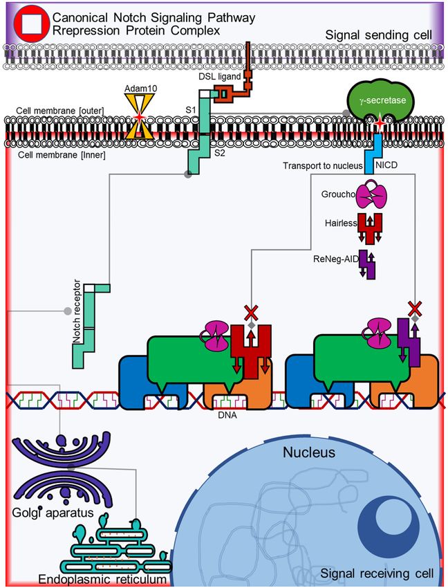
Negative regulation of Notch pathway on mammary cancer cells

Figure S2. NSP repression protein complex. The Hairless protein competes for the union of the CSL transcrip-
tional factor against NICD with a similar binding affinity to form a repression complex with the help of co-repressor
Groucho. The ReNeg-AID peptide having part of the CSL binding domain of the Hairless protein competes in the
same way against NICD to form a repression complex and change the expression pattern of genes dependent on
the Notch pathway.

2
Negative regulation of Notch pathway on mammary cancer cells

Figure S3. Scatter plot profile of the gene expression related to Notch Signaling Pathway on MCF-12F cells. mRNA
was quantified using a real-time PCR method (RT2 ProfileTM PCR Array Human Notch Signaling Pathway, Qiagen).
GAPDH served as internal control and was used to normalize for differences in input RNA. No significant differences
were detected in MCF-12F control vs MCF-12F transfected cells between its respective control (one-way ANOVA and
Tukey’s test) (n=4 individual samples). Only PAX5, PPARG, DLL3, DLL4, IL2RA and IL17B genes were regulated
negatively without significant differences.

3
You can also read


