Establishment of the First Well-differentiated Human Pancreatic Neuroendocrine Tumor Model - Molecular Cancer Research
←
→
Page content transcription
If your browser does not render page correctly, please read the page content below
Published OnlineFirst January 12, 2018; DOI: 10.1158/1541-7786.MCR-17-0163

        Oncogenes and Tumor Suppressors                                                                                                   Molecular
                                                                                                                                          Cancer
                                                                                                                                          Research
     Establishment of the First Well-differentiated
     Human Pancreatic Neuroendocrine Tumor Model
     Daniel Benten1,2, Yasmin Behrang1, Ludmilla Unrau3, Victoria Weissmann4,
     Gerrit Wolters-Eisfeld4, Susanne Burdak-Rothkamm5,6, Felix R. Stahl7,
     Martin Anlauf8, Patricia Grabowski9, Markus Mo€ bs10, Jan Dieckhoff11,
     Bence Sipos , Martina Fahl , Corinna Eggers , Daniel Perez4,
                 12              1               1

     Maximillian Bockhorn4, Jakob R. Izbicki4, Ansgar W. Lohse1, and Jo€ rg Schrader1,4

     Abstract
        Clinical options for systemic therapy of neuroendocrine                          proliferative labeling index, measured by Ki-67, of 14.6%
     tumors (NET) are limited. Development of new drugs re-                              1.0% in NT-3 is akin to the original tumor (15%–20%),
     quires suitable representative in vitro and in vivo model sys-                      and was lower than in BON (80.6%  3.3%) and QGP-1
     tems. So far, the unavailability of a human model with a                            (82.6%  1.0%). NT-3 highly expressed somatostatin recep-
     well-differentiated phenotype and typical growth character-                         tors (SSTRs: 1, 2, 3, and 5). Upon subcutaneous transplan-
     istics has impaired preclinical research in NET. Herein, we                         tation of NT-3 cells, recipient mice developed tumors with
     establish and characterize a lymph node–derived cell line                           an efficient tumor take rate (94%) and growth rate (139%
     (NT-3) from a male patient with well-differentiated pancre-                         13%) by 4 weeks. Importantly, morphology and neuroendo-
     atic NET. Neuroendocrine differentiation and tumor biology                          crine marker expression of xenograft tumors resembled the
     was compared with existing NET cell lines BON and QGP-1.                            original human tumor.
     In vivo growth was assessed in a xenograft mouse model.
     The neuroendocrine identity of NT-3 was verified by expres-                          Implications: High expression of somatostatin receptors and
     sion of multiple NET-specific markers, which were highly                             a well-differentiated phenotype as well as a slow growth rate
     expressed in NT-3 compared with BON and QGP-1. In addi-                             qualify the new cell line as a relevant model to study neuro-
     tion, NT-3 expressed and secreted insulin. Until now, this                          endocrine tumor biology and to develop new tumor treat-
     well-differentiated phenotype is stable since 58 passages. The                      ments. Mol Cancer Res; 16(3); 496–507. 2018 AACR.

     1
       I. Medical Department - Gastroenterology and Hepatology, University Medical       Introduction
     Center Hamburg-Eppendorf, Hamburg, Germany. 2Department of Gastroenter-
     ology, Helios Klinik Duisburg, Duisburg, Germany. 3Department of Stem Cell
                                                                                            Well-differentiated neuroendocrine tumors (NET) are char-
     Transplantation, University Medical Center Hamburg-Eppendorf, Hamburg,              acterized by the cardinal features of slow tumor growth and
     Germany. 4Department of General-, Visceral and Thoracic Surgery, University         potential to secrete functionally active hormones. Because of
     Medical Center Hamburg-Eppendorf, Hamburg, Germany. 5Department of                  their mostly indolent behavior, more than 50% of patients are
     Pathology, University Medical Center Hamburg-Eppendorf, Hamburg, Germany.           diagnosed with metastatic disease not amenable to curative
     6
        Department of Radiotherapy and Radiation Oncology, University Medical            surgery (1). Despite recent advances in the development of
     Center Hamburg-Eppendorf, Hamburg, Germany. 7Institute of Clinical Chemistry
     and Laboratory Medicine, University Medical Center Hamburg-Eppendorf,
                                                                                         medical treatments, long-term disease control is only achieved
     Hamburg, Germany. 8Institute of Pathology, Limburg, Germany. 9Department            in a minority of patients receiving chemotherapy or targeted
     of Gastroenterology, Rheumatology and Infectious Diseases, Charite Campus           therapies in patients with pancreatic NET (2–4). Encouraging
                                                                          – Universi-
     Benjamin Franklin, Berlin, Germany. 10Institute of Pathology, Charite               data on the use of peptide receptor radionuclide therapy show
     taetsmedizin Berlin, Berlin, Germany. 11Department for Interventional and Diag-     long-term disease stabilization but are limited to NET with high
     nostic Radiology and Nuclear Medicine, University Medical Center Hamburg-           somatostatin receptor (SSTR) expression (5). In contrast, for
     Eppendorf, Hamburg, Germany. 12Department of Pathology, University Hospital
        €bingen, Tu€bingen, Germany.
                                                                                         patients with low receptor status identification of alternative
     Tu
                                                                                         NET cell–specific receptor targets is crucial. However, develop-
     Note: Supplementary data for this article are available at Molecular Cancer
                                                                                         ment and evaluation of novel therapeutic targets and optimi-
     Research Online (http://mcr.aacrjournals.org/).
                                                                                         zation of existing NET therapies has been hampered by limited
     D. Benten and Y. Behrang contributed equally to this article.                       availability of preclinical models. Currently, there are only a
     Corresponding Author: Jo  €rg Schrader, I. Medical Department – Gastroenter-        few NET cell lines available (6, 7). Of these, the pancreatic cell
     ology and Hepatology, University Medical Center Hamburg-Eppendorf, Marti-           lines BON and QGP-1 are the most widely used. Despite
     nistrasse 52, Hamburg 20246, Germany. Phone: 4940-7410-53661; Fax: 4940-            expression of some NET markers, BON and QGP-1 do not
     7410-40272; E-mail: j.schrader@uke.de
                                                                                         display a well-differentiated neuroendocrine phenotype. For
     doi: 10.1158/1541-7786.MCR-17-0163                                                  example, cells do not serve as a model to study radionuclide
     2018 American Association for Cancer Research.                                     imaging and therapy, as SSTR expression is very low and

496 Mol Cancer Res; 16(3) March 2018

         Downloaded from mcr.aacrjournals.org on February 3, 2021. © 2018 American Association for Cancer Research.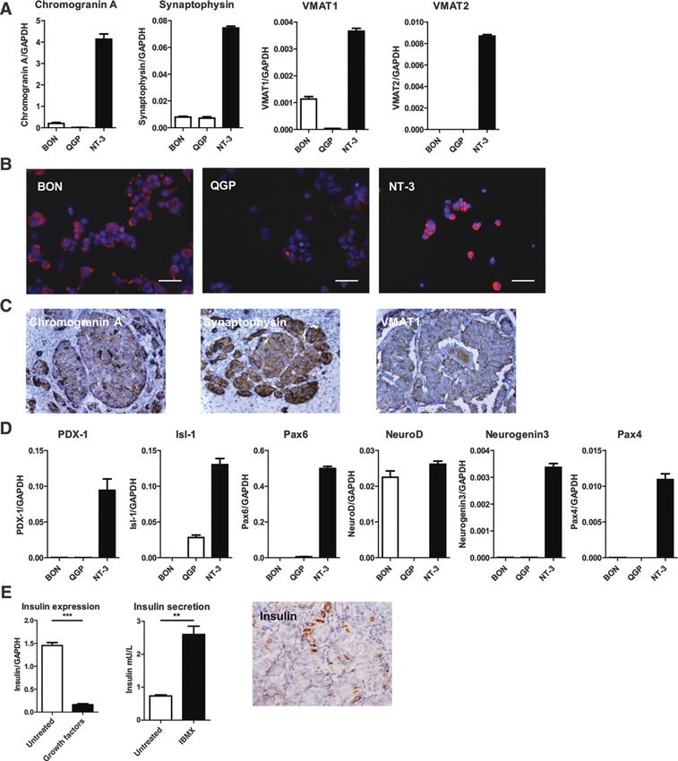
Published OnlineFirst January 12, 2018; DOI: 10.1158/1541-7786.MCR-17-0163

                                                                              Characterization of a Well-differentiated Human NET Model

octreotide radionuclide uptake is only minor (8). Furthermore,        RNA was reverse-transcribed using the High Capacity cDNA
doubling times of less than 48 hours for BON cells, are too fast      Reverse Transcription Kit (Applied Biosystems) and a T3 Thermo-
to represent a relevant model for slow growing NETs (9). In           cycler (Biometra). Prevalidated primers for real-time PCR were
xenotransplantation models, these cell lines show very rapid          purchased from Applied Biosystems (full list in Supplementary
tumor growth with early development of tumor necrosis due to          Methods). Samples in duplicate were subjected to PCR in a Step
poor vascularization (10, 11).                                        One Plus Real-Time PCR System (Applied Biosystems). Values
  Although the establishment of pancreatic neuroendocrine             were expressed as (Ct) values normalized to housekeeping gene
tumor cell lines with a well-differentiated phenotype and low         GAPDH using the 2DDCt method.
proliferation rate would be mandatory to validate new thera-
peutic targets in preclinical settings, extensive research over the   Immunocytochemistry
past 25 years has not resulted in generation of such cell lines.        NT-3, QGP-1, and BON cells were grown on collagen-coated
Accountable for this failure were undefined optimal growth             chamber slides for 48 hours supplemented with or without
conditions for NET cells as well as overgrowth by contaminat-         growth factors (EGF and FGF2) and fixed with 4% paraformal-
ing tumor fibroblasts (12). We here report successful establish-       dehyde. Mouse anti-Ki67 (Clone MIB-1, Dako) and mouse anti-
ment of a new pancreatic NET cell line from a lymph node              Chromogranin-A (Clone DAK-A3, Dako,) were used as primary
metastasis of a patient with functional insulinoma. These cells,      antibodies. Goat anti-mouse Alexa Fluor 555 (Molecular Probes
named NT-3, have now been cultured for more than 3 years              by Life Technologies) was used as secondary antibody. Coverslips
and they still display a stable well-differentiated phenotype,        were mounted using Vectashield Hard Set with DAPI (Vector
including after xenotransplantation. Cell characterization and        Laboratories). Cells were analyzed by fluorescence microscopy
proliferation analyses hold promise that these cells provide a        using a BioRevo BZ-9000 (Keyence).
hitherto unavailable clinically relevant in vitro and in vivo model
of well-differentiated neuroendocrine tumor disease.                  Western blot analysis
                                                                         Western blot analysis was performed as described previously
                                                                      (13). In brief, 20 mg of protein was separated by SDS-PAGE and
Materials and Methods                                                 transferred onto nitrocellulose membranes (Schleicher &
Cell lines                                                            Schuell). After blocking, membranes were probed with primary
   Authenticated BON and QGP-1 cells were cultured in DMEM/           and horseradish peroxidase (HRP)-conjugated secondary antibo-
Ham F12 and RPMI, respectively, supplemented with 10% FCS             dies (full antibody list in Supplementary Methods) and ensuing
and penicillin/streptomycin. Cells regularly tested negative for      bands were visualized using an ECL-kit (Pierce).
mycoplasma contamination. The cells were used within 15 pas-
sages after authentication (DMSZ).                                    ELISA
   The new cell line NT-3 was generated from a surgically                Measurement of VEGF in cell culture supernatants was per-
resected lymph node of a 33-year-old male patient with well-          formed using the R&D Systems VEGF ELISA Development Kit
differentiated NET of the pancreas. The patient had stage IV          following manufacturer's instructions. VEGF concentrations in
disease and a debulking operation had been performed for a            cell culture supernatants were normalized to total protein content
functionally active insulinoma. Histopathology confirmed insu-         of the corresponding cell culture to account for different cell
lin expression and the Ki-67 was determined to be 15%–20%.            numbers of the cell lines.
The local ethical review board approved the use of human tissue
and informed consent from the patient was obtained before             MTT assay
surgery. The study was conducted in accordance with the Dec-             The MTT assay was carried out under standardized condi-
laration of Helsinki. Tissue culture was started after mincing of     tions in 96-well plates. After incubation for 24 hours, cells
the tissue and digestion with collagenase IV (Serva). The ensuing     were treated separately with the following therapeutics: octreo-
primary mixed cell culture containing tumor and stromal cells         tide (Sandostatin, Novartis), streptozotocin (Sigma), 5-fluo-
was cultivated in RPMI medium supplemented with 10% FCS,              rouracil (5-FU; Sigma), everolimus (LC Laboratories), and
penicillin/streptomycin, HEPES, EGF (20 ng/mL), and FGF2              sunitinib (LC Laboratories). All therapeutics were replenished
(10 ng/mL; PeproTech). To eliminate fibroblasts, the primary           once after 48 hours. 120 hours after the first addition of
cultures were passaged by sequential trypsinization and culti-        therapeutics, 20 mL of MTT (5 mg/mL, Fluka Analytical, Sigma)
vation under low-adherent conditions. While fibroblasts                was added to each well and plates were incubated for 2 hours.
attached in all culture conditions, tumor cells preferentially        Using a Tecan infinite F50 spectrophotometer (Tecan), absor-
attached to collagen-coated culture plates. Passage numbering         bance was measured at 540 nm after solubilization of the
of tumor cell culture was started after achieving a pure tumor        formazan dye with DMSO. Results are displayed as mean
cell culture and successful cryopreservation. Cells from passage      absorbance relative to controls (untreated or DMSO control
15–30 have been used for the experiments.                             as indicated).
   For cell culture experiments under hypoxic conditions, cells
were incubated in a nitrogen-supplemented incubator with 0.5%         Cell counting
oxygen for up to 24 hours.                                               Fast growing BON and QGP-1 cells were seeded in 24-well-
                                                                      plates with 1  104 cells per well, and slower growing NT-3 with
Quantitative real-time PCR                                            2  104 cells per well, diluted in 500-mL medium supplemented
  Total RNA was isolated from cell cultures and tumor samples         with or without growth factors (EGF and FGF2). BON and QGP-1
using the Nucleo Spin RNA, DNA, and protein purification Kit           cells were counted on days 1, 3, 7, and 10, whereas NT-3 cells were
(Macherey-Nagel) according to the manufacturer's protocol. Total      counted on days 1, 3, 7, 14, 21, 28, 35, and 42.

www.aacrjournals.org                                                                                   Mol Cancer Res; 16(3) March 2018     497

   Downloaded from mcr.aacrjournals.org on February 3, 2021. © 2018 American Association for Cancer Research.
Published OnlineFirst January 12, 2018; DOI: 10.1158/1541-7786.MCR-17-0163

     Benten et al.

     Soft agar assay                                                        (Dako), and incubated with the following primary antibodies:
       Soft agar assay was performed as described previously (14). In       Chromogranin A, synaptophysin, and Ki-67 (all from Dako).
     brief, 1  105 NT-3 cells were plated in 60-mm Petri dishes            Positive staining was visualized using a corresponding secondary
     between layers of agar mixed with complete growth media. Plates        HRP-coupled antibody and the DAB staining system (Dako).
     were cultured up to 12 weeks with regular growth media addition.       Slides were counterstained with hematoxylin (Sigma). Staining
     Ensuing colonies were counted by light microscopy.                     of primary human tumor tissue was done as described before
                                                                            using the following primary antibodies: Chromogranin A, Synap-
     Insulin measurement                                                    tophysin, VMAT1, SSTR2A, SSTR5, Ki-67, and insulin (16).
        Human insulin content was determined using the ADVIA
     Centaur Insulin Assay (REF 02230141) and ADVIA Centaur XP              Statistical analysis
     analyzer (Siemens Healthcare). This assay is standardized to the           All graphs and statistical analysis have been performed
     first human insulin international reference preparation by the          using GraphPad Prism 4 utilizing standard two-tailed unpair-
     World Health Organization (NIBSC code 66/304). FCS did not             ed Student t test. P values
Published OnlineFirst January 12, 2018; DOI: 10.1158/1541-7786.MCR-17-0163
Characterization of a Well-differentiated Human NET Model
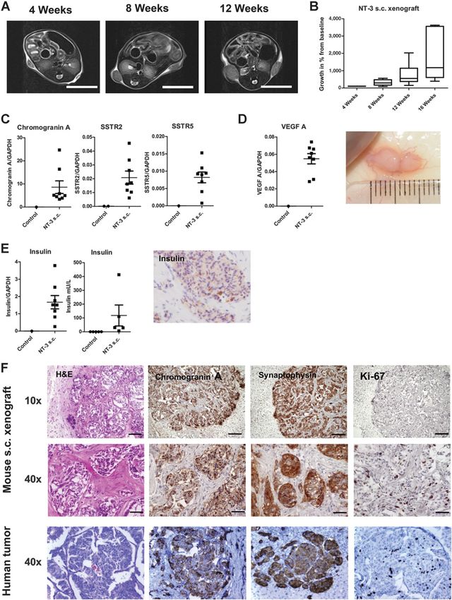
low expression in QGP-1 (Fig. 1A). Correspondingly, NT-3, and physin was expressed in all three cell lines confirming their
to a lesser extent, also BON stained positive for CgA on neuroendocrine origin and again NT-3 showed a much higher
immunofluorescence (Fig. 1B). The neuronal marker synapto- expression than BON and QGP-1 (Fig. 1A). Another prominent
Figure 1.
Neuroendocrine phenotype of NT-3. A, Expression analyses of CgA, synaptophysin, and VMAT1 in BON, QGP-1, and NT-3 cells showed a high degree of
neuroendocrine differentiation in NT-3 cells. B, Strong expression of CgA in NT-3 was confirmed by immunocytochemistry. C, Staining of the primary surgical tumor
specimen for CgA, synaptophysin, and VMAT1 corresponded to the expression profile of NT-3 cells. D, Increased expression of neuroendocrine-related transcription
factors PDX-1, neurogenin3, NeuroD, Isl-1, Pax4 and Pax6 supported the well-differentiated phenotype of NT-3 compared with BON and QGP-1. E, Expression and
IBMX-stimulated release of insulin in NT-3 confirmed functionality. Correspondingly, IHC of the primary human tumor showed insulin expression. Scale bar, 50 mm.
www.aacrjournals.org Mol Cancer Res; 16(3) March 2018 499
Downloaded from mcr.aacrjournals.org on February 3, 2021. © 2018 American Association for Cancer Research.Published OnlineFirst January 12, 2018; DOI: 10.1158/1541-7786.MCR-17-0163
Benten et al.
feature of neuroendocrine cells is their uptake and processing NT-3 expresses and releases insulin, corresponding to the
of monoamines. We detected expression of vesicular mono- functional insulinoma of the patient (Fig. 1E). Insulin expres-
amine transporter (VMAT) 1 and VMAT2 in NT-3, while QGP-1 sion in the original patients' tumor was confirmed by IHC
expressed neither of them and BON only weakly expressed showing moderate staining intensity with some strongly
VMAT1 (Fig. 1A). In addition, all cell lines expressed enzymes stained tumor cells. Interestingly, insulin expression by NT-3
required for serotonin biosynthesis (e.g., tryptophan hydroxy- was modifiable by the omission or addition of the growth
lase 1 and dopa-decarboxylase) to various extent (Supplemen- factors EGF/FGF-2.
tary Fig. S2C). Reassuringly, the original NET tumor of the The finding of growth factor–dependent insulin expression
patient was also positive for CgA, synaptophysin, and VMAT1 prompted us to investigate this effect also for the abovemen-
(Fig. 1C). tioned neuroendocrine markers, and we detected profoundly
Neuroendocrine cell differentiation in the pancreas during reduced expression upon growth factor supplementation for
embryonal development is promoted by multiple transcrip- CgA, synaptophysin, and all neuroendocrine-related transcrip-
tion factors, most of which are still expressed and functionally tion factors (Fig. 2A and data not shown). Correspondingly,
relevant in adult islet cells (17). We observed high expression NT-3 cells showed a reduction in E-cadherin and an increase
of PDX-1, Isl-1, Neurogenin3, NeuroD, Pax4, and Pax6 in in vimentin expression when treated with growth factors,
NT-3 cells, whereas BON only express NeuroD and QGP-1 suggesting epithelial-to-mesenchymal shifting of their pheno-
only expresses Isl-1 (Fig. 1D). As a marker of functionality, type (Fig. 2B). This was, in part, reflected by the appearance of a
Figure 2.
Plasticity of NT-3 cells. A,
Supplementation of the cell culture
media with growth factors EGF and FGF2
decreased expression of neuroendocrine
markers in NT-3 cells. B, This was
accompanied by downregulation of
E-Cadherin and upregulation of
vimentin. C, These changes are reflected
by appearance of a spindle-shaped
subpopulation (arrows) in growth
factor–supplemented cell cultures.
, P < 0.005; , P < 0.001. Scale
bar, 100 mm.
500 Mol Cancer Res; 16(3) March 2018 Molecular Cancer Research
Downloaded from mcr.aacrjournals.org on February 3, 2021. © 2018 American Association for Cancer Research.Published OnlineFirst January 12, 2018; DOI: 10.1158/1541-7786.MCR-17-0163
Characterization of a Well-differentiated Human NET Model
small spindle-shaped subpopulation in NT-3 cultures treated Clinical tumor grading of NET is assessed by IHC staining
with growth factors (Fig. 2C), which completely disappeared for the nuclear proliferation factor Ki-67. BON and QGP-1 have
after subsequent withdrawal of growth factors. In contrast to a Ki-67 labeling index of 80.0% 3.3% and 82.6% 1.0%
this high plasticity of NT-3, QGP-1 showed only minimal respectively, corresponding to highly proliferative G3 carcinomas.
changes upon growth factor treatment, whereas BON cell In contrast, nuclear Ki-67 in NT-3 is only 2.0% 0.2%, corre-
phenotype was completely unaffected by addition or omission sponding to a slowly proliferative G1–G2 tumor. Addition of
of growth factors (data not shown). growth factors slightly increased Ki-67 index in BON by 7%,
compared with a profound increase in NT-3 by 325% to an index
Cell proliferation of 14.6% 1.1% (Fig. 3C and D), now similar to the patients
Well-differentiated NETs are characterized by a low pro- original tumor and xenograft tumors (see below). Cellular
liferation rate and slow tumor growth. Cultured BON and anchorage-independent growth, which represents a sign of malig-
QGP-1 cells had a doubling time of less than two days (BON nancy, was confirmed in NT-3 by soft agar assay. Out of 1 105
1.5 0.4 days, QGP-1 1.6 0.3 days), which was unaffected cells plated in soft agar, 2,200 formed cell clusters of 10–20 cells
by addition of growth factors (Fig. 3A; Supplementary with a latency of 12 weeks, corresponding to a colony-forming
Fig. S3A). In contrast, NT-3 grew slowly with a doubling frequency of approximately 2% (Supplementary Fig. S3B).
time of 10.9 0.7 days (Fig. 3A). Their growth was depen-
dent on the addition of growth factors, as we did not observe Therapeutic targets
growth of NT-3 cells plated at low cell density without growth SSTRs are crucial therapeutic targets for somatostatin ana-
factors (Fig. 3B). logues and peptide radio receptor therapy in NET treatment.
Figure 3.
Proliferation. A, Growth curves of
tumor cells cultured with growth
factor supplementation show a fast
growth rate of BON and QGP-1, while
NT-3 exhibit a very slow growth over a
period of 7 weeks. B, NT-3 plated at
low density without growth factor
addition did not proliferate. C and D,
Staining for Ki-67 was used to
calculate proliferative indices in BON,
QGP-1, and NT-3 cells with or without
growth factor supplementation. Scale
bar, 50 mm.
www.aacrjournals.org Mol Cancer Res; 16(3) March 2018 501
Downloaded from mcr.aacrjournals.org on February 3, 2021. © 2018 American Association for Cancer Research.Published OnlineFirst January 12, 2018; DOI: 10.1158/1541-7786.MCR-17-0163
Benten et al.
NT-3 shows high mRNA expression of SSTR1, SSTR2, SSTR3, of these cell lines expresses SSTR4. Western blotting confirm-
and SSTR5, among those, the expression of SSTR-1 is partic- ed high expression of SSTRs and showed that the most clin-
ularly strong. In contrast, BON and QGP-1 express these ically relevant SSTR2 is exclusively expressed in NT-3, while
receptors either only weakly or not at all (Fig. 4A). None SSTR3 and SSTR5 are also expressed in BON (Fig. 4B).
Figure 4.
Therapeutic targets in NT-3. A and B, Western blot and qPCR analysis of SSTR subtypes revealed higher expression of SSTR1 and SSTR2 in NT-3 compared
with BON and QGP-1. C, Expression of VEGF A, VEGF B and angiopoetin mRNA is at least 3-fold higher in NT-3 cells compared to BON and QGP-1.
The expression of VEGF A is further increased after 6 and 24 hours in low oxygen environment (D) and VEGF release into the supernatant after 24h of
hypoxia was confirmed by ELISA (E). Ensuing hypoxic conditions in tumor cells were verified by increase in carboanhydrase IX (CAIX) expression after
6 and 24 hours by qPCR (F) and after 24-hour by Western blot analysis (G).
502 Mol Cancer Res; 16(3) March 2018 Molecular Cancer Research
Downloaded from mcr.aacrjournals.org on February 3, 2021. © 2018 American Association for Cancer Research.Published OnlineFirst January 12, 2018; DOI: 10.1158/1541-7786.MCR-17-0163
Characterization of a Well-differentiated Human NET Model
Correspondingly, the patient's primary tumor tissue showed studying upcoming novel therapies, we aimed to establish the
positive staining for SSTR2A and SSTR5 on IHC (Supplemen- efficacy of current treatments compared with QGP-1 cells. Upon
tary Fig. S4A and S4B). treatment with 100 nmol/L octreotide, we observed a significant
Another therapeutic target in clinical NET treatment are (34.8%, P < 0.001) reduction in NT-3 cell numbers after a
angiogenic factors. In standard culture conditions, the mRNA treatment for 5 days (Fig. 5A). In contrast, the low SSTR-expres-
expression of VEGF A and B in NT-3 is more than 3-fold sing QGP-1 cells did not show significant reduction of cell
higher than in BON and QGP-1 (Fig. 4C). In addition, NT-3 numbers (4.6%, P ¼ 0.22). Treatment of cell cultures with
cells also express the proangiogenic growth factor angiopoietin 2 50 nmol/L everolimus resulted in a 31.5% and 26.2% reduction
(ANG2) to a higher extent than BON and QGP-1 cells (Fig. 4C). of NT-3 and QGP cell numbers after 5 days (P < 0.001),
Upon cell transfer into a hypoxic environment (0.5% oxygen), respectively (Fig. 5B). Increasing the dose up to 500 nmol/L
we observed a further increase in VEGF (Fig. 4D), but not in did not further increase treatment response (data not shown).
ANG2 expression (not shown). ELISA confirmed secretion of Treatment with sunitinib in doses up to 500 nmol/L did not
VEGF into cell culture supernatants under hypoxic conditions decrease viability or proliferation in either cell line (data not
(Fig. 4E). Establishment of a relevant hypoxic environment was shown). To evaluate treatment response to standard cytotoxic
confirmed by upregulation of carboanhydrase IX, a prototypic chemotherapy, we analyzed the dose-dependent effect on cell
hypoxia-induced gene target (Fig. 4F–G). viability after incubation with streptozotocin and 5-FU. After
These results demonstrate that the new NT-3 cell line should 5 days, streptozotocin treatment at 1 mmol/L resulted in a
meet the criteria for a suitable model to study antiproliferative viability decrease of 43.3% 1.1% in NT-3 (P < 0.001)
and antiangiogenic treatments. compared with a lesser effect of 18.9% 7.5% (P ¼ 0.10)
in QGP-1 (Fig. 5C). Increasing the dose to 10 mmol/L substan-
Treatment response tiated the difference in susceptibility toward streptozotocin.
Current guidelines for the treatment of advanced pancreatic In contrast, both cell lines responded well to 5-FU; treatment
NET recommend somatostatin analogues, chemotherapy with with 10 mmol/L for 5 days resulted in decreased viability of
streptozotocin/5-FU, and the targeted therapies everolimus or 30.1% 8.8% (P ¼ 0,014) in NT-3 compared with 49.7%
sunitinib. To evaluate the potential of our new NT-3 cells for 6.0% in QGP-1 (P ¼ 0.0002; Fig. 5D).
Figure 5.
Treatment response in NT-3
compared with QGP-1. Treatment
efficacy of various treatments was
assessed in NT-3 and QGP-1 cells after
5 days of treatment by MTT assay.
Octreotide at 100 nmol/L (A)
compromised NT-3 viability, whereas
everolimus at 50 nmol/L was equally
effective in NT-3 and QGP-1 (B).
Addition of genotoxic
chemotherapeutics STZ (C) and 5-FU
(D) showed a stronger effect for
streptozotocin in NT-3 than QGP-1
cells at all tested doses, but similar
responses for 5-FU in both cell lines.
www.aacrjournals.org Mol Cancer Res; 16(3) March 2018 503
Downloaded from mcr.aacrjournals.org on February 3, 2021. © 2018 American Association for Cancer Research.Published OnlineFirst January 12, 2018; DOI: 10.1158/1541-7786.MCR-17-0163
Benten et al.
Xenograft animal model or a very benign course without frequent development of dis-
On the basis of our previous experience with BON and seminated tumor disease, such as b-cell–specific MEN1 knockout
QGP-1 xenografts (11), we transplanted 2 106 NT-3 tumor mice (21). Likewise, the reported xenograft models of QGP-1 and
cells into the flanks of NOD/SCID mice and we achieved a BON cells show extremely rapid tumor growth with development
tumor take rate of 94% (15/16). NT-3 tumor growth in vivo of large necrotic areas and severe tumor disease in mice within a
was very slow, with the first palpable nodes detected approx- few weeks (10, 11). Hence, there is great need for a suitable and
imately 6 weeks after cell transplantation. Because of the small clinically relevant in vivo tumor model to assess future therapies
tumor size during the first 12 weeks, traditional caliper mea- in NET disease. The here reported novel pancreatic neuroendo-
surements were unreliable and we employed serial MRI of sub- crine tumor cell line NT-3 closely resembles the patients original
cutaneous tumor nodules to monitor their growth (Fig. 6A). The tumor: it has a well-differentiated phenotype, is functionally
tumors were akin to human neuroendocrine tumors on MRI active, and has a slow growth rate in vitro and in vivo, thereby
imaging, with a strong enhancement in T2-weighted sequences recapitulating the cardinal features of neuroendocrine tumors
(Fig. 6A). Median NT-3 tumor growth rate (as measured by and providing a hitherto unavailable relevant preclinical neuro-
tumor volume) was þ139% 13% per 4 weeks (Fig. 6B). The endocrine tumor model.
proliferative index corresponded well to the proliferative Currently, only few NET cell lines are available. Of these, the
index of the original tumor (15%–20% Ki-67 labeling rate; neuroendocrine cell lines derived from small intestinal NETs
focally up to 25%). mRNA expression profiles of neuroendo- (KRJ-1, GOT1, P-STS) probably display a well-differentiated phe-
crine markers in subcutaneous tumors were in line with expres- notype. Because of their limited availability, a thorough charac-
sion profiles of NT-3 cells in vitro (Fig. 6C). In contrast to our terization of their biology and validation as a relevant NET model
previous xenograft experiments with BON and QGP-1 cells in vivo is still pending. From a clinical point of view, treatment of
(11), we did not detect any necrotic areas within the tumor, pancreatic NET is more challenging and associated with a worse
while macroscopic appearance of NT-3 tumors showed high prognosis than small intestinal NETs. The two available pancreatic
vascularization (Fig. 6D). We detected high mRNA expression neuroendocrine cell lines BON and QGP-1 have so far been
of human CgA, synaptophysin, and the somatostatin receptors widely used as a bona fide NET model. Documented fast growth
SSTR1, 2, 3, and 5. In addition, serum analyses of tumor-bearing rates of these tumor cells have put their usefulness as NET models
mice after 16 weeks demonstrated detectable levels of human into question. Our detailed characterization of BON and QGP-1
insulin (median 118 mU/L, range 6–412 mU/L), and insulin further substantiates such concerns as expression of neuroendo-
expression in xenograft tumor cells was verified by IHC crine markers in these cell lines was low. Given that both cell lines
(Fig. 6E). These results confirmed the functional well-differen- have been in use for more than 25 years, it appears likely that the
tiated phenotype of the tumor xenograft. cells have acquired a more malignant phenotype in culture.
Histology of dissected xenograft tumors at the end of animal Although the currently detected doubling times of 36 and 38
experiments recapitulated the insular and nesting growth pat- hours for BON and QGP-1 cells, respectively, are higher than the
tern of the original patient's tumor with highly abundant originally reported doubling times of 60 hours (BON) (22) and
stroma (Fig. 6F). Positive staining for CgA and synaptophysin 84 hours (QGP-1; ref. 23), these cells proliferate much faster, even
confirmed the neuroendocrine identity of xenografts. Also, IHC in their early passage numbers, compared with NT-3 with a
for human cytokeratins revealed CK18 and CK19 expression in doubling time of more than 240 hours under growth factor–
both the original tumor and in NT-3 xenografts (Supplemen- stimulated conditions. The recent discovery of mutations in RAS
tary Fig. S5). Although not being a focus of this study, initial and TP53 genes in BON and QGP-1 added to these concerns as
pilot experiments using intraportal injection of NT-3 cells led to well-differentiated NET rarely harbor such mutations (24–26). In
development of small liver tumors in one out of three trans- contrast, our new NT-3 cell line does not harbor mutations in RAS
planted mice. These were also rich in stroma tissue and were or TP53 genes thereby resembling the nonmutated status of well-
positive for CgA, indicating that our new cell line will also be differentiated NET for these oncogenes. It is open to speculation,
suitable for future experiments on orthotopic NET liver meta- whether potentially effective new therapeutics for NET patients
stases (Supplementary Fig. S6). have been dismissed or development has been prematurely
stopped due to failure in BON and QGP-1 cells.
We found a missense mutation in the MEN1 gene in NT-3 cells.
Discussion This mutation is listed as a known SNP (rs2959656) with a minor
In vitro and in vivo evaluation of future therapies in preclinical allele frequency between 6% and 16% in different populations.
models remains the mainstay of successful development of novel Although the SNP has been described in sporadic insulinoma,
treatments ultimately improving patient care in cancer patients parathyroid adenoma, and hemangioblastoma (27), the patho-
(18, 19). The use of cell lines has recently been challenged by the genic role of this variant is controversial due to the high frequency
announcement of the National Cancer Institute (NCI) to aban- in the general population. Targeted sequence analysis of "classi-
don their NCI-60 panel of cell lines and to encourage the use of cal" pNET-associated genes, that is, VHL, MEN1, TSC1, TSC2,
patient-derived xenografts. Nevertheless the NCI still recom- DAXX, and ATRX did not reveal any further mutations, which is
mends the use of well-characterized novel primary cell cultures in line with findings from Jiao and colleagues reporting muta-
to explore future therapeutic options, and our novel cell line and tions in either of these genes in only 68% of patients with
the corresponding xenograft model fulfill these criteria. sporadic pancreatic NETs (26). The lack of such mutations in
So far, testing of potentially new targets in neuroendocrine NT-3 offers exciting new opportunities for future analyses as
tumor disease has been impaired by the lack of suitable preclinical targeted knockdown will help to establish the role of either of
models. The available genetic NET models display either a rapid the above-mentioned genes with regard to tumor progression and
and short-term fatal disease course, that is, Rip1-Tag2 mice (20), therapeutic response of pNETs. Furthermore, detailed genetic and
504 Mol Cancer Res; 16(3) March 2018 Molecular Cancer Research
Downloaded from mcr.aacrjournals.org on February 3, 2021. © 2018 American Association for Cancer Research.Published OnlineFirst January 12, 2018; DOI: 10.1158/1541-7786.MCR-17-0163
Characterization of a Well-differentiated Human NET Model
Figure 6.
Xenograft tumor model. Subcutaneous tumors in NT-3 transplanted NOD/SCID mice were first detected by MRI (A). Growth curves were calculated from
serial imaging of tumors over 16 weeks (B). Expression profiles of tumor xenografts resembled the well-differentiated phenotype of NT-3 cells in vitro
(C). Normal murine liver tissue of control mice (no xenograft) served as negative controls. Xenograft tumors were highly vascularized and
correspondingly expressed high levels of human VEGF A (D). Analysis of human insulin content in mouse serum 16 weeks after transplantation of
tumor cells confirmed the functionally active phenotype of NT-3 in vivo. Correspondingly, IHC showed insulin expression in xenografts (E). Histology of
dissected xenograft tumors confirmed neuroendocrine identity and displayed a morphology and proliferation rate akin to the original human tumor (F).
Scale bar, 1 cm for MRI images and 200 mm (10) and 50 mm (40) for microscopic images.
www.aacrjournals.org Mol Cancer Res; 16(3) March 2018 505
Downloaded from mcr.aacrjournals.org on February 3, 2021. © 2018 American Association for Cancer Research.Published OnlineFirst January 12, 2018; DOI: 10.1158/1541-7786.MCR-17-0163
Benten et al.
proteomic analyses could identify yet unknown mechanism for tigate combinations of radionuclide SSTR-targeted therapies
the development and progression of pNETs. and chemotherapies in NET (42). Making well-differentiated
One of the main obstacles in obtaining primary NET cultures patient-derived cells with relevant SSTR expression, that is, our
is the unsolved question of optimal growth conditions for these NT-3 model, widely accessible to translational researchers will
cells (12). We here report an essential role for the supplemen- aid such attempts in providing a platform to optimize timing
tation of growth factors EGF and FGF-2 to establish prolifer- and dosing of therapies. Assessing the effect of cotherapeutics on
ating cell cultures. Earlier studies in BON and QGP-1 cells also SSTR expression in NT-3 will help choosing effective drug
found a growth stimulatory role for EGF, FGF-2, TGF-a, and combinations. As a substantial fraction of NET tumors, espe-
IGF-1 (28–30). Recently, a c-MET stimulating antibody has cially pancreatic NET, do not express sufficient levels of SSTR2
been identified to promote primary NET cell growth in vivo and SSTR5, these patients are currently not amenable to effective
(31). As expression of these growth factors apart from IGF-1 treatments. As NET cells do not only express SSTR, but also
and TGF-a is generally low in NET cells, the tumor microen- many other neuroendocrine-specific cell surface receptors (e.g.,
vironment most likely contributes these growth stimulatory for incretin hormones like GIP and GLP-1), searching for and
factors (28, 32, 33). Indeed, our NT-3 xenograft model of NT-3 evaluating such receptors as therapeutic targets might stimulate
supports this hypothesis as NT-3 cells, surrounded in vivo by development of novel therapies (43).
abundant stroma, show a proliferation rate comparable with In summary, we have successfully established a well-differen-
the in vitro growth rate with growth factor stimulation. For tiated and slow growing pancreatic NET cell line. NT-3 cells and
further exploration of such interesting new aspects in NET the corresponding xenograft animal model will overcome current
tumor biology, we have conserved tumor-associated fibroblasts limitations in developing and testing novel therapies for this
from the original patients tumor, as recently recommended difficult to treat disease.
by the NCI for novel primary tumor cell cultures, to study the
interaction of NET cells with their microenvironment. Disclosure of Potential Conflicts of Interest
The documented insulin expression and secretion of NT-3 D. Benten reports receiving commercial research support as research
cells warrants evaluation as a potential model for human funding. J. Schrader reports receiving a commercial research grant from Novartis
pancreatic beta cells. Until now, no such human model exists and has received speakers bureau honoraria from Novartis and IPSEN. No
potential conflicts of interest were disclosed by the other authors.
and most basic diabetes research has been performed in a rat
insulinoma cell line (INS1E), with obvious limitations such as
differences in protein expression and functional status between Authors' Contributions
Conception and design: D. Benten, J.R. Izbicki, A.W. Lohse, J. Schrader
human and rodent islet cells (34, 35). The only human cell line
Development of methodology: D. Benten, D. Perez, J. Schrader
derived from an insulinoma (called CM) has already lost Acquisition of data (provided animals, acquired and managed patients,
insulin expression and secretion during in vitro culture (36). provided facilities, etc.): D. Benten, Y. Behrang, L. Unrau, V. Weissmann,
Recently, a genetically engineered human beta cell line has G. Wolters-Eisfeld, S. Burdak-Rothkamm, F.R. Stahl, M. Anlauf, P. Grabowski,
been presented, but still needs to prove easy access, handling, M. M€ obs, J. Dieckhoff, B. Sipos, C. Eggers, D. Perez, M. Bockhorn, J. Schrader
and a stable phenotype for basic research (37, 38). Analysis and interpretation of data (e.g., statistical analysis, biostatistics,
computational analysis): D. Benten, Y. Behrang, L. Unrau, G. Wolters-Eisfeld,
Well-differentiated pancreatic NETs are currently treated with
S. Burdak-Rothkamm, M. Anlauf, B. Sipos, M. Bockhorn, A.W. Lohse, J. Schrader
somatostatin analogues or STZ/5-FU, and these substances are Writing, review, and/or revision of the manuscript: D. Benten, Y. Behrang,
also effective for growth reduction in our NT-3 cells in vitro. M. M€ obs, B. Sipos, M. Bockhorn, A.W. Lohse, J. Schrader
Indeed, it has been shown that STZ is particularly potent in Administrative, technical, or material support (i.e., reporting or organizing
SSTR-positive NET (39), pointing toward an increased suscepti- data, constructing databases): J. Dieckhoff, M. Fahl, M. Bockhorn, J.R. Izbicki,
bility in tumors with a well-preserved neuroendocrine phenotype A.W. Lohse
Study supervision: D. Benten, J.R. Izbicki, A.W. Lohse, J. Schrader
due to a hitherto undiscovered mechanism. In contrast, 5-FU,
which is a general cytotoxic chemotherapeutic agent and therefore
also active against dedifferentiated tumors, is also effective in Acknowledgments
This work was funded by Forschungsf€orderungsfond Medizin of Univer-
QGP-1. Everolimus was similarly effective in both, NT-3 and
sity Medical Center Hamburg-Eppendorf (to J. Schrader) and a research grant
QGP-1, as the mTOR pathway is activated in well- and less from the Theranostic Research Network Bad Berka (to J. Schrader). D. Benten
differentiated tumors (40). and L. Unrau received grant support from Deutsche Forschungsgemeinschaft
Two of the most effective treatments for patients with NET (SFB 841, project C7). G. Wolters-Eisfeld received grant support from
(e.g., somatostatin analogues and peptide radionuclide therapy) Deutsche Forschungsgemeinschaft (WO 1967/2-1).
are dependent on expression of SSTRs, in particular, SSTR2 and
SSTR5, on the tumor cell surface (41). Until now, improvements The costs of publication of this article were defrayed in part by the
payment of page charges. This article must therefore be hereby marked
in SSTR targeting have been impaired by the lack of suitable and advertisement in accordance with 18 U.S.C. Section 1734 solely to indicate
widely available preclinical models. Our novel NT-3–derived this fact.
animal model has the potential to overcome these limitations as
we observed high expression of SSTRs in vitro and in xenograft Received March 26, 2017; revised August 28, 2017; accepted December 20,
tumors. Recently, clinical pilot studies were initiated to inves- 2017; published OnlineFirst January 12, 2018.
References
1. Modlin IM, Oberg K, Chung DC, Jensen RT, de Herder WW, Thakker RV, 2. Clewemar Antonodimitrakis P, Sundin A, Wassberg C, Granberg
et al. Gastroenteropancreatic neuroendocrine tumours. Lancet Oncol D, Skogseid B, Eriksson B. Streptozocin and 5-fluorouracil for
2008;9:61–72. the treatment of pancreatic neuroendocrine tumors: efficacy,
506 Mol Cancer Res; 16(3) March 2018 Molecular Cancer Research
Downloaded from mcr.aacrjournals.org on February 3, 2021. © 2018 American Association for Cancer Research.Published OnlineFirst January 12, 2018; DOI: 10.1158/1541-7786.MCR-17-0163
Characterization of a Well-differentiated Human NET Model
prognostic factors and toxicity. Neuroendocrinology 2016;103: 23. Kaku M, Nishiyama T, Yagawa K, Abe M. Establishment of a carcinoem-
345–53. bryonic antigen-producing cell line from human pancreatic carcinoma.
3. Yao JC, Shah MH, Ito T, Bohas CL, Wolin EM, Van Cutsem E, et al. RAD001 Gan 1980;71:596–601.
in advanced neuroendocrine tumors, third trial (RADIANT-3) study group. 24. Boora GK, Kanwar R, Kulkarni AA, Pleticha J, Ames M, Schroth G, et al.
Everolimus for advanced pancreatic neuroendocrine tumors. N Engl J Med Exome-level comparison of primary well-differentiated neuroendocrine
2011;364:514–23. tumors and their cell lines. Cancer Genet 2015;208:374–81.
4. Raymond E, Dahan L, Raoul JL, Bang YJ, Borbath I, Lombard-Bohas C, et al. 25. Vandamme T, Peeters M, Dogan F, Pauwels P, Van Assche E, Beyens M, et al.
Sunitinib malate for the treatment of pancreatic neuroendocrine tumors. Whole-exome characterization of pancreatic neuroendocrine tumor cell
N Engl J Med 2011;364:501–13. lines BON-1 and QGP-1. J Mol Endocrinol 2015;54:137–47.
5. Sabet A, Biersack HJ, Ezziddin S. Advances in peptide receptor radionuclide 26. Jiao Y, Shi C, Edil BH, de Wilde RF, Klimstra DS, Maitra A, et al. DAXX/
therapy. Semin Nucl Med 2016;46:40–6. ATRX, MEN1, and mTOR pathway genes are frequently altered in pancre-
6. Grozinsky-Glasberg S, Shimon I, Rubinfeld H. The role of cell lines atic neuroendocrine tumors. Science 2011;331:1199–203.
in the study of neuroendocrine tumors. Neuroendocrinology 2012; 27. Jyotsna VP, Malik E, Birla S, Sharma A. Novel MEN 1 gene findings in rare
96:173–87. sporadic insulinoma–a case control study. BMC Endocr Disord 2015;
7. Babu V, Paul N, Yu R. Animal models and cell lines of pancreatic neuro- 15:44.
endocrine tumors. Pancreas 2013;42:912–23. 28. Townsend CM Jr, Ishizuka J, Thompson JC. Studies of growth regulation in
8. Taelman V, Radojewski P, Marincek N, Ben-Shlomo A, Grotzky A, Olariu C, a neuroendocrine cell line. Acta Oncol 1993;32:125–30.
et al. Up-regulation of key molecules for targeted imaging and therapy. 29. Siddique ZL, Drozdov I, Floch J, Gustafsson BI, Stunes K, Pfragner R, et al.
J Nucl Med 2016;57:1805–1810. KRJ-I and BON cell lines: defining an appropriate enterochromaffin cell
9. Leu FP, Nandi M, Niu C. The effect of transforming growth factor beta neuroendocrine tumor model. Neuroendocrinology 2009;89:458–70.
on human neuroendocrine tumor BON cell proliferation and differen- 30. Di Florio A, Sancho V, Moreno P, Delle Fave G, Jensen RT. Gastrointestinal
tiation is mediated through somatostatin signaling. Mol Cancer Res hormones stimulate growth of Foregut Neuroendocrine Tumors by trans-
2008;6:1029–42. activating the EGF receptor. Biochim Biophys Acta 2013;1833:573–82.
10. Evers BM, Hurlbut SC, Tyring SK, Townsend CM Jr, Uchida T, Thompson 31. Krampitz GW, George BM, Willingham SB, Volkmer JP, Weiskopf K,
JC. Novel therapy for the treatment of human carcinoid. Ann Surg Jahchan N, et al. Identification of tumorigenic cells and therapeutic targets
1991;213:411–6. in pancreatic neuroendocrine tumors. Proc Natl Acad Sci U S A 2016;
11. Fraedrich K, Schrader J, Ittrich H, Keller G, Gontarewicz A, Matzat V, 113:4464–9.
et al. Targeting aurora kinases with danusertib (PHA-739358) inhibits 32. Chaudhry A, Funa K, Oberg K. Expression of growth factor peptides and
growth of liver metastases from gastroenteropancreatic neuroendocrine their receptors in neuroendocrine tumors of the digestive system. Acta
tumors in an orthotopic xenograft model. Clin Cancer Res 2012;18: Oncol 1993;32:107–14.
4621–32. 33. Briest F, Grabowski P. PI3K-AKT-mTOR-signaling and beyond: the com-
12. Lundqvist M, Oberg K. In vitro culture of neuroendocrine tumors of the plex network in gastroenteropancreatic neuroendocrine neoplasms.
pancreas and gut. Acta Oncol 1989;28:335–9. Theranostics 2014;4:336–65.
13. Schrader J, Gordon-Walker TT, Aucott RL, van Deemter M, Quaas A, Walsh 34. MacDonald MJ, Longacre MJ, Stoker SW, Kendrick M, Thonpho A, et al.
S, et al. Matrix stiffness modulates proliferation, chemotherapeutic Differences between human and rodent pancreatic islets: low pyruvate
response, and dormancy in hepatocellular carcinoma cells. Hepatology carboxylase, atp citrate lyase, and pyruvate carboxylation and high glucose-
2011;53:1192–205. stimulated acetoacetate in human pancreatic islets. J Biol Chem 2011;286:
14. Wege H, Le HT, Chui MS, Liu L, Wu J, Giri R, et al. Telomerase reconstitution 18383–96.
immortalizes human fetal hepatocytes without disrupting their differen- 35. Martens GA. Species-related differences in the proteome of rat and human
tiation potential. Gastroenterology 2003;124:432–44. pancreatic beta cells. J Diabetes Res 2015;2015:549818.
15. Benten D, Follenzi A, Bhargava KK, Kumaran V, Palestro CJ, Gupta S. 36. Gragnoli C. The CM cell line derived from liver metastasis of malignant
Hepatic targeting of transplanted liver sinusoidal endothelial cells in intact human insulinoma is not a valid beta cell model for in vitro studies.
mice. Hepatology 2005;42:140–8. J Cell Physiol 2008;216:569–70.
16. Krieg A, Mersch S, Boeck I, Dizdar L, Weihe E, Hilal Z, et al. New 37. Scharfmann R, Pechberty S, Hazhouz Y, von B€ ulow M, Bricout-Neveu E,
model for gastroenteropancreatic large-cell neuroendocrine carcino- Grenier-Godard M, et al. Development of a conditionally immortalized
ma: establishment of two clinically relevant cell lines. PLoS One 2014; human pancreatic b cell line. J Clin Invest 2014;124:2087–98.
9:e88713. 38. Benazra M, Lecomte MJ, Colace C, M€ uller A, Machado C, Pechberty S, et al.
17. van der Meulen T, Huising MO. Role of transcription factors in the A human beta cell line with drug inducible excision of immortalizing
transdifferentiation of pancreatic islet cells. J Mol Endocrinol 2015;54: transgenes. Mol Metab 2015;4:916–25.
R103–17. 39. Krug S, Boch M, Daniel H, Nimphius W, M€ uller D, Michl P, et al.
18. Barretina J, Caponigro G, Stransky N, Venkatesan K, Margolin AA, Kim S, Streptozocin-based chemotherapy in patients with advanced neuroendo-
et al. The Cancer Cell Line Encyclopedia enables predictive modelling of crine neoplasms–predictive and prognostic markers for treatment stratifi-
anticancer drug sensitivity. Nature 2012;483:603–7. cation. PLoS One 2015;10:e0143822.
19. Day CP, Merlino G, Van Dyke T. Preclinical mouse cancer models: a maze 40. Shida T, Kishimoto T, Furuya M, Nikaido T, Koda K, Takano S, et al.
of opportunities and challenges. Cell 2015;163:39–53. Expression of an activated mammalian target of rapamycin (mTOR)
20. Fendrich V, Lopez CL, Manoharan J, Maschuw K, Wichmann S, Baier in gastroenteropancreatic neuroendocrine tumors. Cancer Chemother
A, et al. Enalapril and ASS inhibit tumor growth in a transgenic Pharmacol 2010;65:889–93.
mouse model of islet cell tumors. Endocr Relat Cancer 2014;21: 41. Oberg KE, Reubi JC, Kwekkeboom DJ, Krenning EP. Role of somatostatins
813–24. in gastroenteropancreatic neuroendocrine tumor development and ther-
21. Jiang X, Cao Y, Li F, Su Y, Li Y, Peng Y, et al. Targeting b-catenin signaling for apy. Gastroenterology 2010;139:742–53.
therapeutic intervention in MEN1-deficient pancreatic neuroendocrine 42. Claringbold PG, Turner JH. Pancreatic neuroendocrine tumor control:
tumours. Nat Commun 2014;5:5809. durable objective response to combination 177Lu-octreotate-capecita-
22. Parekh D, Ishizuka J, Townsend CM Jr, Haber BE, Beauchamp RD, bine-temozolomide radiopeptide chemotherapy. Neuroendocrinology
Rajaraman S, et al. Differential effects of sodium butyrate and hexa- 2016;103:432–9.
methylene bisacetamide on growth and secretion of cultured human 43. Reubi JC, Waser B. Triple-peptide receptor targeting in vitro allows detec-
endocrine tumor cells. Arch Surg 1991;126:467–72. tion of all tested gut and bronchial NETs. J Nucl Med 2015;56:613–5.
www.aacrjournals.org Mol Cancer Res; 16(3) March 2018 507
Downloaded from mcr.aacrjournals.org on February 3, 2021. © 2018 American Association for Cancer Research.Published OnlineFirst January 12, 2018; DOI: 10.1158/1541-7786.MCR-17-0163
Establishment of the First Well-differentiated Human Pancreatic
Neuroendocrine Tumor Model
Daniel Benten, Yasmin Behrang, Ludmilla Unrau, et al.
Mol Cancer Res 2018;16:496-507. Published OnlineFirst January 12, 2018.
Updated version Access the most recent version of this article at:
doi:10.1158/1541-7786.MCR-17-0163
Supplementary Access the most recent supplemental material at:
Material http://mcr.aacrjournals.org/content/suppl/2018/01/12/1541-7786.MCR-17-0163.DC1
Cited articles This article cites 43 articles, 10 of which you can access for free at:
http://mcr.aacrjournals.org/content/16/3/496.full#ref-list-1
Citing articles This article has been cited by 2 HighWire-hosted articles. Access the articles at:
http://mcr.aacrjournals.org/content/16/3/496.full#related-urls
E-mail alerts Sign up to receive free email-alerts related to this article or journal.
Reprints and To order reprints of this article or to subscribe to the journal, contact the AACR Publications Department at
Subscriptions pubs@aacr.org.
Permissions To request permission to re-use all or part of this article, use this link
http://mcr.aacrjournals.org/content/16/3/496.
Click on "Request Permissions" which will take you to the Copyright Clearance Center's (CCC)
Rightslink site.
Downloaded from mcr.aacrjournals.org on February 3, 2021. © 2018 American Association for Cancer Research.You can also read