Bone marrow niche ATP levels determine leukemia-initiating cell activity via P2X7 in leukemic models - JCI
←
→
Page content transcription
If your browser does not render page correctly, please read the page content below
The Journal of Clinical Investigation                                                                                                                             RESEARCH ARTICLE

Bone marrow niche ATP levels determine leukemia-
initiating cell activity via P2X7 in leukemic models
Xiaoxiao He,1 Jiangbo Wan,2 Xiaona Yang,3 Xiuze Zhang,4 Dan Huang,1 Xie Li,4 Yejun Zou,4 Chiqi Chen,1 Zhuo Yu,1 Li Xie,1
Yaping Zhang,1 Ligen Liu,1 Shangang Li,5 Yuzheng Zhao,4 Hongfang Shao,6 Ye Yu,3 and Junke Zheng1,7
Hongqiao International Institute of Medicine, Shanghai Tongren Hospital, Key Laboratory of Cell Differentiation and Apoptosis of Chinese Ministry of Education, Faculty of Basic Medicine, Shanghai Jiao
1

Tong University School of Medicine, Shanghai, China. 2Department of Hematology, Xinhua Hospital, Affiliated to Shanghai Jiao Tong University School of Medicine, Shanghai, China. 3School of Basic Medicine
and Clinical Pharmacy, China Pharmaceutical University, Nanjing, China. 4Optogenetics & Synthetic Biology Interdisciplinary Research Center, State Key Laboratory of Bioreactor Engineering, Research
Unit of Chinese Academy of Medical Sciences, School of Pharmacy, East China University of Science and Technology, Shanghai, China. 5Yunnan Key Laboratory of Primate Biomedicine Research, Institute
of Primate Translational Medicine, Kunming University of Science and Technology, Kunming, China. 6Center of Reproductive Medicine, Shanghai Sixth People’s Hospital, Shanghai, China. 7Shanghai Key
Laboratory of Reproductive Medicine, Shanghai Jiao Tong University School of Medicine, Shanghai, China.

             How particular bone marrow niche factors contribute to the leukemogenic activities of leukemia-initiating cells (LICs)
             remains largely unknown. Here, we showed that ATP levels were markedly increased in the bone marrow niches of mice
             with acute myeloid leukemia (AML), and LICs preferentially localized to the endosteal niche with relatively high ATP levels,
             as indicated by a sensitive ATP indicator. ATP could efficiently induce the influx of ions into LICs in an MLL-AF9–induced
             murine AML model via the ligand-gated ion channel P2X7. P2x7 deletion led to notably impaired homing and self-renewal
             capacities of LICs and contributed to an approximately 5-fold decrease in the number of functional LICs but had no effect on
             normal hematopoiesis. ATP/P2X7 signaling enhanced the calcium flux–mediated phosphorylation of CREB, which further
             transactivated phosphoglycerate dehydrogenase (Phgdh) expression to maintain serine metabolism and LIC fates. P2X7
             knockdown resulted in a markedly extended survival of recipients transplanted with either human AML cell lines or primary
             leukemia cells. Blockade of ATP/P2X7 signaling could efficiently inhibit leukemogenesis. Here, we provide a perspective for
             understanding how ATP/P2X7 signaling sustains LIC activities, which may benefit the development of specific strategies for
             targeting LICs or other types of cancer stem cells.

Introduction                                                                                              understood. We recently reported that several lipid metabolism–
Acute myeloid leukemia (AML) is a severe hematological malig-                                             related regulators, including ANGPTL2 and APOE, can bind to cer-
nant disease that is characterized by the clonal expansion of hema-                                       tain immune inhibitory receptors, including LILRB2 and LILRB4,
topoietic stem/progenitor cells (1). AML is the major type of acute                                       to promote leukemogenesis by enhancing self-renewal and migra-
leukemia in adults and has high mortality (approximately 65%)                                             tion or by suppressing T cell responses (12, 13). We further showed
and relapse (approximately 50%) rates (2). It has been reported                                           that the glycolytic preference of AML LICs determines their hom-
that there is a small population of leukemia-initiating cells (LICs)                                      ing and localization to the endosteal niche rather than the vascular
that can self-renew and give rise to all the bulk leukemia cell proge-                                    niche (14). These findings indicate that there may be certain met-
nies (3). LICs are also resistant to chemotherapy, which leads to the                                     abolic niches that tightly control the fates and activities of LICs,
relapse of AML (4). Nevertheless, the mechanism by which LICs                                             although the detailed mechanisms require further investigation.
maintain their leukemogenic activities remains largely unknown.                                                Nucleotides, mainly ATP, UTP, ADP, and UDP, have been
    Increasing evidence has suggested that bone marrow (BM)                                               reported to be abundantly present in the extracellular niche/
niche factors play important roles in leukemogenesis or resistance                                        microenvironment; for example, it has been found that ATP lev-
to conventional chemotherapy or radiotherapy treatments (5, 6).                                           els in the microenvironment of solid cancers are in the range of
Although many different niche factors, such as CCL3, GDF1, IL-6,                                          100–500 micromoles per liter, which is much higher than that in
selectins, and hyaluronic acid, have been shown to participate in                                         normal tissues (10–100 nanomoles per liter) (15, 16). These nucle-
sustaining the LIC pool (7–11), the details of the niche components                                       otides may play important roles in cell-to-cell communication and
and regulatory networks involved in this process are still poorly                                         signal transduction during many physiological and pathological
                                                                                                          processes by binding to different types of purinergic receptors
                                                                                                          (P2Rs) (17–19). The P2R family can further be subdivided into 2
Authorship note: XH and JW contributed equally to this work.                                              subgroups, namely, the ligand-gated ion channels (P2X, which
Conflict of interest: The authors have declared that no conflict of interest exists.
                                                                                                          only bind ATP) and G protein–coupled membrane receptors (P2Y,
Copyright: © 2021, American Society for Clinical Investigation.
Submitted: May 15, 2020; Accepted: December 9, 2020; Published: February 15, 2021.
                                                                                                          which bind ATP, ADP, UTP, and UDP) (20, 21). Upon ATP stimu-
Reference information: J Clin Invest. 2021;131(4):e140242.                                                lation, the P2X family members (P2X1–P2X7) mainly act as mem-
https://doi.org/10.1172/JCI140242.                                                                        brane channels to mediate the influx of ions into cells, including

                                                                                                                                                                                                        1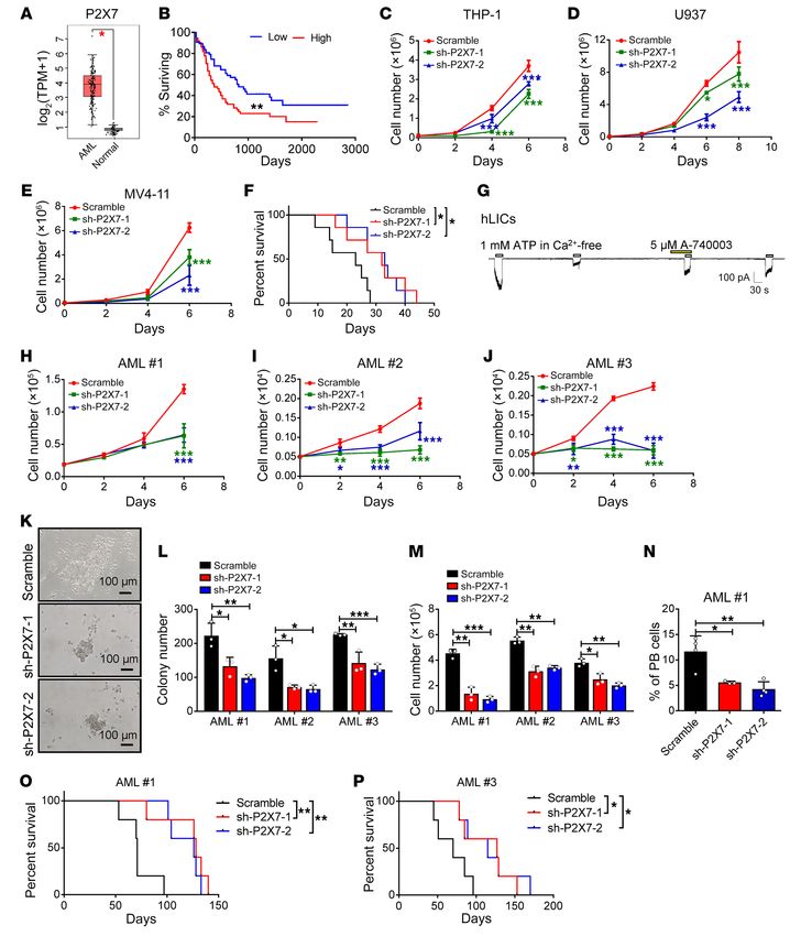
RESEARCH ARTICLE                                                                                      The Journal of Clinical Investigation

cells of the hematopoietic system, to regulate cell proliferation,         mogenic activities in an MLL-AF9–induced murine model of AML
differentiation, migration, and death (22–24). For example, ATP            but is not required for the maintenance of HSC stemness. Extra-
enhances the proliferation of hematopoietic stem cells (HSCs) and          cellular ATP efficiently induces the P2X7-mediated influx of ions,
maintains the size of the hematopoietic progenitor population,             which leads to enhanced calcium signaling–mediated direct trans-
especially during chronic inflammation, mainly through P2X1 and            activation of phosphoglycerate dehydrogenase (Phgdh) expression
P2X4. ATP, in combination with other cytokines, including IL-3             and maintenance of normal serine metabolism to control LIC
and GM-CSF, can also induce myeloid differentiation (25). Both             homing and self-renewal abilities. P2X7 is also required for the
ATP and UTP can promote the proliferation of human HSCs in                 proliferation of human AML cell lines and primary LICs. Treat-
vitro or in vivo (19). Interestingly, extracellular UTP and, to a less-    ment with P2X7 antagonists or depletion of serine can effectively
er extent, ATP, are required for the regulation of HSC motility and        inhibit the proliferation of AML cells both in vitro and in vivo.
homing (26). However, the detailed functions of the different P2X
members in the regulation of HSC stemness or malignant trans-              Results
formation remain largely unknown.                                          The endosteal niche has a much higher ATP level than the vascular
      Several studies have shown that extracellular ATP has a sup-         niche. To determine how the niche component ATP affects LIC
pressive effect in cancers, including hematologic malignancies             fate, we first measured the ATP concentration in the BM fluid from
(16, 27). For example, ATP can inhibit the growth and enhance              leukemic and control mice transplanted with BM cells expressing
the differentiation of human HL-60 or NB4 AML cells (28, 29).              an empty vector 4 months after injection and found that the ATP
However, studies have also shown that P2Xs, especially the P2X7            level in the leukemic mice was approximately 950-fold higher than
subtype, are expressed in many types of cancer, including chron-           that in the control mice (231 vs. 0.24 μM, Figure 1A). To precisely
ic B cell lymphocytic leukemia (B-CLL), prostate cancer, breast            evaluate the dynamic changes in the ATP levels in the BM nich-
cancer, neuroblastoma, and epithelial cancers, and enhance the             es, we took advantage of genetically encoded fluorescent sensor
progression and drug resistance of these cancers (30, 31). P2X7            iATPSnFR for cell surface ATP detection (44, 45) and generated
can also inhibit the antitumor effect of CD8+ T cells to enhance           a ratiometric ATP sensor by fusing the red fluorescent protein
tumorigenesis and metastasis (32, 33). P2X7 or other P2X mem-              mCherry to the N-terminus of iATPSnFR (mCherry-iATPSn-
bers (including P2X1 and P2X4) have also been found to be                  FR, Figure 1B) and a nonresponsive control sensor (mCherry-
highly expressed in myeloid disorders, such as myelodysplastic             cpSFGFP, NC). To test whether iATPSnFR can be used to sensi-
syndrome, chronic myeloid leukemia, and AML (34–36). Recent                tively measure extracellular ATP levels in vitro, we firstly trans-
evidence further indicates that ectopic P2X7 overexpression may            fected iATPSnFR and the control sensor into a human AML cell
enhance AML development but impair normal hematopoiesis                    line, U937 cells, and examined the changes in iATPSnFR fluores-
(37, 38). In light of these seemingly contradictory functions of           cence at excitation wavelengths of 488 nm and 561 nm. As shown
P2Xs, it is essential to use genetic tools, such as P2X-knockout           in Figure 1C, cell surface mCherry-iATPSnFR displayed clear
mice, to delineate how ATP levels are changed or distributed               changes in green fluorescence (to measure the ATP level) in the
in different BM niches and how ATP/P2X–mediated signaling                  presence of ATP, whereas the red fluorescence (to normalize the
affects leukemogenesis.                                                    iATPSnFR sensor protein level in individual cells) of mCherry
      Compared with the other P2X subtypes, P2X7 is unique                 did not. ATP stimulation in vitro led to an approximately 2-fold
because it has the longest C-terminal intracellular domain, and            increase in the fluorescence ratio (488 nm/561 nm) of iATPSnFR
forms a pore that is permeable to large cations upon stimulation.          but did not increase the fluorescence ratio of the control sensor
After extracellular ATP binds to P2X7, the ion channel opens and           mCherry-cpSFGFP (Figure 1, C and D, and Supplemental Fig-
allows the influx of calcium and the efflux of potassium (39).             ure 1A; supplemental material available online with this article;
During repeated or prolonged ATP stimulation, P2X7 continues               https://doi.org/10.1172/JCI140242DS1). Currently, the vascular
to open and form pores that allow larger molecules to enter cells          niche and endosteal niche are usually defined as the regions where
(40). P2X7 can also indirectly trigger voltage-gated calcium chan-         the leukemia cells directly contact or are within one-cell distance
nels to regulate calcium-dependent signal transduction pathways            from the endothelial cells and osteoblasts in the BM, respectively.
(41), and P2X7 plays an important role in a variety of physiological       To test whether iATPSnFR can indicate the distribution of ATP in
and pathological processes (15, 32). Preclinical studies have also         different BM niches, we transplanted iATPSnFR-U937 cells and
shown that P2X7 antagonists can potentially be used for the treat-         NC-U937 cells into NOD-SCID mice and found that the fluores-
ment of many disorders, especially inflammatory disorders and              cence ratio (488 nm/561 nm) of the iATPSnFR-U937 cells resid-
cancers (42, 43). However, the mechanism by which ATP/P2X7                 ing near the endosteal niche was much higher than that of the
signaling regulates the leukemogenic activities of LICs in BM              cells residing near the vascular niche (Figure 1, E–F). Moreover,
niches and its potential therapeutic role in leukemia treatment are        the closer the cells were to the endosteal niche, the higher the
still not fully understood.                                                iATPSnFR fluorescence ratio was (Figure 1G). In contrast, in the
      In this study, we demonstrated that ATP levels are markedly          mice transplanted with the NC-U937 cells, no significant differ-
increased in the BM niches of AML mice and that LICs tend to be            ence was observed in the fluorescence ratio of the cells located
localized to the endosteal niche, which has high ATP levels, by            near the endosteal niche and that of the cells located near the vas-
using a genetically encoded ATP sensor (iATPSnFR). We gener-               cular niche (Supplemental Figure 1, B–D).
ated P2x7-knockout mice and provide several lines of evidence                  To demonstrate whether the ATP level as determined by a
showing that P2X7 is important for the maintenance of LIC leuke-           human AML cell line was applicable to a primary murine AML mod-

   2                                   J Clin Invest. 2021;131(4):e140242 https://doi.org/10.1172/JCI140242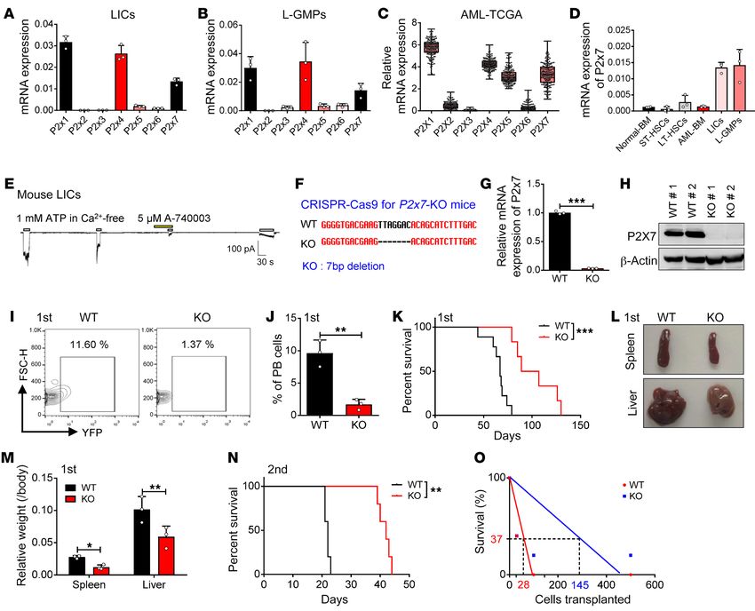
The Journal of Clinical Investigation RESEARCH ARTICLE
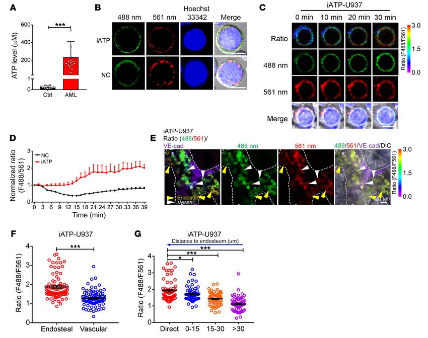
Figure 1. The endosteal niche has a much higher ATP level than the vascular niche. (A) Biochemical analysis of ATP levels in BM fluid of control and
leukemic mice (Ctrl, n = 12; AML, n = 15). ***P < 0.001 by Student’s t test. (B) Representative images of U937 cells infected with the ATP sensor (iATPSnFR)
or the control sensor (mCherry-cpSFGFP, NC). Nuclei were stained with Hoechst 33342. (C) Representative images of the ratio of iATPSnFR fluorescence
(F488/F561 nm) at excitation wavelengths of 488 nm and 561 nm in iATPSnFR-U937 cells at the indicated time points upon ATP treatment. Scale bars (B
and C): 10 μm. (D) Quantification of the fluorescence ratio (F488/F561 nm) of iATPSnFR and the control sensor in iATPSnFR-U937 cells upon ATP treat-
ment (n = 41 cells/group). (E) Representative images of the localization of iATPSnFR-U937 cells in the endosteal niche (dashed lines) and vascular niche
(labeled with anti–VE-cadherin) and their ratios of iATPSnFR fluorescence (488/561 nm) are shown. (F) Quantification of the iATPSnFR fluorescence ratio
in iATPSnFR-U937 cells in the endosteal and the vascular niche in panel E (n = approximately 85 cells/group from 3 biological replicates). (G) Quantification
of the iATPSnFR fluorescence ratio in iATPSnFR-U937 cells and its relationship to the cells’ distance from the endosteum in panel E (n = approximately 65
cells/group from 3 biological replicates). *P < 0.05; ***P < 0.001 by 1-way ANOVA with Tukey’s multiple-comparison test.
el, we overexpressed iATPSnFR or the control sensor in an MLL- observed in the NC-MLL-AF9 leukemic mouse model (Supplemen-
AF9–induced murine AML model to determine whether it was also tal Figure 1, K–M). These results further confirmed that ATP levels
able to efficiently reflect the ATP levels in different BM niches. We have different distribution patterns in different BM niches. Consis-
demonstrated that exogenous ATP could also increase the fluores- tently, Col2.3+ osteoblasts seemed to produce more ATP than either
cence ratio of iATPSnFR-AML cells in vitro by up to 2-fold (Supple- CD31+CD45 –Ter119– endothelial cells or CD31–CD45 –Ter119–Lepr+
mental Figure 1, E–G); however, exogenous ATP did not increase mesenchymal stem cells (MSCs), as determined by an in vitro assay.
the fluorescence ratio of NC-AML cells. In vivo imaging analysis Osteoblasts also had the highest mRNA levels of pannexin 1 (Panx1)
also revealed that the fluorescence ratio of the iATPSnFR-AML cells and connexin 43 (Cx43), which are 2 key regulators (46, 47) that
residing near the endosteal niche was 1.5-fold higher than that of enhance ATP secretion into the extracellular microenvironment
the iATPSnFR-AML cells residing near the vascular niche in the BM (Supplemental Figure 1, N–O). As we have previously shown that
of C57BL/6 leukemic mice (Supplemental Figure 1, H–I). Similar to AML LICs prefer to localize to the endosteal niche rather than the
our observations in the iATPSnFR-U937 cells, the closer the cells vascular niche (14), it is possible that the high ATP levels in the end-
were to the endosteal niche, the higher the iATPSnFR fluorescence osteal niche may contribute to the retention of LICs in specific BM
ratio was (Supplemental Figure 1J); however, this difference was not niches and to the maintenance of their leukemogenic activities.
J Clin Invest. 2021;131(4):e140242 https://doi.org/10.1172/JCI140242 3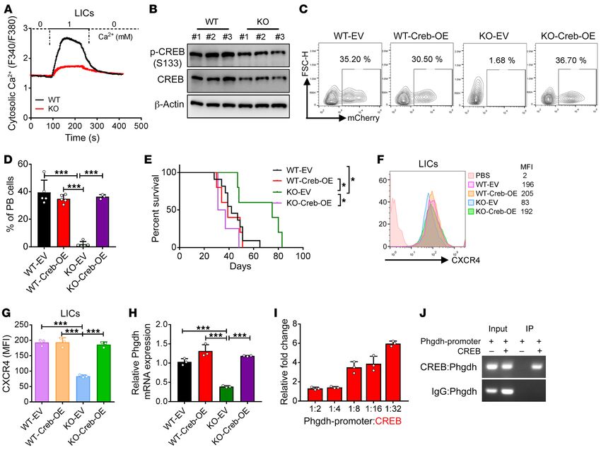
RESEARCH ARTICLE                                                                                               The Journal of Clinical Investigation

Figure 2. P2X7 is highly expressed in LICs and promotes AML development. (A and B) mRNA levels of P2x1–P2x7 were determined in the immunopheno-
typic Mac-1+c-Kit+ LICs (A) or Lin–Sca-1–c-Kit+CD34+CD16/32– L-GMPs (B) by quantitative RT-PCR (n = 3). (C) mRNA levels of P2Xs were analyzed in cells of
AML patients from TCGA database (n = 179). (D) mRNA levels of P2x7 in normal mouse bone marrow cells (Normal-BM), Lin–Sca-1+c-Kit+CD34–Flk2– long-term
HSCs (LT-HSCs), Lin–Sca-1+c-Kit+CD34+Flk2+ short-term HSCs (ST-HSCs), YFP+ leukemia cells (AML-BM), Mac-1+c-Kit+ LICs, and Lin–Sca-1–c-Kit+CD34+CD16/32–
L-GMPs were measured by quantitative RT-PCR (n = 3). (E) P2X7-mediated influx of ions in murine Mac-1+c-Kit+ LICs cells was measured upon sequential
treatments with extracellular ATP and P2X7 antagonist A-740003 by whole-cell patch-clamp recording (n = 3). (F) Nucleotide base sequences deleted in
the P2x7-knockout (P2x7-KO or P2x7-null) mouse using the CRISPR-Cas9 strategy. (G) mRNA level of P2x7 in WT and P2x7-KO BM cells was measured
by quantitative RT-PCR (n = 3). ***P < 0.001 by Student’s t test. (H) P2X7 protein levels in WT and P2x7-KO BM cells were measured by Western blot. (I)
Representative flow cytometric analysis of leukemia cells (YFP+) in the peripheral blood upon primary transplantation. (J) Quantification of data in panel I (n
= 5). PB, peripheral blood. **P < 0.01 by Student’s t test. (K) The overall survival of the recipient mice transplanted with WT or P2x7-KO MLL-AF9+ BM cells
upon primary transplantation (n = 5). ***P < 0.001 by log-rank test. (L) Representative images of the size of spleens and livers of recipients upon primary
transplantation. (M) Quantification of data in panel L (n = 4). *P < 0.05, **P < 0.01 by 2-way ANOVA with Sidak’s multiple-comparison test. (N) The overall
survival of recipient mice transplanted with WT or P2x7-KO MLL-AF9+ BM cells upon secondary transplantation (n = 5). ***P < 0.001 by log-rank test. (O)
Limiting dilution assays for the frequency of the functional LICs of WT and P2x7-KO BM cells. Experiments were repeated independently 3 times.

     P2X7 is highly expressed in LICs and promotes AML development.              Lin– Sca-1+c-Kit+CD34+CD16/CD32+ L-GMP cells (a population
To understand whether the extracellular ATP levels are associated                more highly enriched in LICs; Figure 2, A and B). Consistent with
with LIC activities in the endosteal niche, we first examined the                the expression patterns in the mouse AML LICs, several P2X
mRNA expression levels of all the P2X ATP-gated ion channels                     members, including P2X1, P2X4, P2X5, and P2X7, were high-
(P2X1–P2X7) in the LICs of mice with MLL-AF9–induced AML;                        ly expressed in the human AML cells from The Cancer Genome
in these mice, the AML cells only expressed myeloid cell markers                 Atlas (TCGA) database (Figure 2C). Because P2X7 has been
(Mac-1 and Gr-1) but not lymphoid cell markers (CD3 and B220),                   reported to exhibit completely different patterns of ion flux and
as previously described (48). Interestingly, P2x1, -4, and -7 were               formation of cytolytic pores that are permeable to large cations
abundant in both the immunophenotypic Mac-1+c-Kit+ LICs and                      upon repeated ATP stimulation (40, 49), we next decided to exam-

   4                                      J Clin Invest. 2021;131(4):e140242 https://doi.org/10.1172/JCI140242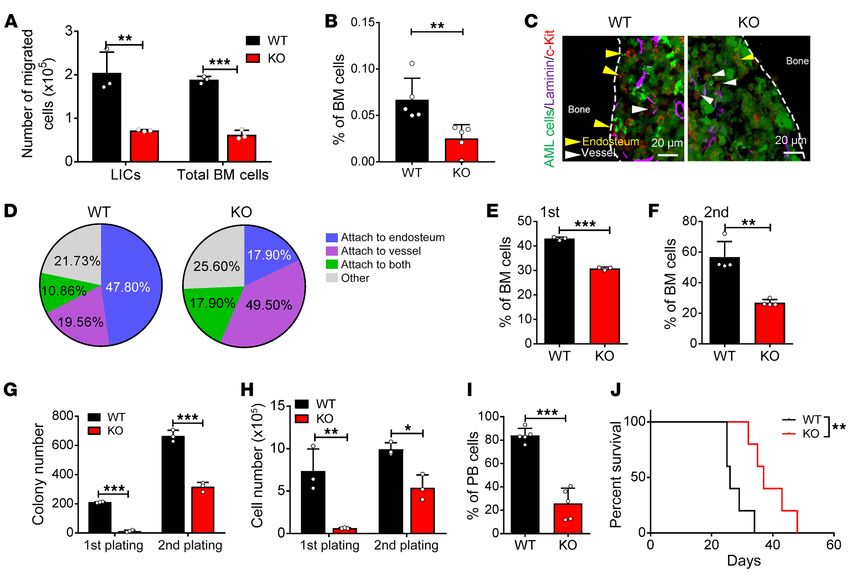
The Journal of Clinical Investigation                                                                           RESEARCH ARTICLE

ine the function of P2X7 during leukemogenesis. We first exam-                ATP/P2X7 signaling promotes the homing and self-renewal of
ined the mRNA levels of P2x7 in both normal hematopoietic cells          LICs. To determine how ATP/P2X7 signaling affects the fate of
and leukemia cells. Interestingly, the immunophenotypic Mac-1+           LICs, we first analyzed the migratory abilities of P2x7-null AML
c-Kit+ LICs and Lin– Sca-1+c-Kit+CD34+CD16/CD32+ L-GMP cells             bulk cells and LICs by using a Transwell assay. As shown in Figure
expressed high levels of P2x7, and the expression of P2x7 in these       3A, the percentages of migrated P2x7-null bulk leukemia cells and
cells was approximately 5- to 10-fold higher than that in the bulk       LICs were much lower after 4 hours of culture. Moreover, we also
AML cells and long-term HSCs (LT-HSCs) (Figure 2D). Moreover,            performed a homing assay to further confirm the change in the
the P2X7-mediated ion flux in the murine AML LICs could be effi-         migratory ability of the P2x7-null AML cells and found that there
ciently induced by stimulation with extracellular ATP and blocked        were approximately 50%–60% fewer cells that homed to the BM
by A-740003, a potent and selective P2X7 inhibitor, as determined        in the recipient mice transplanted with P2x7-null leukemia cells
by whole-cell patch-clamp electrophysiology recording (Figure            than in the recipient mice transplanted with WT cells (Figure 3B
2E); these results indicated that the ATP/P2X7–mediated signal is        and Supplemental Figure 3A). Because the ATP level was much
important for the leukemogenic activities of AML-LICs.                   higher in the endosteal niches (Figure 1, E and F, and Supplemen-
     To address the potential functions of P2X7 in leukemogenesis,       tal Figure 1, H and I), we speculated that the deletion of P2x7 may
P2x7 was knocked down in MLL-AF9+ murine AML cells by shRNA              have affected the localization of the LICs. We performed ex vivo
(Supplemental Figure 2, A and B), and these cells were transplant-       imaging analysis of the cranium of the leukemic mice and found
ed into recipient mice. AML development was much slower in the           that a much higher percentage of WT AML cells resided in the
mice transplanted with the P2x7-knockdown cells than in the con-         endosteal niche than P2x7-null AML cells (35.71% vs. 18.60%;
trol mice, as evidenced by the decreased frequency of leukemia           Supplemental Figure 3, B and C). Immunostaining analysis fur-
cells in the peripheral blood (Supplemental Figure 2, C and D) and       ther showed that approximately 47.80% of the LICs in the WT
the reduced infiltration in the livers and spleens of the transplant     leukemic mice were mainly localized to the endosteal niche, while
recipients (Supplemental Figure 2, E and F). More importantly,           only 19.56% of these cells resided near the vascular niche; how-
the overall survival of the leukemic mice transplanted with the          ever, in the P2x7-null leukemic recipients, approximately 17.90%
P2x7-knockdown cells was markedly extended compared with                 and 49.50% of the LICs were localized to the endosteal niche and
that of the control mice upon either primary (45 or 51 vs. 26 days,      vascular niche, respectively (Figure 3, C and D). These results indi-
Supplemental Figure 2G) or secondary transplantation (29 or 32           cated that ATP/P2X7 signaling may be important for the retention
vs. 25 days, Supplemental Figure 2H), indicating that P2X7 may be        of LICs in the endosteal niche.
required for the self-renewal capacity of LICs.                               We then further evaluated whether ATP/P2X7 signaling is
     To further confirm the function of P2X7, we generated               required for the commitment of LICs to other fates by examining
P2x7-knockout (P2x7-KO) mice (Figure 2F) using the CRISPR-               the LIC frequency after serial transplantation. Interestingly, we
Cas9 strategy and demonstrated that almost no detectable P2x7            found that the frequency of the immunophenotypic Mac-1+c-Kit+
mRNA or protein levels (Figure 2, G and H) were observed in the          P2x7-null LICs was reduced to 70% or 50% of the frequency of the
total BM cells of the P2x7-KO mice. We then established an MLL-          WT LICs upon primary (Figure 3E and Supplemental Figure 3D)
AF9–induced AML model with wild-type (WT) and KO mice and                or secondary transplantation (Figure 3F and Supplemental Fig-
revealed that leukemia development was much slower in the                ure 3E). Moreover, the frequencies of the Lin–CD127– Sca-1– c-Kit+
mice transplanted with P2x7-null AML cells than in the control           CD34+CD16/32+ P2x7-null L-GMP cells, which is a population that
mice, as evidenced by the decreased frequency of YFP+ leuke-             has been indicated to be more highly enriched in immunopheno-
mia cells in the peripheral blood (Figure 2, I and J), the reduced       typic LICs (50), were also much lower than those of WT control
percentages of blast cells in both the peripheral blood and BM           cells (Supplemental Figure 3, F–H). An in vitro surrogate func-
(Supplemental Figure 2, I–L), the dramatically increased overall         tional analysis with methylcellulose medium further revealed a
survival (67 vs. 98 days, Figure 2K), and the reduced infiltration       dramatic decrease in the colony size, colony number, and derived
in the livers and spleens of the transplant recipients after prima-      cell number of the P2x7-null AML cells during both primary plat-
ry transplantation (Figure 2, L and M, and Supplemental Figure           ing and secondary plating (Figure 3, G and H, and Supplemental
2M). The survival of the mice transplanted with primary P2x7-            Figure 3I). We also used an alternative way to exclude the homing
null cells was significantly prolonged compared with that of the         effect by using intratibial injection with AML cells. Interestingly,
mice transplanted with WT cells after secondary transplantation          the leukemia development was still much slower in the recipient
(22 vs. 42 days, Figure 2N). Limiting dilution assays with the           mice receiving P2x7-null AML cells than that of the WT control,
YFP+ AML cells from the primary transplant recipients revealed           as evidenced by the reduced leukemia cell frequency in peripher-
that the frequency of functional LICs was 1 in 28, which was             al blood and extended overall survival (Figure 3, I and J). These
approximately 5.2-fold higher than that in the P2x7-null controls        results suggest that both homing and proliferation defects may
(1 in 145, Supplemental Table 1 and Figure 2O). Taken together,          contribute to the phenotypes in P2x7-KO leukemic mice. In addi-
these results clearly suggest that ATP/P2X7–mediated pathways            tion, there was no significant difference in apoptosis (Supplemen-
enhance leukemogenesis and may be an ideal target in LICs. In            tal Figure 3, J and K) or differentiation (Supplemental Figure 3L),
contrast, ATP/P2X7 signaling seemed to have no effects on the            as measured by annexin V/7-AAD staining or Mac-1/Gr-1 staining
normal repopulation of HSCs (Supplemental Figure 2, N–P),                (Gr-1 expression level represents the degree of differentiation),
suggesting that ATP/P2X7 signaling may be an ideal target for            respectively. Consistently, the LIC frequency was substantial-
LIC-targeted therapy.                                                    ly reduced in the mice transplanted with P2x7-knockdown cells

                                     J Clin Invest. 2021;131(4):e140242 https://doi.org/10.1172/JCI140242                                5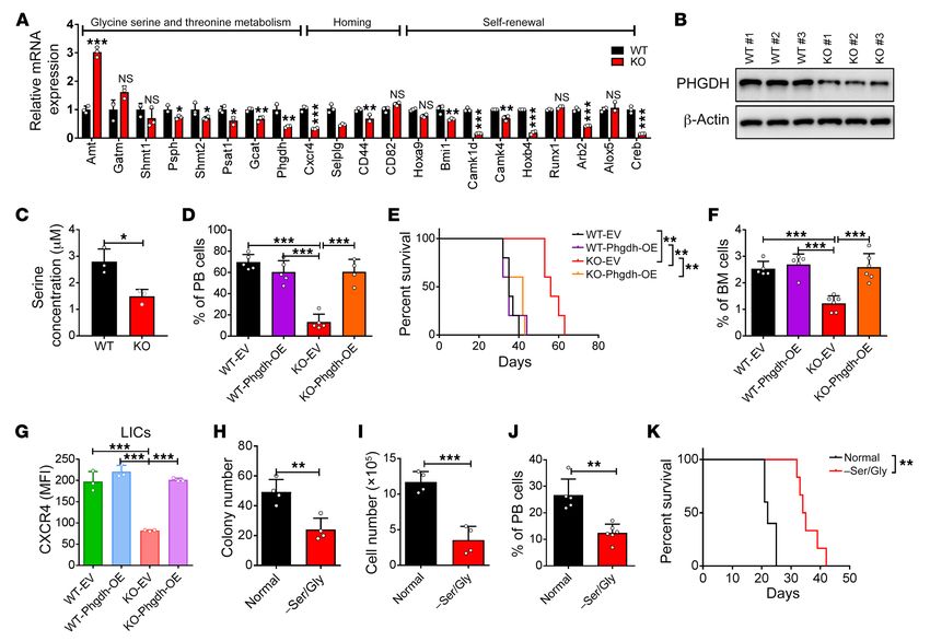
RESEARCH ARTICLE                                                                                              The Journal of Clinical Investigation

Figure 3. ATP/P2X7 signaling maintains the homing and self-renewal abilities of LICs. (A) Quantification of the migratory abilities of WT and P2x7-KO
Mac-1+c-Kit+ LICs or total BM cells evaluated by Transwell assay (n = 3). **P < 0.01, ***P < 0.001 by 2-way ANOVA with Sidak’s multiple-comparison
test. (B) The frequency of homed WT and P2x7-KO leukemia cells (CFSE+) in the BM of the recipients 16 hours after transplantation (n = 5). **P < 0.01
by Student’s t test. (C) Representative images of immunofluorescence staining for the localization of LICs (red) in the laminin+ vascular niche (purple)
or the endosteal niche (dashed lines) in the femur of AML mice. AML cells are indicated by GFP positivity (green). Scale bars: 20 μm. (D) Percentages of
Mac-1+c-Kit+ LICs attached to the endosteal niche, the vascular niche, or both in panel C were calculated (n = ~50 cells/group from 4 biological replicates).
(E and F) The frequency of WT and P2x7-KO Mac-1+c-Kit+ LICs of the recipients upon primary (E) and secondary (F) transplantation (n = 3). ***P < 0.001
by Student’s t test. (G and H) Colony numbers (G) and derived total cell count (H) of WT and P2x7-KO YFP+ AML cells during the first and second plating
were calculated (n = 3). *P < 0.05, **P < 0.01, ***P < 0.001 by 2-way ANOVA with Sidak’s multiple-comparison test. (I) Leukemia cells were transplanted
into recipient mice by intratibial injection. Shown is the quantification of the frequency of the leukemia cells (YFP+) in the peripheral blood of recipient
mice 3 weeks after transplantation (n = 5). ***P < 0.001 by Student’s t test. (J) Overall survival was determined in the leukemic mice in panel I (n = 5).
**P < 0.01 by log-rank test.

compared with the mice transplanted with the WT cells after seri-                high levels in THP-1 (M5), U937 (M5), and MV4-11 (M5) cells but
al transplantation (Supplemental Figure 3, M–O). The colony size,                expressed at low levels in HL60 (M3), NB4 (M3), and K562 (CML)
colony number, and derived cell number of the P2x7-knockdown                     cells (Supplemental Figure 4A). We then constructed 2 shRNAs
AML cells were also dramatically decreased, further indicating a                 to knock down P2X7 in several human AML cell lines to evalu-
remarkable inhibition of the self-renewal ability of LICs (Supple-               ate its roles in human leukemia development. Both the shRNAs
mental Figure 3, P and Q). These results imply that ATP/P2X7                     (sh-P2X7-1 and -2) efficiently downregulated the mRNA level of
signaling promotes the homing and self-renewal abilities of LICs,                P2X7 in THP-1 cells, as measured by quantitative RT-PCR (Sup-
which further contribute to leukemogenesis.                                      plemental Figure 4, B and C). Knockdown of P2X7 in 3 human
     P2X7 is required for the proliferation of human AML cells. To               AML cell lines, THP-1, U937, and MV4-11 cells, resulted in a
further examine the role of P2X7 in human LICs, we first ana-                    marked decrease in cell proliferation in vitro at the indicated time
lyzed the mRNA levels of P2X7 in human AML cells using TCGA                      points (Figure 4, C–E). We then transplanted the P2X7-knock-
database. Strikingly, the P2X7 mRNA level in the AML cells was                   down THP-1 cells into NOD-SCID mice and found that leukemia
approximately 7-fold higher than that in the normal BM cells (Fig-               development was notably delayed in these mice, as evidenced by
ure 4A), and the P2X7 level was negatively correlated with the                   the decreased leukemia cell frequency in peripheral blood (Sup-
overall survival of AML patients (Figure 4B), indicating that P2X7               plemental Figure 4D), prolonged overall survival (32 or 33 vs. 23
may be critical for leukemogenesis.                                              days, Figure 4F), and the smaller size and lower weight of the
     To further evaluate the functions of P2X7 in human AML cells,               spleens and livers (Supplemental Figure 4, E and F) in these mice
we examined the P2X7 levels in several human AML cell lines by                   compared with those of the control mice transplanted with the
quantitative RT-PCR and showed that P2X7 was expressed at                        scrambled shRNA–treated THP-1 cells.

   6                                      J Clin Invest. 2021;131(4):e140242 https://doi.org/10.1172/JCI140242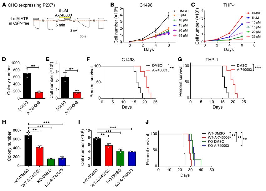
The Journal of Clinical Investigation                                                                                          RESEARCH ARTICLE

Figure 4. P2X7 is required for the proliferation of human AML cells. (A) P2X7 mRNA levels were analyzed in AML cells and normal BM cells from TCGA
database (AML, n = 173; normal n = 70). *P < 0.05 by Student’s t test. TPM, transcripts per million. (B) The relationship between P2X7 level and survival of AML
patients from TCGA database (n = 75/group). **P < 0.01 by log-rank test. (C–E) The numbers of THP-1, U937, and MV4-11 cells were calculated after infection
with shRNAs targeting P2X7 (sh-P2X7-1 and -2) or scrambled shRNA (n = 3). *P < 0.05, ***P < 0.001 by 2-way ANOVA with Sidak’s multiple-comparison
test. (F) The overall survival was examined in the recipients transplanted with P2X7-knockdown (sh-P2X7-1 and -2) THP-1 cells and scrambled shRNA (n = 7).
*P < 0.05 by log-rank test. (G) P2X7-mediated influx of ions into human CD34+ LICs was measured upon the sequential treatment with ATP and the P2X7
antagonist A-740003 (n = 3). (H–J) The numbers of human primary AML cells were counted upon P2X7 knockdown. Three patients’ samples were examined
(AML 1–3) (n = 3). *P < 0.05, **P < 0.01, ***P < 0.001 by 2-way ANOVA with Sidak’s multiple-comparison test. (K) Representative images of colonies derived
from human AML cells upon the knockdown of P2X7 by shRNAs (sh-P2X7-1 and -2) or those treated with scrambled shRNA control. (L and M) Colony numbers
and derived total cell counts were examined in human AML cells upon the knockdown of P2X7 by shRNAs (sh-P2X7-1 and -2) and scrambled shRNA (n = 3). *P
< 0.05, **P < 0.01, ***P < 0.001 by 2-way ANOVA with Sidak’s multiple-comparison test. (N–P) CD45+GFP+ human AML cells in the peripheral blood (N) (AML
1, n = 5) and the survival (O and P) (AML 1 and 2, n = 5) of the recipients transplanted with P2X7-knockdown (sh-P2X7-1 and -2) human AML cells or scrambled
shRNA are shown. PB, peripheral blood. *P < 0.05, **P < 0.01 by 1-way ANOVA with Tukey’s multiple-comparison test (N) or log-rank test (O and P).

                                           J Clin Invest. 2021;131(4):e140242 https://doi.org/10.1172/JCI140242                                           7
RESEARCH ARTICLE The Journal of Clinical Investigation
We then further evaluated the role of P2X7 in human prima- LICs (Figure 5A). These results indicated that these genes might
ry AML cells and found that the P2X7 mRNA level in LICs was serve as downstream targets of P2X7.
approximately 7-fold higher than that in HSCs (Supplemental Fig- Serine has been known to serve as a proteinogenic amino acid
ure 4G). Importantly, ATP supplementation efficiently induced and the source of 1-carbon units important for de novo purine and
the flux of ions into human CD34+ AML LICs, which was com- deoxythymidine synthesis. Because PHGDH is the key rate-lim-
pletely blocked by the P2X7 antagonist A-740003, as measured iting enzyme involved in the first step of serine and glycine syn-
by whole-cell patch-clamp recording (Figure 4G). Knockdown thesis and has been reported to be critical for the development of
of P2X7 in human CD34+ AML LICs resulted in an approximate- several types of cancers (51, 52), we further examined the protein
ly 60%–80% reduction in cell growth in vitro (Figure 4, H–J). An level of PHGDH. We found that the level of the PHGDH protein
in vitro functional colony forming assay further showed that the was markedly decreased in the P2x7-null LICs compared with the
P2X7-knockdown LICs generated approximately 40%–60% few- WT control cells (Figure 5B). This finding was consistent with the
er colony numbers and 40%–80% fewer total cells than the LICs much lower level of serine in the P2x7-null LICs compared with
infected with the scrambled shRNA (Figure 4, K–M). Moreover, in that in the control LICs, as measured by liquid chromatography–
vivo transplantation experiments revealed that P2X7 knockdown tandem mass spectrometry (LC-MS/MS) (Figure 5C). To further
led to a marked delay in AML development, as evidenced by the verify that Phgdh is a downstream target gene of P2X7, we overex-
decreased percentages of leukemia cells in the peripheral blood pressed Phgdh in P2x7-null AML cells and transplanted these cells
(Figure 4N) and the significantly extended overall survival in the into lethally irradiated recipient mice. The overexpression of Phgdh
recipient mice transplanted with P2X7-knockdown AML cells in the P2x7-null AML cells fully reversed the extended survival of
compared with those of the control mice (128 or 126 vs. 71 days in the recipient mice to levels comparable to those of the recipient
Figure 4O, and 127 or 115 vs. 70 days in Figure 4P). AML develop- mice transplanted with WT leukemia cells (Figure 5, D and E, and
ment in the recipient mice was confirmed based on the expression Supplemental Figure 5, E and F). In addition, the overexpression
of several myeloid markers, such as CD33, CD15, and CD34, but of Phgdh had no effect on WT leukemia cell proliferation (Figure
not lymphoid markers, such as CD19 and CD20 (Supplemental 5, D and E, and Supplemental Figure 5, E and F). Importantly, an
Figure 4H). These results indicate that P2X7 is critical for the leu- in vitro Transwell assay also showed that overexpression of Phgdh
kemogenic capacities of human AML LICs. reversed the impaired migratory abilities of the P2x7-deficient
ATP/P2X7 signaling sustains LIC activities by activating the LICs (Supplemental Figure 5, G and H) or bulk AML cells (Supple-
PHGDH pathway. To determine the underlying molecular mech- mental Figure 5, I and J). In vivo homing analysis further revealed
anisms that control the homing and self-renewal activities of that the Phgdh-overexpressing P2x7-null AML cells exhibited
P2x7-null LICs, WT and P2x7-null LICs were analyzed by RNA enhanced homing activities compared with the P2x7-null AML
sequencing. The RNA sequencing data showed that 672 or 576 cells; the homing activities of the Phgdh-overexpressing P2x7-null
genes were significantly up- or downregulated in the P2x7-null AML cells were comparable to those of the WT AML cells (Figure
group compared with the WT group (Supplemental Figure 5A). 5F and Supplemental Figure 5K). Consistently, the CXCR4 pro-
Among the top 15 significant differences in the Gene Ontology tein level was much lower in both P2x7-null LICs and bulk BM
(GO) or Kyoto Encyclopedia of Genes and Genome (KEGG) anal- cells than that of WT controls, which could be significantly upreg-
yses, P2x7 has been shown to be involved in biological process- ulated upon the overexpression of Phgdh in P2x7-null leukemia
es, such as cell migration, regulation of cell proliferation, cellular cells (Figure 5G and Supplemental Figure 5, L–N), indicating that
response to calcium ions, cell adhesion, and ATP metabolic pro- PHGDH regulates the homing activities of LICs. To further con-
cess (Supplemental Figure 5B), or to be associated with several firm that serine metabolism plays a role in leukemogenesis, an in
pathways, including oxidative phosphorylation, focal adhesion, vitro functional colony forming assay was performed in the pres-
and glycine, serine, threonine, and purine metabolism (Supple- ence or absence of serine and glycine. Interestingly, the colony
mental Figure 5C). Because we noticed that several metabolic size, colony numbers, and derived cell numbers of the leukemia
pathways (such as ATP metabolic process, oxidative phosphor- cells were significantly decreased upon the depletion of serine
ylation, and glycine, serine, threonine, and purine metabolism) and glycine (Figure 5, H and I). In vivo transplantation experi-
were markedly downregulated in the P2x7-null LICs and that ments further showed that the depletion of serine/glycine resulted
P2x7 deletion led to the loss of homing to the endosteal niche and in a notable delay in leukemia development, as exhibited by the
self-renewal abilities of the LICs, we first examined the potential decreased frequency of AML cells in the peripheral blood and the
changes in several genes related to glycine, serine, and threonine prolonged overall survival of the recipient mice (Figure 5, J and K).
metabolism (such as Phgdh, Psat1, and Shmt1), homing activities CREB signaling maintains PHGDH levels to enhance leukemo-
(such as Cxcr4, Cd44, Emb, Cc3cr1, and Vcam1), and self-renew- genesis. Because many studies have shown that P2X7 mediates the
al activities (such as Creb, Mef2c, Camk1d, Camk4, and Hoxb4) in influx of calcium and the efflux of potassium (41), and because our
the RNA sequencing data. This analysis showed that several key RNA sequencing data also revealed a significant reduction in the
genes were indeed significantly decreased in the P2x7-null LICs cellular response to calcium ions (Supplemental Figure 5A), we
(Supplemental Figure 5D). Consistently, quantitative RT-PCR further measured the calcium influx capacity of the P2x7-null LICs
further demonstrated that most of the key genes, including using the calcium indicator Fura-2 AM. Interestingly, the constitu-
Phgdh, Psat1, Psph (related to glycine serine metabolism), Cxcr4, tive calcium influx was markedly decreased in both the P2x7-null
Selplg, Cd44 (related to homing), Creb, and Hoxb4 (related to LICs (Figure 6A) and bulk AML cells (Supplemental Figure 6A)
self-renewal), were significantly downregulated in the P2x7-null compared with that of the WT cells. Because the transcription
8 J Clin Invest. 2021;131(4):e140242 https://doi.org/10.1172/JCI140242The Journal of Clinical Investigation RESEARCH ARTICLE
Figure 5. ATP/P2X7 signaling maintains LIC activities by activating the PHGDH pathway. (A) Potential candidates related to glycine, serine, and thre-
onine metabolism, homing, and self-renewal were examined in WT and P2x7-KO Mac-1+c-Kit+ LICs by quantitative RT-PCR (n = 3). *P < 0.05, **P < 0.01,
***P < 0.001 by 2-way ANOVA with Sidak’s multiple-comparison test. (B) Protein levels of PHGDH were determined in WT and P2x7-KO Mac-1+c-Kit+ LICs.
(C) Serine levels were measured in WT and P2x7-KO BM leukemia cells by LC-MS/MS (n = 5). *P < 0.05 by Student’s t test. (D and E) The leukemia cell
(YFP+mCherry+) frequencies in the peripheral blood (D) (n = 5; ***P < 0.001 by 1-way ANOVA with Tukey’s multiple-comparison test) and overall survival (E)
(n = 5; **P < 0.01 by log-rank test) were compared among the recipients transplanted with WT, P2x7-KO, Phgdh-overexpressing WT, or P2x7-KO leukemia
cells. (F) The frequency of homed WT, P2x7-KO, Phgdh-overexpressing WT, or P2x7-KO leukemia cells (CFSE+) in the BM of the recipients 16 hours after
transplantation (n = 5). ***P < 0.001 by 1-way ANOVA with Tukey’s multiple-comparison test. (G) The mean fluorescence intensities (MFIs) of CXCR4 in
WT, P2x7-KO, Phgdh-overexpressing WT, or P2x7-KO Mac-1+c-Kit+ LICs (n = 3). ***P < 0.001 by 1-way ANOVA with Tukey’s multiple-comparison test. (H and
I) The colony formation abilities of leukemia cells were determined in the presence or absence of serine and glycine (normal or –Ser/Gly). Colony numbers
(H) and derived total cell counts (I) were calculated (n = 3). **P < 0.01, ***P < 0.001 by Student’s t test. (J) Leukemia cells were cultured with or without
serine and glycine (normal or –Ser/Gly) for 48 hours, followed by transplantation into the recipient mice. Shown are the leukemia cell frequencies in the
peripheral blood of recipients 3 weeks after transplantation (n = 5–6). **P < 0.01 by Student’s t test. (K) Overall survival was determined in the leukemic
mice in panel J (n = 5–6). **P < 0.01 by log-rank test. EV, empty vector; OE, overexpressing; PB, peripheral blood.
factor CREB serves as a key mediator of calcium signaling and its WT, P2x7-null, Creb-overexpressing WT, or Creb-overexpressing
mRNA level was also markedly decreased upon P2x7 deletion, it is P2x7-null AML cells were confirmed by quantitative RT-PCR
possible that CREB may act as an upstream target of P2X7. Consis- (Supplemental Figure 6D). The overexpression of Creb in the WT
tently, the protein levels of phospho-CREB (p-CREB) (S133) and AML cells had no effect on leukemogenesis (Figure 6, C and D,
CREB in the P2x7-null LICs were significantly reduced compared and Supplemental Figure 6, B and C). Moreover, the overexpres-
with those in the WT control LICs (Figure 6B). Compared with the sion of Creb also upregulated the protein level of CXCR4 in both
P2x7-null control cells, P2x7-null AML cells overexpressing Creb the P2x7-null LICs and bulk AML cells, and these levels were
accelerated the development of leukemia and reversed the pheno- comparable to that observed in the WT cells (Figure 6, F and G,
types resulting from P2x7 deletion, as evidenced by the increased and Supplemental Figure 6, E and F). These results suggest that
frequency of leukemia cells in the peripheral blood (Figure 6, C CREB-mediated calcium signaling is required for the leukemo-
and D), the enhanced infiltration in spleens and livers (Supple- genic activities of P2X7.
mental Figure 6, B and C), and the reduced overall survival; these To determine whether P2X7 is regulated by CREB-medi-
parameters were comparable to those observed in the mice trans- ated signaling, the mRNA level of Phgdh was evaluated in both
planted with WT AML cells (Figure 6E). The levels of Creb in the Creb-overexpressing WT and P2x7-null AML cells by quantita-
J Clin Invest. 2021;131(4):e140242 https://doi.org/10.1172/JCI140242 9RESEARCH ARTICLE The Journal of Clinical Investigation Figure 6. CREB signaling maintains PHGDH levels to enhance leukemogenesis. (A) The constitutive calcium influx of WT and P2x7-KO Mac-1+c-Kit+ LICs was measured using Fura-2 AM (n = 205 cells; 0 = Ca2+-free solution; 1 = solution with 1 mM Ca2+). (B) The protein levels of phospho-CREB (S133) and CREB were determined in both WT and P2x7-KO Mac-1+c-Kit+ LICs by Western blot. (C) Representative flow cytometric analysis of leukemia cells (YFP+mCherry+) in the peripheral blood of recipient mice transplanted with WT, P2x7-KO, Creb-overexpressing WT, or P2x7-KO AML cells. (D) Quantification of data in panel C (n = 5). ***P < 0.001 by 1-way ANOVA with Tukey’s multiple-comparison test. (E) The overall survival was compared among the mice transplanted with WT, P2x7-KO, Creb-overexpressing WT, or P2x7-KO AML cells (n = 5). *P < 0.05 by log-rank test. (F) The expression levels of CXCR4 in WT, P2x7-KO, Creb-overexpressing WT, or P2x7-KO Mac-1+c-Kit+ LICs were determined by flow cytometry. (G) Quantification of the mean fluorescence intensities (MFIs) in panel F (n = 3). ***P < 0.001 by 1-way ANOVA with Tukey’s multiple-comparison test. (H) The mRNA levels of Creb in WT, P2x7-KO, Creb-overexpressing WT, or P2x7-KO AML cells were measured by quantitative RT-PCR (n = 3). ***P < 0.001 by 1-way ANOVA with Tukey’s multiple-comparison test. (I) A lucif- erase reporter assay was used to evaluate the transcriptional activity of CREB on the promoter of Phgdh (n = 3–4). (J) The binding of CREB to the Phgdh promoter region was determined by chromatin immunoprecipitation (ChIP) assay. EV, empty vector; OE, overexpressing; PB, peripheral blood. tive RT-PCR; the results showed that Creb overexpression indeed To further verify that P2X7 exerts its leukemogenic effect on resulted in a significant increase in the Phgdh levels (Figure 6H). human LICs via pathways similar to those used by murine LICs, By using a dual luciferase reporter assay, we further demonstrat- we examined the protein levels of p-CREB, CREB, and PHGDH ed that Creb could directly bind to the Phgdh promoter region and in both the AML cell lines and primary samples. This analysis transactivate the Phgdh expression in a dose-dependent manner showed that P2X7 knockdown led to a marked reduction in CREB/ (Figure 6I); these findings were further validated by subsequent PHGDH signaling (Supplemental Figure 7, A and B). Similarly to ChIP analysis with an antibody against CREB (Figure 6J). Con- the murine P2x7-null AML cells, the P2X7-knockdown human pri- sistently, compared with the knockdown control, knockdown of mary AML cells also had lower levels of serine than the primary P2x7 in primary mouse AML cells also led to a notable reduction in AML cells infected with the scrambled shRNA, further indicating the protein levels of p-CREB, CREB, and PHGDH (Supplemental that impaired serine metabolism may inhibit AML cell prolifer- Figure 6G). These results demonstrated that ATP/P2X7 signaling ation (Supplemental Figure 7C). Depletion of serine and glycine enhances the homing and self-renewal capacities of LICs through from the culture medium also resulted in an approximately 30%– CREB/PHGDH signaling pathways. 70% reduction in the growth of several AML cell lines, THP-1, 10 J Clin Invest. 2021;131(4):e140242 https://doi.org/10.1172/JCI140242
The Journal of Clinical Investigation                                                                                      RESEARCH ARTICLE

Figure 7. Targeting leukemia development by suppressing ATP/P2X7 signaling. (A) The P2X7-mediated influx of ions in CHO cells overexpressing rat P2x7
was measured upon sequential treatments with extracellular ATP and the P2X7 antagonist A-740003 by whole-cell patch-clamp recoding (n = 3). (B and C)
C1498 cells (B) and THP-1 cells (C) were treated with the indicated doses of A-740003 and the numbers were calculated (n = 3). (D and E) Colony numbers
(D) and derived total cell counts (E) were determined in THP-1 cells treated with A-740003 (10 μM) and DMSO (n = 3). **P < 0.01 by Student’s t test. (F and
G) The overall survival was compared in the recipient mice transplanted with C1498 cells (F) or THP-1 cells (G), followed by the treatments with A-740003
and DMSO (n = 5). **P < 0.01, ***P < 0.001 by log-rank test. (H and I) Colony numbers (H) and derived total cell counts (I) of WT and P2x7-KO murine AML
cells were measured upon the treatments with A-740003 (10 μM) and DMSO (n = 3). **P < 0.01, ***P < 0.001 by 1-way ANOVA with Tukey’s multiple-com-
parison test. (J) The overall survival was compared in the recipient mice transplanted with WT and P2x7-KO AML cells, followed by the treatments with
A-740003 and DMSO 7 days after transplantation (50 mg/kg every other day for 2 weeks) (n = 5). **P < 0.01 by log-rank test.

U937, and MV4-11 cells (Supplemental Figure 7, D–F). Moreover,                  PHGDH in normal and AML data sets, which showed that CXCR4
an in vitro functional colony assay revealed that the numbers of                and CREB, but not PHGDH, were upregulated in AML patients
colonies and the total cells derived from the human primary AML                 and consistent with the expression of P2X7 in human AML (Sup-
cells were reduced to approximately 50%–70% of those derived                    plemental Figure 7, M–O). Although the mRNA level of PHGDH
from the control cells when serine and glycine were depleted from               in AML patients was not consistent with that of P2X7 and was
the methylcellulose medium (Supplemental Figure 7, G and H).                    relatively lower than that of normal ones, the overall survival was
We also cultured human cord blood CD34+ cells with and with-                    inversely correlated with PHGDH expression and similar to that
out serine/glycine in vitro and found that impaired serine/glycine              of P2X7 (Supplemental Figure 7P), indicating that PHGDH may
metabolism did not affect the proliferation of normal CD34+ cells               still serve as a critical oncogene in leukemia development. Mean-
(Supplemental Figure 7, I and J), indicating such an effect may be              while, we also found that the protein level of PHGDH was notably
specific to AML cells.                                                          increased in human AML samples compared with that of the nor-
     Consistently, we also revealed that the ATP level in the BM                mal BM cells, indicating that posttranslational modification may
fluid of human AML patients was much higher compared with                       be important for the high protein level and function of PHGDH in
healthy donors (Supplemental Figure 7K), which was consistent                   leukemogenesis (Supplemental Figure 7, Q and R).
with the finding in the murine model. Interestingly, it seems that                    Targeting AML development by suppressing ATP/P2X7 signaling.
the ATP level in several subtypes (M2, M3, and M5) of AML were                  To determine whether the blockade of ATP/P2X7 signaling can
also much higher than the normal ones (Supplemental Figure                      inhibit AML development, we extensively evaluated the poten-
7L). We further analyzed the expression of CREB, CXCR4, and                     tial therapeutic effect of a specific antagonist of P2X7, A-740003,

                                          J Clin Invest. 2021;131(4):e140242 https://doi.org/10.1172/JCI140242                                       11
RESEARCH ARTICLE The Journal of Clinical Investigation
both in vitro and in vivo. Similar to the experiments mentioned inhibited leukemia cell growth (Supplemental Figure 9, A–E),
above (Figure 2E and Figure 4G), we tested the inhibitory effect of which might have resulted from nonspecific cytotoxicity (53–55).
A-740003 by analyzing P2x7-overexpressing CHO cells by whole- However, apoptosis (Supplemental Figure 9, F–J) and the cell cycle
cell patch-clamp technology. This analysis showed that the ion (Supplemental Figure 9, K–O) were not altered upon treatment
flux induced by extracellular ATP could be completely inhibited with low-dose ATP (the changes in the high-dose group may be
by 5 μM A-740003 and gradually recovered thereafter (Figure 7A). due to nonspecific cytotoxicity), indicating that ATP-mediated
A-740003 sufficiently inhibited the in vitro proliferation of both signaling may mainly sustain the self-renewal ability of leukemia
the murine (C1498, Figure 7B) and human (THP-1, Figure 7C) cells and blockade of the ATP/P2X7 pathway is a potential strategy
AML cell lines in a dose-dependent manner. An in vitro function- for leukemia treatment.
al colony forming assay further showed that A-740003 treatment
notably impaired the colony forming capacity of THP-1 cells, as Discussion
exhibited by the smaller colony size and fewer numbers of total In current study, we show a unique pattern of ATP distribution
cells after A-740003 treatment than after control treatment (Sup- in the leukemic BM, as indicated by a genetically encoded ATP
plemental Figure 8A and Figure 7, D and E). These results prompt- sensor (iATPSnFR), namely, the endosteal niche has a much
ed us to evaluate the potential therapeutic effects of A-740003 in higher ATP level than the vascular niche. The high levels of ATP
vivo. Recipient mice were transplanted with either C1498 or THP-1 can mediate the flux of calcium via the ion channel P2X7 to sus-
cells and intraperitoneally injected with A-740003 for 2 weeks tain the homing and self-renewal capacities of LICs. Calcium
(Supplemental Figure 8B). Interestingly, A-740003 treatment signaling further enhances the phosphorylation of CREB, which
resulted in a significant reduction in the frequency of leukemia transactivates PHGDH to maintain the leukemogenic activities of
cells in the peripheral blood (Supplemental Figure 8, C–F) and a LICs. Blocking ATP/P2X7 signaling with the specific antagonist
notably extended survival time of the recipient mice compared A-740003 efficiently delays leukemia development both in vitro
with the control mice (Figure 7, F and G), indicating that A-740003 and in vivo (Supplemental Figure 9P). Our studies show that ATP
can indeed be used to effectively target AML cells. may serve as an important niche factor that participates in the con-
We then examined the therapeutic effect of A-740003 in an trol of the fates of LICs via activation of P2X7, which may be an
MLL-AF9–induced aggressive AML model. An in vitro function- ideal therapeutic target in LICs or other types of cancer stem cells.
al colony formation analysis revealed that A-740003 treatment Increasing evidence shows that BM niches play key roles in sus-
caused an approximately 30% reduction in colony formation and taining LIC activities. However, the mechanisms by which niche
cell count compared with the control treatment (Figure 7, H and factors affect the fates of different types of LICs remain elusive.
I). More importantly, after 2 weeks of intraperitoneal injection Here, we provide unexpected data showing that ATP levels are
with A-740003, the recipient mice transplanted with the WT AML markedly increased in leukemic BM niches compared with normal
cells, but not the P2x7-null AML cells, exhibited a notable delay BM niches and support the homing and self-renewal abilities of
in leukemogenesis compared with the control-treated mice, as LICs, which may be derived from the remodeling of the leukemic
evidenced by the decreased percentages of both YFP+ leukemia niche due to enhanced hypoxia or stress during leukemogenesis.
cells in the peripheral blood (Supplemental Figure 8, G and H) and Strikingly, we show that ATP levels in the leukemic BM niche are
the significantly extended overall survival (Figure 7J). There was in the range of 59–723 μM, which is approximately 950-fold higher
no difference in early/late apoptosis in both murine and human than that of the control (Figure 1A). Therefore, it is possible that
primary AML cells (Supplemental Figure 8, I–L), which was con- such high ATP levels in leukemic BM niches may further induce
sistent with the findings in WT and P2x7-KO AML cells (Supple- P2X7-mediated ion flux to enhance AML development, although
mental Figure 3, J and K). These results indicate the antagonist P2X7 has been shown to have a notably high threshold for activa-
A-740003 may target AML development by inhibiting the self- tion by ATP (ranging from 0.1 to 1.0 mM ATP) (56–58). Moreover,
renewal ability LICs. A-740003 treatment also significantly based on the ATP sensor (iATPSnFR), we establish a sensitive
reduced the p-CREB, CREB, and PHGDH protein levels in the WT method for monitoring the dynamic changes in ATP levels both in
AML cells but not in the P2x7-null AML cells, which may contrib- vitro and in vivo; this ATP sensor will be useful to precisely deter-
ute to the marked delay in leukemogenesis (Supplemental Figure mine the ATP levels in the different leukemic BM niches.
8M). Consistently, an in vitro colony formation assay showed that In the current study, we also showed that the endosteal niche
the colony sizes, numbers, and derived total cell counts of the has a much higher ATP level than the vascular niche, indicating
human primary AML cells were markedly reduced upon the addi- that the osteoblasts in the niche may generate more extracellular
tion of A-740003 to the methylcellulose medium (Supplemental ATP during leukemia development; however, other niche cells,
Figure 8, N–P). CREB phosphorylation and PHGDH levels were such as osteoclasts, may also be involved in the secretion of ATP.
significantly reduced in human AML samples upon A-740003 It is also possible that LICs autonomously and substantially con-
treatment (Supplemental Figure 8Q). tribute to the extracellular ATP pools to maintain their leukemic
Alternatively, we treated both murine and human primary capacities. Currently, extracellular ATP is reported to be mainly
AML cells with different doses of ATP in vitro and revealed that secreted by infected cells or cells undergoing certain stresses,
low doses of ATP (10–100 μM) indeed could enhance the prolif- and this secretion is regulated via pannexin 1 (Panx1) channels
eration of leukemia cells of WT, but not P2x7-KO, murine leuke- or connexin 43 (Cx43) hemichannels (46, 47). Whether Panx1- or
mia cells (Supplemental Figure 9, A and B) or human ones (Sup- Cx43-mediated ATP secretion contributes to the enhanced ATP
plemental Figure 9, C–E). In contrast, a high dose of ATP (1 mM) level in the leukemic BM niches requires further investigation.
12 J Clin Invest. 2021;131(4):e140242 https://doi.org/10.1172/JCI140242You can also read