Curcumin and Hispolon as Potential Antibacterial Agents
←
→
Page content transcription
If your browser does not render page correctly, please read the page content below
Montclair State University
Montclair State University Digital Commons
Theses, Dissertations and Culminating Projects

5-2019

Curcumin and Hispolon as Potential Antibacterial
Agents
Haleigh Elizabeth Sullivan
Montclair State University

Follow this and additional works at: https://digitalcommons.montclair.edu/etd
     Part of the Biology Commons

Recommended Citation
Sullivan, Haleigh Elizabeth, "Curcumin and Hispolon as Potential Antibacterial Agents" (2019). Theses, Dissertations and Culminating
Projects. 285.
https://digitalcommons.montclair.edu/etd/285

This Thesis is brought to you for free and open access by Montclair State University Digital Commons. It has been accepted for inclusion in Theses,
Dissertations and Culminating Projects by an authorized administrator of Montclair State University Digital Commons. For more information, please
contact digitalcommons@montclair.edu.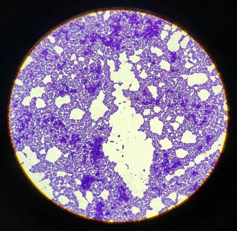
Abstract
  Antibiotic resistant bacteria are becoming an increasing threat worldwide, particularly in the
healthcare setting. This has led researchers and healthcare providers to begin looking elsewhere
for solutions. Research suggests that curcumin, a phenolic compound from the spice turmeric,
has antibacterial properties that may be able to treat potentially life-threatening hospital
infections, such as those caused by Staphylococcus aureus and Pseudomonas aeruginosa.
Turmeric has been used in Asian medicine for thousands of years as a general antimicrobial.
Curcumin was utilized in this study, along with hispolon, another phenolic compound isolated
from various mushrooms, such as Inonotus hispidus and Phellinus linteus, a medicinal
mushroom. There is less prior data on hispolon as an antibacterial agent, but it has been found to
be a potentially effective antiviral and antitumor treatment. Promising research done so far with
hispolon as an antitubercular drug suggests that it may have some antibacterial properties as
well. In this study, curcumin, hispolon mono methylether (HME), and hispolon pyrazole (HP)
were tested on Staphylococcus aureus, Pseudomonas aeruginosa, Enterococcus faecalis,
Escherichia coli, and Mycobacterium smegmatis. The results obtained through colony forming
unit assays, % inhibition calculations, growth curves, biofilm assays, and live-dead fluorescent
microscopy suggest that HME has significant antibacterial effects on all of the microorganisms
used except for E. faecalis, where the effects are only moderate, while curcumin has moderate
antibacterial effects on all but M. smegmatis. HP did not have strong antibacterial effects on
gram positive or gram negative bacteria, and only seemed to be effective against M. smegmatis.

                                                                                                1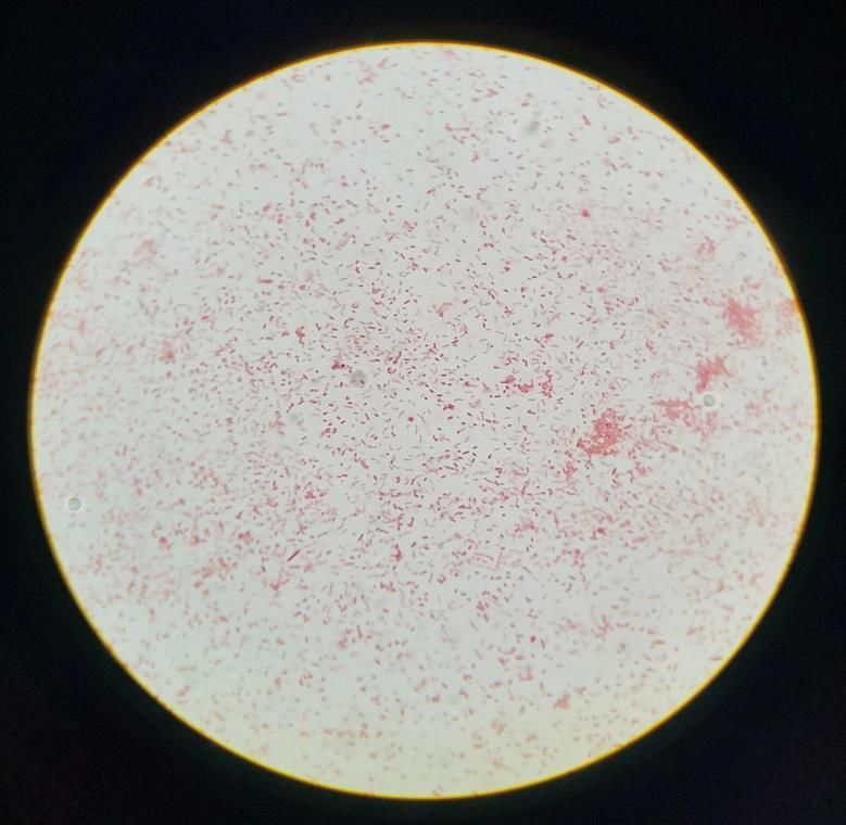
Master of Science

  College of Science and Mathematics

Biology

                                        2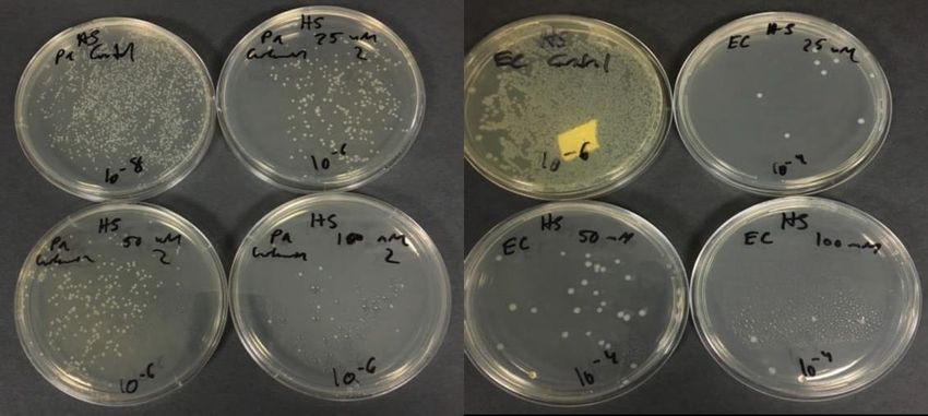
CURCUMIN AND HISPOLON AS POTENTIAL ANTIBACTERIAL AGENTS

                              A THESIS

          Submitted in partial fulfillment of the requirements
                 For the degree of Master of Science

                                  by
                HALEIGH ELIZABETH SULLIVAN

                      Montclair State University
                            Montclair, NJ
                                 2019

                                                                 3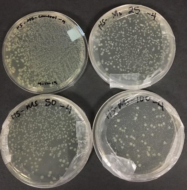
Copyright C 2019 by Haleigh Elizabeth Sullivan. All rights reserved.

                                                                       4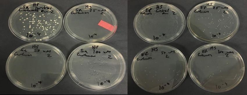
Acknowledgments
 I would like to thank Dr. Lee and Dr. Adams at Montclair State University for their amazing
mentorship and patience throughout this project. I would also like to thank Dr. DiLorenzo as a
member of my thesis committee.

  Thank you to Adam Parker and Rose Lipala for help with machine tutorials and media
preparation. Thank you to Theresa Aponte for always being around to help everyone in the lab.
Thank you to Diana Sanchez Castellanos for help with this project. Thank you to Summer
Elsayed for the neverending emotional support during this experience.

  Thank you to all of the members of Dr. Lee’s lab. Thank you to all of the Biology GAs, and
thank you to the biology department for their support these past two years.

 Last but not least, thank you to my friends and family for their unwavering support throughout
my academic career.

                                                                                                 5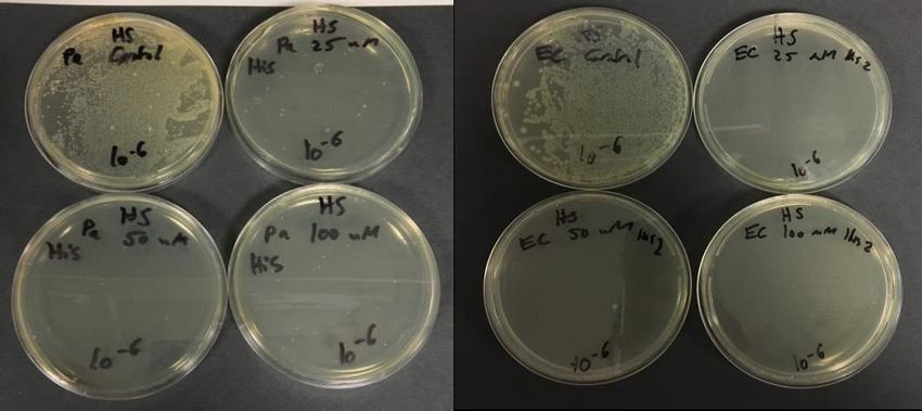
Table of Contents
Abstract… ........................................................................................... 1
Acknowledgments................................................................................5
List of Figures… ................................................................................. 7
List of Tables… ................................................................................... 8
Introduction: Literature Review… ....................................................... 9
Materials and Methods ........................................................................ 26
Results and Discussion… ....................................................................33
Conclusions ......................................................................................... 72
References… ....................................................................................... 76

                                                                                                           6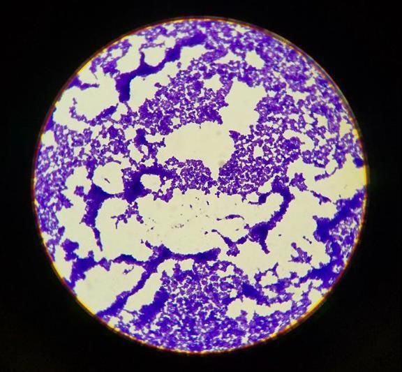
List of Figures:
Figure 1: Factors of Pathogenicity in Bacteria… .............................................10
Figure 2: Antibiotic Modes of Action… ......................................................... 11
Figure 3: Antibiotic Resistance in Gram Positive and Negative Bacteria… .. 13
Figure 4: Gram Stain of Staphylococcus aureus .............................................15
Figure 5: Gram Stain of Enterococcus faecalis .............................................. 16
Figure 6: Gram Stain of Pseudomonas aeruginosa ........................................ 17
Figure 7: Gram Stain of Escherichia coli ....................................................... 18
Figure 8: Acid-fast Stain of Mycobacterium smegmatis................................19
Figure 9: Structure of Curcumin… ................................................................. 20
Figure 10: Structure of Hispolon… ................................................................ 22
Figure 11: BLAST Results for Staphylococcus aureus .................................. 33
Figure 12: BLAST Results for Enterococcus faecalis .................................... 34
Figure 13: BLAST Results for Escherichia coli ............................................. 34
Figure 14: BLAST Results for Pseudomonas aeruginosa .............................. 35
Figure 15: BLAST Results for Mycobacterium smegmatis ............................ 35
Figure 16: Screening of Curcumin Effectiveness ........................................... 37
Figure 17: Screening of HME Effectiveness .................................................. 39
Figure 18: Screening of HP Effectiveness ...................................................... 41
Figure 19: CFU Assays for Curcumin Treated Bacteria… ............................. 43
Figure 20: CFU Assays for HME Treated Bacteria… .................................... 44
Figure 21: CFU Assays for HP Treated Bacteria .............................................45
Figure 22: % Inhibition for Curcumin Treated Bacteria… ............................. 47
Figure 23: % Inhibition for HME Treated Bacteria… .................................... 49
Figure 24: % Inhibition for HP Treated Bacteria.............................................51
Figure 25: Growth Curve for Staphylococcus aureus......................................53
Figure 26: Growth Curve for Enterococcus faecalis ....................................... 54
Figure 27: Growth Curve for Escherichia coli ................................................ 55
Figure 28: Growth Curve for Pseudomonas aeruginosa ................................. 56
Figure 29: Growth Curve for Mycobacterium. smegmatis .............................. 57
Figure 30: Congo Red Assay. .......................................................................... 58
Figure 31: Crystal Violet Assay… ................................................................... 60
Figure 32: Resazurin Assay… .......................................................................... 62
Figure 33: Live-Dead Assay of Staphylococcus aureus ................................... 64
Figure 34: Live-Dead Assay of Enterococcus faecalis......................................66
Figure 35: Live-Dead Assay of Escherichia coli .............................................. 68
Figure 36: Live-Dead Assay of Pseudomonas aeruginosa................................70
Figure 37: Live-Dead Assay of Mycobacterium smegmatis ............................. 71

                                                                                                            7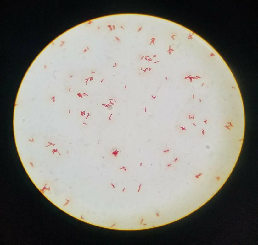
List of Tables
Table 1: % Inhibition Values for Curcumin Treated Bacteria ....................... 48
Table 2: % Inhibition Values for HME Treated Bacteria… .......................... 50
Table 3: % Inhibition Values for HP Treated Bacteria… .............................. 52
Table 4: % Inhibition Values for Crystal Violet Assay… ............................. 61
Table 5: % Inhibition Values for Resazurin Assay… .................................... 63

                                                                                            8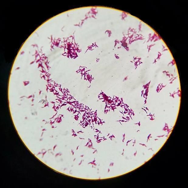
1. Introduction
   Many kinds of bacteria are beneficial in various aspects of life. Healthy humans tend to have

about 1014 bacteria just in their gut alone (Zhang et al., 2015). These bacteria play many

important roles in health, such as helping with digestion and providing certain nutrients (Zhang

et al., 2015). However, not all bacteria are beneficial, and there is a significant number that can

cause dangerous illnesses in humans when introduced into the body. Bacterial infections have

been a problem for people for as long as humans have existed, and they can be caused by various

type of pathogenic bacteria.

Pathogenicity

 There are many factors that make certain bacteria pathogenic. Many bacteria produce toxins,

such as the lipopolysaccharide endotoxin in gram negative bacteria and teichoic acid in gram

positive bacteria (Wilson et al., 2002). Toxins are generally classified into different categories,

including A-B toxins, commonly produced by gram negative bacteria such as Escherichia coli,

and toxins that form pores in host cell membranes (Wilson et al., 2002). Some pathogenic

bacteria are enclosed in capsules. These give them extra protection from destruction by

macrophages in the host immune system, making it easier for these bacteria to evade host

defenses (Wilson et al., 2002). The cell wall can also determine different virulence factors. As

previously mentioned, most bacteria are classified as gram negative or gram positive, depending

on the peptidoglycan composition in their cell wall (Wilson et al., 2002). Some bacteria also

have the ability to break down host tissues in order to invade them. Staphylococcus aureus is an

example of a pathogenic bacterium that uses this method, and once the tissues are invaded, it is

easier for the bacteria to spread to other areas of the body, which then puts the host at risk for

sepsis (Wilson et al., 2002). There are many mechanisms that pathogenic bacteria use to evade

                                                                                                      9
the host immune system and successfully cause disease. Therefore, there need to be treatments to
either kill these bacteria, or help the immune system eliminate them from the body (Figure 1).
Figure 1. Some features that contribute to pathogenicity in bacteria (Wilson et al., 2002)
Antibacterial Treatments
Since the discovery of penicillin as an antibacterial agent by Alexander Fleming, antibacterial
compounds called antibiotics have been an effective treatment for many bacterial infections
(Lobanovska & Pilla, 2017). The mode of action for this particular antibiotic is to prevent
peptidoglycan formation, which can then potentially result in the bacterial cell bursting
(Lobanovska & Pilla, 2017). The discovery of penicillin has led to the development of other
antibiotics over time, since different bacteria are susceptible to different ones. For example, gram
positive and gram negative bacteria may respond differently to various antibacterial compounds
(Lobanovska & Pilla, 2017). Some of these antibiotics include quinolones, which block bacterial
DNA replication, aminoglycosides, which inhibit the 30S ribosomal subunit, and macrolides,
which prevent protein synthesis (Kapoor et al., 2017). There are currently many different
10antibiotics used in modern medicine that have a wide range of actions against pathogenic
bacteria (Figure 2). Unfortunately, despite the fact that antibiotics have been in use for less than
one hundred years, they are quickly becoming less effective.
Figure 2. Various modes of action of different antibiotics (Kapoor et al., 2017)
Antibiotic Resistance
Bacteria that are no longer responding to various types of antibiotics are a growing problem in
the medical field, leading doctors and scientists to search elsewhere for a solution. Unfortunately,
a fairly wide range of bacteria are becoming antibiotic resistant, including those that are both
gram positive and gram negative (Tyagi et al., 2015). In hospitals, where these super bug
infections are especially a concern, bacteria such as Pseudomonas aeruginosa and
Staphylococcus aureus are becoming an increasing problem (Tyagi et al., 2015). Bacterial
infections that can no longer be treated with antibiotics pose a serious threat to future humans.
The rise in antibiotic resistance has been attributed mainly to over prescribing antibiotics, as well
as people misusing them, such as stopping the medication before instructed by their physician
11(Ventola, 2015). Scientists project that by the year 2050, as many as 10 million people each year
could die from infections caused by antibiotic resistant bacteria (Banin et al., 2017). This issue
has led researchers to begin investigating other potential antibacterial compounds to eventually
treat some of these infections.
There are various mechanisms that different bacteria use to become resistant to antibiotics.
Gram negative bacteria are generally more resistant to antibacterial agents than gram positive
bacteria due to the fact that they have an extra cell wall (Zgurskaya et al., 2015). A fairly
common mechanism that leads to antibiotic resistance is horizontal gene transfer (HGT), where
one bacterium receives foreign segments of DNA from a different bacterium (Munita & Arias,
2016). Through this, a gene for resistance to certain antibiotics can be passed on to bacteria that
did not previously have them. HGT is a major reason for evolution in bacteria as well (Munita &
Arias, 2016). Gram negative bacteria do have some mechanisms that are different from gram
positive bacteria. Many gram negative bacteria have started producing enzymes known as β-
lactamases, which can break down β-lactam rings in certain antibiotics, while a common reason
for antibiotic resistance in gram positive bacteria includes them modifying sites where antibiotics
would normally bind (Munita & Arias, 2016). In addition to these methods, gram negative and
gram positive bacteria both may have efflux pumps, which they can use to quickly pump the
antibiotics out of the cell before it can be killed (Munita & Arias, 2016) (Figure 3). Therefore,
regardless of gram classification, antibiotic resistance is becoming a very significant issue
worldwide.
12Figure 3. A visual representation of various efflux pumps used in gram positive and gram
negative bacteria (Munita & Arias, 2016).
Antibiotic resistance is also a growing problem in Mycobacterium tuberculosis, which is acid-
fast instead of gram positive or negative (Balaji et al., 2016). Acid-fast positive bacteria, like
gram positive and gram negative bacteria, do have peptidoglycan in their cell wall (Alderwick et
al., 2015). However, this is not the main component of their cell wall. Acid-fast cell walls are set
apart by the large number of mycolic acids they contain, making the cell wall thick, waxy, and
difficult for various drugs to permeate (Takayama et al., 2005, Smith et al., 2013). Gram positive
bacteria have a thick peptidoglycan cell wall, while gram negative bacteria have only a thin layer
of peptidoglycan but also an additional membrane that assists them in drug resistance (Mai-
Prochnow et al., 2016). The ability for bacteria to develop methods for antibiotic resistance
regardless of cell wall composition shows what a truly widespread problem this is. In
comparison to gram negative and gram positive bacteria, there is already a limited range of
antibiotics that can treat diseases caused by acid-fast bacteria, such as tuberculosis (Smith et al.,
2013). Horizontal gene transfer is also a common way for acid-fast bacteria like M. tuberculosis
13to become antibiotic resistant, as well as the very slow passage of antibacterial molecules
through the very thick cell wall (Smith et al., 2013). These bacteria can also modify drugs to
make them less harmful, as well as using various enzymes to break them down. The particularly
thick cell wall of acid-fast bacteria already makes it difficult for most antibiotics to permeate
(Smith et al., 2013). These many mechanisms of antibiotic resistance make it clear that disease
causing acid-fast bacteria are just as much of an increasing problem as gram negative and gram
positive bacteria are.
Staphylococcus aureus
Staphylococcus aureus (S. aureus) is a gram positive coccus that can cause significant
infections, such as sepsis and infective endocarditis (Teow et al., 2016). These bacteria are
currently most well-known for its growing resistance to many types of antibiotics and its ability
to cause deadly skin infections (Teow et al., 2016). S. aureus related diseases that are difficult to
treat with antibiotics are becoming an increasing problem in healthcare settings, specifically
methicillin-resistant Staphylococcus aureus (MRSA) (Kong et al., 2016). Many strains of S.
aureus are also exhibiting resistance to the antibiotic vancomycin (McGuinness et al., 2017).
Unfortunately, in addition to the strains of S. aureus resistant to one specific drug, there are also
numerous strains that are multidrug resistant, making them extremely possible to treat and the
infections they cause very life-threatening (Onanuga & Temedie, 2011). The lack of response
this pathogen has to certain antibiotics is leading healthcare professionals to start to look
elsewhere for treatments. MRSA is transmittable from person to person, and based on recent
studies, has been found to be evolving at a fast rate (Kong et al., 2016). Due to its growing threat
to the world, S. aureus is a popular target for research.
14Figure 4. Gram Stain of Gram Positive Coccus Staphylococcus aureus (Aponte 2018)
Enterococcus faecalis
Enterococcus faecalis (E. faecalis) is a gram positive coccus that can be found in the gut as
part of normal flora of many organisms, including mammals (Van Tyne et al., 2013). Due to
shedding through feces, E. faecalis can also be found in nature, although it prefers environments
that are low in oxygen, such as the intestines (Van Tyne et al., 2013). However, as in the case of
other types of bacteria, overuse of antibiotics has resulted in E. faecalis becoming a growing
health threat. Virulent strains of E. faecalis are currently one of the main causes of multidrug
resistant infections, such as bacteremia. These infections are made more dangerous by the
presence of an exotoxin called cytolysin, which can burst the cells of the host (Van Tyne et al.,
2013). Since E. faecalis is becoming more resistant to traditional antibiotics, it is also often
included in research searching for new antibacterial treatments.
15Figure 5. Gram Stain of Gram Positive Coccus Enterococcus faecalis (Aponte 2018)
Pseudomonas aeruginosa
Pseudomonas aeruginosa (P. aeruginosa) is a gram negative bacillus often found in soil
(Gellatly & Hancock, 2013). It also can cause various infections in humans, and these infections
are growing increasingly difficult to treat with current antibiotics. Like other difficult to treat
infections, pseudomonal infections are especially a threat in healthcare settings (Gellatly &
Hancock, 2013). Some of these infections include bacteremia, ear infections, and, most
commonly, respiratory tract infections (Gellatly & Hancock, 2013). The respiratory infections
are considered highly dangerous, but unfortunately are difficult to treat. P. aeruginosa also has
the ability to form biofilms that are difficult to break up. Researchers are looking for treatments
that will affect quorum sensing and virulence genes (Gellatly & Hancock, 2013). Drug resistance
in P. aeruginosa is mainly due to a poorly permeable outer membrane (Gellatly & Hancock,
162013).
Figure 6. Gram Stain of Gram Negative Bacillus Pseudomonas aeruginosa (Aponte 2018)
Escherichia coli
Escherichia coli (E. coli) is a gram negative bacillus that is very commonly found in the
healthy gut of humans (Jang et al., 2017). However, certain strains are also associated with
gastrointestinal illnesses in humans. The most well-known E. coli serovar is O157:H7, which is
often in the news for severe food poisoning cases that result in stomach cramping, vomiting, and
bloody diarrhea (Jang et al., 2017). While food is a common source of pathogenic E. coli, water
can contain the bacteria as well. When testing water quality, the presence or absence of E. coli is
used to determine whether or not the water is contaminated with feces (Jang et al., 2017). Like
many other types of bacteria in recent years, E. coli has been developing resistance to many of
the commonly used antibiotics. This is mainly due to the fact that E. coli found within the gut is
17subject to the antibiotics consumed by their host (Jang et al., 2017). This poses a threat to human
health and is leading researchers to look for new antibacterial treatments.
Figure 7. Gram Stain of Gram Negative Bacillus Escherichia coli (Aponte 2018)
Mycobacterium smegmatis
Mycobacterium smegmatis (M. smegmatis) is an acid-fast bacteria that does not take up any
regular gram staining dyes due to its extremely waxy cell membrane, a result of the presence of
mycolic acids (Wu et al., 2018). M. smegmatis is the less dangerous relative of M. tuberculosis,
the causative agent of tuberculosis (Smith, 2003). Researchers can use M. smegmatis as a safer
way to learn about M. tuberculosis. However, it is important to note that they are different from
one another. M. smegmatis has become a research target because of the growing incidence of
antibiotic resistance in cases of tuberculosis (Sotgiu et al., 2015). This is making it necessary for
new anti-tubercular drugs to be discovered.
18Figure 8: Acid-fast Stain of Acid-fast Positive Bacillus Mycobacterium smegmatis (Aponte
2018)
Curcumin
A compound that has provided promising results in terms of being a future antibacterial agent
is curcumin, a polyphenolic compound that comes from the spice turmeric (Tyagi et al., 2015)
(Figure 9). Turmeric can be consumed and is often used in various foods or mixed into liquids,
such as teas. Turmeric is a rhizome, and its scientific name is Curcuma longa (Tyagi et al.,
2015). Records show that people have been using this rhizome as a broad antimicrobial for quite
a long time, and that it does have potential antifungal, antibacterial, and antiviral activity
(Moghadamtousi et al., 2014). The earliest known use of turmeric as an herbal medicine goes as
far back as about 4000 years ago, where it was used in India. Eventually its use spread
throughout Asia, and it was a popular spice in trading (Prasad & Aggarwal, 2011). The antiviral
19properties of curcumin have been thoroughly investigated, and it has been determined to work
against several viruses. Some of these include human immunodeficiency virus (HIV), where
curcumin is believed to inhibit viral replication, and herpes simplex virus 1 (HSV-1), where host
cells treated with curcumin are less vulnerable to infection (Kutluay et al., 2008, Flores et al.,
2016). Now, scientists look to determine just how effective curcumin can be on different types of
drug resistant bacteria.
Figure 9. Structure of a Curcumin Molecule (Moghadamtousi et al., 2014)
Currently, methicillin-resistant Staphylococcus aureus (MRSA) poses a serious threat to
patients who are hospitalized (Gupta et al., 2015). This illness has a very high mortality rate,
leaving healthcare professionals and researchers desperate for another solution. Staphylococcus
aureus that is resistant to antibiotic treatment is thought to be one of the most common causes of
infection in the United States (Green et al., 2012). Fortunately, research so far has found
curcumin to be especially effective against Staphylococcus aureus, which is incredibly important
for the future of medicine and the growing issue of antibiotic resistance (Teow et al., 2016).
However, much further research is needed before curcumin is ready to be tested as a new
treatment. This is also the case for a variety of antibiotic resistant bacteria, including
Enterococcus faecalis, Pseudomonas aeruginosa, and Escherichia coli, which have all been
20found to be susceptible to curcumin so far (Tyagi et al., 2015). It has been determined that
curcumin’s mode of action in both gram positive and gram negative bacteria is to compromise
the bacterial membrane, killing the cell (Tyagi et al., 2015). There is also potential for curcumin
to be effective against biofilms. Research done with P. aeruginosa, another threat to patients
staying in a hospital, suggests that curcumin may inhibit genes associated with biofilm
formation, such as quorum sensing genes (Moghadamtousi et al., 2014). However, it is also
cautioned that it cannot yet be determined definitively that curcumin is effective against P.
aeruginosa. Overall, however, research done with curcumin as an antibacterial agent shows
promising results.
Hispolon
Hispolon, a compound similar in structure to curcumin with only a difference in one aromatic
ring, is also being explored by various researchers as a potential antibacterial agent (Ravindran et
al., 2010). Hispolon is a polyphenolic compound that has been isolated from various mushrooms,
such as Inonotus hispidus, and the medicinal mushrooms Phellinus linteus and Phellinus
igniarius (Chethna et al., 2018). It has various derivatives, including hispolon monomethyl ether
(HME) (Figure 10) and hispolon pyrazole (HP) (Chethna et al., 2018). It is also considered a
curcuminoid derivative (Amalraj et al., 2016). So far, its role as an antibacterial agent is less
certain than curcumin. Hispolon is already a known antitumor agent, and it has been suggested to
have antiviral properties as well (Chethna et al., 2018). Using this compound as a potential
antitumor drug has been the most researched, with much work still needed in terms of hispolon
being an antiviral and antibacterial agent. Currently, hispolon has been shown to stop cell
proliferation and induce apoptosis in a wide variety of cancers (Kim et al., 2016). The
antibacterial effects of hispolon have not been widely explored, unlike the antibacterial effects of
21curcumin, especially in terms of how they would work on antibiotic resistant bacteria. However,
some early research suggests that various hispolon derivatives may be effective against the
Mycobacterium genus, leaving it as a possible new treatment for tuberculosis in the future (Balaji
et al., 2016). Despite the similarity in structure of hispolon to curcumin, past studies have not
found curcumin to be effective against Mycobacterium (Balaji et al., 2016). Currently, the
hispolon pyrazole derivatives have been found to be especially effective against Mycobacterium,
likely through preventing synthesis of mycolic acids (Balaji et al., 2019). It is uncertain as to
whether or not these specific derivatives have another mechanism to make them active against
gram positive and gram negative bacteria. Hispolon derivatives such as hispolon mono
methylether are thought to possibly inhibit the production of the enzyme fatty acid synthase II, as
well as preventing the synthesis of mycolic acid in Mycobacterium (Balaji et al., 2016). This
could indicate that these derivatives could be active against gram positive and negative bacteria
to some extent, as well as being active against acid-fast bacteria.
Figure 10. Structure of a Hispolon Derivative Hispolon Mono Methylether (Balaji et al., 2016)
Tuberculosis is a very serious respiratory illness that still affects millions of people worldwide
every year. It is caused by the bacterium Mycobacterium tuberculosis (Sulis et al., 2014). These
bacteria are highly virulent and often cause an inflammatory response in the lungs, resulting in
tissue damage. They also have an extremely thick, waxy cell wall, made up of mycolic acid,
making treatment difficult (Smith, 2003). Current treatment includes a rigorous antibiotic
22regimen, with some of the antibiotics effective against tuberculosis including isoniazid, rifampin,
pyrazinamide, kanamycin, and amikacin (Sotgiu et al., 2015). However, despite the wide range
of antibiotics available, deaths from tuberculosis are still fairly high, suggesting the need for
different antibacterial agents that may work more effectively. This is why it is significant that
some research suggests hispolon may be effective against Mycobacterium (Balaji et al., 2016).
Interestingly enough, even though curcumin is similar in structure, it does not appear to be
effective against the Mycobacterium genus (Balaji et al., 2016). The true antibacterial effect of
hispolon derivatives on other bacteria, such as S. aureus, E. faecalis, P. aeruginosa, and E. coli,
has yet to be determined.
Biofilms
Biofilms, especially in the healthcare setting, have the ability to be very dangerous to humans.
Biofilms result from bacteria forming a thick clump on some hard surface and then shielding
themselves with extracellular matrix (ECM) (Lόpez et al., 2010). The ECM is produced by the
bacteria in the biofilm and include polysaccharides, proteins, and some DNA (Lόpez et al.,
2010). There are many places biofilms can form, and although some may lead to persistent
infections in humans, there are some that are not necessarily harmful. An example of a neutral
biofilm would be in dental plaques (Lόpez et al., 2010). However, there are many cases where
the formation of biofilms can lead to infections that are difficult to treat. Many biofilms contain
persister cells, which contain toxins thought to block antibiotics from being effective (Lόpez et
al., 2010). Therefore, biofilms that contain these non-dividing cells are often resistant to a wide
range of antimicrobials, making them hard to cure (Lόpez et al., 2010).
Biofilm bacteria use a process called quorum sensing in order to communicate with one
another (Høiby et al., 2011). Through quorum sensing they are able to turn on genes that allow
23for the production of virulence factors, which help make the biofilm more aggressive (Høiby et
al., 2011). Mutation is also common in biofilms, increasing the probability of antibiotic
resistance. Unfortunately, biofilms are not only resistant to many antibiotics, but they are often
able to evade the response by the immune system as well (Høiby et al., 2011). Persister cells are
considered one main reason why antibiotic resistance occurs. The ECM itself is also thought to
be a source of antibiotic resistance, suggesting that it prevents effective diffusion of drugs into
the biofilm (Høiby et al., 2011). Most types of bacteria are able to form biofilms if given the
proper environment. Pseudomonas aeruginosa, one of the bacteria used in this study, is a
common cause of biofilms that threaten human health, particularly in cystic fibrosis patients
(Høiby et al., 2011). P. aeruginosa often causes lung infections in these patients, and it is
capable of causing skin infections as well (Lόpez et al., 2010). Naturally, biofilms threaten
health are an area of interest in research as scientists try to identify potential treatments that will
work more effectively than many of the current antimicrobials.
There is some evidence that antibiotics used in synergy with other antimicrobial compounds,
such as curcumin, may be more effective against biofilms than just antibiotics or curcumin alone
(Kali et al., 2016). Results from this research so far suggest that when antibiotics and curcumin
are used together, curcumin is able to increase antibiotic susceptibility in biofilms, so then the
antibiotics are more successful at ending the infection as opposed to when either are working on
their own (Kali et al., 2016). Instead of current antibiotics being phased out all together, the
solution may be to just use them along with another antibacterial compound. Examples of this
have been seen specifically in regard to Staphylococcus aureus. Researchers have looked at the
use of curcumin and antibiotics together against methicillin resistant Staphylococcus aureus
(MRSA) strains to see if the curcumin is able to reduce antibiotic resistance (Mun et al., 2013).
24S. aureus in particular is an antibiotic resistance threat, making it very popular in research
looking for new antimicrobials. Thus far research has found that the use of curcumin with
antibiotics such as quinolones and β-lactams against MRSA does in fact make the bacteria more
susceptible to the antibiotics by decreasing minimal inhibitory concentration (MIC) (Mun et al.,
2013). It is cautioned that more research in this area is needed, particularly with other resistant
bacteria besides S. aureus, but early results are promising.
Mode of Action
The exact mechanisms by which the antibacterial activity of curcumin and hispolon occur are
still to be determined. Since curcumin has been a much larger antibacterial target in current
research, scientists do have some ideas as to how this compound stops bacterial growth.
Currently results show that curcumin may act by causing leaking in the cell membrane, both in
gram negative and gram positive bacteria (Tyagi et al., 2015). This eventually causes the cell to
burst and results in death, preventing further reproduction (Tyagi et al., 2015). There is more of a
question as to exactly how hispolon works in killing bacteria that are not acid-fast. Studies that
have focused on using hispolon compounds to treat tuberculosis suggest that both hispolon and
hispolon pyrazole derivatives may target enzymes required to synthesize mycolic acid, which is
essential in the Mycobacterium cell wall (Balaji et al., 2016, Balaji et al., 2019). If the ability to
synthesize mycolic acid is hindered, that will prevent the replication of the bacteria. It is thought
that this is done through inhibiting mtFABH, the Mycobacterium specific version of the beta-
ketoacyl-synthase III enzyme that plays a role in fatty acid synthesis (Balaji et al., 2016, Brown
et al., 2005). mtFABH is essential for the synthesis of mycolic acids in acid-fast bacteria, and its
inhibition would pose a serious problem for replication (Balaji et al., 2016). Studies suggest that
this is one mode of action by hispolon derivatives, and so far the main mode of action by
25hispolon pyrazoles (Balaji et al., 2016, Balaji et al., 2019). However, this does not explain the
mode of action hispolon compounds may have in gram positive and gram negative bacteria,
since their cell walls are made differently. It has also been suggested that the regular hispolon
derivatives may block the action of fatty acid synthase II, which could affect fatty acid synthesis
in all bacteria (Balaji et al., 2016). This is the mode of action of some current antibiotics (Zhang
et al., 2006). Further research focusing on hispolon compounds as antibacterial agents is needed
in order to determine its mode of action on these bacteria.
Objectives of This Study:
1) Determine the antibacterial effects of curcumin on S. aureus, E. faecalis, E. coli, P.
aeruginosa, and M. smegmatis.
2) Determine the antibacterial effects of HME on S. aureus, E. faecalis, E. coli, P. aeruginosa,
and M. smegmatis.
3) Determine the antibacterial effects of HP on S. aureus, E. faecalis, E. coli, P. aeruginosa, and
M. smegmatis.
4) Determine if curcumin and HME are effective against biofilms at 50 µM concentrations.
5) Determine if one compound is more effective than the other.
2. Materials and Methods
Preparation of Bacterial Cultures:
Stock cultures of S. aureus, E. faecalis, E. coli, and P. aeruginosa were prepared by
inoculating 15 ml test tubes of previously prepared Brain Heart Infusion (BHI) broth with pure
cultures prepared on nutrient agar plates. M. smegmatis stock cultures were prepared by
inoculating 15 ml test tubes of previously prepared 7H9 neat media with pure cultures prepared
26on LB CB+CHX agar plates. All tubes were incubated overnight at 37°C. Stock cultures were
then stored at 4°C. The purity of the cultures was checked periodically.
Preparation of Curcumin Stock:
A 10X stock solution of curcumin was prepared by dissolving curcumin powder in
dimethylsulfoxide (DMSO). The stock was wrapped in aluminum foil and stored at 4°C. The
10X solution was further diluted as required to produce final concentrations of 25 μM, 50 μM,
and 100 μM.
Preparation of HME Stock:
A 10X stock solution of HME was prepared by dissolving HME powder in dimethylsulfoxide
(DMSO). The stock was wrapped in aluminum foil and stored at 4°C. The 10X solution was
further diluted as required to produce concentrations of 25 μM, 50 μM, and 100 μM.
Preparation of HP Stock:
A 10X stock solution of HP was prepared by dissolving HP powder in dimethylsulfoxide
(DMSO). The stock was wrapped in aluminum foil and stored at 4°C. The 10X solution was
further diluted as required to produce concentrations of 25 μM, 50 μM, and 100 μM.
DNA Extraction, PCR, and Sequencing to Confirm the Purity of the Cultures
Methods from Lee et al., 2018: In order to determine if the five bacterial cultures were pure and
correct, polymerase chain reaction (PCR) was performed using 16S forward and reverse primers,
27F (5’- AGA GTT TGA TCC TGG CTC AG-3’) and 1492R (5’- ACG GCT ACC TTG TTA
CGA CTT-3’) respectively. The PCR products were then sequenced and the microorganisms
were determined by BLAST search. DNA extraction was first performed on all five
microorganisms by adding 500 µl of each bacterium to five separate 1.5 ml microcentrifuge
27tubes each containing 100 µl of InstaGene. The tubes were vortexed for 30 seconds and then
placed in a 100°C heating block for ten minutes. After the heating period, tubes were
immediately placed on ice for one minute and then vortexed for 30 seconds. Tubes were then
centrifuged at maximum speed for one minute to separate the extracted DNA into the
supernatant. PCR reaction tubes were then prepared for each of the five microorganisms. To each
tube, 20 µl of Taq MasterMix, 2 µl of forward primer, 2 µl of reverse primer, 14 µl of nuclease
free sterile water, and 2 µl of microbial DNA were added for a total volume of 40 µl (Lee et al.,
2018). PCR was completed by the thermalcycler under the following conditions: 95°C for one
minute for initial denaturation, 25 cycles of 95°C (10 seconds), 55°C (10 seconds), and 72 °C (10
seconds), and then 72°C for 10 minutes. The PCR products were confirmed through gel
electrophoresis and samples were sent out for sequencing. Sequences were copy and pasted into
the BLAST computer program to see if the correct bacteria were isolated.
Curcumin Colony Forming Unit (CFU) Assay:
Curcumin treatments were prepared from the 10X stock solution by diluting it to the desired
final concentrations of 25 µM, 50 µM, and 100 µM. Untreated cultures were used as a control.
Three repeatings were carried out for each condition and each microorganism. For each of the
microorganisms, 99 µl of bacteria were treated with 1 µl of each of the three concentrations in
microcentrifuge tubes. Treated bacteria were incubated at room temperature for 2 hours (120
minutes). Serial dilutions were prepared using 990 μl of water in 1.5 ml microcentrifuge tubes.
For each dilution, 10 μl of treated bacteria were added to the 990 μl of water. Final dilutions for
S. aureus, E. coli, and E. faecalis were 10-4. Final dilutions for P. aeruginosa and M. smegmatis
were 10-6. For each of the 15 samples, 100 μl of each sample was plated onto 15 different
nutrient agar plates (3 plates for each microorganism: 25 μM, 50 μM, 100 μM). Control plates
28were prepared for each of the five microorganisms, where serial dilutions were performed as
mentioned previously and 100 μl of each of the untreated microorganisms were plated on
previously prepared nutrient agar plates. Final dilutions for S. aureus, E. coli, and E. faecalis,
were 10-4. Final dilutions for M. smegmatis were 10-6. Final dilutions for P. aeruginosa were 10-
8
. All plates were inverted and incubated overnight at 37°C. M. smegmatis plates were incubated
for an additional 6 days. Colony forming units (CFUs) were counted. There were three
repeatings for each microorganism. The mean and standard deviation for each microorganism at
each treatment concentration were obtained to prepare graphs for CFU data. Each of the five
microorganisms were also treated with 2% DMSO to determine the effect of DMSO at this
concentration and lower on the bacteria.
HME Colony Forming Unit (CFU) Assay:
HME treatments were prepared from the 10X stock solution by diluting it to the desired final
concentrations of 25 µM, 50 µM, and 100 µM. Untreated cultures were used as a control. Three
repeatings were carried out for each condition and each microorganism. For each of the
microorganisms, 99 µl of bacteria were treated with 1 µl of each of the three concentrations in
microcentrifuge tubes. Treated bacteria were incubated at room temperature for 2 hours (120
minutes). The following procedures are the same as described above.
HP Colony Forming Unit (CFU) Assay:
Curcumin treatments were prepared from the 10X stock solution by diluting it to the desired
final concentrations of 25 µM, 50 µM, and 100 µM. Untreated cultures were used as a control.
Three repeatings were carried out for each condition and each microorganism. For each of the
microorganisms, 99 µl of bacteria were treated with 1 µl of each of the three concentrations in
29microcentrifuge tubes. Treated bacteria were incubated at room temperature for 2 hours (120
minutes). The following procedures are the same as described above.
% Inhibition Calculations
CFU data collected from all five microorganisms treated with different concentrations of
curcumin, HME, and HP was averaged to calculate % inhibition using the formula:
% inhibition= (control-treatment)/control X100. This was calculated for each microorganism
at each treatment concentration for all three compounds. Values were then graphed to compare
% inhibition between concentrations and compounds for each of the five microorganisms.
The Effect of Curcumin and HME on the Growth of Microorganisms
Growth curves were performed on S. aureus, E. faecalis, P. aeruginosa, E. coli, and M.
smegmatis untreated, treated with curcumin (50 µM), and treated with HME (µM). A 48-well
plate was used, and three repeatings were carried out for each of the five bacteria under each of
the three conditions (untreated, treated with curcumin, and treated with HME). All bacterial
samples were diluted with nutrient broth to approximately 0.1 optical density (OD) at 600 nm.
The growth was monitored for the five microorganisms untreated, treated with 50 μM curcumin,
and treated with 50 μM HME. To each of these wells, 450 μl of bacteria were added. For the
untreated, 50 μl of media was added. For the curcumin treated, 50 μl of a 1X curcumin solution
at a final concentration of 50 μM was added. For the HME treated, 50 μl of a 1X HME solution
at a final concentration of 50 μM HME was added. The three remaining wells were used for
negative controls: one with only media (nutrient broth), one with media and curcumin, and one
with media and HME. Readings were done every hour, starting at 0 hours and going to 10 hours
using the microplate reader. A final reading was taken after 24 hours. The three repeatings for
each microorganism under each condition were averaged and graphed on Excel to determine the
30overall growth over the 10 hour span. Standard deviation was calculated and shown on the graph
as well.
Biofilm Evaluation- Congo Red Assay:
A 24-well Congo Red plate was utilized to test curcumin and HME effectiveness on biofilms.
90 μl of S. aureus, E. faecalis, E. coli, P. aeruginosa, and M. smegmatis were each incubated
with 10 μl of a 1X curcumin solution (100 μM) at room temperature for two hours. In addition,
90 μl of S. aureus, E. faecalis, E. coli, P. aeruginosa, and M. smegmatis were each incubated
with 10 μl of 100 μM HME at room temperature for two hours. Four wells were utilized for each
microorganism: a negative control, the microorganism without treatment, the microorganism
treated with 50 μM curcumin for two hours, and the microorganism treated with 50 μM HME for
two hours. A total of 50 μl of each sample was inoculated into each well. The plate was then
incubated at 37°C and checked at 24, 48, and 72 hours.
Biofilm Evaluation- Crystal Violet Assay
For this assay to evaluate action against biofilms, a 48-well plate was prepared. Three
repeatings were done for each of the five bacteria (S. aureus, E. faecalis, E. coli, P. aeruginosa,
and M. smegmatis) under each of the three conditions (untreated, treated with curcumin, and
treated with HME). All bacterial samples were diluted with nutrient broth to approximately 0.1
optical density (OD). The plate included the five microorganisms untreated, treated with 50 μM
curcumin, and treated with 50 μM HME. To each of these wells, 450 μl of bacteria were added.
For the untreated samples, 50 μl of media was added. For the curcumin treated, 50 μl of 50 μM
curcumin was added. For the HME treated, 50 μl of 50 μM HME was added. The three
remaining wells were used for negative controls: one with only media (nutrient broth), one with
media and curcumin, and one with media and HME. The plate was incubated at 37°C for four
31days to allow formation of biofilms. On the fourth day, liquid was aspirated from all wells and
discarded. The wells were washed three times with 250 μl 1X PBS. To each well, 500 μl of 0.1%
crystal violet was added and allowed to stain for 30 minutes. The crystal violet was removed and
the wells were washed again with 250 μl 1X PBS. The plate was left at room temperature
overnight for the stain to dry. Excess stain was wiped up with sterile cotton swabs, and 1 ml of
30% acetic acid was added to each well. The microplate reader was then used to determine
optical density at 595 nm for each sample. Averages were taken for each microorganism under
each of the three conditions.
Biofilm Evaluation- Resazurin Assay
For this assay to evaluate biofilms, 48-well plate was used, and three repeatings were done for
each of the five bacteria under each of the three conditions (untreated, treated with curcumin, and
treated with HME). All bacterial samples were diluted with nutrient broth to approximately 0.1
optical density (OD). This growth curve included the five microorganisms untreated, treated with
50 μM curcumin, and treated with 50 μM HME. To each of these wells, 450 μl of bacteria were
added. For the untreated, 50 μl of media was added. For the curcumin treated, 50 μl of a 1X
curcumin solution at a final concentration of 50 μM was added. For the HME treated, 50 μl of a
1X HME solution at a final concentration of 50 μM was added. The three remaining wells were
used for negative controls: one with only media (nutrient broth), one with media and curcumin,
and one with media and HME. The plate was incubated at 37°C and 5% CO2 for four days in
order to allow biofilms to grow. After the four days, the liquid was aspirated from each well and
discarded. Each well was washed three times with 250 μl 1X PBS. An additional 200 μl 1X PBS
was added to each well, along with 200 μl of 20 μM Resazurin. The plate was wrapped in
aluminum foil and placed at 4°C overnight. The microplate reader was then used to measure
32excitation at 560 nm and emission at 590 nm. Averages were taken for each microorganism
under each of the three conditions.
Live-Dead Assay Using Fluorescent Microscopy
Stock plates were prepared for all five microorganisms on nutrient agar plates. To prepare
treatment tubes, colonies from the plates were added to 1.5 ml microcentrifuge tubes,
resuspended in 5 µl of nutrient broth, and treated with 2 µl of of a 1X curcumin or HME solution
at a final concentration of 50 μM for two hours. To prepare the control slides, a loopful of
bacteria was taken from the plate for each of the five microorganisms and smeared on the slide.
Following this step, 5 µl of 60 Red: 40 Green Live-Dead dye was added directly over the
bacterial sample on the slide. For the treatment slides for both curcumin and HME, 5 µl of the
treated bacteria were added to the slide, along with 5 µl of Syto®9-Green and Propidium iodide-
Red (live-dead dye). All slides were allowed to dry before being focused under the fluorescent
microscope and imaged at 40X. This was done for all five microorganisms including a control
slide, a curcumin treated slide, and an HME treated slide.
3. Results and Discussion
Bacterial Purity and Species Determination
Following DNA extraction and PCR of each of the five bacteria used in this study, the PCR
products were sequences. The sequences obtained were run through the BLAST computer
program at the NCBI website in order to determine if the stock samples were pure and correct.
Sequencing determined that the proper microorganisms were in fact being used (Figures 11-15).
Figure 11: BLAST Sequencing Results for S. aureus
CCCAATAATTCCGGATAACGCTTGCCACCTACGTATTACCGCGGCTGCTGGCACGTAGT
TAGCCGTGGCTTTCTGATTAGGTACCGTCAAAATGTGCACAGTTACTTACACATATGTTCTTCCC
TAATAACAGAGTTTTACGATCCGAAGACCTTCATCACTCACGCGGCGTTGCTCCGTCAGGCTTTC
GCCCATTGCGGAAGATTCCCTACTGCTGCCTCCCGTAGGAGTCTGGACCGTGTCTCAGTTCCAG
33TGTGGCCGATCACCCTCTCAGGTCGGCTATGCATCGTTGCCTTGGTAAGCCGTTACCTTACCAAC
TAGCTAATGCAGCGCGGATCCATCTATAAGTGACAGCAAGACCGTCTTTCACTTTTGAACCATGC
GGTTCAAAATATTATCCGGTATTAGCTCCGGTTTCCCGAAGTTATCCCAGTCTTATAGGTAGGTT
ATCCACGTGTTAC GCTTCTCTGATGTTAGCGGCGGACGGGTGAGTAACACGTGG
ATAACCTACCTATAAGACTGGGATAACTTCGGGAAACCGGAGCTAATACCGGATAATATTTTGAA
CCGCATGGTTCAAAAGTGAAAGACGGTCTTGCTGTCACTTATAGATGGATCCGCGCTGCATTAG
CTAGTTGGTAAGGTAACGGCTTACCAAGGCAACGATGCATAGCCGACCTGAGAGGGTGATCGGC
CACACTGGAACTGAGACACGGTCCAGACTCCTACGGGAGGCAGCAGTAGGGAATCTTCCGCAAT
GGGCGAAAGCCTGACGGAGCAACGCCGCGTGAGTGATGAAGGTCTTCGGATCGTAAAACTCTG
Figure 11. Staphylococcus aureus sequence (via NCBI, blast.ncbi.nlm.nih.gov).
Figure 12: BLAST Sequencing Results for E. faecalis
GTTACCGGGGCTGATCTGCGAGTCGTACGCTTCTTTCCTCCCGAGTGCTTGCACTCAATTGGAG
AGAGGAGTGGCGGCCGGGCGAGTATCACGTATGCGGCCTACCCATCAGAGCGGGATTACACTT
GGAAACAGATGCTTATACCGCATAACAGTTTATGCCGCATGCCATATGAGTGAAAGGCGCTTTC
GGGTGTCGCTGATGGATGGCCCCGCGGTGCATTAGCTAGTTGGTGAGATAACGGCTCACCAAGG
CCACCATGCATAGCCGACCTGAAACAGGATTCAACTCTACTGGGACTGACACAGGGTTGAGACT
CCTACGGGAGGCAGCAGTAGGGAATCTTCGGCAATGGACGAAAGTCTGACCGAGCAACGCCGC
GTGAGTGAAGAAGACATTCGGATCGTAAAACTCTGTTGTTAGAGAAGAACAAGGACGTTACTGA
CCTGAACGTCCCCTGACGGTATCTAACCAGAAAGCCACGGCTAACTACGTGCCAGCAGCCGCGG
GCAATACGTACGGAGGCAAGCGTTGTGCTGGATTTGATTGGGCGTAAAGCGAGCGCATGGCGT
GTGACTTACGTCTGATGTGACAGCACTCCGGCTTCAACCGGTGCAGGATCATTGTACACTCAGC
ACACTAGAGTGCAACACGAGGCAGAGTGGTATTCACATGATGCTAGCGACTGGAAATGGCACTA
CCTAGTGATGGTAGGGATCACGGGTTGGCTGCACCGACGGCTTCTCTAGGCTCTGGATAACCTC
GGACTGCCTGAAGACCTACGAAAAGGCCGTGGTTCAGCAACTGAGGGATTTCAGTATAACCCTT
GGCTAGGTCAATCGTCAGTAGAACGGAATGGTGGGACTAACGTGGTTCTTGCCATGCGTTCTGC
CGACTTCTATAGCAGTTGGCCTAGCAGTCAAACTGCAACTCCATGCCACGTGCCTGCACTGGCG
GGAGATAACTGTATACTCAGCTACACAGAGATTCGCTCA
Figure 12. Enterococcus faecalis sequence (via NCBI, blast.ncbi.nlm.nih.gov).
Figure 13: BLAST Sequencing Results for E. coli
CAGCACCAGCTACCATGCAGTCGACGGTACAGGAGCAGCTTGCTGCTTCGCTGACGAGTGGCGG
ACGGGTGAGTAATGTCTGGGAAACTGCCTGATGGAGGGGGATAACTACTGGAAACGGTAGCTAA
TACCGCATAACGTCGCAAGACCAAAGAGGGGGACCTTCGGGCCTCTTGCCATCGGATGTGCCCA
GATGGGATTAGCTAGTAGGTGGGGTAACGGCTCACCTAGGCGACGATCCCTAGCTGGTCTGAGA
GGATGACCAGCCACACTGGAACTGAGACACGGTCCAGACTCCTACGGGAGGCAGCAGTGGGGA
ATATTGCACAATGGGCGCAAGCCTGATGCAGCCATGCCGCGTGTATGAAGAAGGCCTTCGGGTT
GTAAAGTACTTTCAGCGGGGAGGAAGGGAGTAAAGTTAATACCTTTGCTCATTGACGTTACCCG
CAGAAGAAGCACCGGCTAACTCCGTGCCAGCAGCCGCGGTAATACGGAGGGTGCAAGCGTTAA
TCGGAATTACTGGGCGTAAAGCGCACGCAGGCGGTTTGTTAAGTCAGATGTGAAATCCCCGGGC
34TCAACCTGGGGAACTGCATCTGATACTGGCAAGCTTGAGTCTCGTAGAGGGGGGGTAGAATTCC
AGGTGTAGCGGTGAAATGCGTAGAGATCTGGAGGAATACCGGTGGCGAAGGCGGCCCCCTGCA
CGAAGACTGACGCTCAAGGTGCGAAAGCGTGGCGAGCAAACAGGATTAGATACCCTGGGTAGTT
CCACGCCGTAAAACGATGTCGACTTGGGAGGTTTGTGCCCTTGAGGCGTGCCTTCCGGGAGCTA
ACGCGTTTAAGTCGACCGCCATGGCGGAAGTACGGCTGCAAGGCCTAAAAACTTCTAACTGTAA
TGGAACTGGAGGGGAACTGCACAACCTCTGT
Figure 13. Escherichia coli sequence (via NCBI, blast.ncbi.nlm.nih.gov).
Figure 14: BLAST Sequencing Results for P. aeruginosa
CGGGGGACAGCTACACATGCAGTCGAGCGGAGAAGGGAGCTTGCTCCTGGATTCAGCGGCGGA
CGGGTGAGTAATGCCTAGGAATCTCGCCTGGTAGTGGGGGATAACGTCCGGAAACGGGCGCTA
ATACCGCATACGTCCTGAGGGAGAAAGTGGGGGATCTTCGGACCTCACGCTATCAGATGAGCCT
AGGTCGGATTAGCTAGTTGGTGGGGTAAAGGCCTACCAAGGCGACGATCCGTAACTGGTCTGAG
AGGATGATCAGTCACACTGGAACTGAGACACGGTCCAGACTCCTACGGGAGGCAGCAGTGGGG
AATATTGGACAATGGGCGAAAGCCTGATCCAGCCATGCCGCGTGTGTGAAGAAGGTCTTCGGAT
TGTAAAGCACTTTAAGTTGGGAGGAAGGGCAGTAAGTTAATACCTTGCTGTTTTGACGTTACCAA
CAGAATAAGCACCGGCTAACTTCGTGCCAGCAGCCGCGGTAATACGAAGGGTGCAAGCGTTAAT
CGGAATTACTGGGCGTAAAGCGCGCGTAGGTGGTTTCAGCAAGTTGGATGTGAAATCCCCGGGC
TCAAACCTGGGAACTGCATCCCAAAACTACTGAGCTAGAGTACGGGTAGAGGGTGGTGGAATTT
CCCTGTGTAGCCGGTGAAATGCGTAGAATAATAGGTAGGAACATCCAGTGTCGGAAGCGACCAC
CTTGGACTGATACCTGACACTGAAGTGGGCGAAAGCGTGGGGAGCAACAGAATAGATACCTTGC
TAGTCCACGCCGTAACGATGTCCGACTAGTCGTCGGAATCCTGGAACTTTATGCGCAGCTTAAC
GCATAGTGCACCGCTGGTATACGTCGCAGGGGTTTAC
Figure 14. P. aeruginosa sequence (via NCBI, blast.ncbi.nlm.nih.gov).
Figure 15: BLAST Sequencing Results for M. smegmatis
CTGAGATACGGCCCAGACTCCTACGGGAGGCAGCAGTGGGGAATATTGCACAATGGGCGC
AAGCCTGATGCAGCGACGCCGCGTGAGGGATGACGGCCTTCGGGTTGTAAACCTCTTTCAGCAC
AGACGAAGCGCAAGTGACGGTATGTGCAGAAGAAGGACCGGCCAACTACGTGCCAGCAGCCGC
GGTAATACGTAGGGTCCGAGCGTTGTCCGGAATTACTGGGCGTAAAGAGCTCGTAGGTGGTTTG
TCGCGTTGTTCGTGAAAACTCACAGCTTAACTGTGGGCGTGCGGGCGATACGGGCAGACTAGAG
TACTGCAGGGGAGACTGGAATTCCTGGTGTAGCGGTGGAATGCGCAGATATCAGGAGGAACAC
CGGTGGCGAAGGCGGGTCTCTGGGCAGTAACTGACGCTGAGGAGCGAAAGCGTGGGGAGCGAA
CAGGATTAGATACCCTGGTAGTCCACGCCGTAAACGGTGGGTACTAGGTGTGGGTTTCCTTCCT
TGGGATCCGTGCCGTAGCTAACGCATTAAGTACCCCGCCTGGGGAGTACGGCCGCAAGGCTAAA
35ACTCAAAGGAATTGACGGGGGCCCGCACAAGCGGCGGAGCATGTGGATTAATTCGATGCAACG
CGAAGAACCTTACCTGGGTTTGACATGCACAGGACGCCGGCAGAGATGTCGGTTCCCTTGTGGC
CTGTGTGCAGGTGGTGCATGGCTGTCGTCAGCTCGTGTCGTGAGATGTTGGGTTAAGTCCCGCA
ACGAGCGCAACCCTTGTCTCATGTTGCCAGCACGTTATGGTGGGGACTCGTGAGAGACTGCCGG
GGTCAACTCGGAGGAAGGTGGGGATGACGTCAAGTCATC
Figure 15. M. smegmatis sequence (via NCBI, blast.ncbi.nlm.nih.gov).
Screening Effectiveness of Curcumin on Bacterial Cells by CFU Assays
To determine whether or not curcumin would be at all effective against the various types of
bacteria used in this study, each of the five microorganisms were initially treated with varying
concentrations of curcumin (25 μM, 50 μM, and 100 μM) for two hours (120 minutes) at room
temperature. The plates were incubated overnight, and colonies were counted on each plate,
including all control and treated plates. This was repeated for each microorganism three times.
The results indicated that limited colonies were seen on treated S. aureus, E. faecalis, P.
aeruginosa, and E. coli for all concentrations compared to all control plates. M. smegmatis did
not seem to be inhibited by curcumin (Figure 16). These results suggest that curcumin is
effective against four of the five studied microorganisms, and that acid-fast bacteria may be
resistant to curcumin treatment.
36You can also read