Molecular typing and antimicrobial resistance profiling of 33 mastitis related Staphylococcus aureus isolates from cows in the Comarca Lagunera ...
←
→
Page content transcription
If your browser does not render page correctly, please read the page content below
www.nature.com/scientificreports
OPEN Molecular typing and antimicrobial
resistance profiling of 33
mastitis‑related Staphylococcus
aureus isolates from cows
in the Comarca Lagunera region
of Mexico
Y. Mora‑Hernández1, E. Vera Murguía1, J. Stinenbosch1, P. Hernández Jauregui2, Jan
Maarten van Dijl1* & G. Buist1
Mastitis in cows is a major cause of economic losses and it is commonly associated with Staphylococcus
aureus. Little is known about the S. aureus lineages causing mastitis in Mexican cattle. The aim of this
study was to type S. aureus isolates causing mastitis in cows from the Comarca Lagunera region in
Mexico in 2015–2016. Multi-locus variable number tandem repeat fingerprinting (MLVF) of 33 S. aureus
isolates obtained from 210 milk samples revealed the MLVF clusters A (n = 1), B (n = 26), C (n = 5) and
D (n = 1). Spa-typing showed that clusters A and B represent the spa-type t224, cluster C includes spa-
types t3196 and t416, and cluster D represents spa-type t114. The different spa-types were mirrored
by the masses of protein A bands as detected by Western blotting. Antimicrobial susceptibility testing
showed that one isolate was susceptible to all antimicrobials tested, whereas all other strains were
resistant only to benzylpenicillin. These findings show that only four S. aureus lineages, susceptible
to most antimicrobials, were responsible for causing mastitis at the time of sampling. Lastly, many
isolates carried the same small plasmid, designated pSAM1. The high prevalence of pSAM1 amongst
the antimicrobial-susceptible isolates suggests an association with bovine colonization or mastitis
rather than antimicrobial resistance.
Mastitis is the disease that causes the highest economic loss in the dairy industry worldwide1. The prevalence
of mastitis amongst cattle in Mexico was reported to be up to 35%, depending on the region, the raining season
and the system of cattle farming2,3.
Mastitis is an inflammation of the mammary gland, due to the invasion and destruction of milk-producing tis-
sue by bacteria. This can be caused by Gram-positive and Gram-negative bacteria. The main reason for infection
is poor hygiene due to which these bacteria are most often transmitted via milk machines, udder cloths, milkers’
hands, or calves4. The most common bacteria causing mastitis are staphylococci, streptococci, Escherichia coli
and Klebsiella pneumoniae5. The Gram-positive bacterium Staphylococcus aureus is most frequently isolated from
infected udders in dairy c attle5. Mastitis is usually treated with antimicrobials, such as penicillin G, cloxacillin,
macrolides, lincosamides, and c ephalosporins6.
Staphylococci are part of the normal bacterial flora of the skin, mucous membranes and urogenital tract in
mammals and birds7. S. aureus is a facultative anaerobic bacterial pathogen carried by humans and many dif-
ferent mammals. It is currently not well understood what makes these bacteria switch from a commensal to a
pathogenic lifestyle in susceptible individuals. However, once S. aureus has breached the skin or mucosal barriers,
it can infect almost every part of the human body. S. aureus can also cause various types of infections in domestic
animals8. In veterinary medicine, S. aureus is notorious for causing mastitis in cattle, sheep, goats, and horses;
dermatitis in sheep and goats; botryomycosis in pigs and horses; and comb necrosis, bacterial chondronecrosis,
1
Department of Medical Microbiology, University of Groningen, University Medical Center Groningen, Hanzeplein
1, P.O. Box 30001, 9700 RB Groningen, The Netherlands. 2Cyta Labs, Puebla, Mexico. *email: j.mvan.dijl01@
umcg.nl
Scientific Reports | (2021) 11:6912 | https://doi.org/10.1038/s41598-021-86453-2 1
Vol.:(0123456789)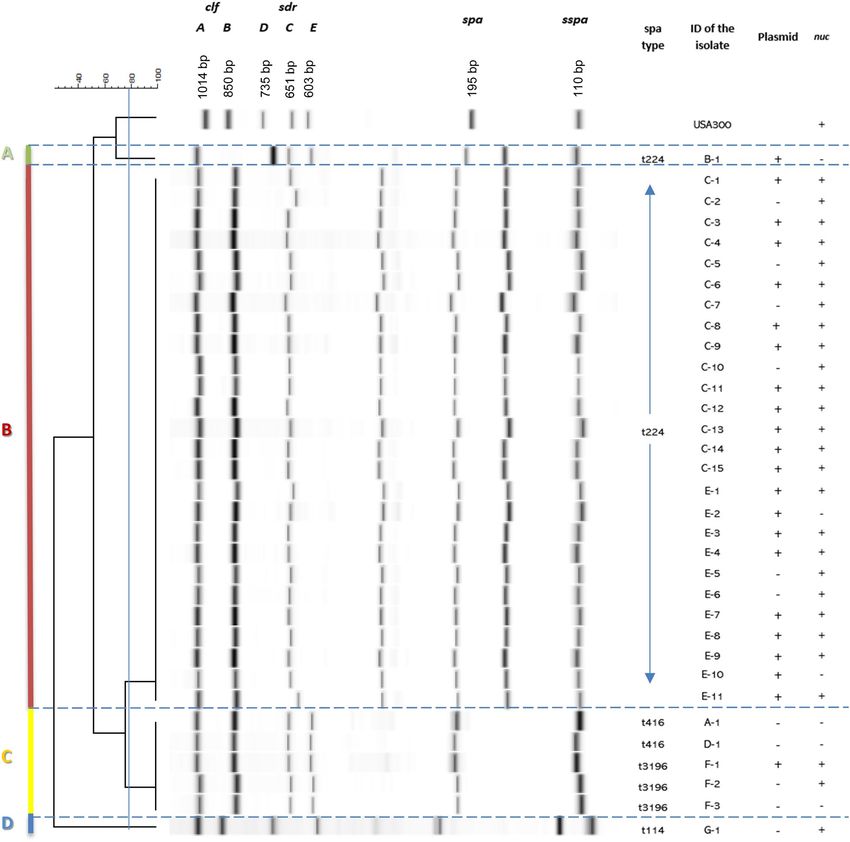
www.nature.com/scientificreports/
and septicemia in p oultry7. Boss et al. demonstrated that in animals the majority of S. aureus strains evolved from
human strains. For example, strains with the sequence type 8 (ST8) and strains belonging to the clonal complexes
(CC) CC5, CC8, CC59, CC97, and CC398 encountered in cows were of human o rigin9.
Resistant S. aureus can easily be transmitted between humans and animals, as has been shown for strains with
the ST398 that were transmitted from pigs to humans resulting in major health p roblems7. However, the zoonotic
transfer of S. aureus from milk and intramammary infections to humans seems to be very l ow9.
To determine the level of bacterial spread among animals and humans, various typing methods are used.
As such, the distinction of organisms within a species by typing has become a very important epidemiological
tool. The currently available typing methods can be classified into phenotyping and genotyping (“molecular”
typing), the latter one being the more sensitive and more appropriate approach to study the bacterial population
genetics10.
Several typing methods have been described to genotype S. aureus11. Pulsed-field gel electrophoresis (PFGE)
was for many years considered as the “gold standard” among the typing methods, because it has a high power
of discrimination, is highly reproducible and not expensive. However, this technique is labor-intensive, subjec-
tive and technically demanding11. Today, whole-genome sequencing (WGS) is gradually becoming the new
gold standard for typing and studying bacterial population genetics, especially because it has a much higher
discriminatory power than any other bacterial typing technique. Nevertheless, there is still a need to standardize
and unify data obtained from typing tools based on WGS for straightforward inter-laboratory comparisons12.
The latter issue has been overcome in another commonly applied sequencing-based S. aureus typing technique
named spa-typing, which involves the sequencing of the repeat region of the spa gene encoding the immuno-
globulin G-binding protein A 13. Although this method has less discriminatory power than PFGE and WGS, there
are several advantages that make it one of the most frequently used typing techniques for the classification of S.
aureus. These advantages include high reproducibility, low costs, user friendliness, quickness, portability of data
and high-throughput11. Another convenient method to type S. aureus isolates is multi-locus variable number
tandem repeat (VNTR) fingerprinting (MLVF), which consists of a multiplex PCR-based assay to determine the
polymorphism of VNTRs in 7 individual genes of S. aureus (sspA, spa, sdrC, sdrD, sdrE, clfA, and clfB)14. It has
been reported that this technique is quicker, cheaper and easier to use than PFGE and spa-typing, even though
it is difficult to compare the results between laboratories. For this reason, it was recommended to use MLVF
combined with a complementary typing method that provides portable data, like spa-typing or multi-locus
sequence typing (MLST)15.
Very limited information is available about the S. aureus lineages causing diseases in Mexican cattle, as well
as their antimicrobial resistances. One study classified the collected isolates using a phenotypic method based on
the production of staphylokinase, the type of hemolysis displayed and the clotting of bovine plasma16. Another
phenotypic typing study involved the classification of isolates by their hemolysis p atterns17. In a third study, a
genotypic characterization of different virulence factors was performed, including cell surface-associated and
secreted proteins, and different classes of the accessory gene regulator agr were d istinguished18. More recently,
Valdez-Alarcón et al. performed MLST on S. aureus strains isolated from 3 different regions of M exico19.
Most S. aureus isolates obtained from bovine mastitis have been shown to contain plasmid DNA20. The
presence of differently sized plasmids was associated with the carriage of multiple antimicrobial r esistances21.
Staphylococcal plasmids have been shown to range from 1 to 60 kb in size. In S. aureus, two main classes of
plasmids were identified, which contribute to the resistance against antimicrobials and/or virulence. All of these
plasmids bear the features of conjugative or mobilizable p lasmids21.
The aim of the present study was to investigate the diversity of 33 S. aureus isolates causing mastitis in cows
from the Mexican Comarca Lagunera region by MLVF and spa-typing, as well as to determine the resistance of
these strains against a variety of antimicrobials with possible links to the presence of plasmid DNA.
Results
Isolation of S. aureus mastitis strains. In total, 331 samples collected from milk of Holstein dairy cows
(n = 226) and nasal swabs (n = 105) collected from their calves were collected from 14 farms in the region called
Comarca Lagunera in Mexico (Fig. 1). The milk samples were taken from single quarters with clinical masti-
tis, where the criteria for mastitis were a somatic cell count > 400,000 SCC/mL. After a combination of Gram-
staining, coagulase testing and MALDI-TOF analysis, 33 isolates of S. aureus obtained from individual cows at
seven different farms (designated A-G) were identified (Table 1). To determine whether the potential presence
of S. aureus strains resulted in transmission to calves, nasal samples (n = 105) were collected from calves that had
contact with the cows with mastitis. All of the nasal swabs from calves tested negative for S. aureus, which sug-
gests that transmission events between cows and calves were infrequent or remained undetected.
MLVF and PCR analysis. The 33 mastitis-related S. aureus isolates were subjected to PCR analysis for the
presence of genes of the micrococcal nuclease MN (nuc) and the methicillin resistance gene mecA. The nuc gene
was detected in 27 isolates (Fig. 2). None of the isolates contained the mecA resistance gene. This matched with
the antimicrobial susceptibility testing, because none of the isolates were resistant against cefoxitin or oxacillin.
It was therefore concluded that all isolates were methicillin-susceptible S. aureus (MSSA)22.
For typing of the isolates, the total DNA was subjected to MLVF. The data obtained with the Bioanalyzer was
used to create the MLVF dendrogram shown in Fig. 2. Altogether, the MLVF analysis of S. aureus isolates from
milk samples revealed 4 different clusters, which were designated A (n = 1), B (n = 26), C (n = 5) and D (n = 1).
Judged by the expected lengths of the PCR fragments for the genes sspA (110-bp), spa (195-bp), sdrE (603-
bp), sdrC (651-bp), sdrD (735-bp), clfB (850-bp), clfA (1014-bp) from the control strain S. aureus USA300, a
clear variation in the lengths of several PCR fragments was observed among the four identified mastitis isolate
Scientific Reports | (2021) 11:6912 | https://doi.org/10.1038/s41598-021-86453-2 2
Vol:.(1234567890)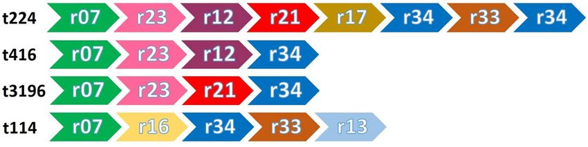
www.nature.com/scientificreports/
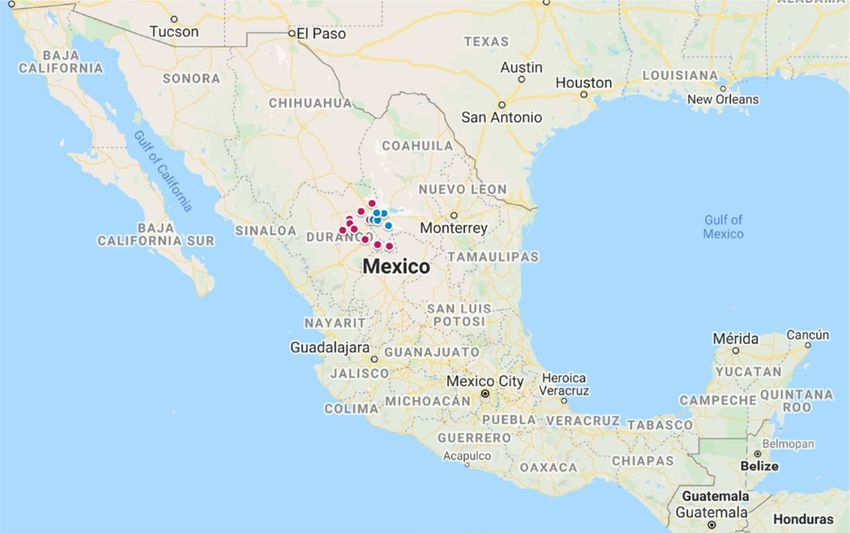
Figure 1. Map of Mexico indicating the Comarca Lagunera region from which the mastitis isolates
were collected. The Comarca Lagunera region is marked with blue and pink dots. This region includes 5
municipalities of the Coahuila state (blue dots) and 11 municipalities of the Durango state (pink dots). The map
was created from Google Maps (Map data Copyright 2020 Google) and edited in Microsoft PowerPoint 2016.
clusters. While the PCR fragments representing the sspA and the clfAB genes appeared nearly identical for all
isolates, the PCR fragments representing the sdrDE and spa genes showed clear variations between the clusters.
Further, for all mastitis isolates the spa product was smaller compared to that of the control strain USA300. The
amplified spa fragments from isolates belonging to clusters A and B appeared to be similar in length, while those
for isolates in Cluster C seemed to be even smaller. Based on the band intensity, we assume that the spa and sspA
PCR products had an identical size for isolates in cluster C. Lastly, the PCR products tentatively attributed to
sdrD and sdrE seemed to be smallest for isolates from cluster B.
Spa‑typing. The 33 S. aureus isolates were also characterized by spa-typing, yielding 4 different spa-types
(Figs. 2 and 3). Spa-type t224 was found to be the most common as it was assigned to 23 isolates. Spa-type t3196
was assigned to 3 isolates, t416 to 2 isolates and t114 was identified for only one isolate. Notably, all isolates
belonging to the MLVF clusters A and B had the spa-type t224, whereas spa-type 114 was unique for MLVF
cluster D. In contrast, isolates belonging to MLVF cluster C had either spa-type t3196 or t416. The variation in
the number of repeats within the spa genes of the different isolates was in agreement with the variations in length
that were observed for the respective spa-specific PCR fragments in the MLVF (Figs. 2 and 3). When comparing
the composition of the different types of identified repeats within the spa gene, it seems that isolate G-1 (t114)
includes two repeat sequences (r16 and r13) that are not present in the spa genes of the other mastitis isolates
(Fig. 3).
Western blotting analysis. To verify the observed size differences of the spa and sdrE PCR products in
the MLVF pattern, a Western blotting analysis was performed to detect the respective protein A and SdrE pro-
teins. For this analysis the covalently cell wall-bound proteins of 6 different mastitis isolates were extracted. As
controls, the S. aureus strain Newman, which was originally isolated in 1952 from a human infection, and the
mastitis-related S. aureus strain RF122 were used. The strain Newman is known to produce protein A (55.6 kDa)
and SdrE (126.5 kDa), while the RF122 strain lacks expression of protein A. From the Mexican mastitis isolates,
one strain of each MLVF cluster was selected. Specifically, these included isolates F-1 (t3196), B-1 (t224), G-1
(t114) and E-3 (t224). Using a polyclonal antibody against SdrE, both the expression of SdrE and protein A could
be determined by Western blotting (Fig. 4). In the case of protein A, this relates to the fact that it efficiently binds
the Fc region of rabbit polyclonal antibodies. The results of the blot are congruous with the VNTR repeats of
the different spa-types. The VNTR repeats of the isolates B-1 and E-3 with spa-type t224 (MLVF clusters A and
B, respectively) were equal in size and slightly smaller than those of the control strain Newman. For isolate G-1
with spa-type 114 (MLVF cluster D), a smaller size for protein A was detected. The smallest form of protein A
was detected for isolate F-1 with spa type t3196 and belonging to the MLVF cluster C (Figs. 3 and 4).
Scientific Reports | (2021) 11:6912 | https://doi.org/10.1038/s41598-021-86453-2 3
Vol.:(0123456789)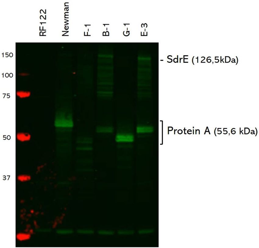
www.nature.com/scientificreports/

                                             Isolatesa   MLVF cluster; spa-type   Relevant phenotype or genotype                                   References
                                                                                                                                                   48
                                             Newman                               Clinical isolate
                                                                                                                                                   49
                                             USA300                               Community-acquired MRSA isolate
                                                                                                                                                   37
                                             RF122                                Mastitis-associated clinical isolate
                                             B-1         A; t224                  Mexican mastitis-associated isolate; orange-pinkish colonies
                                             C-1
                                             C-2
                                             C-3
                                             C-4
                                             C-5
                                             C-6                                  Mexican mastitis-associated isolates; white-yellowish colonies
                                             C-7
                                             C-8
                                             C-9
                                             C-10
                                             C-11
                                             C-12                                 Mexican mastitis-associated isolates; orange-pinkish colonies
                                             C-13
                                                         B; t224
                                             C-14
                                             C-15
                                             E-1                                                                                                   This study
                                             E-2
                                             E-3
                                             E-4
                                                                                  Mexican mastitis-associated isolates; white-yellowish colonies
                                             E-5
                                             E-6
                                             E-7
                                             E-8
                                             E-9
                                             E-10
                                             E-11
                                             A-1
                                                         C; t416                  Mexican mastitis-associated isolates; orange-pinkish colonies
                                             D-1
                                             F-1
                                             F-2         C; t3196
                                                                                  Mexican mastitis-associated isolates; white-yellowish colonies
                                             F-3
                                             G-1         D; t114

                                            Table 1.  Bacterial strains used in this study. a The letters A to G in the names of the isolates refer to the
                                            different farms from which they were collected.

                                                Unexpectedly, no protein band corresponding to SdrE was detected for strain RF122 (Fig. 4). In the sample
                                            of strain Newman a clear SdrE signal at the expected height of 126.5 kDa was detected together with multiple
                                            degradation bands. Similar patterns were observed for isolates F-1, B-1 and E-3, indicating that these isolates do
                                            express SdrE. Yet, no SdrE signal was detectable for isolate G-1 (Fig. 4), indicating that it does not express the
                                            SdrE protein, even though a possible sdrE PCR fragment appeared to be detectable in the MLVF analysis (Fig. 2).

                                            Antimicrobial resistance profiling. To determine possible variations in antimicrobial resistance and
                                            possible association of resistance phenotypes with the MLVF and/or spa-types of the investigated mastitis iso-
                                            lates, all 33 isolates were examined for antimicrobial susceptibility. The analysis of antimicrobial susceptibility
                                            revealed that for the antimicrobials with clinical breakpoints all isolates were susceptible to erythromycin, oxa-
                                            cillin, and tetracycline. Thirty-two isolates were resistant to benzylpenicillin (97%). Only strain G-1 (3%) was
                                            susceptible to all tested antimicrobials. Lastly, all isolates tested negative in the screen for cefoxitin resistance.

                                            Plasmid profiling and sequence analysis. To determine the presence of plasmids, plasmid DNA was
                                            isolated from all mastitis isolates. Using agarose gel electrophoresis it was shown that 22 out of the 33 isolates
                                            contain a plasmid of identical size (Supplemental Fig. S1). No plasmid DNA was detected in strains with the spa-
                                            types t114 and t416. On basis of the electrophoresis results, the plasmid DNA seemed to be of the same size in
                                            all strains. The plasmid DNA extracted from the isolates F-1 (t3196), E-3 (t224) and C-11 (t224) was sequenced.

          Scientific Reports |   (2021) 11:6912 |                   https://doi.org/10.1038/s41598-021-86453-2                                                  4

Vol:.(1234567890)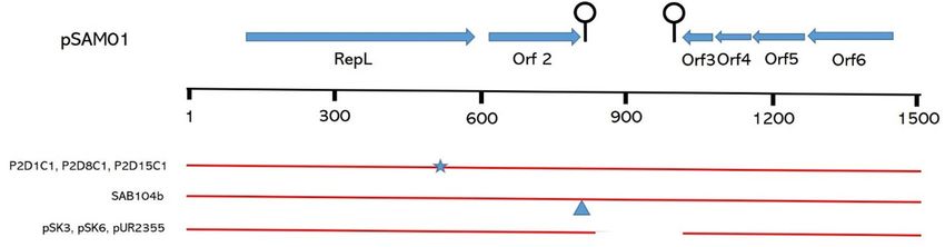
www.nature.com/scientificreports/
Figure 2. MLVF dendrogram of 33 S. aureus isolates from cows with mastitis. PCR fragments of seven VNTRs
of S. aureus were separated on a Bioanalyzer 2100 with microfluidic DNA 7500 chips. The dendrogram was
constructed in silico from CSV files of the Bioanalyzer runs using GelCompar II software (Applied Maths;
https://www.applied-maths.com/gelcompar-ii). The PCR fragments (clfA [1014-bp], clfB [850-bp], sdrD
[735-bp] sdrC [651-bp] sdrE [603-bp], spa [195-bp] sspA [110-bp]) obtained for the control DNA of S. aureus
USA300 are indicated. For the generation of the dendrogram, a cut-off value of 81% was used, yielding a
concordance of 0.980. The respective MLVF clusters (A-D), the respective spa-types, and the presence of the nuc
gene and plasmid pSAM1 are specified. The Figure was created with Microsoft PowerPoint 2016.
Interestingly, the respective plasmid sequences were found to be identical, consisting of 1508-bp. Accordingly,
the respective plasmid was named pSAM1. Searches for open reading frames (ORF) combined with Blast analy-
ses showed the presence of several ORFs in pSAM1 (Fig. 5).
The plasmid sequences showed a G + C content of 32.6 mol%, which is in agreement with the G + C content
determined for S. aureus (33 mol%)23. Based on DNA sequence similarity, plasmid pSAM1 can be classified as a
rolling circle replicating (RCR) plasmid that belongs to the pSN2 Family21. In total, six ORFs were identified on
pSAM1 (Fig. 5). The largest identified orf of pSAM1 encodes a protein of 154 amino acid (AA) residues which,
on the basis of sequence similarity, corresponds with the replication protein RepL. Downstream of the repL gene,
the orf2 gene is located, which encodes a protein of 62 residues with unknown function. An inverted repeat with a
∆G° of − 12.6 kcal/mol that may function as a rho-independent terminator is located downstream of orf2. 136-bp
Scientific Reports | (2021) 11:6912 | https://doi.org/10.1038/s41598-021-86453-2 5
Vol.:(0123456789)www.nature.com/scientificreports/
Figure 3. Schematic representation of the polymorphic VNTRs associated with identified spa-types. The order
of identified VNTRs of the identified spa-types t224, t416, t3196 and t114 as detected in the present study
isolates is schematically represented. The Figure was created with Microsoft PowerPoint 2016.
Figure 4. Western blotting analysis to detect the presence of SdrE and protein A. Proteins in the cellular
fractions of S. aureus strains RF122 and Newman, and the mastitis isolates F-1, B-1, G-1 and E-3 were separated
by LDS-PAGE and transferred to a nitrocellulose membrane. To visualize the presence of SdrE and protein
A, the membrane was incubated with SdrE-specific rabbit antibodies (1:5000) and, subsequently, with an
IRDye800CW-labelled goat anti-rabbit IgG secondary antibody (1:10,000). Note that protein A is detected
by its capacity to bind rabbit IgG. The sizes of the marker proteins are indicated in kDa at the left side of the
membrane. On the right side, the position of SdrE- and protein A-specific protein bands is marked. The
complete original Western blot is presented as Supplemental Fig. S2. The Figure was created with Microsoft
PowerPoint 2016.
Figure 5. Schematic representation of open reading frames on plasmid pSAM1. The relative positions of open
reading frames (repL, 2, 3, 4, 5 and 6) are marked by arrows. Predicted stem-loop structures are indicated by
lollypops. Regions of sequence identity between different plasmids are indicated by red lines, in which mutations
(*) and insertions (∆) are marked. The names of the homologous plasmids are indicated on the left. The Figure
was created with Microsoft PowerPoint 2016.
Scientific Reports | (2021) 11:6912 | https://doi.org/10.1038/s41598-021-86453-2 6
Vol:.(1234567890)www.nature.com/scientificreports/
downstream of this stem-loop another stem-loop with a ∆G° of − 10.2 kcal/mol is located, which could function
as a terminator for the orf3. Blast analysis shows that the sequence similarity with other plasmids stops directly
after either of these two stem loops, suggesting that they are potential sites for recombination. Blast analyses
also showed that in S. capitis a nucleotide sequence is present encoding a protein of 73 residues with unknown
function, which overlaps with orfs 3, 4 and 524. Also, in other S. aureus plasmids like pSK3, pSK6 and pUR2355,
mutations have been identified for this ORF (Fig. 5), suggesting that the presently identified orfs 3, 4 and 5 do
not encode a functional protein in S. aureus. The last orf, orf6, has a G + C content of 46%, which is much higher
than the average G + C content of 32.6% observed for the rest of the plasmid.
Overall, plasmid pSAM1 shows a high degree of sequence similarity with the plasmids P2D1C1, P2D8C1
and P2D15C1 that were previously identified in other S. aureus strains25. Compared to these plasmids small
mutations are located in the repL gene. The overall homology with a plasmid from S. aureus strain SAP104B
(1552-bp) was also very high although the SAP104B-derived plasmid contained some extra nucleotide sequences
in between orf3 and the downstream located stem loop. On basis of its repL gene plasmid pSAM1 was grouped
in rep family 10b26.
Discussion
In the present study, we describe the diversity of 33 S. aureus isolates causing mastitis in Mexican cows by MLVF
and spa-typing, as well as their susceptibility for a range of commonly used antimicrobials.
Our typing analysis shows that the S. aureus isolates associated with mastitis group into four MLVF clusters,
which overlap with the determined spa-types. The predominantly encountered spa-type t224 was determined
for all isolates of the MLVF clusters A and B. In recent studies, this spa-type has been reported for S. aureus
isolates from milk of cows with intra-mammary infections in European countries, China, Japan and Egypt9,27,28.
The present identification of mastitis-associated S. aureus isolates with spa-type t224 in Mexico implies that
this staphylococcal type has now been detected on all continents except Australia. However, in other studies,
t224 was not the predominantly encountered spa-type. Of note, the spa-type t224 is frequently observed for
isolates belonging to the ST97 group that is known to be carried by cows, humans and swine. In swine, such
isolates are occasionally M RSA29. Whether the isolates used in this study also belong to the ST97 remains to be
investigated. The MLVF cluster C includes the spa-types t416 and t3196. S. aureus isolates with spa-type t416
have been reported in Austria and China, mainly for human MRSA isolates30,31. According to the Ridom Spa
Server (https://spaser ver.ridom.de/), S. aureus isolates with the spa-type t3196 have been identified in Belgium,
Denmark, and the Netherlands. However, no information is currently available concerning the respective host
species, or whether these t3196 isolates were associated with bovine mastitis. The MLVF cluster D is formed
by a singleton with spa-type t114, which has been described for S. aureus isolates from the milk of cows with
mastitis in Brazil and C hina32,33.
The nuc gene, which encodes a thermostable nuclease, has been described as a specific marker for direct detec-
tion of S. aureus involved in infection of humans34. Nonetheless, in 4 of the 33 presently investigated S. aureus
isolates the nuc gene was apparently not present. Interestingly, Javid et al. reported that nuc could only be detected
in a fraction of S. aureus isolates collected from cows with mastitis in I ndia35. These observations are intriguing,
as it has been proposed that the micrococcal nuclease is involved in the evasion of the human immune system,
by preventing the capture and elimination of S. aureus in neutrophil extracellular t raps36.
It was previously reported that the spa, clfA and sdrC genes of the bovine mastitis-related S. aureus strain
RF122 are pseudogenes, because of the presence of premature stop c odons37. This phenomenon was not observed
for the spa genes of the presently investigated S. aureus isolates from Mexico, because protein A was detectable for
all isolates by Western blotting. On the other hand, RF122 contains a complete sdrE gene, which was confirmed
in a Western blot using an antibody against the region A of the SdrE protein of S. aureus strain Newman38. The
sequence similarity of region A in the SdrE proteins of strains RF122 and Newman is only 76%. This difference
in the sequence could be the reason that SdrE was not detected by Western blotting in the sample for covalently
cell wall-bound proteins of S. aureus RF122. Another possible explanation could be the use of whey permeate
as a growth medium for S. aureus in the present study, which may have lowered the expression of SdrE in the
RF122 strain.
Antimicrobial resistance testing showed that the majority of the isolates (32/33) were resistant to penicillin.
This is not surprising, as this antimicrobial is the first option to treat bovine mastitis. In many dairy farms it is
common practice to use intramammary infusions of antimicrobials as a prophylactic approach to prevent and
control mastitis in all dairy cows during the dry period, primarily with penicillins, cephalosporins, or other
beta-lactam drugs6.
In 67% of the investigated isolates, the presence of a single plasmid was detected. Based on agarose gel elec-
trophoresis and sequencing data, it seems that all plasmid-bearing strains contained the same plasmid, which
was named pSAM1. Sequence analysis of the repL gene showed that pSAM1 belongs to the pSN2 family, which
encompasses the smallest RCR plasmids identified in staphylococci21. The presence of pSAM1 was evidently not
linked to any of the MLVF clusters, a spa-type or a specific antimicrobial resistance. Besides the putative replica-
tion protein RepL, no function has so far been assigned to any of the five other proteins encoded by pSAM1. The
high prevalence of pSAM1 amongst the presently investigated S. aureus isolates could suggest an association of
this plasmid and its encoded proteins with bovine colonization or even mastitis. However, the latter remains to
be investigated, preferably in the context of follow-up studies on possible adaptations that allowed the present
mastitis isolates to thrive in the bovine mammary gland.
Scientific Reports | (2021) 11:6912 | https://doi.org/10.1038/s41598-021-86453-2 7
Vol.:(0123456789)www.nature.com/scientificreports/
Materials and methods
Sample collection and selection. Cases of mastitis were identified using the California Mastitis Test
(CMT), which is a simple on-farm method to indirectly indicate the level of the somatic cell count (SCC)39. Two
hundred and twenty six individual quarter milk samples were collected in 2015–2016 from 14 different farms in
the Comarca Lagunera region in Mexico. Foremilk was discarded after which a little amount of milk was drawn
in the CMT paddle. An equal amount of the CMT reagent was added and gently mixed. After a few seconds, the
reaction was scored following the scale described by Easterday et al.39. The weakly positive samples with 400,000
to 1,000,000 SCC/mL (mixture is slightly mucous, but still without gel formation), positive samples with 800,000
to 5,000,000 SCC/mL (distinct gel formation), and the samples with more than 5,000,000 SCC/mL (strongly
positive with strong gel formation that adheres to the paddle) were collected for plating. For transmission analy-
sis single nasal swabs were collected from 105 calves of these cows.
Identification of isolates and culture conditions. All milk samples and nasal swabs collected from
calves were directly plated at the farm on mannitol salt agar (Becton Dickinson de Mexico). Subsequently, the
plates were incubated at 37 °C overnight to determine the presence of bacteria. From each plate one potential S.
aureus colony was selected, depending on the following colors: golden-yellow, creamy-yellow, yellowish, orange,
pinkish and white (Table 1). After Gram-staining, all the Gram-positive cocci were subjected to the c atalase40
and Voges Poskauer41 tests. Finally, all colonies that tested potentially positive as S. aureus in the biochemical
tests were collected and shipped in mannitol salt agar to the University Medical Center Groningen, the Nether-
lands.
The potential S. aureus isolates were cultured on Blood Agar (BA) plates containing 5% sheep blood and
aztreonam (Media products, Groningen, the Netherlands). Firstly, a quick screen of the strain was done using the
Pastorex Staph Plus test (Bio-Rad, Marnes-la-Coquette, France). To select and identify S. aureus strains, single
colonies were analyzed by matrix-assisted laser desorption ionization-time of flight mass spectrometry (MALDI-
TOF MS) with a Microflex LT Biotyper (Bruker Daltonics, Bremen, Germany) as previously d escribed42. Only
S. aureus strains with log scores ≥ 2 were selected. Thirty-three isolates were positively identified as S. aureus.
The isolates were cultured overnight in tryptic soy broth (TSB, Oxoid, Hampshire, UK) at 37 °C with shaking
(250 rpm) and, subsequently, stored in 12% glycerol (Sigma-Aldrich, Zwijndrecht, the Netherlands) at −80 °C
for further analyses.
Antimicrobial susceptibility testing. Antimicrobial susceptibility was measured with the VITEK 2 sys-
tem (ID. Card: AST-GP69, bioMerieux Corporate, Marcy l’Etoile, France) in accordance with the manufac-
turer’s instructions. For quality control of the card, the strain S. aureus ATCC 29,213 was used as suggested
by the supplier. The following antimicrobials were evaluated: benzylpenicillin, cefoxitin, enrofloxacin, erythro-
mycin, kanamycin, oxacillin and tetracycline. The minimum inhibitory concentrations (MIC) obtained from
the VITEK analysis were validated using the Advanced Expert System following the Clinical and Laboratory
Standards Institute (CLSI) of the veterinary g uidelines43, which employed the human interpretive data taken
from the CLSI M100-S series44. The MIC breakpoints are specific for S. aureus udder isolates and are expressed
as μg/ml in parentheses. These breakpoints are for benzylpenicillin (Susceptible [S] ≤ 0.12, Resistance [R] ≥ 0.25);
erythromycin (S ≤ 0.5, R ≥ 8); oxacillin (S ≤ 2, R ≥ 4) and tetracycline (S ≤ 4, R ≥ 16). For cefoxitin only negative or
positive test results were obtained. For the veterinary antimicrobials enrofloxacin and kanamycin, interpretative
data are not available for mastitis-associated S. aureus. All results are presented in the Supplementary Table S1.
MLVF and PCR analysis. Total DNA was extracted from S. aureus colonies picked from BA plates by bead-
beating according to Glasner et al.14.
MLVF typing was performed following the adjusted protocol described by Sabat et al.15 and Glasner et al.14.
Briefly, 1 μL of total DNA was subjected to a multiplex PCR of seven VNTRs of S. aureus (sdrC, sdrD, sdrE,
clfA, clfB, sspA and spa) (Eurogentec, Maastricht, the Netherlands). Subsequently, 1 µL aliquots of the resulting
amplicons were separated employing a Bioanalyzer 2100 (Agilent Technologies, Palo Alto, USA) with microflu-
idic DNA 7500 chips following the manufacturer’s instructions. The strain USA 300 was used as the technical
control in each Bioanalyzer run to ensure the reproducibility of the data. Data was analyzed with the GelCompar
II software (Applied Maths, Kortrijk, Belgium). For this purpose, the data generated in the Bioanalyzer were
imported as CSV files. The position tolerance and optimization were set at 0.9% and 0.5% respectively, as with
these settings the control isolate (USA300) displayed identical MLVF banding patterns. To calculate the pairwise
similarity coefficients, the dice formula was used. A dendrogram was created with the unweighted pair group
method using average linkages (UPGMA). A cut-off of 81% was set to allow a differentiation between clusters.
Identical banding patterns were assigned to the same subtype; when one or more bands differed in the MLVF
pattern, they were assigned to different clusters.
All S. aureus strains were also screened for the presence of the mecA gene encoding methicillin resistance and
the nuc gene for the micrococcal nuclease MN produced by S. aureus, as previously described34,45.
Spa‑typing. Spa-typing was performed as described by Harmsen et al.13 using the Ridom Staph Type soft-
ware version 2.2.1 (Ridom GmbH, Würzburg, Germany). An ABI Prism 3130 genetic analyzer (Applied Biosys-
tems, Foster City, USA) was used to obtain the DNA sequences.
Plasmid sequencing. Plasmid DNA was extracted using the Innu PREP Plasmid Mini Kit (Analytik Jena,
Jena, Germany) following the special protocol of the supplier, where in the second step the cell pellet was resus-
Scientific Reports | (2021) 11:6912 | https://doi.org/10.1038/s41598-021-86453-2 8
Vol:.(1234567890)www.nature.com/scientificreports/
pended in resuspension buffer with added lysostaphin (final concentration 200 µg/mL; AMBI Products, Law-
rence, USA) and lysozyme (final concentration 3 mg/mL; Sigma‐Aldrich). The bacteria were incubated at 37 °C
for 10–30 min until complete lysis was obtained. Plasmid DNA was analyzed using 1.0% agarose gels. Nucleotide
sequence analyses of the plasmids were performed by an Illumina MiSeq system generating paired-end reads of
300 bp (Illumina, CA, USA). De novo assembly of paired-end reads was performed using the CLC Genomics
Workbench v11.0.1 (QIAGEN, Hilden, Germany). Homology comparisons were performed using the basic logi-
cal alignment tool (https://blast.ncbi.nlm.nih.gov/Blast.cgi). Dyad symmetries, Delta G calculation and Open
Reading Frames (ORFs) were determined using the program CloneManager 9 (Sci-Ed software, Westminster,
Colorado, USA). The GenBank accession number of plasmid pSAM1 is 2,388,794.
Western blotting. S. aureus strains were grown in TSB overnight at 37 °C, 250 rpm. Bacterial cultures were
diluted to an optical density at 600 nm (OD600) of 0.05 in 4% whey permeate medium containing 0.2% casein
hydrolysate (ICN Biochemicals, Ohio, USA) and 1.9% glycerolphosphate (SIGMA Life Science, Missouri, USA).
Growth was continued under the same conditions until the exponential phase ( OD600 = 0.5) was reached. After
this pre-culture, the cells were diluted once more in whey permeate medium46 and growth was continued for
2 h. Bacterial culture samples of 2 mL were pelleted and washed once in phosphate-buffered saline (PBS, Media
products). The obtained cell pellet was disrupted with 0.1 µM glass beads in PBS with a protease inhibitor cock-
tail (complete, Mini, EDTA-free, Roche) in a Precellys 24 homogenizer (Bertin Technologies, Saint Quentin en
Yvelines Cedex, France). After centrifugation, cell wall fragments were resuspended in 4% sodium dodecyl sul-
phate (Sigma-Aldrich), incubated for 3 min at 100 °C, and washed six times with P BS47. Afterwards, the cell wall
fragments were incubated in 50 mM Tris (pH 7.5, Sigma-Aldrich) with lysostaphin (final concentration 200 µg/
mL) and protease inhibitor cocktail (Roche) for 3 h at 37 °C. The original Western blotting image is presented
as Supplemental Fig. S2.
Covalently cell wall-bound proteins were separated by lithium dodecyl sulphate (LDS)-PAGE using NuPAGE
gels (Life Technologies, California, USA) and subsequently transferred to a nitrocellulose membrane (Protran
nitrocellulose transfer paper, Whatman, Germany). Membranes were incubated with polyclonal rabbit antibodies
against SdrE (1:5,000) and thereafter with an IRDye800CW-labelled goat anti-rabbit IgG secondary antibody
(1:10,000; LI-COR Biosciences, Lincoln, USA). Finally, the membranes were scanned with an Odyssey Infrared
Imaging System (LI-COR Biosciences) for fluorescence at 800 and 700 nm.
Biological and chemical safety. S. aureus is a biosafety level 2 (BSL-2) microbiological agent and was
accordingly handled following appropriate safety procedures. All experiments involving live S. aureus bacteria
were performed under appropriate BSL-2 containment as approved by the biological safety officers of the Uni-
versity Medical Center Groningen (UMCG). All chemicals and reagents used in this study were handled accord-
ing to the local and international guidelines and protocols for safe usage and protection of the environment.
Ethics. For the isolation of bacteria from waste milk samples of mastitic cows and for the non-invasive nasal
swabs from calves of mastitic cows, in Mexico no ethical permission was required at the time of sampling. Since
all investigated bacteria were collected in Mexico, no ethical approval was required for the investigations per-
formed at the University Medical Center Groningen (UMCG). The research project was performed with adher-
ence to the Nagoya Protocol on Access and Benefit Sharing (ABS).
Received: 13 October 2020; Accepted: 10 March 2021
References
1. Halasa, T., Huijps, K., Østerås, O. & Hogeveen, H. Economic effects of bovine mastitis and mastitis management: A review. Vet.
Q. 29, 18–31 (2007).
2. Valle-Aguilar, M., Lopez-Gonzalez, F., Sainz-Ramirez, A. & Arriaga-Jordan, C. M. Prevalence subclinical mastitis in small-scale
dairy farms under grazing or in total confinement in the central highlands of Mexico. Indian J. Dairy Sci. 73, 73–76 (2020).
3. Olivares-Pérez, J. et al. Prevalence of bovine subclinical mastitis, its etiology and diagnosis of antibiotic resistance of dairy farms
in four municipalities of a tropical region of Mexico. Trop. Anim. Health Prod. 47, 1497–1504 (2015).
4. Zadoks, R. N., Middleton, J. R., McDougall, S., Katholm, J. & Schukken, Y. H. Molecular epidemiology of mastitis pathogens of
dairy cattle and comparative relevance to humans. J. Mammary Gland Biol. Neoplasia 16, 357–372 (2011).
5. Barkema, H. W., Green, M. J., Bradley, A. J. & Zadoks, R. N. Invited review: The role of contagious disease in udder health. J. Dairy
Sci. 92, 4717–4729 (2009).
6. Barlow, J. Mastitis Therapy and Antimicrobial Susceptibility: a Multispecies Review with a Focus on Antibiotic Treatment of
Mastitis in Dairy Cattle. J. Mammary Gland Biol. Neoplasia 16, 383–407 (2011).
7. Haag, A. F., Fitzgerald, J. R. & Penadés, J. R. Staphylococcus aureus in Animals. Microbiol. Spectr. 7, (2019).
8. Weese, J. S. & van Duijkeren, E. Methicillin-resistant Staphylococcus aureus and Staphylococcus pseudintermedius in veterinary
medicine. Vet. Microbiol. 140, 418–429 (2010).
9. Boss, R. et al. Bovine Staphylococcus aureus: Subtyping, evolution, and zoonotic transfer. J. Dairy Sci. 99, 515–528 (2016).
10. van Belkum, A. et al. Guidelines for the validation and application of typing methods for use in bacterial epidemiology. Clin.
Microbiol. Infect. 13, 1–46 (2007).
11. Sabat, A. J. et al. Overview of molecular typing methods for outbreak detection and epidemiological surveillance. Euro surveill.
18, 20380 (2013).
12. Uelze, L. et al. Typing methods based on whole genome sequencing data. One Heal. Outlook https://doi.org/10.1186/s42522-020-
0010-1 (2020).
Scientific Reports | (2021) 11:6912 | https://doi.org/10.1038/s41598-021-86453-2 9
Vol.:(0123456789)www.nature.com/scientificreports/
13. Harmsen, D. et al. Typing of methicillin-resistant Staphylococcus aureus in a university hospital setting by using novel software for
spa repeat determination and database management. J. Clin. Microbiol. 41, 5442–5448 (2003).
14. Glasner, C. et al. Rapid and high-resolution distinction of community-acquired and nosocomial Staphylococcus aureus isolates
with identical pulsed-field gel electrophoresis patterns and spa types. Int. J. Med. Microbiol. 303, 70–75 (2013).
15. Sabat, A. J. et al. Microfluidic-chip-based multiple-locus variable-number tandem-repeat fingerprinting with new primer sets for
methicillin-resistant Staphylococcus aureus. J. Clin. Microbiol. 50, 2255–2262 (2012).
16. Manjarrez López, A. M. et al. Identificación de biotipos de Staphylococcus aureus en vacas lecheras de producción familiar con
mastitis subclínica en la región centro-este del Estado de México. Rev. Mex. ciencias Pecu. 3, 265–274 (2012).
17. Velázquez Ordoñez, V. et al. Human and bovine Staphylococcus aureus biotypes associated with haemolisin production and resist-
ance to oxacillin in cows with subclinical mastitis in family dairy farms. J. ISAH. Warsaw. Pol. 300–333 (2005).
18. El-Sayed, A. et al. Comparative study on genotypic properties of Staphylococcus aureus isolated from clinical and subclinical
mastitis in Mexico. Vet. Méx. 37, 165–179 (2006).
19. Valdez-Alarcón, J. J. Estructura genética de la población de Staphylococcus aureus asociada a mastitis bovina en granjas familiares
de Tarímbaro, Michoacán, mediante análisis de secuencias multilocus (Multilocus sequence typing; MLST). Biológicas Rev. la DES
Ciencias Biológico Agropecu. Univ. Michoacana San Nicolás Hidalgo 16, 1–10 (2015).
20. Hoque, M. N., Das, Z. C., Rahman, A. N. M. A., Haider, M. G. & Islam, M. A. Molecular characterization of Staphylococcus aureus
strains in bovine mastitis milk in Bangladesh. Int. J. Vet. Sci. Med. 6, 53–60 (2018).
21. Kwong, S. M., Ramsay, J. P., Jensen, S. O. & Firth, N. Replication of staphylococcal resistance plasmids. Front. Microbiol. 8,
2170–2178 (2017).
22. Skov, R. et al. Phenotypic detection of mecC-MRSA: Cefoxitin is more reliable than oxacillin. J. Antimicrob. Chemother. https://
doi.org/10.1093/jac/dkt341 (2014).
23. Muto, A. & Osawa, S. The guanine and cytosine content of genomic DNA and bacterial evolution. Proc. Natl. Acad. Sci. USA 84,
166–169 (1987).
24. Lymperopoulou, D. S. et al. Draft genome sequences of eight bacteria isolated from the indoor environment: Staphylococcus capitis
strain H36, S. capitis strain H65, S. cohnii strain H62, S. hominis strain H69, Microbacterium sp. strain H83, Mycobacterium
iranicum strain H39, Plantib. Stand. Genom. Sci. 12, (2017).
25. Liu, J., Gefen, O., Ronin, I., Bar-Meir, M. & Balaban, N. Q. Effect of tolerance on the evolution of antibiotic resistance under drug
combinations. Science (80-. ). 367, 200–204 (2020).
26. Lozano, C. et al. Expansion of a plasmid classification system for gram-positive bacteria and determination of the diversity of
plasmids in Staphylococcus aureus strains of human, animal, and food origins. Appl. Environ. Microbiol. 78, 5948–5955 (2012).
27. Hata, E. et al. Genetic variation among Staphylococcus aureus strains from bovine milk and their relevance to methicillin-resistant
isolates from humans. J. Clin. Microbiol. 48, 2130–2139 (2010).
28. El-Ashker, M. et al. Antimicrobial resistance pattern and virulence profile of S. aureus isolated from household cattle and buffalo
with mastitis in Egypt. Vet. Microbiol. 240, 108535 (2020).
29. Feltrin, F. et al. A livestock-associated, multidrug-resistant, methicillin-resistant Staphylococcus aureus clonal complex 97 lineage
spreading in dairy cattle and pigs in Italy. Appl. Environ. Microbiol. 82, 816–821 (2016).
30. Ruppitsch, W. et al. Classifying spa types in complexes improves interpretation of typing results for methicillin-resistant Staphy-
lococcus aureus. J. Clin. Microbiol. 44, 2442–2448 (2006).
31. Yu, F. et al. Virulence gene profiling and molecular characterization of hospital-acquired Staphylococcus aureus isolates associated
with bloodstream infection. Diagn. Microbiol. Infect. Dis. 74, 363–368 (2012).
32. Bonsaglia, E. C. R. et al. Molecular epidemiology of methicillin-susceptible Staphylococcus aureus (MSSA) isolated from milk of
cows with subclinical mastitis. Microb. Pathog. 124, 130–135 (2018).
33. Yi, Y. et al. Analysis of the genetic diversity in methicillin-resistant Staphylococcus aureus isolates from bovine subclinical mastitis
case in Xinjiang China. Foodborne Pathog. Dis. 15, 568–575 (2018).
34. Brakstad, O. G., Aasbakk, K. & Maeland, J. A. Detection of Staphylococcus aureus by polymerase chain reaction amplification of
the nuc gene. J. Clin. Microbiol. 30, 1654–1660 (1992).
35. Javid, F. et al. Molecular typing of Staphylococcus aureus based on coagulase gene. Vet. World 11, 423–430 (2018).
36. Berends, E. T. M. et al. Nuclease expression by Staphylococcus aureus facilitates escape from neutrophil extracellular traps. J. Innate
Immun. 2, 576–586 (2010).
37. Herron-Olson, L., Fitzgerald, J. R., Musser, J. M. & Kapur, V. Molecular correlates of host specialization in Staphylococcus aureus.
PLoS ONE 2, e1120 (2007).
38. O’Brien, L. et al. Multiple mechanisms for the activation of human platelet aggregation by Staphylococcus aureus: Roles for the
clumping factors ClfA and ClfB, the serine-aspartate repeat protein SdrE and protein A. Mol. Microbiol. 44, 1033–1044 (2002).
39. Easterday, B. C., Simon, J. & Hanson, R. P. The use of the modified Whiteside test as a screen test for bovine mastitis. J. Am. Vet.
Med. Assoc. 133, 470–473 (1958).
40. Painter, K. L. et al. Staphylococcus aureus adapts to oxidative stress by producing H2O2-resistant small-colony variants via the
SOS response. Infect. Immun. https://doi.org/10.1128/IAI.03016-14 (2015).
41. Yang, S. J., Dunman, P. M., Projan, S. J. & Bayles, K. W. Characterization of the Staphylococcus aureus CidR regulon: Elucidation
of a novel role for acetoin metabolism in cell death and lysis. Mol. Microbiol. https://doi.org/10.1111/j.1365-2958.2006.05105.x
(2006).
42. Veloo, A. C. M. et al. Validation of MALDI-TOF MS Biotyper database optimized for anaerobic bacteria: The ENRIA project.
Anaerobe 54, 224–230 (2018).
43. CLSI. Performance Standards for Antimicrobial Disk and Dilution Susceptibility Tests for Bacteria Isolated from Animals, 3rd
edition. Clin. Lab. Stand. Inst. (2015).
44. CLSI. Performance Standards for Antimicrobial Susceptibility Testing: 26th Informational Supplement, Document M100-S26.
Clin. Lab. Stand. Inst. (2016).
45. Murakami, K. et al. Identification of methicillin-resistant strains of staphylococci by polymerase chain reaction. J. Clin. Microbiol.
29, 2240–2244 (1991).
46. Buist, G., Venema, G. & Kok, J. Autolysis of Lactococcus lactis is influenced by proteolysis. J. Bacteriol. 180, 5947–5953 (1998).
47. Romero Pastrana, F. et al. Human antibody responses against non-covalently cell wall-bound Staphylococcus aureus proteins. Sci.
Rep. 8, 3234 (2018).
48. Lorenz, L. L. & Duthie, E. S. Staphylococcal coagulase: Mode of action and antigenicity. Microbiology 6, 95–107 (1952).
49. McDougal, L. K. et al. Pulsed-field gel electrophoresis typing of oxacillin-resistant Staphylococcus aureus isolates from the United
States: Establishing a national database. J. Clin. Microbiol. 41, 5113–5120 (2003).
Author contributions
Y.M-.H, P.H.J., J.M.v.D. and G.B. conceived the study. P.H.J., J.M.v.D. and G.B. supplied materials. Y.M-.H.,
E.V.M. and J.S. performed experiments and analyzed the data. Y.M-.H., J.M.v.D. and G.B. wrote the manuscript.
All authors reviewed and approved the manuscript.
Scientific Reports | (2021) 11:6912 | https://doi.org/10.1038/s41598-021-86453-2 10
Vol:.(1234567890)www.nature.com/scientificreports/
Competing interests
The authors declare no competing interests.
Additional information
Supplementary Information The online version contains supplementary material available at https://doi.org/
10.1038/s41598-021-86453-2.
Correspondence and requests for materials should be addressed to J.v.D.
Reprints and permissions information is available at www.nature.com/reprints.
Publisher’s note Springer Nature remains neutral with regard to jurisdictional claims in published maps and
institutional affiliations.
Open Access This article is licensed under a Creative Commons Attribution 4.0 International
License, which permits use, sharing, adaptation, distribution and reproduction in any medium or
format, as long as you give appropriate credit to the original author(s) and the source, provide a link to the
Creative Commons licence, and indicate if changes were made. The images or other third party material in this
article are included in the article’s Creative Commons licence, unless indicated otherwise in a credit line to the
material. If material is not included in the article’s Creative Commons licence and your intended use is not
permitted by statutory regulation or exceeds the permitted use, you will need to obtain permission directly from
the copyright holder. To view a copy of this licence, visit http://creativecommons.org/licenses/by/4.0/.
© The Author(s) 2021
Scientific Reports | (2021) 11:6912 | https://doi.org/10.1038/s41598-021-86453-2 11
Vol.:(0123456789)You can also read