Preparation and characterizations of antibacterial-antioxidant film from soy protein isolate incorporated with mangosteen peel extract
←
→
Page content transcription
If your browser does not render page correctly, please read the page content below
e-Polymers 2021; 21: 575–589
Research Article
Xin Zhou, Qingyin Dai, Xi Huang, and Zhiyong Qin*
Preparation and characterizations of
antibacterial–antioxidant film from soy protein
isolate incorporated with mangosteen peel
extract
https://doi.org/10.1515/epoly-2021-0058
received May 16, 2021; accepted July 12, 2021
1 Introduction
Abstract: The mangosteen peel extract (MPE) was used to Petroleum-based plastics with the advantages of light-
obtain soy protein isolate (SPI) films. The results show that ness, barrier characteristics, processability, low cost, and
MPE exhibited a high content of total phenolics and anti- good physical properties have been widely used in the
oxidant activity. Moreover, the MPE can enhance the anti- consumer products packaging (1,2). But the problem is
bacterial–antioxidant properties, UV-visible light barrier that these discarded plastics not only pollute soil and
properties, and water-resistant properties of the SPI films. water, but also produce unwanted by-products (3). Con-
The presence of MPE resulted in an increase in water vapor cerns about the environmental problems caused by non-
permeability and hydrophobicity. The extract addition biodegradable plastics and the depletion of natural
also reduced the film’s crystallinity along with a decrease resources have prompted people to develop environmen-
in the mechanical property and lowering of the maximum tally friendly and green composites using biodegradable
degradation temperature. Attenuated total reflectance Fourier and renewable materials.. Biopolymer packaging, as an
transform infrared spectroscopy revealed that the polyphe- effective solution to reduce food waste, has additional
nols in MPE could interact with SPI through hydrogen bonds
functions such as biodegradability, improved food quality
and hydrophobic interactions, and the addition of MPE
and shelf life (4). Antioxidant and antimicrobial functions
changed the secondary structure of SPI with a decrease
are the main developmental direction in active bioplastic
in β-sheets and an increase in β-turns and random coils.
packaging (4,5). Using antimicrobials in food packaging
Scanning electron microscopy showed that all the films
have greater advantages than adding them directly to
exhibited smooth and homogenous morphology on the
the food because antimicrobials added to the food surface
surface and on some layers through cross-sectional images.
via sprays or drops are not effective enough to inhibit
Our results suggested that the MPE would be a promising
microorganisms (6). This is due to the rapid spread of
ingredient to make SPI films used as an active packaging
antimicrobials into foods and the denaturation of active
material.
compounds in food ingredients that reduce the activity of
Keywords: SPI film, MPE, antibacterial–antioxidant functional agents. Antimicrobials provide a slow and con-
tinuous migration of these agents from the packaging
material to the food surface, which can maintain high
concentrations of antimicrobial agents over a long period
of time (7). Natural antimicrobial ingredients such as poly-
* Corresponding author: Zhiyong Qin, School of Resources, phenol-rich natural extracts are volatile and cannot be
Environment and Materials, Guangxi University, Nanning 530000, used alone. In order to improve the efficacy of these mate-
China; MOE Key Laboratory of New Processing Technology for Non- rials, active films are used (8).
Ferrous Metals and Materials, Guangxi University, Nanning 530000, As a natural substance, soy protein isolate (SPI) has
China, e-mail: q_zhiyong@163.com
higher protein content than other soybean protein pro-
Xin Zhou, Qingyin Dai, Xi Huang: School of Resources, Environment
and Materials, Guangxi University, Nanning 530000, China; MOE
ducts (9), and SPI film has good antioxidant and oil-proof
Key Laboratory of New Processing Technology for Non-Ferrous performances at low relative humidity (RH) (10). However,
Metals and Materials, Guangxi University, Nanning 530000, China due to the inherent hydrophilicity of natural proteins and
Open Access. © 2021 Xin Zhou et al., published by De Gruyter. This work is licensed under the Creative Commons Attribution 4.0 International
License.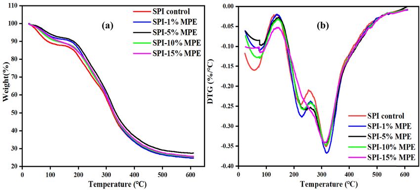
576 Xin Zhou et al.
strong molecular interactions, SPI films do not exhibit Mw = 9,000) was purchased from Chengdu Kelong
satisfactory mechanical properties or water vapor barrier Chemical Co., Ltd. Glycerol was procured from Tianjin
properties, which become worse under highly humid con- Aopusheng Chemical Co., Ltd. Mangosteen was bought
ditions (11). In order to improve the mechanical properties from local market, and the waste pulp was removed,
of SPI films, various studies have reported that the blending dried, and smashed for further use.
of SPI and biodegradable polymers will be a promising 2,2-Diphenyl-1-picrylhydrazyl (DPPH) and Folin–
method of structural modification, and the improvement Ciocalteau reagents were obtained from Shanghai Macklin
in the basic properties of SPI films has been widely paid Biochemical Co., Ltd. All the other chemical reagents
attention (12). Polyvinyl alcohol (PVA) (13) is a biodegrad- used in the experiment were of analytical grade. Escherichia
able and non-toxic synthetic polymer used to develop blend coli ATCC 25922 (E. coli) and Staphylococcus aureus ATCC
films. It imparts good tensile strength and biodegradability 25923 (S. aureus) were provided by Guangdong Huankai
and hence has been widely used in biomaterial fields. Microbial Sci. & Tech. Co., Ltd.
Therefore, PVA is expected to improve the mechanical prop-
erty of SPI films and dispersibility of antibacterial agent. In
addition to serving as protective barriers, these films can
also serve as carriers for bioactive compounds with antiox- 2.1 Preparation of MPE
idant or antimicrobial properties. Waste from fruit proces-
sing can be considered as an economic source of antiox- The mangosteen peel was washed three times, dried until
idants or antimicrobial agents, which may reduce the no water existed, ground into powder, and screened by a
demand for synthetic preservatives. 60-mesh sieve. Then, 20 g of the powder were added into
Mangosteen (14,15) is a tropical fruit that is widely 140 mL of 70% ethanol solution and stirred continuously
grown in Thailand, Malaysia, Indonesia, and South China. at a temperature of 70°C for 120 min. The supernatant was
The skin of the mangosteen accounts for about two-thirds obtained by centrifuging at 8,000 rpm for 10 min, filtered,
of its weight. The pericarp contains rich bioactive com- and condensed under rotary evaporator. The MPE was
pounds, which can be used as therapeutic drugs (16) or obtained after freeze-drying to a constant weight. The
functional food additives (17). Mangosteen peel extract dried MPE were stored under low temperature (4°C) and
(MPE) (18,19), a natural yellow-orange bioactive com- dark conditions for further use.
pound extracted from the mangosteen, is non-toxic and
considered safe even at high concentrations and has a
variety of functional properties, such as antioxidant, anti-
inflammatory (20), antitumor, anticancer, and antibacterial 2.2 Film preparation
activities (21). In recent years, antioxidant capacity and anti-
bacterial activity of MPE have been frequently researched. The SPI films were prepared by using solution-casting
However, no previous investigations have been conducted method (13). The SPI (5% w/w) solution was prepared
on MPE incorporation in SPI active packaging. in distilled water. The SPI solution mixing 30 wt% of gly-
Therefore, this research was aimed to study the anti- cerol was magnetically stirred at a temperature of 80°C
oxidant properties and antibacterial activities of MPE, for 30 min to denature the protein fraction. The PVA solu-
and antibacterial–antioxidant ability, mechanical proper- tion was prepared by adding 5% of PVA in 95°C water in a
ties, water vapor barrier capacity, water solubility, water 100 mL capped beaker where it was maintained for 2 h,
contact angle, and optical properties of the SPI films incor- while being constantly shaken for good film-forming
porated with MPE. In addition, the films were also ana- properties. The PVA solution at 5 wt% and MPE at con-
lyzed with attenuated total reflectance Fourier transform centrations of 0%, 1%, 5%, 10%, and 15% based on SPI
infrared (ATR-FTIR) spectroscopy, thermal gravimetric ana- were added to the SPI solution at room temperature. After
lyzer (TGA), scanning electron microscopy (SEM), and X-ray stirring for 2 h, the large particles were removed by cen-
diffractometer (XRD). trifugation at 6,000 rpm for 10 min, and the supernatant
was collected in the glass bottle. Ultrasound technique
(40 W for 10 min) was used to improve the solubility of
2 Materials mixture and to remove air bubbles. Twenty gram of the
prepared solution were poured in petri dishes (9 cm dia-
SPI powder (more than 90% of dry protein) was provided meter) and dried at a temperature of 45°C for 24 h at a RH
by Henan Yuzhou Biotechnology Co., Ltd. PVA (1799, of 45% to obtain the films. Before use, the film was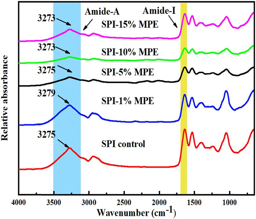
Preparation and characterizations of antibacterial–antioxidant film from SPI 577
Table 1: Experimental formulations of the SPI films with and room temperature. Meanwhile, the absorbance of 1 mL
without MPE ethanol solution mixed with DPPH ethanol solution was
determined for comparison. The formula used for calcula-
Films 5% 5% Glycerol (g)c MPE (g)d tion is as follows (Eq. 2):
SPI (g)a PVA (g)b
A1 − A2
SPI control 25 1.25 0.375 0 Scavenging rate (%) = × 100% (2)
A1
SPI-1% MPE 25 1.25 0.375 0.0125
SPI-5% MPE 25 1.25 0.375 0.0625 where A1 is the absorbance of the blank DPPH solution
SPI- 25 1.25 0.375 0.125 and A2 is the absorbance of the sample extract or the
10% MPE
test film.
SPI- 25 1.25 0.375 0.1875
15% MPE
a
5% (w/w) solution; b5% (w/w) solution, 5 wt% based on SPI dry
weight; c30 wt% based on SPI dry weight; d0%, 1%, 5%, 10%, and
2.4 Determination of antimicrobial activity
15% based on SPI.
The antimicrobial activity of MPE on E. coli (ATCC 25922)
and S. aureus (ATCC 25923) was tested by using the agar
removed from the petri dish and stored at room tempera- diffusion method (Oxford cup method) (24,25). All the
ture and 57% of RH for 72 h. The final formulations of the strains were incubated in 50 mL of Luria–Bertani broth
SPI and MPE-SPI films are shown in Table 1. at a temperature of 37°C until microbial tests. After 20 h of
incubation, 100 μL of the diluted culture broth prepared
through ten-fold serial dilutions were coated onto MH
2.3 Determination of antioxidant activity agar media and then left to dry for 2 min. Sterile stainless
steel drill was used to make wells (diameter of 6 mm) in
The total phenolic (TP) content and DPPH radical scaven- the medium. Each well was filled with 100 μL diluted
ging ability were determined to evaluate its antioxidant extract at concentrations of 0, 0.75, 1.5, 3, 6, and 12 mg/mL.
activity. 0.4 g MPE and 0.4 g SPI film samples containing The plates were left in a refrigerator for overnight to allow
different levels of MPE were solubilized in ethanol solu- diffusion of active components in the media. 0 mg/mL MPE
tion (10 mL, 10%) and placed in a thermostatic oscillator was used as negative control under the same conditions. The
at a temperature of 30°C for 16 h, followed by centrifuga- plates were incubated at a temperature of 37°C for 12–48 h
tion at 6,000 rpm for 10 min. The final volume was diluted to until there was significant microbial growth on the control
100 mL. The supernatants were employed by Folin–Ciocalteu plate. The inhibition zone around the well was measured in
method (22). Briefly, the test solution (1 mL) was mixed mm (including well diameter). The antimicrobial activity was
with Folin phenol reagent (1 mL) and reacted at room tem- indicated by the diameter of the inhibition zones produced
perature for 4 min. After the incorporation of 2 mL of 10% by the extract on the test microorganism. All the experi-
sodium carbonate solution, the mixture was reacted for 2 h ments were repeated 3 times.
at room temperature. The absorbance was measured at The macrodilution method was used to evaluate the
760 nm. Distilled water was blank and TP content was biocide property of the SPI films (26). All film samples
expressed as gallic acid equivalents (GAE) using a standard were stored in a biosafety cabinet and sterilized by UV
curve of the gallic acid and was calculated using Eq. 1: irradiation overnight. Same weight of each film specimen
a×V was added to the test tubes containing E. coli and S. aureus
TP (mg GAE/g) = (1) culture. The bacterial suspensions were incubated at a
W
temperature of 37°C for 1 h. Then, 100 μL of the sample
where a (mg/mL) is the concentration of the polyphenols were taken and spread on a MH agar plate, which was
in the test solution, V (mL) is the volume of the test incubated at a temperature of 37°C for 24 h. The number
solution, and W (mg) is the mass of the extract or the of colonies was counted with a colony counter. The inhibi-
dried film. tion of bacterial growth was calculated using Eq. 3:
The free radical scavenging activities of the extract
a−b
and the films were estimated using the DPPH method (23). Reduction (%) = × 100% (3)
Different concentrations of test solution (1 mL) were mixed a
with 4 mL of DPPH ethanol solution (25 mg/L). The absor- where a and b are the number of bacterial colonies of the
bance was measured at 517 nm after reaction for 30 min at control and test films, respectively.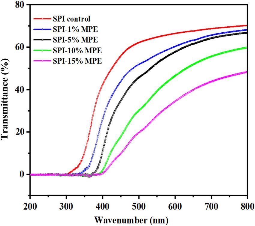
578 Xin Zhou et al.
2.5 Mechanical properties weighing bottle and sealed with a film. The weighing
bottles were then placed at room temperature with a
The thickness of each sample was measured randomly at RH of 90%. The bottles were weighed every 1 h for 12 h.
different positions with an Electronic digital micrometer WVP is calculated using Eq. 5 as follows:
(EVERTE, awt-chy01, Zhejiang) having a accuracy of 1 μm
ΔW × d
and the average value was reported (27). Three repeti- WVP = (5)
t × A × ΔP
tions were used for each sample.
Mechanical properties of the films were determined where WVP (g/cm s MPa) is the water vapor permeability,
according to ASTM D882 (28) using an Instron universal ΔW (g) is the mass of water passing through the films,
testing machine (Instron 8801, Britain). The film samples d (m) is the film thickness, t (s) is the time interval, A (m2)
were placed in a desiccator at room temperature and 57% is the effective film area, and ΔP (MPa) is the partial
of RH for at least 2 days prior to measurement. All per- pressure of water vapor over the film samples. All experi-
formance measurements were done immediately after the ments were performed in triplicate.
film samples were removed from the laboratory to mini-
mize humidity differences. Each sample was repeated five
times. To determine the tensile strength (TS, MPa) and 2.8 Water contact angle (WCA)
elongation at break (EB, %), the films were cut into strips
of 60 mm × 10 mm and installed between the tensile clamps. The contact angle of water on the surface of the films
The initial separation of the tool head was 50 mm, and the were measured to estimate the hydrophobicity. The films
crosshead speed was 0.8 mm/s. Three repetitions were used were cut into rectangular pieces (3 cm × 10 cm) and
for each sample. placed on a horizontal movable platform (black Teflon
coated steel, 7 cm × 11 cm) equipped with a WCA analyzer
(Fangrui, JCY-1, Shanghai). Then, a micro-syringe was
used to drop 2 μL of water on the surface of the films.
2.6 Water solubility (WS) The WCA on both sides of the water drop was measured
and the average value was taken. The liquid used was
The WS of the samples was measured according to a
distilled water and the experiments were performed at
method described by Rambabu et al. (29). In short, the
room temperature and 57 ± 2% of RH.
film samples were dried in an oven at a temperature of
103°C for 24 h, cut into a size of 40 mm × 10 mm, and
weighed to determine the initial solid content (Wi ). The
2.9 Color measurement
pre-weighed film samples were immersed in 50 mL of
distilled water for 24 h at a temperature of 30°C under
A colorimeter was used to determine the color of the film
constant stirring. The remaining films were then filtered
and the white board was used as a reference. Each treat-
and dried in a dry oven at a temperature of 103°C for 24 h.
ment was measured at least 3 times, and the values of L*,
The WS of each film was calculated using Eq. 4:
a*, b*, and ΔE were recorded. Three replicate film sam-
Wi − Wf ples were tested.
WS (%) = × 100% (4)
Wi
where Wi is the initial weight of the dried film (g) and Wf
2.10 Films opacity
is the final weight of the dried film (g).
Five repetitions were used for each film, and the
The opacity of SPI films was measured with the absor-
arithmetic mean was reported as solubility percentage.
bance of 600 nm per mm thickness by a Shimadzu UV-
2450 UV-Visible (UV-Vis) spectrophotometer. Each sample
was cut into rectangular pieces and placed in the UV spectro-
2.7 Water vapor permeability (WVP) photometer test cell, and an empty test cell as a reference
for measurement. The following formula was used to cal-
The WVP of the films was determined by the modified
culate the optical properties of the films (Eq. 6):
method of Qin et al. (30). Anhydrous calcium chloride
(CaCl2) and weighing bottle were dried at a temperature Abs 600
Opacity (%) = (6)
of 105°C for 24 h. Then 10 g of CaCl2 were poured into a L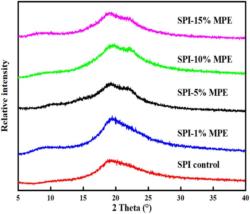
Preparation and characterizations of antibacterial–antioxidant film from SPI 579
where Abs600 is the spectrophotometric absorbance value The results are expressed as mean values ± standard
at 600 nm wavelength and L is the thickness of the deviation (SD). If P < 0.05, the significance level was
film (mm). defined.
2.11 Films transmittance 3 Results
The transmittance spectra of the SPI films were obtained
using -Vis spectrophotometer. The spectra were recorded 3.1 TP content and antioxidant activity
at room temperature in steps of 1 nm, in the range of
200–800 nm. For this, a rectangular piece of film (4.5 cm × The Folin–Ciocalteu method is used to evaluate the anti-
1 cm) cut from each film sample was previously conditioned oxidant activity of MPE both individually and when
in a dry desiccator at room temperature for 48 h, and three incorporated into the SPI matrix. Since the order of addi-
measurements were performed. tion of extracts directly reflects the availability of phe-
nolic hydroxyl groups, it can be used as an effective
method to evaluate the effect of the order of addition of
extracts on the antioxidant capacity of final materials.
2.12 Characterizations
The results obtained by the Folin–Ciocalteu method
showed that the MPE presented the TP content of
The thermal properties of the films were measured by
81.34 mg GAE/g, which were lower than that in other
TGA (NETZSCH, STA 449 F3, Germany). Approximately
similar studies (31). Emam-Djomeh et al. (32) reported
5 mg samples were added to a standard aluminum pan
that the total amount of phenolic compounds in pome-
and heated from 30°C to 600°C at a rate of 10°C/min.
granate fruit peel extract was 186 mg GAE/g.
Nitrogen was used as the purge gas at a flow rate of
These differences may be due to that TP content in
20 mL/min.
different extracts is not only related to the variety, species,
The ATR-FTIR spectra of SPI films were analyzed by
and seasonality of the fruit, but also related to the extrac-
ATR-FTIR spectrometer (Shimadzu, IRTracer-100, Japan).
tion method, the type of solvent used, the temperature, or
The samples were placed on the X-ray exposure table and
stirring (33).
the scanning frequency was 4,000–650/cm with a spec-
The incorporation of MPE had a significant effect
tral resolution of 4/cm. The measurements were carried
on the TP contents of the formulated SPI film as shown
out at room temperature. All the data treatments were
in Figure 1 (P < 0.05). The TP content increased from
performed with Peakfit software version 4.12 (SYSTAT
Software, Richmond, CA, USA).
The surface and cross section of SPI films at 10,000×
and 1,500× were observed by SEM (Phenom Pro, 800-07334,
Netherlands) at 15 kV voltage, respectively. Before scan-
ning, the film sample was cut into 5 mm × 1 mm pieces
and installed on the bronze stub with double-sided carbon
tape. Before visualization, the films were gold-plated with
a sputter coater.
XRD (Rigaku, D/max, 2,500 V, Japan) was used to
study the crystal structure of the films at 40 kV and
40 mA in 2θ = 5–40°. The scanning speed was 2°/min.
2.13 Statistical analysis
Figure 1: TP content and DPPH radical scavenging activity of the SPI
The SPSS statistical software was used for analysis of films with and without MPE. All data are expressed as mean values ±
ANOVA and Duncan triplicate range test for significance standard deviation (SD). Different letters indicated significant
analysis at P < 0.05 level (SPSS Inc. Chicago, IL, USA). differences (P < 0.05).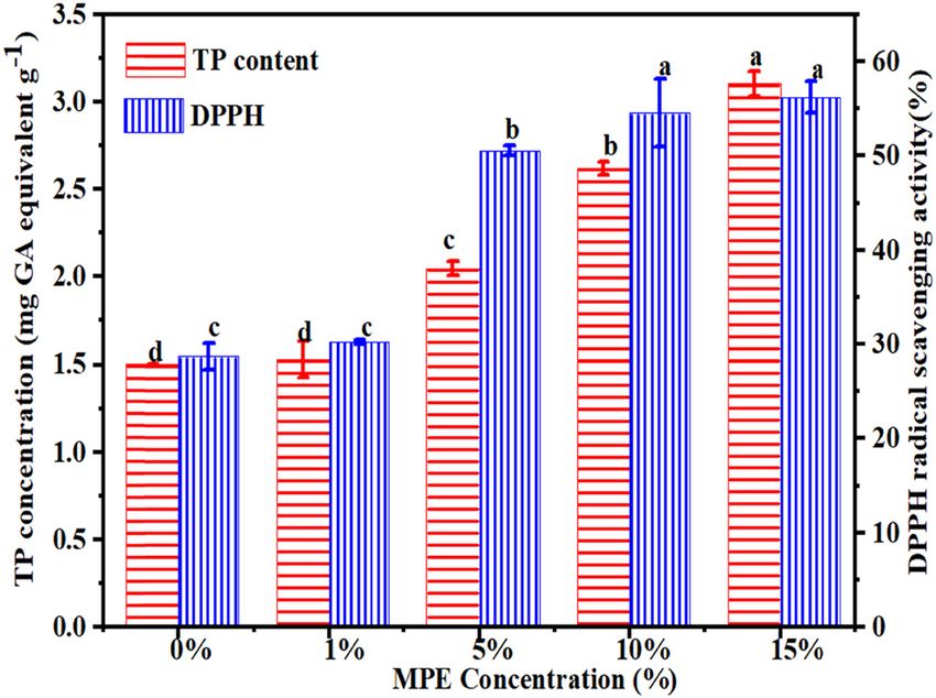
580 Xin Zhou et al.
1.5 mg GAE/g for the peel free film to 3.0 mg GAE/g for the Table 2: Antimicrobial effect of MPE on the tested organisms
film sample that contained 15% of MPE. The low level of
TP observed in the control film may be due to the pre- Mangosteen extracts Diameter of inhibition zone (mm)
sence of amino acid residues such as tyrosine and histi- dilutions (mg/mL)
E. coli ATCC S. aureus ATCC
dine in SPI, which can react with Folin-Ciocalteu reagent. 25922 25923
The TP detected in the film was due to the compounds
12 16.3 17.6
present in MPE. The phenolic compounds are active 6 14.9 16.5
hydrogen donors and are good antioxidants, which may 3 13.6 15.2
be responsible for the oxidative activity of mangosteen 1.5 12.6 13.9
pericarp containing phenol (34), xanthone (35), tannin, 0.75 12.2 13.6
0 0 0
anthocyanin (36), and carotenoids (37).
Antioxidant ability of hydrogen atom to absorb DPPH
free radical was determined by the DPPH radical scaven-
ging activity (38). When the DPPH solution is quenched films was significantly enhanced (P < 0.05). The enhanced
by antioxidants, its color changes from dark purple to antioxidant capacity of SPI film was mainly due to the
light yellow, and its absorbance decreases at 514 nm presence of polyphenols in MPE, which could capture
(3). DPPH assay is a rapid and simple detection method, free radicals by providing phenol hydrogen. The DPPH
which is an effective method to evaluate the antioxidant radical scavenging activity was slightly lower than that
free radical scavenging activity. Figure 2 indicates that of similar studies reported previously. The DPPH activity
MPE has antioxidant and health-promoting potential due of khorasan wheat starch film containing 1% of moringa
to its content of phenolic compounds. Compared with leaf extract has been reported to be 37.9% (33). The DPPH
vitamin C, MPE had similar DPPH radicals scavenging activity of the SPI film with 1% of MPE was 32% (Figure 2).
ability, that is, 0.2 mg/mL. Therefore, MPE could be used These differences may be attributed to different key ingre-
effectively as an antioxidant. dients and film matrix.
DPPH scavenging assay was used to indicate anti-
oxidant activity of the film. The antioxidant activity of
the film was determined by DPPH scavenging assay. As 3.2 Determination of antimicrobial activity
shown in Figure 2, all the films tested showed DPPH of SPI films
radical scavenging activity in a dose-dependent manner.
Due to the lack of a hydrogen atom donor, SPI films The antimicrobial activity of MPE against S. aureus and
showed the lowest antioxidant capacity. With the increase E. coli was determined using agar well diffusion method
in MPE content, the DPPH radical scavenging ability of SPI (Table 2 and Figure 3). Table 2 presents diameters of
inhibition zones exerted by the different concentrations
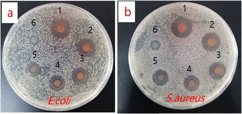
of MPE toward tested microorganisms. The results showed
that the MPE had antibacterial activity against both the
strains. 0 mg/mL MPE had no activity on the tested
Figure 3: Photos of the inhibitory zones of the extracts. (1) 12
mg/mL, (2) 6 mg/mL, (3) 3 mg/mL, (4) 1.5 mg/mL, (5) 0.75 mg/mL,
and (6) 0 mg/mL concentration extracts against (a) E. coli and
Figure 2: DPPH radical scavenging activity of MPE and vitamin C. (b) S. aureus.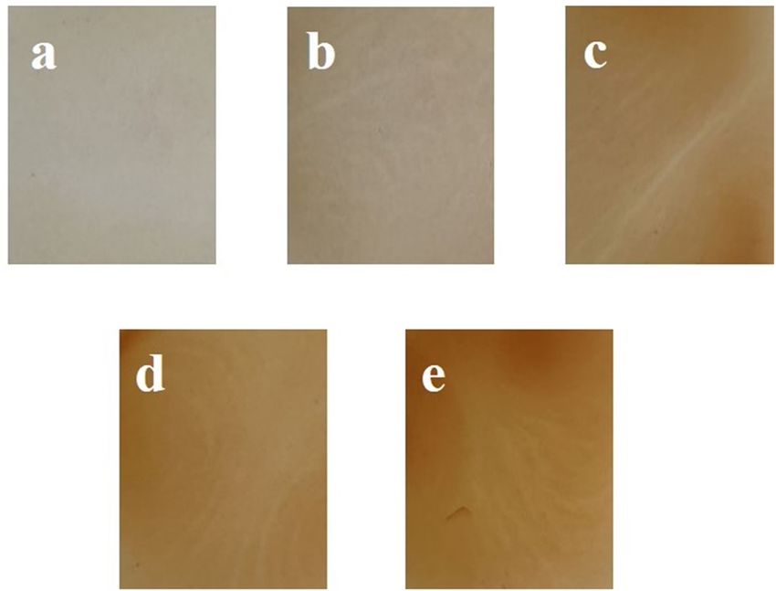
Preparation and characterizations of antibacterial–antioxidant film from SPI 581
microorganisms, which indicated that the obvious inhibi- 3.3 Mechanical properties
tory effect was only related to the extract. The difference in
the antibacterial activity of MPE could be partly explained Table 4 shows that the thickness of the SPI films con-
by the change in the phenol content in the extracts. taining 0%, 1%, 5%, 10%, and 15% MPE were 0.103,
The toxic mechanism of phenols in microorganisms was 0.108, 0.110, 0.123, and 0.129 mm, respectively. Obviously,
related to the reaction of protein sulfhydryl groups and the as the concentration of MPE increased, the thickness of the
inability of the microorganisms to utilize substrates (11). film increased.
MPE interfered with the bacterial protein secretions (15). The mechanical properties of biopolymer-based films,
Liang and Wang (39) showed that there was no inhibition particularly TS and EB, have a great influence on packa-
zone against E. coli on the cortex phellodendron extract ging materials (40). All the samples were preconditioned
but showed clear inhibition zones against S. aureus. The at medium constant RH of 45 ± 5% because the plasticizing
differences in the activities of the peel extracts between the effect of water produce positive and negative changes in
studies could be explained in part as due to the changes in film elasticity and mechanical stress, respectively. In gen-
the phenol content of the extracts, the strain sensitivity, eral, both TS and EB of the SPI films increased with the
and the antibacterial procedures used in the tests. concentration of MPE, and the film containing the highest
Table 3 shows the antibacterial activities of the dif- MPE concentration (15 g/100 g SPI) reached the compar-
ferent SPI films against E. coli and S. aureus. The SPI film able levels in the TS and EB when compared with those
containing 15% of MPE showed the highest antimicrobial from the control film (Table 4). The result was due to the
potency towards both E. coli (66.87%) and S. aureus phenol–protein interaction, which is mainly stabilized by
(80.43%). The antibacterial effect increased with the the hydrogen bonds and hydrophobic interactions. How-
increasing amount of MPE. Phenols react with oxidative ever, a small amount of MPE may cause lower formation of
compounds such as sulfhydryl groups or form non-specific hydrogen bonding in the SPI films, hinder the interaction
complexes with proteins, thereby causing enzyme inhibi- between molecules, and thereby reduce TS and EB. The
tion. In short, all these compounds prevent oxidative strength of SPI films were improved than that in the study
phosphorylation by inhibiting the enzymatic mechanism by Wang et al. (41) because of cross-linking between SPI
of microorganisms, thereby contributing to the rupture of and PVA.
microbial cell walls (26). As the antibacterial activity test
was performed on both gram-negative bacteria and gram-
positive bacteria, the results showed that the SPI film com-
bined with MPE has activity against a broad spectrum of 3.4 WS
microorganisms. Natural analogies in plant extracts with
different bioactivity and potency are highly likely to be The WS of a film in water may also provide insight into
retained for bioactivity. the behavior of the films in an aqueous environment,
which is a measure of its water resistance (42). Since
the weakness of SPI-based films is their higher suscept-
ibility to moisture, many studies are targeted to improve
hydrophilicity, which is highly desirable for food
Table 3: Antibacterial properties of SPI films with and without MPE
Films Bacteria Viable colony Antibacterial
Table 4: Thickness and mechanical properties of SPI films as
numbers potency (%)
affected by MPE incorporation
(CFU/mL)
SPI E. coli 492.0 0 Films Thickness (mm) TS (MPa) EB (%)
control S. aureus 92.0 0
SPI- E. coli 474.0 3.7 SPI control 0.10 ± 0.03b 4.1 ± 0.3a 186.1 ± 10.5a
1%MPE S. aureus 74.0 19.6 SPI-1% MPE 0.11 ± 0.08b 3.1 ± 0.1b 85.8 ± 15.1b
SPI- E. coli 327.0 33.5 SPI-5% MPE 0.11 ± 0.04b 3.7 ± 0.1a 143.6 ± 39.8a
5% MPE S. aureus 41.0 55.4 SPI-10% MPE 0.12 ± 0.02a 3.8 ± 0.1a 159.2 ± 14.8a
SPI- E. coli 280.0 43.1 SPI-15% MPE 0.13 ± 0.06a 4.1 ± 0.3a 187.5 ± 23.7a
10% MPE S. aureus 24.0 73.9
All data are shown as mean values ± standard deviation (SD).
SPI- E. coli 163.0 66.9
Different superscript letters in the column indicate significant dif-
15% MPE S. aureus 18.0 80.4
ferences (P < 0.05).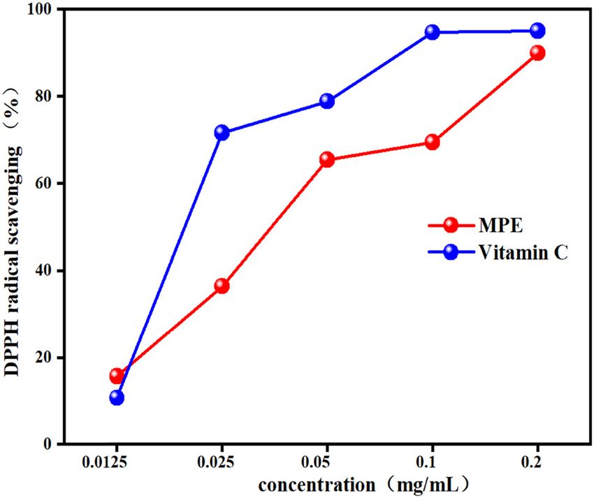
582 Xin Zhou et al.
Table 5: WVP, WS, and WCA of SPI films as affected by MPE incorporation
Films WVP × 10−14 (g/cm s MPa) WS (%) WCA (°)
SPI control 6.92 ± 0.02c
22.38 ± 0.49ab
58.8 ± 0.7a
SPI-1% MPE 7.14 ± 0.06c 23.58 ± 0.12a 55.8 ± 1.8ab
SPI-5% MPE 7.7 ± 0.02b 21.74 ± 0.69b 54.4 ± 2.3b
SPI-10% MPE 8.34 ± 0.01a 22.16 ± 0.62ab 52.7 ± 2.6bc
SPI-15% MPE 8.6 ± 0.01a 22.34 ± 0.58ab 50.1 ± 1.8c
All data are shown as mean values ± standard deviation (SD). Different superscript letters in the column indicate significant differences
(P < 0.05).
packaging application (10). The solubility of the film the polymer layers, which in turn increased the free
remains the same, with only a small difference (P < 0.05) volume and inter-chain distance. Therefore, more chain
(Table 5). Similar results were obtained by other authors mobility and free movement space could promote the
(43). Based on our findings, SPI films were much more transfer of water vapor.
water-resistant, the probable reason behind considerable
differences in WS of the films could be the difference in the
methods applied. The resistance of the film to water, which
is determined by the solubility of the film in water, is
3.6 WCA
critical to the potential application of the film. Originally,
The surface hydrophobicity of the SPI film was evaluated
the WS of polymers is determined by their molecular struc-
by measuring. Generally, the higher the WCA, the more
tures. Therefore, thermal stability information can be obtained
the hydrophobicity of the film surface (45). The addition
by dissolution test.
of MPE had a profound effect on the hydrophobicity of the
resulting films (P < 0.05), which may be due to the hydro-
philicity of MPE (Table 5). Compared with the composite
3.5 Water vapor permeability films, the control film has a higher WCA (58.78 ± 0.38°).
When an increasing concentration of MPE was added to
The WVP values for the samples are listed in Table 5. the SPI films, a decrease in WCA was observed (P < 0.05).
WVP is a parameter that is affected by multiple factors,
such as roughness, hydrophilic tightness, and the distri-
bution and orientation of particles in the film, so it is 3.7 Color measurement
difficult to account for all the changes observed in this
parameter for the samples under study (44). Compared to It is usually found that the addition of various plant
others (30), the WVP of SPI film was reduced because the extracts will change the original color of the biopolymer-
new hydrogen bond between SPI and PVA would reduce based film to some extent, depending on the source and
the free hydrogen group in SPI films. Compared to the concentration of the plant extracts (46). L (0 = black and
control films, incorporation of MPE increased the WVP 100 = white), a (−60 = greenness to + 60 = redness), b (−60 =
of the SPI films significantly (P < 0.05). It was possible blueness to + 60 = yellowness), and total color difference
that the introduction of MPE led to the recombination of (ΔE) of these films are presented in Table 6. The addition
Table 6: Color and opacity of SPI films as affected by MPE incorporation
Films L a b ΔE Opacity
SPI control 90.94 ± 0.22a 2.52 ± 0.47d 7.69 ± 0.23e 3.46 ± 0.07a 1.75 ± 0.02d
SPI-1%MPE 85.31 ± 0.49b 4.88 ± 0.36c 15.09 ± 0.51d 6.85 ± 0.54b 1.78 ± 0.08d
SPI-5% MPE 84.01 ± 0.90c 5.94 ± 0.33b 17.7 ± 2.16c 9.79 ± 1.88c 2.21 ± 0.02c
SPI-10% MPE 80.85 ± 1.22d 8.31 ± 1.58a 22.25 ± 1.93 16.78 ± 2.93d 2.52 ± 0.08b
SPI-15% MPE 77.20 ± 1.85e 10.87 ± 1.30a 23.66 ± 2.23b 19.09 ± 2.87e 2.93 ± 0.01a
All data are shown as mean values ± standard deviation (SD). Different superscript letters in the column indicate significant differences
(P < 0.05).Preparation and characterizations of antibacterial–antioxidant film from SPI 583
observed in the control. The films with higher concentra-
tion of MPE showed substantial (P < 0.05) higher opacity.
This indicated that the opacity of the SPI film was caused
by the polyphenol compound added in the SPI film which
caused light scattering and refraction, resulting in dar-
kening of the film (29). Although food packaging mate-
rials are mostly transparent and colorless, colored films
may also be an advantage in protecting against exposure
to UV-Vis light, which can affect food quality (48).
3.9 Film transmittance
Figure 5 shows the UV-Vis light transmission patterns of
Figure 4: Appearance of SPI films: (a) SPI control, (b) SPI-1% MPE,
all samples in the range of 200–800 nm. The spectrum
(c) SPI-5% MPE, (d) SPI-10% MPE, and (e) SPI-15% MPE.
of all films showed transmittance values of 0–42% in the
UV range (200–400 nm). Therefore, the SPI film effec-
of MPE to SPI films significantly reduced their whiteness tively prevents the transmission of UV light at these
and increased their reds and yellows (P < 0.05). The wavelengths, independent of the addition of MPE. The
changes in ΔE of SPI films were increased by the addition visible light transmittance (400–800 nm) of all films
of MPE which could be confirmed visually in Figure 4. In was between 0% and 70%. By adding different contents
general, the phenolic pigments in plant extracts may be of MPE, a significant change in the light transmittance of
contributors to the various colors observed in these bio- the obtained film could be observed in the visible light
polymer films (47). range. The transmittance value was greatly reduced in
SPI films containing MPE. This also showed that MPE-
SPI films had higher UV-Vis light barrier performance
3.8 Film opacity than SPI control film. In addition, the light barrier per-
formance of the SPI film increased with the increase in
The opacity at different concentrations of MPE in SPI the MPE content. Therefore, it could be concluded that
films are shown in Table 6. The lowest opacity was the UV-Vis light barrier properties of the SPI film might
Figure 5: UV-Vis spectra of SPI films with and without MPE.584 Xin Zhou et al.
Table 7: Thermal degradation temperature (Td, °C) and weight loss
(Δw, %) of films incorporated without and with MPE
Films Δ1 Δ2 Δ3
Td1,onset Δw1 Td2,onset Δw2 Td3,onset Δw3
SPI control 56 0.44 222 1.10 319 2.70
SPI-1% MPE 78 0.37 228 1.21 320 3.16
SPI-5% MPE 758 0.30 243 1.04 313 2.75
SPI-10% MPE 65 0.39 231 1.16 316 2.77
SPI-15% MPE 75 0.35 230 1.13 316 2.55
be due to the UV-absorbing ability of the polyphenolic
compounds in MPE as researchers reported earlier (49).
3.10 TGA Figure 7: ATR-FTIR spectra of SPI films with and without MPE.
TGA was used to measure the thermal properties of the
SPI films, and the results are shown in Table 7 and same as the control film. This result shows that the incor-
Figure 6. As shown in the TGA thermograms, all the films poration of MPE cannot improve the thermal stability of
showed three main stages of weight loss. The first thermal the SPI film.
degradation (30–150°C) was mainly attributed to the eva-
poration of water remaining in SPI films. The second
(150–280°C) and third (280–600°C) stages of degradation 3.11 ATR-FTIR
could be due to the degradation of residual glycerol
and the thermal decomposition of SPI matrix and MPE, The ATR-FTIR spectra of SPI films showed hydrogen
respectively. It is worth noting that the addition of MPE interaction between components (Figure 7). For SPI con-
did not significantly change the TGA curve and derivative trol film, the absorption bands are associated with C]O
thermogravimetric (DTG) curve of the film (50). As shown stretching of 1,634 cm−1 (amide I) and N–H bending of
in Table 6, the weight loss and onset thermal degradation 1,539 cm−1 (amide II). The wide absorption band (amide – A)
temperature of the film containing MPE was almost the observed at 3,275 cm−1 is attributable to the stretching
Figure 6: TGA (a) and DTG (b) patterns of SPI films with and without MPE.Preparation and characterizations of antibacterial–antioxidant film from SPI 585
Table 8: Percentage of secondary structures of SPI films with and without MPE
Films α-helix (%) β-sheet (%) β-turn (%) Random coil (%)
SPI control 24.2 45.8 18.3 11.6
SPI-1% MPE 27.3 40.8 15.5 16.4
SPI-5% MPE 30.5 37.4 18.0 14.1
SPI-10% MPE 25.1 43.3 18.3 13.4
SPI-15% MPE 26.6 42.4 16.9 14.1
vibrations of the free and bonded O–H and N–H groups structures of protein in the SPI films containing MPE. For
(9,51,52). the control film, the main structures resulted from α-helix,
The amide – A shifted slightly to higher wavenum- β-sheet, β-turn, and random coil corresponded to the percen-
bers (3,275–3,281 cm−1) in SPI films incorporated with tage areas of around 24.21%, 45.81%, 18.34%, and 11.64%,
1% and 5% of MPE. This result indicated that the unfolding respectively. The addition of MPE as an antioxidant to the SPI
of soy protein may lead to new hydrophobic interactions film did not significantly affect the β-turn and caused the
and weak hydrogen bonds. Generally, the intensity of β-sheet to transform into random coils and α-helix, in which
amide – A in SPI film added with MPE was weaker than the areas of random coils and α-helix increased. In general,
the film control. This observation may be attributed to the content of β-sheet contributed to the strength of SPI films,
the hydrogen bonding between SPI and MPE. Therefore, while the flexibility of SPI films was related to the content of
hydrogen bond interactions between polyphenols and pro- α-helix and β-turn (54). In the SPI film, the addition of MPE
teins may be enhanced at higher MPE concentrations. The resulted in the conversion of β-sheets to α-helix, and the
amide I band is composed of several bands corresponding percentage area of α-helix increased. Meanwhile, it was
to different secondary structures of the protein, e.g., α-helix believed that the heating of the MPE-SPI solution may
(1,646–1,664/cm), β-sheet (1,615–1,637 and 1,682–1,700/cm), destroy the internal hydrogen bonds of the peptide groups,
β-turn (1,664–1,681/cm), and random coil (1,637–1,645/cm) resulting in random coils. Random coils could provide com-
(53). Table 8 shows the percentage of different secondary plexing sites where the carbonyl group of the polypeptide
Figure 8: SEM images of surface and cross section of the SPI films with and without MPE. (a) SPI control, (b) SPI-1% MPE, (c) SPI-5% MPE,
(d) SPI-10% MPE, and (e) SPI-15% MPE.586 Xin Zhou et al.
the SPI films, peak around 19° became sharper as the
concentration of MPE increased to 15%. Moreover, when
the ratio of MPE was 5%, one peak around 22° appeared.
When MPE was added to SPI film, the peak intensity was
much lower than that of the control film. This indicated
that the crystalline structure of SPI films had collapsed
after the addition of MPE. Compared with the control film,
the XRD peak of SPI film was gentler when MPE was
added. It showed that the crystallinity of SPI film was
decreased by MPE. The results showed that MPE inserted
the macromolecular structure of the SPI film and destroyed
the crystallization of the SPI film. These findings are in
compliance with previous reports where “Pequi” peels
addition reduced the crystallinity of chitosan film (56).
Figure 9: XRD diffraction patterns of SPI films with and without MPE.
4 Conclusion
could interact with the polyphenols in MPE. Overall, it could MPE contains a lot of total polyphenols which causes
be included that the interaction between SPI and MPE attrib- the significant antioxidant and antibacterial properties.
uted to hydrogen bonds and hydrophobic interactions low- When they were added into the SPI film, the bio-activities
ered the mechanical properties of SPI films. of the film increased as extract level increased in film.
Compared with the control film, the UV-Vis barrier and
hydrophobicity of the film were improved with the addi-
3.12 SEM tion of MPE. And the TS and EB properties of the SPI films
containing MPE were changed. SEM and TGA analyses
The characterization of biopolymer microstructure is an indicated that the addition of MPE did not change the
important factor to understand its behavior. Figure 8 microstructure and thermal stability of the films. XRD
shows SEM micrographs of cross section of all the films results showed that the crystalline structure of SPI films
prepared in this study. All films have similar morpholo- was reduced by MPE. ATR-FTIR analysis showed that the
gical images, and the effect of MPE addition was not reaction between polyphenol and protein changed the
detected by analysis. However, some layers were detected secondary structure through hydrogen bonds and hydro-
for all the samples in cross section which is an indication phobic interactions. These results indicate that the MPE
of the phase separation between the component. Sun would be a promising ingredient to use the SPI films as an
et al. (55) reported similar findings. The micrographs of active packaging material.
the fractured cross sections showed slight differences
between the samples. evidencing small changes in the Acknowledgement: The authors would like to thank
mechanical properties and WVP. Guangxi University. This research was supported by the
Fundamental Research Funds for the National Natural
Science Foundation of China (Project No. 31790188).
3.13 XRD Funding information: National Natural Science Foundation
of China (Project No. 31790188).
In order to explore the crystal structure of the control SPI
film and reveal the effect of incorporation of MPE, XRD Author contributions: Xin Zhou: writing – original draft,
analysis was performed. It is evident from the spectrum writing – review and editing, formal analysis; Qinying
in Figure 9 that SPI peaks about 9° and 19° at 2θ reflect Dai: writing – original draft; Xi Huang: editing; Zhiyong
the α-helix and β-sheet structures of the secondary con- Qin: resources, methodology.
formation of SPI, respectively. Similar results have been
reported by Liang and Wang (39). After MPE was added to Conflict of interest: Authors state no conflict of interest.Preparation and characterizations of antibacterial–antioxidant film from SPI 587
References mechanical properties, and thermal stability. J Appl Polym Sci.
2008;110:3706–16. doi: 10.1002/app.28979.
(1) Gutiérrez TJ, Morales NJ, Pérez E, Tapia MS, Famá L. Physico- (14) Zhang X, Liu J, Yong H, Qin Y, Liu J, Jin C. Development of
chemical properties of edible films derived from native and antioxidant and antimicrobial packaging films based on chit-
phosphated cush-cush yam and cassava starches. Food osan and mangosteen (Garcinia mangostana L.) rind powder.
Int J Biol Macromol. 2020;145:1129–39. doi: 10.1016/
Packag Shelf. 2015;3:1–8. doi: 10.1016/j.fpsl.2014.09.002.
j.ijbiomac.2019.10.038.
(2) Kalkan S, Otag MR, Engin MS. Physicochemical and bioactive
(15) Sze Lim Y, Sze Hui Lee S, Chin Tan B. Antioxidant capacity and
properties of edible methylcellulose films containing Rheum
antibacterial activity of different parts of mangosteen
ribes L. extract. Food Chem. 2020;307:125524. doi: 10.1016/
(Garcinia mangostana Linn.) extracts. Fruits. 2013;68:483–9.
j.foodchem.2019.125524.
doi: 10.1051/fruits/2013088.
(3) Rasid NAM, Nazmi NNM, Isa MIN, Sarbon NM. Rheological,
(16) Robinson AN, Scully C. Behcet disease: A new therapeutic
functional and antioxidant properties of films forming solution
agent. [corrected]. Br Dent J. 2015;218:557. doi: 10.1038/
and active gelatin films incorporated with Centella asiatica (L.)
sj.bdj.2015.396.
urban extract. Food Packag Shelf. 2018;18:115–24.
(17) Lee J-S, Park J-M, Wi S-H, Ahn YB, Kim NK, Moon K-W, et al.
doi: 10.1016/j.fpsl.2018.10.002.
Improving consumer recognition and awareness of food
(4) Genskowsky E, Puente LA, Pérez-Álvarez JA, Fernandez-
additives through consumer education in South Korea. Food
Lopez J, Muñoz LA, Viuda-Martos M. Assessment of anti-
Sci Biotechnol. 2014;23:653–60. doi: 10.1007/s10068-014-
bacterial and antioxidant properties of chitosan edible films
0089-1.
incorporated with maqui berry (Aristotelia chilensis). LWT
(18) Suttirak W, Manurakchinakorn S. In vitro antioxidant proper-
Food Sci Technol. 2015;64:1057–62. doi: 10.1016/
ties of mangosteen peel extract. J Food Sci Technol.
j.lwt.2015.07.026.
2014;51:3546–58. doi: 10.1007/s13197-012-0887-5.
(5) Nunes MR, de Souza Maguerroski Castilho M, de Lima
(19) Garcia ML, Carrion MH, Escobar S, Rodriguez A, Cortina JR.
Veeck AP, da Rosa CG, Noronha CM, Maciel M, et al.
Optimization of the antioxidant capacity of mangosteen peels
Antioxidant and antimicrobial methylcellulose films con-
taining Lippia alba extract and silver nanoparticles. Carbohydr (Garcinia mangostana L.) extracts: Management of the drying
Polym. 2018;192:37–43. doi: 10.1016/j.carbpol.2018.03.014. extraction processes. Food Sci Technol Int. 2020. doi: 10.1177/
(6) Hamedi H, Kargozari M, Shotorbani PM, Mogadam NB, 1082013220962630.
Fahimdanesh M. A novel bioactive edible coating based on (20) Pedraza-Chaverri J, Cardenas-Rodriguez N, Orozco-Ibarra M,
sodium alginate and galbanum gum incorporated with Perez-Rojas JM. Medicinal properties of mangosteen (Garcinia
essential oil of Ziziphora persica: the antioxidant and anti- mangostana). Food Chem Toxicol. 2008;46:3227–39.
microbial activity, and application in food model. Food doi: 10.1016/j.fct.2008.07.024.
Hydrocoll. 2017;72:35–46. (21) Zhou H-C, Lin Y-M, Wei S-D, Tam NFY. Structural diversity and
(7) Kowalczyk D, Kordowska-Wiater M, Karaś M, Zięba E, antioxidant activity of condensed tannins fractionated from
Mężyńska M, Wiącek AE. Release kinetics and antimicrobial mangosteen pericarp. Food Chem. 2011;129:1710–20.
properties of the potassium sorbate-loaded edible films made doi: 10.1016/j.foodchem.2011.06.036.
from pullulan, gelatin and their blends. Food Hydrocoll. (22) Cheok CY, Chin NL, Yusof YA, Law CL. Extraction of total phe-
2020;101. doi: 10.1016/j.foodhyd.2019.105539. nolic content from garcinia mangostana Linn. hull. I. Effects of
(8) Roy S, Rhim JW. Preparation of antimicrobial and antioxidant solvents and UV-Vis spectrophotometer absorbance method.
Food Bioprocess Tech. 2012;5:2928–33. doi: 10.1007/s11947-
gelatin/curcumin composite films for active food packaging
011-0627-2.
application. Colloids Surf B. 2020;188:110761. doi: 10.1016/
(23) Zhang W, Li X, Jiang W. Development of antioxidant chitosan
j.colsurfb.2019.110761.
(9) Zhang Y, Chen S, Qi B, Sui X, Jiang L. Complexation of ther- film with banana peels extract and its application as coating in
mally-denatured soybean protein isolate with anthocyanins maintaining the storage quality of apple. Int J Biol Macromol.
and its effect on the protein structure and in vitro digestibility. 2020;154:1205–14. doi: 10.1016/j.ijbiomac.2019.10.275.
Food Res Int. 2018;106:619–25. doi: 10.1016/ (24) Wafa BA, Makni M, Ammar S, Khannous L, Hassana AB,
j.foodres.2018.01.040. Bouaziz M, et al. Antimicrobial effect of the Tunisian Nana
(10) Rani S, Kumar R. A review on material and antimicrobial variety Punica granatum L. extracts against Salmonella
properties of soy protein isolate film. J Polym Environ. enterica (serovars Kentucky and Enteritidis) isolated from
2019;27:1613–28. doi: 10.1007/s10924-019-01456-5. chicken meat and phenolic composition of its peel extract. Int J
(11) Wang SN, Marcone M, Barbut S, Lim LT. The impact of antho- Food Microbiol. 2017;241:123–31. doi: 10.1016/
cyanin-rich red raspberry extract (ARRE) on the properties of j.ijfoodmicro.2016.10.007.
edible soy protein isolate (SPI) films. J Food Sci. (25) Othman M, Loh HS, Wiart C, Khoo TJ, Lim KH, Ting KN. Optimal
2012;77:C497–505. methods for evaluating antimicrobial activities from plant
(12) Su J-F, Huang Z, Yuan X-Y, Wang X-Y, Li M. Structure and extracts. J Microbiol Meth. 2011;84:161–6. doi: 10.1016/
properties of carboxymethyl cellulose/soy protein isolate j.mimet.2010.11.008.
blend edible films crosslinked by Maillard reactions. Carbohyd (26) Sharma C, Bhardwaj NK. Fabrication of natural-origin anti-
Polym. 2010;79:145–53. doi: 10.1016/j.carbpol.2009.07.035. bacterial nanocellulose films using bio-extracts for potential
(13) Su J-F, Huang Z, Yang C-M, Yuan X-Y. Properties of soy protein use in biomedical industry. Int J Biol Macromol.
isolate/poly(vinyl alcohol) blend “green” films: compatibility, 2020;145:914–25. doi: 10.1016/j.ijbiomac.2019.09.182.588 Xin Zhou et al.
(27) Song HY, Shin YJ, Song KB. Preparation of a barley bran (40) Hajivand P, Aryanejad S, Akbari I, Hemmati A. Fabrication and
protein–gelatin composite film containing grapefruit seed characterization of a promising oregano‐extract/psyllium‐
extract and its application in salmon packaging. J Food Eng. seed mucilage edible film for food packaging. J Food Sci.
2012;113:541–7. doi: 10.1016/j.jfoodeng.2012.07.010. 2020;85:2481–90. doi: 10.1111/1750-3841.15331.
(28) Gómez-Estaca J, Bravo L, Gómez-Guillén MC, Alemán A, (41) Wang H, Hu D, Ma Q, Wang L. Physical and antioxidant prop-
Montero P. Antioxidant properties of tuna-skin and bovine- erties of flexible soy protein isolate films by incorporating
hide gelatin films induced by the addition of oregano and chestnut (Castanea mollissima) bur extracts. LWT Food Sci
rosemary extracts. Food Chem. 2009;112:18–25. doi: 10.1016/ Technol. 2016;71:33–9. doi: 10.1016/j.lwt.2016.03.025.
j.foodchem.2008.05.034. (42) Peng Y, Wu Y, Li Y. Development of tea extracts and chitosan
(29) Rambabu K, Bharath G, Banat F, Show PL, Cocoletzi HH. Mango composite films for active packaging materials. Int J Biol
leaf extract incorporated chitosan antioxidant film for active Macromol. 2013;59:282–9. doi: 10.1016/
food packaging. Int J Biol Macromol. 2019;126:1234–43. j.ijbiomac.2013.04.019.
doi: 10.1016/j.ijbiomac.2018.12.196. (43) Hoque MS, Benjakul S, Prodpran T. Properties of film from
(30) Qin Z, Mo L, Liao M, He H, Sun J. Preparation and character- cuttlefish (Sepia pharaonis) skin gelatin incorporated with
ization of soy protein isolate-based nanocomposite films with cinnamon, clove and star anise extracts. Food Hydrocoll.
cellulose nanofibers and nano-silica via silane grafting. 2011;25:1085–97. doi: 10.1016/j.foodhyd.2010.10.005.
Polymers (Basel). 2019;11. doi: 10.3390/polym11111835. (44) Rodrigues MAV, Bertolo MRV, Marangon CA, Martins V,
(31) Yong H, Liu J, Qin Y, Bai R, Zhang X, Liu J. Antioxidant and pH- Plepis AMG. Chitosan and gelatin materials incorporated with
sensitive films developed by incorporating purple and black phenolic extracts of grape seed and jabuticaba peel:
rice extracts into chitosan matrix. Int J Biol Macromol. Rheological, physicochemical, antioxidant, antimicrobial and
2019;137:307–16. doi: 10.1016/j.ijbiomac.2019.07.009. barrier properties. Int J Biol Macromol. 2020;160:769–79.
(32) Emam-Djomeh Z, Moghaddam A, Yasini Ardakani SA. doi: 10.1016/j.ijbiomac.2020.05.240.
Antimicrobial activity of pomegranate (Punica granatum L.) (45) Kanmani P, Rhim JW. Antimicrobial and physical-mechanical
peel extract, physical, mechanical, barrier and antimicrobial properties of agar-based films incorporated with grapefruit
properties of pomegranate peel extract-incorporated seed extract. Carbohydr Polym. 2014;102:708–16.
sodium caseinate film and application in packaging for doi: 10.1016/j.carbpol.2013.10.099.
ground beef. Packag Technol Sci. 2015;28:869–81. (46) Wang S, Marcone MF, Barbut S, Lim L-T. Fortification of dietary
doi: 10.1002/pts.2145. biopolymers-based packaging material with bioactive plant
(33) Ju A, Baek SK, Kim S, Song KB. Development of an antioxida- extracts. Food Res Int. 2012;49:80–91. doi: 10.1016/
tive packaging film based on khorasan wheat starch con- j.foodres.2012.07.023.
taining moringa leaf extract. Food Sci Biotechnol. (47) Han Y, Yu M, Wang L. Preparation and characterization of
2019;28:1057–63. antioxidant soy protein isolate films incorporating licorice
(34) Sungpud C, Panpipat W, Sae Yoon A, Chaijan M. Tuning of residue extract. Food Hydrocoll. 2018;75:13–21. doi: 10.1016/
virgin coconut oil and propylene glycol ratios for maximizing j.foodhyd.2017.09.020.
the polyphenol recovery and in vitro bioactivities of mango- (48) Adilah AN, Jamilah B, Noranizan MA, Hanani ZAN. Utilization of
steen (Garcinia mangostana L.) pericarp. Process Biochem. mango peel extracts on the biodegradable films for active
2019;87:179–86. doi: 10.1016/j.procbio.2019.08.023. packaging. Food Packag Shelf. 2018;16:1–7. doi: 10.1016/
(35) Tjahjani S. Antimalarial activity of Garcinia mangostana L j.fpsl.2018.01.006.
rind and its synergistic effect with artemisinin in vitro. (49) Kaya M, Ravikumar P, Ilk S, Mujtaba M, Akyuz L, Labidi J, et al.
BMC Compl Altern Med. 2017;17:131. doi: 10.1186/s12906-017- Production and characterization of chitosan based edible films
1649-8. from Berberis crataegina’s fruit extract and seed oil. Innov
(36) Kotepong P, Paull RE, Ketsa S. Anthocyanin accumulation and Food Sci Emerg Technol. 2018;45:287–97. doi: 10.1016/
differential gene expression in wild-type and mutant Syzygium j.ifset.2017.11.013.
malaccense fruits during their growth and ripening. Biol Plant. (50) Kalaycioglu Z, Torlak E, Akin-Evingur G, Ozen I, Erim FB.
2019;63:710–20. doi: 10.32615/bp.2019.068. Antimicrobial and physical properties of chitosan films incor-
(37) Sonawane A, Pathak S, Pradhan RC. Bioactive compounds in porated with turmeric extract. Int J Biol Macromol.
bael fruit pulp waste: ultrasound-assisted extraction, charac- 2017;101:882–8. doi: 10.1016/j.ijbiomac.2017.03.174.
terization, modeling, and optimization approaches. Biointerf (51) Mauri AN, Añón MC. Effect of solution pH on solubility and
Res Appl Chem. 2020;11:9318–34. doi: 10.33263/ some structural properties of soybean protein isolate films.
briac112.93189334. J Sci Food Agr. 2006;86:1064–72. doi: 10.1002/jsfa.2457.
(38) Pothitirat W, Chomnawang MT, Supabphol R, Gritsanapan W. (52) Wang HX, Hu DY, Ma QY, Wang LJ. Physical and antioxidant
Free radical scavenging and anti-acne activities of mango- properties of flexible soy protein isolate films by incorporating
steen fruit rind extracts prepared by different extraction chestnut (Castanea mollissima) bur extracts. Lwt-Food Sci
methods. Pharm Biol. 2010;48:182–6. doi: 10.3109/ Technol. 2016;71:33–9.
13880200903062671. (53) Sui X, Sun H, Qi B, Zhang M, Li Y, Jiang L. Functional and
(39) Liang SM, Wang LJ. A natural antibacterial-antioxidant film conformational changes to soy proteins accompanying
from soy protein isolate incorporated with cortex phelloden- anthocyanins: Focus on covalent and non-covalent inter-
dron extract. Polymers. 2018;10. doi: 10.3390/ actions. Food Chem. 2018;245:871–8. doi: 10.1016/
polym10010071. j.foodchem.2017.11.090.Preparation and characterizations of antibacterial–antioxidant film from SPI 589
(54) Yang H, Wen XL, Guo SG, Chen MT, Jiang AM, Lai LS. Physical, apple polyphenols as an active packaging material. Carbohydr
antioxidant and structural characterization of blend films Polym. 2017;163:81–91. doi: 10.1016/j.carbpol.2017.01.016.
based on hsian-tsao gum (HG) and casein (CAS). Carbohydr (56) Breda CA, Morgado DL, Assis OBG, Duarte MCT. Processing
Polym. 2015;134:222–9. doi: 10.1016/j.carbpol.2015.07.021. and characterization of chitosan films with incorporation of
(55) Sun L, Sun J, Chen L, Niu P, Yang X, Guo Y. Preparation and ethanolic extract from “pequi” peels. Macromol Res.
characterization of chitosan film incorporated with thinned young 2017;25:1049–56. doi: 10.1007/s13233-017-5143-4.You can also read