Effects of a herbal formulation, KGC3P, and its individual component, nepetin, on coal fly dust induced airway inflammation - Nature
←
→
Page content transcription
If your browser does not render page correctly, please read the page content below
www.nature.com/scientificreports
OPEN Effects of a herbal formulation,
KGC3P, and its individual
component, nepetin, on coal fly
dust‑induced airway inflammation
Evelyn Saba 1, Young‑sil Lee2, Won‑Kyung Yang 3,6, Yuan Yee Lee 1, MinKi Kim1,
Su‑Min Woo9, KilSoo Kim1, Young‑Sam Kwon 1, Tae‑Hwan Kim1, Dongmi Kwak1,
Yang‑Chun Park 3, Han Jae Shin4, Chang Kyun Han5, Jae‑Wook Oh7, Young Cheol Lee 8
,
Hyung‑Sik Kang9, Man Hee Rhee 1* & Seung‑Hyung Kim 6*
Coal fly dust (CFD)-induced asthma model is used as an ambient particulate matter model of serious
pulmonary damage. We aimed to evaluate the effects of a combination of ginseng and Salvia plebeia
R. Br extract (KGC-03-PS; KG3P) and its individual components (hispidulin, nepetin and rosmarinic
acid) in a CFD-induced mouse model of airway inflammation (asthma). We also evaluated signal
transduction by KG3P and its individual components in the alveolar macrophage cell line, MH-S
cells. In vitro, KG3P and its individual components inhibited nitric oxide production and expression
of pro-inflammatory mediators and cytokines (iNOS, COX-2, IL-1β, IL-6 and TNF-α) through the
NF-κB and MAPK pathways in coal fly ash (CFA)-induced inflammation in MH-S cells. Moreover, in
the CFD-induced asthma model in mice, KG3P and its predominant individual component, nepetin,
inhibited Asymmetric Dimethyl arginine (ADMA) and Symmetric Dimethyl arginine (SDMA) in serum,
and decreased the histopathologic score in the lungs. A significant reduction in the neutrophils and
immune cells in BALF and lung tissue was demonstrated, with significant reduction in the expression
of the pro-inflammatory cytokines. Finally, IRAK-1 localization was also potently inhibited by KG3P
and nepetin. Thus, KG3P extract can be considered as a potent candidate for amelioration of airway
inflammation.
Presently, air pollution is a serious environmental problem encountered by living organisms. Extensive industri-
alization and a sedentary life style, aggravated by the increased usage of automobiles than the physical methods
of transportation, are the etiological factors associated with this p roblem1. Increased usage of automobiles and
the installation of industrial units near urban areas are directly related to increased production of smoke, which
consists of ambient particulate matters such as coal, asbestos, and combustion particles. Although acute inhala-
tion of these nanoparticles does not pose any serious side effects to health, constant and chronic exposure can
lead to the settling of these particles in the respiratory tract, resulting in serious chronic pulmonary aliments such
as asthma, chronic bronchitis, and lung cancer2–4. Industrial combustion is considered a well-studied process
and the fly ash produced by combustion is frequently used as a model particle for studying fine and nano-fine
1
Laboratory of Physiology and Cell Signalling, Department of Veterinary Medicine, College of Veterinary Medicine,
Kyungpook National University, Daegu 41566, Republic of Korea. 2Herbal Medicine Research Division, Korea
Institute of Oriental Medicine, 1672 Yuseong‑daero, Yuseong‑gu, Dajeon 34054, Republic of Korea. 3Division
of Respiratory Systems, Department of Internal Medicine, College of Korean Medicine, Daejeon University,
Daejeon, Republic of Korea. 4KT&G Research Institute, Daejeon 34128, Republic of Korea. 5KGC Research
Institute, Daejeon 34128, Republic of Korea. 6Institute of Traditional Medicine and Bioscience, Daejeon University,
Daejeon 34520, Republic of Korea. 7Department of Stem Cell and Regenerative Biotechnology, Konkuk University,
Seoul 05029, Republic of Korea. 8Department of Herbology, College of Korean Medicine, Sangji University, 83
Sangjidae‑gil, Wonju, Gangwon‑do 26339, Republic of Korea. 9School of Biological Sciences and Technology,
Chonnam National University, Gwangju 500‑757, Republic of Korea. *email: rheemh@knu.ac.kr; sksh518@dju.kr
Scientific Reports | (2020) 10:14036 | https://doi.org/10.1038/s41598-020-68965-5 1
Vol.:(0123456789)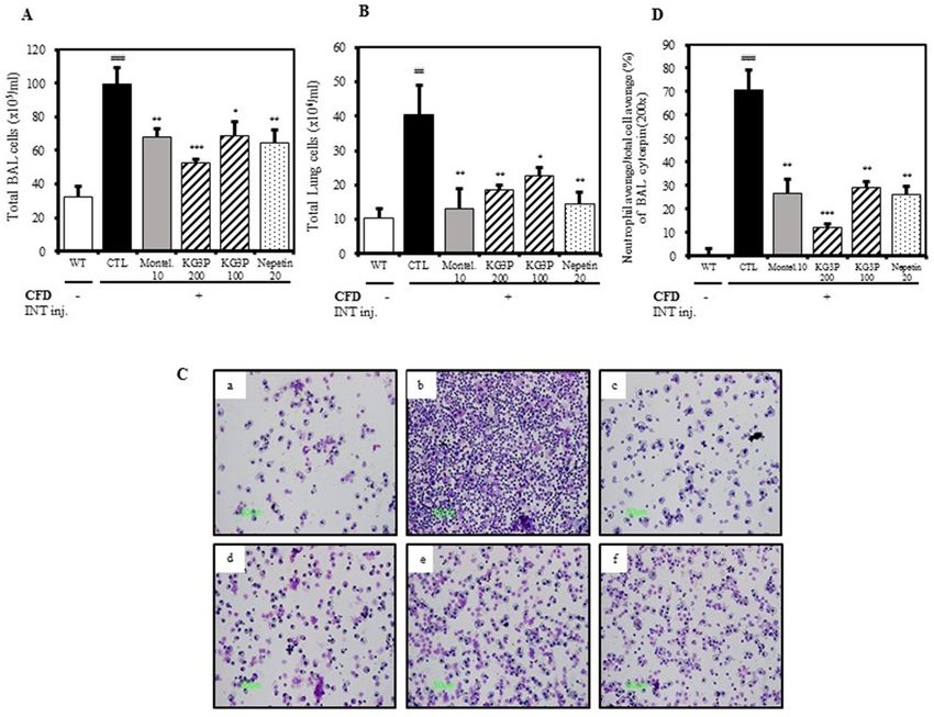
www.nature.com/scientificreports/
components in toxicological r esearch5,6. Since these fine particles can cause chronic, fatal pulmonary diseases,
there has been a shift in research focus towards the discovery of remedies (both allopathic and herbal).
Ginseng, popularly considered a panacea, has been used for centuries in the Korean peninsula owing to its
outstanding health promoting effects against minor to major diseases, including inflammation and cancer7–9.
Extensive data are available on the health enhancing properties of whole ginseng extract as well as for the indi-
vidual compounds in its extract, termed g insenosides10. To date, however, no data exists on the effect of ginseng
in coal fly ash (CFA) or combustion particle-induced inflammation.
Salvia plebeia R BR (SPR-Br) is a common herbal medicine used in China and is often used to treat urinary
tract infections. SPR-Br is known to possess anti-inflammatory, anti-oxidant, anticancer, anti-hypertensive,
and immune boosting effects11,12. The plant is found near streams and mountains that are > 2,800 m above sea
level13. SPR-Br is composed of flavones, organic acids, amino acids, and alcohol phenols14. Most importantly, it
consists of many single compounds such as hispidulin, nepetin, eupafolin, ursoic acid, and caffeic a cid15. Owing
to previous reports, SPR-Br has been confirmed to possess strong biological properties; however, there is no data
available on the effects of this herb on airway inflammation induced by combustion particles.
We aimed to develop a herbal formulation consisting of ginseng extract and SPR-Br (KG3P) to verify the
effects of the extract and its individual components on MH-S alveolar macrophage cells induced by CFA, and
mice with airway inflammation induced by coal fly dust (CFD). Based on the HPLC analysis performed, KG3P
and its three most abundant components potently suppressed inflammation by decreasing the levels of nitric
oxide (NO), iNOS, COX-2, IL-1β, IL-6, and TNF-α via the NF-κB and MAPK pathways in MH-S cells. Moreover,
our in vivo results showed that KG3P and its component, nepetin, downregulated the inflammatory cytokines
in BALF and lung tissues, and decreased the fluorescent intensity of interleukin-1 receptor associated kinase
1 (IRAK-1) in lung tissues. These results suggest that this mixture can be used as an excellent remedy in the
prevention of pulmonary disorders caused by air pollution.
Results
Effects of KG3P and nepetin in vitro on MH‑S cells and signal transduction via the NF‑κB and
MAPK pathways. Based on our UPLC results as shown if Fig. 1, KG3P possessed three abundant indi-
vidual components: hispidulin, nepetin, and rosmarinic acid. Before we start with the in vivo work, we sought
to elucidate the in vitro effects of various concentrations of KG3P in Alveolar macrophage cells (MH-S cells)
stimulated with CFA. We selected alveolar macrophage cells for our study because in case of asthma induction,
it’s the airway inflammation that is responsible for the pathophysiology, and since macrophage cells are the major
players in this process16,17 therefore we checked their levels in response to asthma induction and treatment with
KG3P and nepetin along with the positive control Montelukast. As shown in Fig. 2A,B, KG3P potently inhibited
nitrite production and expression of pro-inflammatory mediators and cytokines in a dose dependent manner;
the individual components, especially nepetin, also exhibited this potent inhibition. In addition, the highest dose
of KG3P and nepetin, mainly, inhibited the phosphorylation of all downstream factors in the NF-κB and MAPK
pathways in MH-S cells (Fig. 2C,D). Since nepetin was demonstrated as the most abundant and effective of the
three individual components, the in vivo study was performed using only nepetin and KG3P.
Inhibitory effects of KG3P and nepetin on serum asymmetrical dimethyl arginine (ADMA)
and symmetric dimethyl arginie (SDMA) levels, and restoration of histopathological
lesions. Asymmetrical dimethyl arginine (ADMA) and symmetric dimethyl arginie (SDMA) are involved
in the inflammation, endothelial dysfunction and oxidative stress. Basically they are the structural analogues
of l-arginine, which competitively regress NO synthase, ultimately leading to decreased basal NO production
with the fact that basal NO production is essential for cellular proliferation, vasodilation and migration18–20.
Therefore we had checked the effects of the KG3P treatment on the serum ADMA and SDMA levels. As shown
in Fig. 3B,C, both ADMA and SDMA were potently reduced by the positive control, montelukast, and by higher
doses of KG3P and nepetin. As observed in Fig. 3D,E, higher doses of KG3P and nepetin restored the histology
of lungs toward normal and decreased the histopathological score.
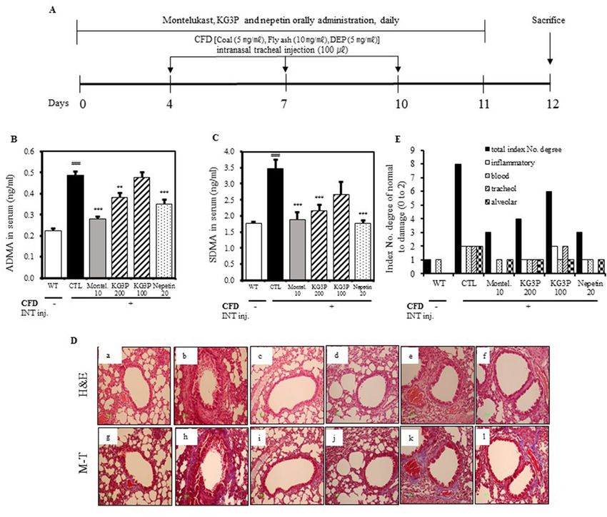
Decreased number of immune cells in BALF and lung tissue. Generally, there is an increase
in immune cells during the invasion of foreign particles in the body which is the natural adaptive immune
response21. We therefore sought to check the immune cell levels in the lungs and BALF. As shown in Fig. 4A‒D,
montelukast, both doses of KG3P, and nepetin potently suppressed the number of total immune cells and neu-
trophils in BALF and lung samples. Moreover, using FACS analysis (Table 1 and Fig. 5, A–G), C D4+, CD8+, and
+
CD11b cells were significantly decreased in BALF and lungs cells, indicating that the over activation of the
immune system caused by CFD was positively suppressed by the higher doses of KG3P and nepetin, decreasing
the aggravation of inflammation.
Suppression of pro‑inflammatory cytokines expression in BALF and lung tissue. Pro-inflam-
matory cytokines are secreted in response to inflammation and timely control is essential; this is because an
uncontrolled secretion of these chemicals can irreversibly damage the tissue22. And it is a renowned fact that the
major contributors in the pathology of asthma are the cytokines that are released from lung cells. Keeping this
in mind, we had investigated the levels of these pro-inflammatory cytokines in the BALF and lung tissue. From
Fig. 6A‒C, the higher doses of KG3P and nepetin are shown to significantly suppress the levels of IL-17,which
is naturally elevated in asthma causing allergic rhinitis23, TNF-α, which is found to be potently involved in
many aspects related to airway pathology in asthma and which is also one of the target for asthma treatment24,
and MIP2 which is activated by IL-1725. In addition to these, CXCL-1 which attracts neutrophils to the sites
Scientific Reports | (2020) 10:14036 | https://doi.org/10.1038/s41598-020-68965-5 2
Vol:.(1234567890)
www.nature.com/scientificreports/
Figure 1. Chromatogram of four compounds purified from the KGC-03-PS mixture (KG3P). Rg1, Rb1, Rg3s,
and nepetin were identified as marker compounds of the Korean Red Ginseng and Salvia plebeia R. Br. mixture
(KG3P). (A) UPLC-PDA chromatograms of three standard ginsenoside mixtures at 203 nm. (B) UPLC-PDA
chromatogram for KG3P mixture at 203 nm. (C) HPLC chromatograms for standard nepetin at 342 nm and
(D) HPLC chromatograms for KG3P mixture at 342 nm. Rg1 (0.22 mg/g ± 0.03), Rb1 (2.06 mg/g ± 0.06), Rg3
(0.51 mg/g ± 0.05), and nepetin (5.42 mg/g ± 0.39) appeared at a retention time of approximately 29.8, 41.2, 45.1,
and 26.9 min, respectively. UPLC-PDA ultra-performance liquid chromatography-photodiode array detector,
HPLC high-performance liquid chromatography.
of airway inflammation26 in BALF samples from mice was also downregulated by KG3P treatment (Fig. 6D).
Moreover, montelukast, both doses of KG3P, and nepetin significantly inhibited the expression of IL-17, IL-1β,
IL-6 and TNF-α which are considered to be the top prioritized pro-inflammatory a gents27 (Fig. 6E–H). Lastly,
CCR3 which is present on the T-cells co-localizing with eosinophils in the allergic asthma condition28, and
MUC5AC which is the major component of mucous causing airway obstruction in asthma29 (Fig. 6I,J) were also
ameliorated by KG3P and nepetin, indicating that the extract and the individual component are indeed potent
anti-inflammatory agents.
Inhibition of fluorescence intensity of IRAK‑1 and signal transduction of KG3P and nepetin
via the NF‑κB and MAPK pathways. Interleukin 1 receptor associated kinase (IRAK-1) is the negative
regulator of Toll like receptor 1 and it is key activator of NF-κB and MAPK pathways. In fact, continuous acti-
vation of IRAK-1 causes the onset of persistent asthma in humans30. We therefore checked lung tissues which
were stained with IRAK-1 antibody to visualize the presence of IRAK-1 positive cells with KG3P and nepetin.
As shown in Fig. 7A,B, IRAK-1 positive cells were potently inhibited in the lung tissue by montelukast, both
doses of KG3P, and nepetin. This was also confirmed via western blot analysis of lung tissue protein where the
higher dose of KG3P and nepetin potently inhibited the expression of IRAK-1, p-TAK1, p-NF-κB, p-ERK, and
p-JNK (Fig. 7C,D). These results strongly suggest that KG3P and the single compound, nepetin, exhibit their
anti-inflammatory effects via the NF-κB and MAPK pathways during CFD-induced airway inflammation.
Discussion
Numerous epidemiological studies are available on the effects of fine and ultrafine particulate matter (PM) on
health, especially those relating to pulmonary disorders that in the long-term, lead to extensive morbidity and
mortality. The composition of PM include coal, ash, oil, and diesel exhaust particles (DEP)31,32. The incidence of
Scientific Reports | (2020) 10:14036 | https://doi.org/10.1038/s41598-020-68965-5 3
Vol.:(0123456789)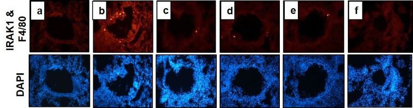
www.nature.com/scientificreports/
Figure 2. Effects of KG3P and nepetin in vitro on MH-S cells and signal transduction via the NF-κB and
MAPK pathways. (A) Nitrite production was inhibited by dose dependent concentrations of KG3P (25, 50 and
100 μg/mL) and hispidulin (HP; 20 μM), nepetin (NP; 20 μM) and rosmarinic acid (RA; 20 μM) as determined
by Griess method. Values in the bar graphs represent means ± SEM of three independent experiments.
***p < 0.001 and **p < 0.05 are considered significant compared to the CFA-only group. (B) Real-Time PCR
for KG3P and single compounds. Values in the bar graphs represent means ± SEM of three independent
experiments. ***p < 0.001 and **p < 0.05 are considered significant compared to the CFA-only group. (C, D)
Signal transduction of KG3P and single compounds through the NF-κB and MAPK pathways in MH-S cells
via western blot analysis. Values in the bar graphs represent means ± SEM of three independent experiments.
***p < 0.001 and **p < 0.05 are considered significant compared to the CFA-only group. KG3P1 KG3P 25 μg/mL,
KG3P2 KG3P 50 μg/mL, KG3P3 KG3P 100 μg/mL, B Basal, CFA Coal Fly ash, HP Hispidulin, NP Nepetin and
RA Rosmarinic acid. Full length western blots are shown in Supplementary Fig. 2a,b.
chronic pulmonary diseases, especially lung cancer, that occur due to chronic deposits of these fine particles in
tissues over a long period of time, is a major concern of s cientists2. The current lifestyle trend inevitably exposes
living organisms to the inhalation of these particles; thus, many scientists are attempting to devise various preven-
tive and therapeutic remedies (of chemical and herbal origin) to avoid incidence of PM-induced chronic diseases.
Few studies exist on the effects of natural herbal extracts or compounds on CFD-induced airway inflamma-
tion using a mice model. However, several studies have been performed using lung epithelial cells to examine
the effects of CFD on reactive oxygen species or pro-inflammatory cytokines33. A study reported that iron, which
constitutes up to 14% of CFA, was responsible for the expression and secretion of the pro-inflammatory cytokine,
IL-8, in A549 human lung epithelial cells34. Another study examined the initiation and secretion of reactive oxy-
gen species and cytokines, primarily IL-6, TNF-α, and IL-8 in human bronchial epithelial cells (BEAS-2B) and
RAW 264.7 cells exposed to CFA or P Ms35. Similar to this study, we found increased expression of NO, iNOS,
COX-2, and pro-inflammatory cytokines such as IL-1β, IL-6, and TNF-α in the murine macrophage MH-S
cell line when stimulated with CFA. However, as expected, these elevated levels of NO and pro-inflammatory
mediators and cytokines were decreased by treatment with KG3P and its individual components (Fig. 2A,B).
Based on the mechanistics, various studies have shown that these PMs can stimulate toll like receptor (TLR) 2
and 4. Once these particulate matters stimulate TLR4 receptors, the classical NF-κB pathway is mainly activated.
This includes the activation of factors downstream of TLR4, which includes IRAK-1, transforming growth factor
beta-activated kinase 1 (TAK-1), etc., which ultimately causes translocation of NF-κB from the cytoplasm to the
nucleus to activate the NF-κB p athway36. Certainly, several studies have shown that in alveolar macrophage cells,
treatment with a TLR4 antagonist results in minimal release of pro-inflammatory c ytokines37. Importantly, in
our study, CFA activated the NF-κB and MAPK pathways, both of which are primarily activated during foreign
invasion, in this case the PMs relative to previously reported studies38, 39. We hypothesize that CFA may bind to
TLR4 receptors, as shown in Fig. 2C,D, as all downstream factors for NF-κB [mainly IRAK-1 and TAK-1, and
MAPK (ERK, JNK and P38)] were activated by CFA and simultaneously inhibited by KG3P and the individual
components. Among the individual components identified in the KG3P extract, nepetin was the most abundant.
Previous reports on the biological activities such as the anti-microbial, anti-oxidant, anti-tumour, and anti-
inflammatory of nepetin have been presented; however, its effect on airway inflammation or asthma remained
un-investigated40–42. Therefore, we continued our in vivo study with nepetin from the KG3P extract.
Many studies have reported the effects of either PMs, CFA, or CFD in various murine and rodent models of
pulmonary ailments such as asthma and airway inflammation43. Walters et al.44 reported that PMs induced an
increase in the amount of immune cells in BALF and lung cells and increased the expression of pro-inflammatory
Scientific Reports | (2020) 10:14036 | https://doi.org/10.1038/s41598-020-68965-5 4
Vol:.(1234567890)www.nature.com/scientificreports/
Figure 3. Inhibition of airway inflammation by the KG3P and nepetin in a CFD-induced murine model
of airway inflammation. (A) Scheme for the CFD sensitization and challenge protocol. Mice were exposed
to 100 μL of CFD [Coal (5 mg/mL), Fly ash (10 mg/mL), Diesel exhaust particles (DEP, 5 mg/mL)] mixed
solution by intranasal tracheal injection thrice in 3 day intervals for 12 days. (B, C) KG3P and nepetin inhibited
asymmetric dimethyl-arginine (ADMA) and symmetric dimethly-arginine (SDMA) production in serum
obtained from CFD mice by ELISA kit. (D) Effect of KG3P and nepetin treatment on lung histopathology in
CFD-CTL mice as visualized by H&E and Masson’s Trichrome staining. . Representative sections from each
treatment group are shown. (a) BALB/c normal Wild type control (WT), (b) CFD-sensitized control mice
(CTL), (c) 10 mg/kg montelukast-treated CFD-sensitized mice, (d) 200 mg/kg KG3P-treated CFD-sensitized
mice, (e) 100 mg/kg KG3P-treated CFD-sensitized mice, and (f) 20 mg/kg nepetin-treated CFD-sensitized mice.
M–T staining pictures have the same order for groups in H&E staining (g–l). (E) Quantitative analyses of the
degree of lung tissue damage in the sections. Data are from individual mice, with arithmetic mean points shown
in histograms. Values are expressed as mean ± SEM (n = 8 mice). #p < 0.05, ##p < 0.01, and ###p < 0.001 (compared
to WT), and *p < 0.05, **p < 0.01, and ***p < 0.001 (compared to CTL).
cytokines in lung tissues. Takano et al.45 reported that the inhalation of DEP exacerbates allergen-related eosino-
phil recruitment and airway hyper-responsiveness in mice. Similar to these studies, we found that exposure of
mice to CFD resulted in elevated levels of ADMA and SDMA (both of which are analogues of NO during inflam-
mation), causing histological changes in lung tissues including infiltration of inflammatory cells, collagenous
fibre production, and increased mucous production due to downstream secretion of pro-inflammatory cytokines
such as interleukins and TNF-α. Various immune cells in BALF and the amounts of neutrophils in lung tissues
(Figs. 3, 4, 5) were positively inhibited by KG3P and nepetin, respectively.
We also observed elevated expression of pro-inflammatory cytokines, primarily the interleukins (i.e., IL-17,
IL-1β, IL-6, and TNF-α) (Fig. 6) in BALF and lung tissues; this is similar to previous studies that indicated the
activation of pro-inflammatory cytokines during airway inflammation and our in vitro results using the CFA-
induced MH-S cells46. Macrophage inflammatory protein-2 (MIP2), chemokine (C-X-C motif) ligand 1 (CXCL1),
Scientific Reports | (2020) 10:14036 | https://doi.org/10.1038/s41598-020-68965-5 5
Vol.:(0123456789)www.nature.com/scientificreports/
Figure 4. The effects of KG3P and nepetin on airway immune cell number and neutrophilic airway
inflammation in CFD-induced airway inflammation murine model. (A) Total BALF cells from each treatment
group. (B) Total lung cells from each treatment group. For A&B, cells were counted using a haemocytometer.
(C) Photomicrograph of BALF cytospins from mice with CFD-induced airway inflammation (magnification,
×200), (a) BALB/c normal Wild type control (WT), (b) CFD-sensitized control mice (CTL), (c) 10 mg/kg
montelukast-treated CFD-sensitized mice, (d) 200 mg/kg KG3P-treated CFD-sensitized mice, (e) 100 mg/kg
KG3P-treated CFD-sensitized mice, and (f) 20 mg/kg nepetin-treated CFD-sensitized mice. (D) KG3P and
nepetin suppressed neutrophilia in BALF. Data are from individual mice, with arithmetic mean points shown in
histograms. Values in bar graphs are expressed as mean ± SEM (n = 8 mice). #p < 0.05, ##p < 0.01, and ###p < 0.001
(compared to WT), and *p < 0.05, **p < 0.01, and ***p < 0.001 (compared to CTL).
and CC- chemokine receptor 3 (CCR3) are major inflammatory factors expressed in airway inflammation and
asthma47–51. In addition, polymeric mucin gene (MUC5AC) is expressed and its activation causes disruption in
the secretion and storage of mucin, thus leading to mucus gel formation. This mucus gel obstructs the airway
epithelium resulting in asthma and b ronchitis29,52. In our mouse model, these factors were elevated; however,
as expected, oral treatment with KG3P and nepetin significantly decreased the levels of these parameters, and
the effects were comparable to those observed on treatment of mice with the positive control, montelukast
(administered to treat a sthma53). This indicates that the decreased levels of expression of these genes inhibited
the infiltration of inflammatory cells and decreased the thickness of the airway epithelium. We also confirmed
the signal transduction of KG3P and nepetin via the IRAK-1-NK-κB pathway using immunohistofluorescence
and immunoblot experiments. KG3P and nepetin potently inhibited the localization of IRAK-1 in lungs and
inhibited the phosphorylation of IRAK-1, p-TAK-1 and NF-κB in lungs tissue, and ERK and JNK from the
MAPK pathways (Fig. 7). These results confirm the in vitro data obtained using the CFA-induced MH-S cells.
Therefore, we demonstrated the novel anti-inflammatory/anti-asthmatic effects of a herbal formulation with
KG3P and those exhibited by its individual component, nepetin, on murine alveolar macrophage MH-S cells and
CFD-induced airway inflammation mouse model. Using an even greater mechanistic study, this formulation may
be proven to be a promising herbal remedy for the prevention of PM-/CFA-/DEP-/CFD-induced airway ailments.
Scientific Reports | (2020) 10:14036 | https://doi.org/10.1038/s41598-020-68965-5 6
Vol:.(1234567890)www.nature.com/scientificreports/
CFD-induced airway inflammation murine model (Absolute No.)
Cell phenotypes in Lung, Normal BALB/c Montel. (10 mg/ KG3P (200 mg/ KG3P (100 mg/ Nepetin (20 mg/
BALF (WT) CTL 0 kg) kg) kg) kg)
CD3+/CD4+
9.8 ± 4.73 79.1 ± 9.5### 27.6 ± 12.1** 29.4 ± 5.05*** 37.1 ± 0.55*** 25.0 ± 8.89***
(× 106 cells)
+ +
CD3 /CD8
(× 105 cells) 12.5 ± 1.40 73.3 ± 4.16### 15.6 ± 0.82*** 25.7 ± 6.13*** 25.1 ± 1.66*** 21.5 ± 0.97***
Lung
CD4+/CD69+
(× 105 cells) 0.4 ± 0.02 9.10 ± 2.65## 1.40 ± 0.37** 4.80 ± 2.21 2.70 ± 0.69* 5.40 ± 1.03
Gr-1+/CD11b+ ##
16.6 ± 3.1 78.2 ± 16.4 23.5 ± 6.9** 32.6 ± 0.57** 36.3 ± 6.2* 33.7 ± 6.5**
(× 105 cells)
+ +
CD3 /CD4
1.30 ± 0.22 9.0 ± 0.36### 3.4 ± 0.11*** 3.5 ± 1.54*** 2.4 ± 0.46*** 2.8 ± 1.01***
(× 104 cells)
+ +
CD3 /CD8
BALF 1.40 ± 0.17 6.7 ± 2.43# 2.8 ± 0.09 2.8 ± 1.40 2.5 ± 1.49 2.1 ± 1.30
(× 104 cells)
+ +
Gr-1 /CD11b
2.10 ± 0.25 53.6 ± 3.12### 22.5 ± 4.49*** 21.5 ± 1.94*** 20.9 ± 2.78*** 26.4 ± 3.04***
(× 104 cells)
Table 1. Fluorescence-activated cell sorting analysis (FACS) of immune cell subtypes in lung and BALF. Each
point represents the mean ± SEM values for 6 mice. # p < 0.05, ##p < 0.01, and ###p < 0.001 (compared to WT), and
*p < 0.05, **p < 0.01, and ***p < 0.001 (compared to CFD).
Figure 5. Reduction in the numbers of Immune cell subtypes in BALF and Lung cells by Fluorescent activated
cell sorting analysis (FACS). (A, E) Lung and BALF cells were stained with anti-CD3+/CD4+ in lung and BALF,
(B, F) anti-CD3+/CD8+ in lung and BALF, (C) CD4+/CD69+, (D, G) Gr-1+/CD11b+ in lung and BALF, mAbs.
(BALB/c normal Wild type control (WT), CFD-sensitized control mice (CTL), 10 mg/kg montelukast-treated
CFD-sensitized mice (Montel. 10), 200 mg/kg KG3P-treated CFD-sensitized mice (KG3P 200), 100 mg/kg
KG3P-treated CFD-sensitized mice (KG3P 100), and (20 mg/kg nepetin-treated CFD-sensitized mice (Nepetin.
20). Values in bar graphs are expressed as mean ± SEM (n = 8 mice). #p < 0.05, ##p < 0.01, and ###p < 0.001
(compared to WT), and *p < 0.05, **p < 0.01, and ***p < 0.001 (compared to CTL).
Materials and methods
Chemicals and reagents. The detailed chemicals and reagents are given in Supplementary information 1.
Sample preparation for KGC‑03‑PS (KG3P). Korean red ginseng extract powder was procured from
Korea Ginseng Corporation. Dried SPR-Br was purchased from Dongjin Farm (Buan, Jeonbuk, Korea) and
extracted twice with a 15-fold volume of 30% alcohol/water for 4 h at 80 ℃. Thereafter, the extract was filtered
Scientific Reports | (2020) 10:14036 | https://doi.org/10.1038/s41598-020-68965-5 7
Vol.:(0123456789)www.nature.com/scientificreports/
Figure 6. The effects of KG3P and nepetin on pro-inflammatory cytokines in BALF and lung tissue. KG3P and
nepetin reduced (A) IL-17A, (B) TNF-α, (C) MIP2, (D) and CXCL1 levels in the BALF obtained from serum
collected from WT, CFD, Montelukast (10 mg/kg), KG3P (200 mg/kg, 100 mg/kg), and nepetin (20 mg/kg)
treated mice; these levels were measured using ELISA. (E) Effects of KG3P and nepetin on the expression of
cytokine mRNA in lung tissues. IL-17, (F) IL-1β, (G) IL-6 (H) TNF-α, (I) CCR3, (J) and MUC5AC expression
was determined by Real-Time PCR. Expression is presented as relative quantitation (RQ). Data are from
individual mice, with arithmetic mean points shown in histograms. Values in bar graphs are expressed as
mean ± SEM (n = 8 mice). #p < 0.05, ##p < 0.01, and ###p < 0.001 (compared to WT), and *p < 0.05, **p < 0.01, and
***p < 0.001 (compared to CTL).
through a 1 µm pore size, and the resultant mixture concentrated using an evaporator prior to preparing the
powdered form by spray-drying. KGC-03-PS (KG3P) was prepared by a mixture of Korean red ginseng:SPR-Br,
(1:3). High performance liquid chromatography (HPLC) was then used to analyse the mixture and the following
composition obtained: Ginsenoside Rg1 (0.22 ± 0.03 mg/g), Rb1 (2.06 ± 0.06 mg/g), 20(S)-Rg3 (0.51 ± 0.05 mg/g),
and nepetin (5.42 ± 0.39 mg/g) using standard operating procedure.
Ginsenoside Rg1, Rb1, and 20(S)-Rg3 standards were purchased from Chromadex (Irvine, USA) and 2 g
of KGC-03-PS powder extracted with 10 ml of 30% aq. methanol in an ultrasonic chamber for 30 min. After
ultrasonic extraction, centrifugal separation (Legand Mach 1.6R, Thermo, Germany) was performed for 10 min
at 3,000 rpm. The resulting supernatant solution was filtered (0.2 µm, Acrodisk, USA) and injected into the
UPLC-PDA system (Waters Co., USA).
The instrumental conditions of UPLC-PDA were as follows: the chromatographic separation was obtained
using a BEH C18 column (100 mm × 2.1 mm, 1.7 µm, Waters Co., USA) and the column temperature set to 40 °C.
The binary gradient elution system consisted of 0.001% phosphoric acid in water (A) and 0.001% phosphoric
acid in acetonitrile (B). Separation was achieved using the following protocol: 0–25.0 min (15% B), 35.0 min
(20% B), 39.5 min (30% B), 40.5 min (32% B), 42.5 min (40% B), 46.0 min (65% B), 47.0–49.0 min (90% B),
and 51.0–53.0 min (15% B). The flow rate and sample injection volume were set to 0.6 mL/min and 2.0 µL,
respectively. The ginsenosides were determined at a UV wavelength of 203 nm using a photodiode array detec-
tor (Waters Co., USA).
Nepetin standard was purchased from Avention (Seoul, Korea). Half gram of KGC-03-PS powder was
extracted with 20 mL of methanol in an ultrasonic chamber for 30 min, and diluted to 50 mL with methanol.
Scientific Reports | (2020) 10:14036 | https://doi.org/10.1038/s41598-020-68965-5 8
Vol:.(1234567890)www.nature.com/scientificreports/
Figure 7. Immunohistofluorescence (IHF) staining of IRAK1 and signal transduction of KG3P and nepetin
via the NF-κB and MAPK pathways in lung tissue. (A) IRAK1 (green), F4/80 (red) and DAPI (DNA) (grey)
in lung tissue as determined from Immunofluorescence microscope. (a) BALB/c normal Wild type control
(WT), (b) CFD-sensitized control mice (CTL), (c) 10 mg/kg montelukast-treated CFD-sensitized mice, (d)
200 mg/kg KG3P-treated CFD-sensitized mice, (e) 100 mg/kg KG3P-treated CFD-sensitized mice, and (f)
20 mg/kg nepetin-treated CFD-sensitized mice. (B) Data are from individual mice, with arithmetic mean
points shown in bar graphs. Values in bar graphs are expressed as mean ± SEM (n = 8). #p < 0.05, ##p < 0.01, and
###
p < 0.001 (compared to WT), and *p < 0.05, **p < 0.01, and ***p < 0.001 (compared to CTL). (C-D) Signal
transduction of KG3P and nepetin through the NF-κB and MAPK pathways in lung tissue in the CFD mice
airway inflammation model by western blotting. Values in the bar graphs are presented as mean ± SEM of three
independent experiments. In the bar graphs and blots, BALB/c normal Wild type control (WT), CFD-sensitized
control mice (CTL), 10 mg/kg montelukast-treated CFD-sensitized mice (Montel. 10), 200 mg/kg KG3P-treated
CFD-sensitized mice (KG3P 200), 100 mg/kg KG3P-treated CFD-sensitized mice (KG3P 100), and 20 mg/
kg nepetin-treated CFD-sensitized mice (Nepetin 20). ***p < 0.001 and **p < 0.05 are considered significant
compared to the CTL group. Full length blots are shown in Supplementary Fig. 2c,d.
After extraction, the sample solution was filtered (0.2 µm, Acrodisk, USA) and injected into the HPLC–PDA
system (Waters Co., USA).
The instrumental conditions of HPLC–PDA were as follows: chromatographic separation was performed
using a Zorbax eclipse XDB C18 column (250 mm × 4.6 mm, 5 µm, Agilent Co., USA). The binary gradient
elution system consisted of 0.05% acetic acid in water (A) and methanol (B). Separation was achieved using the
following protocol: 0.0 min (38% B), 14.0 min (42% B), 17.0 min (45% B), 17.1 min (48% B), 32.0 min (50% B),
40.0 min (85% B), 45.0 min (38% B). The flow rate and sample injection volume were set to 1.0 mL/min and
10.0 µL, respectively. Nepetin was determined at a UV wavelength of 342 nm using a photodiode array detector
(Waters Co., USA). All HPLC chromatograms are presented in Fig. 1A‒D.
Cell culture. The detailed method is given in Supplementary information 1.
Nitric oxide (NO) assay. The detailed method is given in Supplementary information 1.
Cell Viability (MTT) assay. The detailed method is given in Supplementary information 1.
RNA extraction and polymerase chain reaction (PCR). The detailed method is given in Supplemen-
tary information 1 (Table 2).
Western blot analysis. The detailed method is given in Supplementary information 1.
Scientific Reports | (2020) 10:14036 | https://doi.org/10.1038/s41598-020-68965-5 9
Vol.:(0123456789)www.nature.com/scientificreports/
Gene Primer Oligonucleotide sequence (5′–3′)
F 5′-CAATGAATACGGCTACAGCAAC-3′
GAPDH
R 5′-AGGGAGATGCTCAGTGTTGG-3′
F 5′-CCCTTCCGAAGTTTCTGGCAGCAGC-3′
iNOS
R 5′-GGCTGTCAGAGCCTCGTGGCTTTGG-3′
F 5′-TCTCAGCACCCACCCGCTCA-3′
COX-2
R 5′-GCCCCGTAGACCCTGCTCGA-3′
F 5′-CAGGGTGGGTGTGCCGTCTTTC-3′
IL-1β
R 5′-TGCTTCCAAACCTTTGACCTGGGC-3′
F 5′-TTGACCTCAGCGCTGAGTTG-3′
TNF-ɑ
R 5′-CCTGTAGCCCACGTCGTAGC-3′
F 5′-GTACTCCAGAAGACCAGAGG-3′
IL-6
R 5′-TGCTGGTGACAACCACGGCC-3′
F 5′-TCTCATCCAGCAAGAGATCC-3′
IL-17
R 5′-AGTTTGGGACCCCTTTACAC-3′
F 5′-CCCGAACTGTGACTTTGCT-3′
CCR3
R 5′-CCTCTGGATAGCGAGGACTG-3′
F 5′-AGAATATCTTTCAGGACCCCTGCT-3′
MUC5AC
R 5′-ACACCAGTGCTGAGCATACTTTT-3′
Table 2. Primer sequences for RT-PCR and real-time PCR.
Animal experiment and treatment regimen. Male Balb/c mice (6–8 weeks old; 20‒22 g) were
obtained from The Jackson Laboratory (Bar Harbor, ME, USA). The mice were housed in a specific-pathogen-
free barrier facility at 21 ± 2 °C with a relative humidity of 60 ± 10% under a 12-h light/dark cycle. Feed and water
were provided ad libitum. The animal protocol was approved by the committee for animal welfare at Daejeon
University (DJUARB2017-024). This study was performed in strict accordance with the Guide for the Care
and Use of Laboratory Animals of the National Institute of Health. All animal procedures were conducted in
accordance with the guidelines of the Institutional Animal Care and Use Committee of South Korea, Research
Institute of Bioscience and Biotechnology (Daejeon, Republic of Korea). Mice were divided into 6 treatment
groups (n = 8 for each group): (a) BALB/c normal, (b) CFD-sensitized control mice, (c) positive control- 10 mg/
kg montelukast-treated CFD-sensitized mice, (d) 200 mg/kg KG3P-treated CFD-sensitized mice, (e) 100 mg/
kg KG3P-treated CFD-sensitized mice, and (f) 20 mg/kg nepetin-treated CFD-sensitized mice. Following the
acclimatization period, all groups except group 1 were administered 100 μL of CFD (Coal (5 mg/ml, Fly ash
(10 mg/ml), and diesel exhaust particles (DEP; 5 mg/ml) in saline by intratracheal instillation thrice at 3 day
intervals for 12 days using bronchial tubes, as previously d escribed54. Montelukast, KG3P and nepetin were
orally administered daily for 11 days at the above mentioned dosages. On day 12, all mice were euthanized and
blood, bronchoalveolar lavage fluid (BALF), and lungs tissues collected for further experiments. The schematic
diagram for the experimental protocol is presented in Fig. 3A.
Collection of bronchoalveolar lavage fluid (BALF) and lung cells. BALF was collected 24 h after
the last oral injection of samples. Mice were anesthetized by an i.p. injection of 10% urethane (100 µL; Sigma-
Aldrich, Korea). A tracheotomy was then performed, and a cannula inserted into the trachea. Ice-cold DMEM
was instilled into the lungs, and BALF collected. Total cell counts were measured with a haemocytometer. For
cytological examination, cells were prepared with a Cytospin (Hanil Science, Korea), fixed, and stained with a
modified Diff-Quick stain. Differential cell counts were determined using at least 500 cells on each cytospin
slide. Blood was collected by cardiac puncture, allowed to clot, then centrifuged; aliquots of serum were stored
at − 70 °C for ELISA.
Flow cytometric analysis. For FACS analysis of lung tissues, enzymatic digestion of the lungs was per-
formed as previously d escribed55. Briefly, mice were anesthetized, and the lungs carefully removed. After three
washes, the lung tissue was cut into small pieces and transferred to a 15-mL conical tube containing 20 mL of
HBSS with 2% FBS (Gibco-BRL, Grand Island, NY) and 1 mM EDTA (Sigma) for 30 min at 20–25 °C. After
washing, the lung pieces were incubated with 1 mg/mL collagenase (type IV; Sigma), with shaking. The lung
mixture was then filtered through a 70-µm pore size nylon Cell Strainer (BD Falcon, Bedford, MA, USA) and
centrifuged for 20 min at 2,000 rpm. The cell pellet was collected, and the cells washed twice. BALF was col-
lected as described in the previous section and thereafter, cells were incubated with monoclonal antibodies
(mAbs) against CD3e (145-2C11, hamster IgG), CD4 (RM4-5, rat IgG2a), CD8 (53-6.7, rat IgG2a), CD19 (ID3,
rat IgG2a), CD25 (3C7, Rat IgG), and Gr-1 (RB6-8C5, rat IgG2b). All fluorochrome-labelled mAbs and isotype
control IgGs were purchased from BD Biosciences (San Diego, CA, USA), and CCR3 (83103, Rat IgG2a) was
purchased from R&D system (Minneapolis, MN, USA). Cells from the lungs and BALF were incubated with
FITC- and PE-labelled mAbs for 30 min, washed with PBS, fixed with 4% paraformaldehyde (Sigma-Aldrich,
Scientific Reports | (2020) 10:14036 | https://doi.org/10.1038/s41598-020-68965-5 10
Vol:.(1234567890)www.nature.com/scientificreports/
Korea) for 20 min, washed with PBS, and then stored at 4 °C until analysis by two-colour flow cytometry on a
FACS Caliber (BD Biosciences, Mountain View, CA, USA).
BALF and cytokine measurements. Mice were anesthetized by an i.p. injection of urethane (100 µL;
Sigma-Aldrich, Korea), and their lungs gently lavaged with 1 mL of 0.9% saline via a tracheal cannula. Total and
escribed56. Samples were centrifuged at 2,000 rpm
differential BALF cell counts were determined as previously d
for 10 min, and the supernatants stored at − 80 °C. Asymmetric dimethyl-arginine (ADMA), symmetric dime-
thyl-arginine (SDMA), TNF-α, IL-6, IL-1β, IL-17, CXCL-1, CCR3 and MUC5AC in serum, BALF and lungs
tissue were measured by ELISA using a monoclonal antibody-based mouse interleukin ELISA kit (R&D system),
according to the manufacturer’s instructions.
Histological examination. Lungs were infused via the trachea with 1 mL of 10% neutral formalin and
immersed in the same fixative for at least 24 h. Tissues were paraffinized, and 6-µm sections were cut and stained
with H&E and Mason trichrome staining (both obtained from Sigma-Aldrich) to assess cell infiltration and fibre
formation, respectively, under a light microscope. To determine the severity of inflammatory cell infiltration,
peribronchial cell counts, extent of mucus production and goblet cell hyperplasia in the airway epithelium were
blindly quantified using the 5-point (0–4) grading system described by Tanaka et al.57 .
Immunohistofluorescent (IHF) staining. The detailed method is given in Supplementary information 1.
Statistical analysis. The detailed method is given in Supplementary information 1.
Data availability
The data required for this study is present in this main manuscript file and the Supplementary information 1
respectively.
Received: 3 January 2019; Accepted: 25 June 2020
References
1. Ghorani-Azam, A., Riahi-Zanjani, B. & Balali-Mood, M. Effects of air pollution on human health and practical measures for
prevention in Iran. J. Res. Med. Sci. 21, 65. https://doi.org/10.4103/1735-1995.189646 (2016).
2. Zhang, W., Qian, C. N. & Zeng, Y. X. Air pollution: A smoking gun for cancer. Chin. J. Cancer 33, 173–175. https: //doi.org/10.5732/
cjc.014.10034(2014).
3. Yamamoto, S. S., Phalkey, R. & Malik, A. A. A systematic review of air pollution as a risk factor for cardiovascular disease in
South Asia: Limited evidence from India and Pakistan. Int. J. Hyg. Environ. Health 217, 133–144. https://doi.org/10.1016/j.ijheh
.2013.08.003 (2014).
4. Biggeri, A., Bellini, P. & Terracini, B. Meta-analysis of the Italian studies on short-term effects of air pollution—MISA 1996–2002.
Epidemiol. Prev. 28, 4–100 (2004).
5. Diabate, S., Bergfeldt, B., Plaumann, D., Ubel, C. & Weiss, C. Anti-oxidative and inflammatory responses induced by fly ash par-
ticles and carbon black in lung epithelial cells. Anal. Bioanal. Chem. 401, 3197–3212. https://doi.org/10.1007/s00216-011-5102-4
(2011).
6. van Maanen, J. M. et al. In vitro effects of coal fly ashes: Hydroxyl radical generation, iron release, and DNA damage and toxicity
in rat lung epithelial cells. Inhal. Toxicol. 11, 1123–1141. https://doi.org/10.1080/089583799196628 (1999).
7. Dai, D., Zhang, C. F., Williams, S., Yuan, C. S. & Wang, C. Z. Ginseng on cancer: Potential role in modulating inflammation-
mediated angiogenesis. Am. J. Chin. Med. 45, 13–22. https://doi.org/10.1142/S0192415X17500021 (2017).
8. Yuan, S. M. Potential cardioprotective effects of Ginseng preparations. Pak. J. Pharm. Sci. 28, 963–968 (2015).
9. Kiefer, D. & Pantuso, T. Panax ginseng. Am. Fam. Phys. 68, 1539–1542 (2003).
10. Yu, S. E., Mwesige, B., Yi, Y. S. & Yoo, B. C. Ginsenosides: The need to move forward from bench to clinical trials. J. Ginseng Res.
43, 361–367. https://doi.org/10.1016/j.jgr.2018.09.001 (2019).
11. Peng, M. M., Fang, Y., Hu, W. & Huang, Q. The pharmacological activities of Compound Salvia Plebeia Granules on treating urinary
tract infection. J. Ethnopharmacol. 129, 59–63. https://doi.org/10.1016/j.jep.2010.02.029 (2010).
12. Gu, L. & Weng, X. Antioxidant activity and components of Salvia plebeia R. Br.—a Chinese herb. Food Chem. 73, 299–305 (2001).
13. Jin, X.-F., Lu, Y.-H., Wei, D.-Z. & Wang, Z.-T. Chemical fingerprint and quantitative analysis of Salvia plebeia R. Br. by high-
performance liquid chromatography. J. Pharm. Biomed. Anal. 48, 100–104 (2008).
14. Xie, C., Veitch, N. C., Houghton, P. J. & Simmonds, M. S. Flavone C-glycosides from Viola yedoensis MAKINO. Chem. Pharm.
Bull. (Tokyo) 51, 1204–1207 (2003).
15. Ravn, H., Nishibe, S., Sasahara, M. & Xuebo, L. Phenolic compounds from Plantago asiatica. Phytochemistry 29, 3627–3631 (1990).
16. Liu, C. F., Drocourt, D., Puzo, G., Wang, J. Y. & Riviere, M. Innate immune response of alveolar macrophage to house dust mite
allergen is mediated through TLR2/-4 co-activation. PLoS ONE 8, e75983. https://doi.org/10.1371/journal.pone.0075983 (2013).
17. Fricker, M. & Gibson, P. G. Macrophage dysfunction in the pathogenesis and treatment of asthma. Eur. Respir. J. https://doi.
org/10.1183/13993003.00196-2017 (2017).
18. Kahraman, A., Mutlu, E. & Aldag, M. ADMA, SDMA and l-arginine may be novel targets in pharmacotherapy for complications
due to cardiopulmonary bypass. J. Med. Biochem. 36, 8–17. https://doi.org/10.1515/jomb-2016-0025 (2017).
19. Staab, E. B. et al. Asymmetric dimethyl-arginine metabolism in a murine model of cigarette smoke-mediated lung inflammation.
J. Immunotoxicol. 12, 273–282. https://doi.org/10.3109/1547691X.2014.961619 (2015).
20. Arlouskaya, Y. et al. Asymmetric dimethylarginine (ADMA) and symmetric dimethylarginine (SDMA) concentrations in patients
with obesity and the risk of obstructive sleep apnea (OSA). J. Clin. Med. https://doi.org/10.3390/jcm8060897 (2019).
21. Nicholson, L. B. The immune system. Essays Biochem. 60, 275–301. https://doi.org/10.1042/EBC20160017 (2016).
22. Zhang, J. M. & An, J. Cytokines, inflammation, and pain. Int. Anesthesiol. Clin. 45, 27–37. https: //doi.org/10.1097/AIA.0b013e 3180
34194e (2007).
23. Lv, H., Lu, B., Qian, X. J., Huang, J. A. & Qiu, T. F. Serum IL-17 and eotaxin levels in asthmatic patients with allergic rhinitis. Pak.
J. Med. Sci. 32, 700–704. https://doi.org/10.12669/pjms.323.9914 (2016).
Scientific Reports | (2020) 10:14036 | https://doi.org/10.1038/s41598-020-68965-5 11
Vol.:(0123456789)www.nature.com/scientificreports/
24. Brightling, C., Berry, M. & Amrani, Y. Targeting TNF-alpha: A novel therapeutic approach for asthma. J. Allergy Clin. Immunol.
121, 5–10. https://doi.org/10.1016/j.jaci.2007.10.028 (2008) (quiz 11–12).
25. Ano, S. et al. Transcription factors GATA-3 and RORgammat are important for determining the phenotype of allergic airway
inflammation in a murine model of asthma. J. Immunol. 190, 1056–1065. https://doi.org/10.4049/jimmunol.1202386 (2013).
26. Liu, C. et al. Role of epithelial chemokines in the pathogenesis of airway inflammation in asthma (review). Mol. Med. Rep. 17,
6935–6941. https://doi.org/10.3892/mmr.2018.8739 (2018).
27. Chung, K. F. & Barnes, P. J. Cytokines in asthma. Thorax 54, 825–857. https://doi.org/10.1136/thx.54.9.825 (1999).
28. Bertrand, C. P. & Ponath, P. D. CCR3 blockade as a new therapy for asthma. Expert Opin. Investig. Drugs 9, 43–52. https://doi.
org/10.1517/13543784.9.1.43 (2000).
29. Bonser, L. R. & Erle, D. J. Airway mucus and asthma: The role of MUC5AC and MUC5B. J. Clin. Med. https://doi.org/10.3390/
jcm6120112 (2017).
30. Balaci, L. et al. IRAK-M is involved in the pathogenesis of early-onset persistent asthma. Am. J. Hum. Genet. 80, 1103–1114. https
://doi.org/10.1086/518259 (2007).
31. Smith, K. R., Veranth, J. M., Kodavanti, U. P., Aust, A. E. & Pinkerton, K. E. Acute pulmonary and systemic effects of inhaled coal
fly ash in rats: Comparison to ambient environmental particles. Toxicol. Sci. 93, 390–399. https://doi.org/10.1093/toxsci/kfl062
(2006).
32. Aust, A. E. et al. Particle characteristics responsible for effects on human lung epithelial cells. Res. Rep. Health Eff. Inst. 110, 1–65
(2002) (discussion 67–76).
33. Voelkel, K., Krug, H. F. & Diabate, S. Formation of reactive oxygen species in rat epithelial cells upon stimulation with fly ash. J.
Biosci. 28, 51–55 (2003).
34. Smith, K. R., Veranth, J. M., Hu, A. A., Lighty, J. S. & Aust, A. E. Interleukin-8 levels in human lung epithelial cells are increased
in response to coal fly ash and vary with the bioavailability of iron, as a function of particle size and source of coal. Chem. Res.
Toxicol. 13, 118–125 (2000).
35. Mitkus, R. J., Powell, J. L., Zeisler, R. & Squibb, K. S. Comparative physicochemical and biological characterization of NIST Interim
Reference Material PM2.5 and SRM 1648 in human A549 and mouse RAW264.7 cells. Toxicol. In Vitro 27, 2289–2298 (2013).
36. Veronesi, B., Oortgiesen, M., Carter, J. D. & Devlin, R. B. Particulate matter initiates inflammatory cytokine release by activation
of capsaicin and acid receptors in a human bronchial epithelial cell line. Toxicol. Appl. Pharmacol. 154, 106–115. https://doi.
org/10.1006/taap.1998.8567 (1999).
37. Becker, S., Mundandhara, S., Devlin, R. B. & Madden, M. Regulation of cytokine production in human alveolar macrophages and
airway epithelial cells in response to ambient air pollution particles: Further mechanistic studies. Toxicol. Appl. Pharmacol. 207,
269–275. https://doi.org/10.1016/j.taap.2005.01.023 (2005).
38. Quay, J. L., Reed, W., Samet, J. & Devlin, R. B. Air pollution particles induce IL-6 gene expression in human airway epithelial cells
via NF-kappaB activation. Am. J. Respir. Cell Mol. Biol. 19, 98–106. https://doi.org/10.1165/ajrcmb.19.1.3132 (1998).
39. Ramos-Nino, M. E., Haegens, A., Shukla, A. & Mossman, B. T. Role of mitogen-activated protein kinases (MAPK) in cell injury
and proliferation by environmental particulates. Mol. Cell Biochem. 234–235, 111–118 (2002).
40. Osman, W. J. A., Mothana, R. A., Basudan, O., Mohammed, M. S., & Mohamed, M. S. Antibacterial effect and radical scavenging
activity of hispidulin and nepetin; a two flvaones from Tarconanthus camphoratus L. World J. Pharm. Res. 4, 424–433 (2014).
41. Clavin, M. et al. Anti-inflammatory activity of flavonoids from Eupatorium arnottianum. J. Ethnopharmacol. 112, 585–589. https
://doi.org/10.1016/j.jep.2007.04.007 (2007).
42. Militao, G. C. et al. Cytotoxic activity of nepetin, a flavonoid from Eupatorium ballotaefolium HBK. Pharmazie 59, 965–966 (2004).
43. Harkema, J. R. et al. Effects of concentrated ambient particles and diesel engine exhaust on allergic airway disease in Brown Norway
rats. Res. Rep. Health Eff. Inst. 145, 5–55 (2009).
44. Walters, D. M., Breysse, P. N. & Wills-Karp, M. Ambient urban Baltimore particulate-induced airway hyperresponsiveness and
inflammation in mice. Am. J. Respir. Crit. Care Med. 164, 1438–1443. https://doi.org/10.1164/ajrccm.164.8.2007121 (2001).
45. Takano, H. et al. Diesel exhaust particles enhance antigen-induced airway inflammation and local cytokine expression in mice.
Am. J. Respir. Crit. Care Med. 156, 36–42 (1997).
46. Nakajima, H. & Takatsu, K. Role of cytokines in allergic airway inflammation. Int. Arch. Allergy Immunol. 142, 265–273. https://
doi.org/10.1159/000097357 (2007).
47. Driscoll, K. E. TNFalpha and MIP-2: Role in particle-induced inflammation and regulation by oxidative stress. Toxicol. Lett.
112–113, 177–183 (2000).
48. Gosset, P. et al. Production of tumor necrosis factor-alpha and interleukin-6 by human alveolar macrophages exposed in vitro to
coal mine dust. Am. J. Respir. Cell Mol. Biol. 5, 431–436. https://doi.org/10.1165/ajrcmb/5.5.431 (1991).
49. Humbles, A. A. et al. The murine CCR3 receptor regulates both the role of eosinophils and mast cells in allergen-induced airway
inflammation and hyperresponsiveness. Proc. Natl. Acad. Sci. U. S. A. 99, 1479–1484. https://doi.org/10.1073/pnas.261462598
(2002).
50. Moon, K. A. et al. Allergen-induced CD11b+ CD11c(int) CCR3+ macrophages in the lung promote eosinophilic airway inflam-
mation in a mouse asthma model. Int. Immunol. 19, 1371–1381. https://doi.org/10.1093/intimm/dxm108 (2007).
51. Kurai, J. et al. A muscarinic antagonist reduces airway inflammation and bronchoconstriction induced by ambient particulate
matter in a mouse model of asthma. Int. J. Environ. Res. Public Health. https://doi.org/10.3390/ijerph15061189 (2018).
52. Bonser, L. R., Zlock, L., Finkbeiner, W. & Erle, D. J. Epithelial tethering of MUC5AC-rich mucus impairs mucociliary transport
in asthma. J. Clin. Investig. 126, 2367–2371. https://doi.org/10.1172/JCI84910 (2016).
53. Paggiaro, P. & Bacci, E. Montelukast in asthma: A review of its efficacy and place in therapy. Ther. Adv. Chronic Dis. 2, 47–58. https
://doi.org/10.1177/2040622310383343 (2011).
54. Ortiz-Munoz, G. & Looney, M. R. Non-invasive intratracheal instillation in mice. Bio Protoc. 5, e1504 (2015).
55. Hamada, S. et al. Immunosuppressive effects of gallic acid and chebulagic acid on CTL-mediated cytotoxicity. Biol. Pharm. Bull.
20, 1017–1019 (1997).
56. Kim, S. H., Kim, B. K. & Lee, Y. C. Antiasthmatic effects of hesperidin, a potential Th2 cytokine antagonist, in a mouse model of
allergic asthma. Mediat. Inflamm. 2011, 485402. https://doi.org/10.1155/2011/485402 (2011).
57. Tanaka, H. et al. The effect of allergen-induced airway inflammation on airway remodeling in a murine model of allergic asthma.
Inflamm. Res. 50, 616–624. https://doi.org/10.1007/PL00000243 (2001).
Acknowledgements
This study was supported by the Korea Institute of Planning and Evaluation for Technology in Food, Agriculture,
Forestry and Fisheries (IPET) through the High Value-Added Food Technology Development Program, funded
by the Ministry of Agriculture, Food and Rural Affairs (MAFRA) (Grant number 1150002-03).
Author contributions
E.S. performed some parts of experiment and wrote the manuscript. Y.-S.L., W.-K.Y., Y.Y.L., M.K.K., K.S.K.,
Y.-S.K., S.-M.W. performed individual parts of experiments and helped with writing the manuscript. T.-H.K.,
Scientific Reports | (2020) 10:14036 | https://doi.org/10.1038/s41598-020-68965-5 12
Vol:.(1234567890)www.nature.com/scientificreports/
D.-M.K., Y.-C.P., H.J.S., C.K.H., J.-W.O., Y.C.L., H.-S.K., M.H.R. and S.-H.K. conceived the idea, supervised the
experiments and proofread the manuscript.
Competing interests
The authors declare no competing interests.
Additional information
Supplementary information is available for this paper at https://doi.org/10.1038/s41598-020-68965-5.
Correspondence and requests for materials should be addressed to M.H.R. or S.-H.K.
Reprints and permissions information is available at www.nature.com/reprints.
Publisher’s note Springer Nature remains neutral with regard to jurisdictional claims in published maps and
institutional affiliations.
Open Access This article is licensed under a Creative Commons Attribution 4.0 International
License, which permits use, sharing, adaptation, distribution and reproduction in any medium or
format, as long as you give appropriate credit to the original author(s) and the source, provide a link to the
Creative Commons license, and indicate if changes were made. The images or other third party material in this
article are included in the article’s Creative Commons license, unless indicated otherwise in a credit line to the
material. If material is not included in the article’s Creative Commons license and your intended use is not
permitted by statutory regulation or exceeds the permitted use, you will need to obtain permission directly from
the copyright holder. To view a copy of this license, visit http://creativecommons.org/licenses/by/4.0/.
© The Author(s) 2020
Scientific Reports | (2020) 10:14036 | https://doi.org/10.1038/s41598-020-68965-5 13
Vol.:(0123456789)You can also read