Molecular diversity and evolutionary trends of cysteine rich peptides from the venom glands of Chinese spider Heteropoda venatoria - Nature
←
→
Page content transcription
If your browser does not render page correctly, please read the page content below
www.nature.com/scientificreports
OPEN Molecular diversity
and evolutionary trends
of cysteine‑rich peptides
from the venom glands of Chinese
spider Heteropoda venatoria
Jie Luo1,4, Yiying Ding1,4, Zhihao Peng1, Kezhi Chen1, Xuewen Zhang1, Tiaoyi Xiao2 &
Jinjun Chen1,3*
Heteropoda venatoria in the family Sparassidae is highly valued in pantropical countries because the
species feed on domestic insect pests. Unlike most other species of Araneomorphae, H. venatoria uses
the great speed and strong chelicerae (mouthparts) with toxin glands to capture the insects instead
of its web. Therefore, H. venatoria provides unique opportunities for venom evolution research. The
venom of H. venatoria was explored by matrix-assisted laser desorption/ionization tandem time-of-
flight and analyzing expressed sequence tags. The 154 sequences coding cysteine-rich peptides (CRPs)
revealed 24 families based on the phylogenetic analyses of precursors and cysteine frameworks in
the putative mature regions. Intriguingly, four kinds of motifs are first described in spider venom.
Furthermore, combining the diverse CRPs of H. venatoria with previous spider venom peptidomics
data, the structures of precursors and the patterns of cysteine frameworks were analyzed. This work
revealed the dynamic evolutionary trends of venom CRPs in H. venatoria: the precursor has evolved
an extended mature peptide with more cysteines, and a diminished or even vanished propeptides
between the signal and mature peptides; and the CRPs evolved by multiple duplications of an
ancestral ICK gene as well as recruitments of non-toxin genes.
Spiders (Araneae), including 48,504 described species grouped into 4156 genera and 120 families https://wsc.
nmbe.ch/ (December 2019), have high biological and ecological diversity. Most of spiders employ chemical and
pharmacological complicated venoms to subdue their prey rapidly. Spider venoms contain rich compounds,
including proteins, peptides, and low-molecular-weight components. The predominant peptides of spider venoms
are secretory cysteine-rich peptides (CRPs) with multiple disulfide bonds that provide stability and resistance
to protease degradation1. There has been much interest in studying the biochemical and structural properties2,3,
pharmacological applications4, evolution and diversification of secretory cysteine-rich peptide toxin5–9. Up to
now, the venoms of snakes, scorpions, and Conus snails are much more deeply understood than those of spiders
partly since spiders have considerable species and complicated v enoms10,11.
The huntsman spider (Heteropoda venatoria Carl Linnaeus, 1767) is a member of family Sparassidae, a com-
mon name giant crab spider, and takes more than 250 days to complete their life cycle, which leads to the col-
lection of natural venoms is l imited12. Adult specimens have a flattened body length of about 25 mm with eight
long, slightly hairy legs spanning 60 to 120 mm. A yellow to cream clypeus just in front of the eyes is one of the
main features of H. venatoria. The female has a larger abdomen with an overall brown body. Usually, an egg sac
up to about 25 mm wide was carried with her pedipalps under its body. The male has a slender body and long
legs, a distinctive pattern on his carapace. This species is found in many tropical and subtropical regions globally
and can’t survive outside during sub-freezing temperatures. In the present study, the spiders were found from
basements, barns, and greenhouses of our scientific research farm in summer. The huntsman spiders do not spin
1
College of Bioscience and Biotechnology, Hunan Agricultural University, Changsha 410128, People’s Republic
of China. 2College of Animal Science and Technology, Hunan Agricultural University, Changsha 410128, People’s
Republic of China. 3Hunan Provincial Engineering Technology Research Center for Cell Mechanics and Function
Analysis, Changsha 410128, People’s Republic of China. 4These authors contributed equally: Jie Luo and Yiying
Ding. *email: chhncjj@126.com
Scientific Reports | (2021) 11:3211 | https://doi.org/10.1038/s41598-021-82668-5 1
Vol.:(0123456789)www.nature.com/scientificreports/
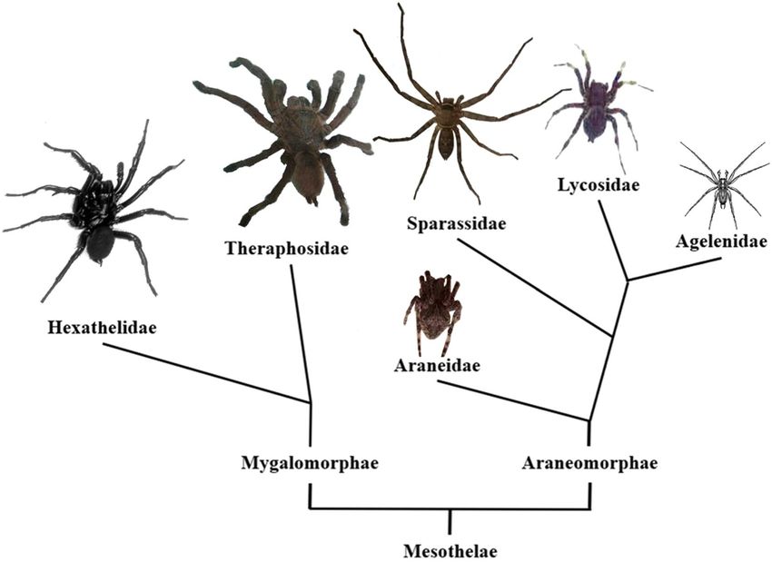
Figure 1. Simplified phylogenetic tree of spider families with the physique in the relative size of the
representative species of each family. Modified from Kuhn-Nentwig et al. (2011)11. Hadronyche versuta15
in Hexathelidae, Chilobraechys guangxiensis in Theraphosidae, Araneus trifolium in Araneidae, Heteropoda
venatoria in Sparassidae, Lycosa singoriensis16 in Lycosidae, and Agelenopsis potteri17 in Agelenidae are
mentioned in the present work.
webs. They are known to hunt and feed on living insects with their exceptional agility and speed at night. They
can stay and run on a smooth vertical surface and contort and squeeze their large body to fit into surprisingly
small cracks and crevices, which give them a strong advantage both in predation and evading predators. Almost
as soon as they catch their prey, the spiders paralyze them by injecting with the venoms, from glands extending
from the chelicerae into the cephalothorax. The spider is considered a beneficial resident of households because it
can hunt pests efficiently and does no harm to people13. The venom of H. venatoria contains hundreds of peptides
with severe toxicities on Periplaneta americana (LD50: 28.18 mg/g of body weight), and venom neurotoxins target
specific types of insect ion channels and receptors, which have broadly applied potential as insecticides in pest
control14. Many studies have focused on the activity and composition of the venom of H. venatoria. However,
the diversity of CRPs in the venom, and the evolutionary relationship in the phylogenetic framework has not
been explored.
The phylogenetic relationships amongst the spider families whose venom CRPs have been well described
show H. venatoria (in Sparassidae) locate between Theraphosidae and Lycosidae (Fig. 1). Comparing Mygalo-
morphae species (Cyriopagopus schmidti, Cyriopagopus hainanus, Grammostola rosea, Chilobrachys guangxiensis)
to Araneomorphae species (Lycosa singoriensis, Dolomedes mizhoanus, Araneus ventricosus), the body size of
H. venatoria is medium between the two suborders, and its family, Sparassidae, belongs to the more primitive
Araneomorphae11. However, H. venatoria subdues prey by long legs, strong chelicerae, and complex venom,
which seems similar to tarantula’s predation behavior. Unlike web-forming spiders (A. ventricosus, A. orientalis
et al.) that weave cobwebs before prey and then eat after wrapping its prey with cobwebs.
Based on the comparisons of morphology and predation habits of H. venatoria along with other spiders, as
well as its phylogenetic classification, we believe that the investigation of the genetic coding products in its venom
can contribute to the understanding of Araneae toxins evolution in the context of ecology, and likely facilitate
exploration of the popular spider resources in pantropical regions. In the study, the complex toxin repertoire of
H. venatoria venom is revealed by combining mass spectrum and Sanger sequencing for the venom gland cDNA
library. Furthermore, the dynamic evolution of CRPs is explored by combining the data from well-described
venom gland ESTs retrieved from ArachnoServer18,19. The present results contribute to understanding the evo-
lutionary trends and diversity of CRPs in spider venom.
Scientific Reports | (2021) 11:3211 | https://doi.org/10.1038/s41598-021-82668-5 2
Vol:.(1234567890)www.nature.com/scientificreports/
Family Signal peptide mode Length of propeptides/PQM Mature peptide mode C-terminal propeptides
1 MKh12S h5 19/DEQR XCIX6CIIX5CIIICIVX4CVX3CVIX4-6 G(K)
2 MKT h11S h5 15/AVAR X2CIX6CIIX5CIIICIVX4CVX3CVIX3 G
3 MKh18 15/VAAR X5CIX6CIIX6CIIICIVX3CVX3CVIX5 GK(K)
4 MKIT h15 11/VQAR XCIX6CIIX6CIIICIVX4CVX4CVIX6 GK
5 MKTTh3Th6Sh5 15–17/ATGR X3CIX6CIIX5CIIICIVX4CVX5CVIX W/O
6 MKTTh3Th6Sh5 15/VTGR X2-4CIX6CIIX5CIIICIVX4CVX5CVIX3 RKX4-5
7 MKh5Th6Sh5 15/ITVR X2-3CIX6CIIX5CIIICIVX4CVX9CVIX4 G
8 MKSSh7Th4Sh2EFTRS 12/VQER X2CIX6CIIX4CIIICIVX4CVX8CVIX3 W/O
Table 1. Sequence diversity of the predicted 6-cys ICK motif toxins in H. venatoria. In the signal peptide
mode, ‘h’ indicates hydrophobic residue, and the Arabic numerals denote the number of residues. Capital
letters indicate the corresponding amino acids. ‘C’ is cystine, ‘X’ is any residue but cystine, and ‘W/O’ denotes
no putative C-terminal propeptides.
Results
Transcriptomics uncovers the diversity of CRPs in H. venatoria venom. From the cDNA library
of H. venatoria venom gland, 912 sequenced ESTs, and 154 predicted novel CRP precursors were obtained. The
CRPs from H. venatoria were named according to the nomenclature proposed previously, which describes the
toxin’s activity, biological source, and relationship to other t oxins20. The cDNA sequences of CRPs have been sub-
mitted into the public database (http://www.ncbi.nlm.nih.gov/entrez, GenBank accession numbers: KC145575-
KC145728). The presence and location of signal peptide cleavage sites in the amino acid sequences were pre-
dicted with SignalP 4.1 and SpiderP program (http://www.arachnoserver.org/spiderP.html). A full-length CRPs
precursor absolutely contains both a signal peptide and a mature peptide, while some CRPs precursors contain
an additional propeptide preceding the mature toxin sequence just as the precursors of most of spider peptide
toxins reported b efore21. The alignment of the resultant amino acid sequences revealed extensive variation in the
molecular structure of the transcripts for most CRP types.
Family/cluster identification. In the present study, CRP toxins were classified into 24 families based on
the alignment of precursors and their known or predicted cysteine framework. The formation of disulfide bonds
stabilizes the three-dimensional (3D) structures of toxins, and is commonly used to classify toxins.
Family 1–8. The full primary sequences of the CRPs in the Family 1–8 are comprised of a signal sequence
(19–25 residues) and a propeptide (11–19 residues) preceding the mature toxin sequence. The N- and C-terminus
of mature peptides are highly variable regions. The 11 members of Family 1 are homologs of Kappa-SPRTX-
Hv1c, including its five different precursors (κ-SPRTX-Hv1c_1-5). Signal peptide mode of CRPs in the family
is ‘MKh12Sh5’, where ‘h’ indicates hydrophobic residue, the Arabic numerals denote the number of residues, and
capital letters indicate the corresponding amino acids. The propeptides of Family 1 are 19 residues with highly
conserved DEQR as an endoproteolytic site preceding the mature peptides, named the Processing Quadruplet
Motif (PQM)22. Mature peptide mode of Family 1 is ‘XCIX6CIIX5CIIICIVX4CVX3CVIX4–6’, where ‘X’ is any amino
acid. On the C-terminal of the mature peptides, there is ‘GK’ as the amidation site. The characters of the signal
peptides, propeptides and mature peptides of Family 2–8 are compared with those of Family 1, shown in Table 1.
According to homology analysis, the mature peptides of Family 1–8 are speculated as the ‘classical’ Inhibitory
Cystine Knot (ICK) motif containing three disulfide bonds with I–IV, II–V and III–VI connectivity. The first two
disulfide bonds (I–IV and II–V) form an embedded ring which is threaded by the third disulfide bond (III–VI).
The backbone regions between successive Cys residues are referred to as loops, numbered starting with loop 1
between Cys I and Cys II23,24. There is less amino acid sequence divergence in loop 1 and 3 than in the much more
variable loop 2, 4, N- and C-terminus in mature peptides. The precursors of Family 1–7 have higher similarity
with those from the same species than others. Only the sequences of CRPs in the Family 8 show high homology
with U23-ctenitoxin-Pn1a and U4-agatoxin-Ao1a from Phoneutria nigriventer and Agelena orientalis respectively
(Supplementary Information File 1, Suppl-Fig. 1). Recently, there are also 8 superfamilies of 6-cys ICK motif
toxins identified in Hadronyche infensa25.
Family 9. The precursors of Family 9 have a high content of acidic amino acid in putative mature peptides
with a novel Cys scaffold ‘CIX5CIIX3CIIIX5CIVXCVXCVI’. Since no significant homologous sequence has been
found in public protein databases, posttranscriptional processes such as alternative splicing or post-translational
modifications remain uncertain. In this case, the dotted border indicates the putative short propeptides (Sup-
plementary Information File 1, Suppl-Fig. 2). The motif of Family 9 is the first time reported from spider venom.
Family 10. Family 10 includes eleven homologous sequences which are characterized by two consecutive Cys
residues in the middle of signal peptide and that is straight followed by the mature region with a cys-scaffold
‘CIX13CIIX2CIIIX12CIVX3CVX8CVI’. The scaffold in mature peptide has been identified as a conserved domain
pfam01147 (representative proteins gi: 221468699, 5921747 shown in Supplementary Information File 1, Suppl-
Fig. 3), which includes all known crustacean hyperglycemic hormones (CHHs) found in the sinus gland of
isopods and d ecapods26 and the molt-inhibiting hormone (MIH) of the lobster Homarus americanus27. The three
disulfide bridges are CI–CV, CII–CIV, and CIII–CVI28. In addition, the amino acid sequences of several translated
cDNA (gi: 304306070, 304307035, 304306844, 304306583) from Loxosceles intermedia venom gland library29
Scientific Reports | (2021) 11:3211 | https://doi.org/10.1038/s41598-021-82668-5 3
Vol.:(0123456789)www.nature.com/scientificreports/
are also similar to that of Family 10 (Supplementary Information File 1, Suppl-Fig. 3). The latrodectins which
are identified in widow spider venom glands, also share six conserved cysteines that adopt the same disulfide
bond pairing in the mature peptide30,31.
Family 11, 12, 13 and 14. The four families have eight cysteines with a typical motif ‘CIX6CIIXnCIIICIVX4CVX-
CVIXnCVIIXCVIII’ where X is any residue but cystine. However, the amounts and properties of residues in the loops
between CII and CIII, CVI and CVII and at the C-terminus vary greatly. The sequences of the signal and precursor
proteins, as well as endoproteolytic sites, are also diverse. In Family 14, there is a long loop between C VI and
CVII and a very short propeptide preceding the mature region. Significantly, there is no propeptide predicted in
the precursor of U32-sparatoxin-Hv1a. The amino acid sequences of Family 11, 12, 13, and 14 are aligned with
the most similar known homologs (Supplementary Information File 1, Suppl-Fig. 4). The cysteine-frame is also
employed by superfamily 3 and 23 of H. infensa venom. The eight cysteines are arranged in four disulfide bonds
(CI–CIV, CII–CV, CIII–CVIII, CVI–CVII), which form an extended ICK m otif25.
Family 15. Family 15 includes 20 unique sequences that are highly homologous. It is noteworthy that Family
15 includestranscripts coding a new venom peptide type with a high mRNA expression level in H. venatoria.
There are 20 orthologs identified coding full-length cysteine frame in Family 15. The transcripts of U25-spa-
ratoxin-Hv1c and U25-sparatoxin-Hv1j are 30 and 29 copies, respectively, which are the top two precursors
identified in the H. venatoria venom cDNA library. There are twelve residues between the signal peptides and
the first Cys, but no usual PQM. The mature region is characterized by a novel eight-Cys scaffold ‘CIX21CIIX-
4CIIIX9CIVX10CVX11CVICVIIX4CVIII’. The transcript of a secretory protein with the identical Cys-frame has been
identified from the black widow spider (gi: 318087504). However, it is hypothesized to be involved in wrapping
silk fibers. Moreover, several hypothetical non-secretory proteins from Amblyomma maculatum also adopt the
same eight-Cys framework (Supplementary Information File 1, Suppl-Fig. 5).
Family 16. The six precursors are highly homologous with Omega-agatoxin-1A (gi: 2507406) from Agelenopsis
aperta containing a ten-Cys scaffold ‘CIX6/8CIIXCIIIX6CIVXCVX7/11CVIXCVIIX7CVIIIX5CIXX19/20CX’, so they belong
to the omega-agatoxin superfamily, which has a particularly exciting feature of the prepropeptide with the occur-
rence of two glutamate-rich sequences interposed between the signal sequences, the major peptide toxin, and
the minor toxin peptide. The heterodimer of the two subunits is linked by a disulfide bond32 (Supplementary
Information File 1, Suppl-Fig. 6).
Family 17 and 18. Family 17 is homologous with U19-ctenitoxin-Pn1a (gi: 50401390), Hainantoxin-XIV-7 (gi:
310946827), HWTX-XIVa2 (gi: 166007861) precursor, and a toxin-like peptide (gi: 380692240) from G. rosea.
Family 18 is homologous with U3-aranetoxin-Ce1a (gi: 27805756). The precursors in both families contain a
signal peptide and a mature peptide with a ten-Cys-scaffold -like ‘CIXnCIIX4CIIICIVXnCVX9CVIXnCVIIXCVIIIX-
5CIXXnCX’. However, the residues are very different in the loops between cysteines. The loops of CIV–CV, CVI–CVII,
and CIX–CX are longer in Family 18 than those in Family 17 (Supplementary Information File 1, Suppl-Fig. 7).
Recently, the ten-Cys scaffold was reported as superfamily 2 in Australian funnel-web spiders H. infensa, named
mamba intestinal toxin 1 (MIT1)-like toxin. The precursors of SF2 also have been identified with no propeptide
region25.
Family 19, 20 and 21. All the three families are composed of a signal peptide and a mature region with
a ten-Cys framework ‘C IX 7C IIX 8C IIIC IVX 4C VX 5C VIC VIIX 3C VIIIX 3C IX X 17C X’, which has a high degree of
similarity to the Cysteine frame of U7-agatoxin-Ao1a (gi: 74845728) and U20-lycotoxin-Ls1a/c/d (gi:
313471673/313471696/313471677) from A. orientalis and L. singoriensis respectively. The amino acids in the
loop between CVIII and CIX are conserved in the peptides even from different spider families (Sparassidae,
Agelenidae and Lycosoidea). The sequences between C IV and C
V are also conserved in the three families from
the same spider H. venatoria. However, those in other spaces are less homologous, especially, the aminoacid
sequences in the N- and C-terminus are much various and diverse in Family 19, 20, and 21 (Supplementary
Information File 1, Suppl-Fig. 8).
Family 22. The predicted peptide sequences in Family 22 have a similar disulfide bonding pattern and struc-
ture to U9-agatoxin-Ao1a (gi: 74845712). Their typical Cys bonding pattern is ‘CIX6CIIX3CIIIXCIVCVX5CVIX-
CVIIX4CVIIIXCIXX8CXX6CXIX12CXII’. However, the sequences are much different in signal peptides, propeptides,
and loops between the cysteines, and PQM is apparent in U9-agatoxin-Ao1a, rather than in the precursors of
Family 22 from H. venatoria (Supplementary Information File 1, Suppl-Fig. 9).
Family 23. There are three secretory proteins with a long 12-Cys framework in Family 23 as follows: ‘CIX-
7CIIX23CIIIX9CIVX7CVX22CVIX15CVIIX11CVIIIX11CIXX8CXX8CXIX22CXI’. The precursors had no homologs when they
were aligned against the Database of GenBank, EMBL, and DDBJ. However, two sequences from the spider EST
database were matched using TBLASTN, which have not been identified as toxins. The amino acid sequences
of gi: 304306221 and gi: 189216028, which are in the cDNA library from L. intermedia venom gland and Acan-
thoscurria gomesiana, respectively, are homologous to U28-sparatoxin-Hv1a with 43% (E-value is 5e−07) and
48% (E-value is 1e−06) positives (Supplementary Information File 1, Suppl-Fig. 10).
Family 24. The two precursors in Family 24 with only two cysteines ‘CIX6CIIX16′ in the mature region have no
significant sequence homolog in the Database of GenBank, EMBL, and DDBJ. The propeptides were predicted
by using SpiderP (Supplementary Information File 1, Suppl-Fig. 11). Noteworthy, there are two different prob-
able cleavage modes.
Mass spectrometry reveals complex PTM in spider peptide toxins. The peptide elution from RP-
HPLC separation was collected and analyzed by MALDI-TOF mass spectrometry. There are several distinct
components in each eluent on the retention time (RT). About 140 different peptide masses were detected, most
of which fall in the 2800–5000 Da mass range and only 19 between 5000 and 7000 Da. Given the low abundance
peptides may vanish in the process of isolation and purification, the crude venom was directly analyzed by
Scientific Reports | (2021) 11:3211 | https://doi.org/10.1038/s41598-021-82668-5 4
Vol:.(1234567890)www.nature.com/scientificreports/
MALDI-TOF mass spectrometry and resulted in 227 peptide masses, a considerable part of which are in the
5000–8000 Da mass range. Intriguingly, only a few of peptide masses (22) match to the theoretical molecular
weights directly even though the disulfide bonds and C-terminal amidation are considered (Supplementary
Information File 2 and Supplementary Information File 3). Significantly, there are many abundant long precur-
sors cannot match to any mass, which suggests that the post-translational modifications are prevalent and to be
unscrambled in H. venatoria venom. There are several characters observed: Firstly, the C-terminal loops of most
CRPs precursors (101 out of 154) are ≥ 5 amino acid residues. Secondly, the equivocal hydrolysis sites of pro-
peptide are not the usual motifs recognized by SpiderP. For example, U1-sparatoxin-Hv1c and U23-sparatoxin-
Hv are predicted with a long N-terminal loop (> 22 amino acids), respectively. Both the long C-terminal and
N-terminal loops have more probabilities for processing. Thirdly, the propeptide occurs between the cysteines.
For example, the precursor of U21-sparatoxin-Hv1a is speculated under post-translational proteolytic process-
ing by proprotein convertases at two sites, one (EQAR) following the signal peptide, and the other (REEDELER)
between the ninth and tenth cysteines (Supplementary Information File 1, Suppl-Fig. 6).
Phylogenetic study of the CRPs in H. venatoria. The 151 precursor sequences of CRPs from H.
venatoria venom gland were aligned using Clustal X 2.0. The resulting alignment was imported into MEGA X
software to construct the phylogenetic tree with the neighbor-joining method. All ambiguous positions were
removed for each sequence pair (pairwise deletion option). There were a total of 205 positions in the final data-
set. Most of the 6-cys ICK motif precursors (Family 1, 2, 3, 5, 6, and 7) were defined as the relatively original
clade. Only Family 4 and 8, with shorter propeptides (12 aa), were placed outside the “older” clade. Remarkably,
Family 8, whose signal peptides are different from and longer than other families with speculative 6-cys ICK
motif, was located far away from the original clade precursors (Family 1, 2, 3, 5, 6, and 7). Intriguingly, four 8-cys
ICK-like motif families (Family 12, 13, 14 and 15) were put in different clades. It is reasonable to put them in
four families, although the cys-scaffold of the mature peptide looks similar. Family 17 and 18, which both adopt
a ten-Cys-frame ‘CIXnCIIX4CIIICIVXnCVX9CVIXnCVIIXCVIIIX5CIXXnCX’ in the mature peptide domains, were also
arranged in two far-away phylogenetic clades (Fig. 2). The three loops (CIV–CV, CVI–CVII, and C IX–CX) are longer
in Family 18 than in Family 17.
The evolutionary selection of each Family was also conducted in MEGA X. The sequences of each family of
H. venatoria venoms were analyzed and aligned, and the gap positions were omitted in the subsequent analysis.
The nucleotide sequences of signal peptides, propeptides, and mature peptides of Family 1–10 and 15–23 were
compared, respectively. The original Nei-Gojobori model (p-distance) was used to estimate the number of
synonymous substitutions (Ds) of each synonymous site, the number of non-synonymous substitutions (Dn)
of each non-synonymous site, and calculate ω (Dn/Ds) values. The p-value of each family was calculated and
analyzed (Table 2).
The ω values of both pro and mature peptide coding genes are over 1.0 only in Family 6, which suggests that
the rate of non-synonymous mutation is significantly higher than that of synonymous mutation, and positive
selection may increase the diversity of venoms in Family 6. The ω values of signal peptide, propeptide and mature
peptide coding genes are under 1.0 with p-value ≤ 0.05 in almost all CRPs families except Family 6, which means
most of CRPs are under negative selection. We also noticed that the ω values of some families are unreliable as
their p-value ≥ 0.05, which may attribute to very few samples.
Common types of spider venom CRPs from multiple families. In the present study, the spider CRPs
discovered with Sanger sequencing and Edman degradation sequencing methods were gathered from Arach-
noserver spider toxins database18. There are more than 28 scaffolds of the CRPs from 19 families, 55 species
of spiders. The short CRPs with 6-cys are popular in Mygalomorphae, which present account for 82.5% and
mainly include three kinds of scaffolds (ICK, disulfide-directed β-hairpin (DDH) and Kunitz). Relatively, the
6-cys peptides account for 22.2% in Araneomorphae which are mainly ICK-like motif peptides, a few CHH and
low molecular weight protein (LMWP) motifs. The long CRPs with more than 6 cysteines account for 17.5% in
Mygalomorphae, which present six scaffolds with 8-cys and one with 10-cys. However, the long CRPs are much
more popular in Araneomorphae, which present account for 77.8% and include diversified scaffolds (Table 3).
Evolution trend analyses of the propeptides of CRPs and cysteines in mature peptide. The
high-quality cDNA libraries and full-length EST sequences from eight spiders, 4 of Mygalomorphae and 4 of
Araneomorphae, were used for the analysis of propeptide and cysteines in the mature domain. The length of
propeptide varies among the species. There are longer propeptides in Mygalomorphae than in Araneomorphae.
The propeptides longer than 25 aa account for 72.7%, 90%, 53.6% and 53.8% in C. schmidti, C. hainanus, G. rosea
and C. guangxiensis; respectively. By contrast, there are only 5%, 13.2% and 2.5% propeptides longer than 25 aa
in L. singoriensis, D.mizhoanus and A.ventricosus, respectively, and none is longer than 25 aa in H. venatoria. By
contrast, the percentages of precursors with a propeptide less than 10 residues are 4.6%, 5.7%, 17.8% and 29.2%
in C. schmidti, C. hainanus, G. rosea and C. guangxiensis, respectively, and 47.7%, 43.4% and 92.5% in three spe-
cies (H. venatoria, D. mizhoanus and A. ventricosus) of Araneomorphae. Although the ratio percentage of pre-
cursors with the shortest propeptide is only 10.4% in L. singoriensis, the precursors with a 10–25 aa propeptide
account for 84.7% (Fig. 3).
As for the cysteines in the mature domain, there are 69.3%, 83.6%, 75%, 76.9% peptides with 6-cys motif in
the four species of Mygalomorphae (C. schmidti, C. hainanus, G. rosea and C. guangxiensis), respectively. How-
ever, there is no 6-cys CRPs described in L. singoriensis and D. mizhoanus so far and 55% and 10% peptides with
6-cys motif in H. venatoria and A. ventricosus, respectively. On the contrary, there are more peptides with ≥ 8-cys
Scientific Reports | (2021) 11:3211 | https://doi.org/10.1038/s41598-021-82668-5 5
Vol.:(0123456789)www.nature.com/scientificreports/
Figure 2. Evolutionary relationships of CRPs from H. venatoria venom glands. The evolutionary history was
inferred using the Neighbor-Joining method33. The tree is drawn to scale, with branch lengths in the same
units as those of the evolutionary distances used to infer the phylogenetic tree. The evolutionary distances were
computed using the Poisson correction method34 and are in the units of the number of amino acid substitutions
per site. This analysis involved 151 amino acid sequences. All ambiguous positions were removed for each
sequence pair (pairwise deletion option). There were a total of 205 positions in the final dataset. Evolutionary
analyses were conducted in MEGA X 35.
motif, and the ratios percentages are 45%, 100%, 100% and 90% in H. venatoria, L. singoriensis, D. mizhoanus
and A. ventricosus, respectively (Fig. 4).
The styles of cysteine frames of CRPs with more than 6 cysteines in Mygalomorphae are much fewer than
those in Araneomorphae. The evolution of CRPs expressed in the venom of H. infensa, a spider in Mygalomor-
phae, was recently shown largely by duplication and elaboration of a single ancestral knottin g ene25. However,
the CRPs in the venom of H. venatoria, a spider in Araneomorphae, evolved by duplication as well as recruit-
ment. In the present study, Family 1–8, 11–14, 17 and 18 are predicted to comprise of simple or highly derived
knottins and evolve by multiple duplications of an ancestral ICK gene followed by periods of diversification,
which is the similar style employed by CRPs in funnel-web spider v enom25. Furthermore, ten families with dif-
ferent cysteine frame predicted beyond the ICK scaffold are also identified in the venom of H. venatoria. Among
them, four novel cysteine scaffolds (Family 9, 15, 23 and 24) are uncovered for the first time in spider venom.
Scientific Reports | (2021) 11:3211 | https://doi.org/10.1038/s41598-021-82668-5 6
Vol:.(1234567890)www.nature.com/scientificreports/
ω p-value
Gene name Signal Pro Mature Signal Pro Mature
Family 1 0.478615148 0.510231237 0.332445114 0.005929225 0.940316982 0.000128405
Family 2 0 0 0 1.65377E−11 7.03411E−08 2.9917E−12
Family 3 0.049919485 0 5.008187135 0.095276259 7.03411E−08 0.110095282
Family 4 0.095238095 0 0.125 0.433820531 7.03411E−08 0.000117168
Family 5 0.195828255 0 0.241523218 0.625979649 7.03411E−08 5.29103E−05
Family 6 0.090301003 1.489437121 1.125851979 0.000295395 0.006537796 3.61297E−06
Family 7 0.452407683 1.188707564 0.922396864 0.027520806 0.125681993 0.044007869
Family 9 0.527353422 0.788144231 0.422801919 0.000274795 0.046929982 0.004300409
Family 10 0 – 0.425 1.65377E−11 – 0.23625743
Family 15 0.014619883 – 0.840435754 3.35828E−10 – 0.184069688
Family 16 0 0.497297297 0.22173913 1.65377E−11 0.873999133 0.000927842
Family 17 0 – 0.113043478 1.65377E−11 – 0.010813093
Family 18 0.3 – 0.255672269 0.721660871 – 0.005900781
Family 19 0.487702917 – 0.44471645 0.010113167 – 0.046432457
Family 20 0.427655367 – 2.154982007 0.067588108 – 0.109231216
Family 21 0 – 0 1.65377E−11 – 2.9917E−12
Family 22 0 0.240072202 0.26 1.65377E−11 0.333780885 0.054726703
Family 23 0 0 0 1.65377E−11 7.03411E−08 2.9917E−12
Table 2. Evolution analysis of each family of H. venatoria venoms. The bold background cell represents
p-value ≥ 0.1.
Mygalomorphae Araneomorphae
Total number of 6-Cys CRPs/percentage 597/82.5% 140/22.2%
Total number of > 6-Cys CRPs/percentage 127/17.5% 520/77.8%
ICK-like/476 ICK-like/118
Main motifs = 6-Cys/ number DDH/99 CHH/20
Kunitz/22 LMWP/2
C…CXC…CXC…CXC…C/5
C…CXC…CXC…CXC…C…C…C/12
C…CXC…CXC…C…C…C/1
C…C…CC…C…CC…C…C…C/74
C…C…CC…C…C…CXC/1
C…C…CC…CXC…CXC/254
C…C…CXCC…C…C…C/36
C…C…CC…C…C…CXC…C…C/38
C…C…CCC…C…C…C/6
C…CXC…CC…CXC…CXC…C/1
C…C…C…CC…C…C…C/2
C…C…CXCC…CXC…CXC…C/34
C…C…CC…CC…C…C/9
Main motifs > 6-Cys/ number C…C…CXCC…CXC…CXC…C…C…C/18
C…C…CC…C…C…C…C/15
C…C…CXCC…C…C…CXC…C…C…C/7
C…C…CC…CXC…CXC/23
C…C…CXCC…C…C…C…C…C/12
C…C…CC…C…C…CXC…C…C/35
C…C…C…C…C…C…CXC…C…C/2
CRISP > 14C/1
C…C…C…CXC…CXC…C…C…CXC…C/24
C…C…CXCCXC…CXC…CXC…C…C/8
C…C…CC…CXC…C…C…CXC…CXC/7
C…C…CXCC…CXC…CXC…C…C…C…C…C/7 C…C…C…C…C…C…C…C…C…C…
C…C/3
CRISP > 14C/14
Table 3. Characteristics and distribution of common motifs of CRPs in Mygalomorphae and Araneomorphae.
Capital letters indicate the corresponding amino acids. ‘C’ is cystine, and ‘X’ is any residue but cystine. The
sequences are identified by deducing from Sanger sequencing and/or by Edman sequencing.
The homogeneous analyses indicate that CRPs in both Family 15 and Family 10 of H. venatoria are evolved by
recruitment events from non-toxin genes followed by limited extensive duplication. Our study overall contributes
to the understanding of the molecular diversity and combinatorial evolutionary trends of CRPs in spider toxins.
Discussion
The construction of the cDNA library with ESTs Sanger sequencing approach has been proved to be a rapid
and reliable method for discovering new genes and obtaining data on the gene expression of CRPs in venom
glands, which are characterized as multigene displaying high similarity in part of their sequence36. In our group,
second-generation sequencing technologies were applied to explore the diverse peptide toxins in venom of C.
schmidti37 and Latrodectus tredecimguttatus38, the sequencing assembly of which strongly relied on the ESTs
Scientific Reports | (2021) 11:3211 | https://doi.org/10.1038/s41598-021-82668-5 7
Vol.:(0123456789)www.nature.com/scientificreports/
100%
4 8
21
6
90% 10
20 19
80% 23
71
70%
15
11
60% The length of
74 Propeptide 25 aa
10%
7 4
10
0% 2
sis
en
xi
a ng
gu
C.
Figure 3. The distribution of the length of propeptide in eight spider species. The species of Mygalomorphae
are C. schmidti, C. hainanus, G. rosea and C. guangxiensis, and the species of Araneomorphae are L. singoriensis,
D. mizhoanus, A. ventricosus and H. venatoria. The figure was conducted in Microsoft Excel 2010.
100% 2 8 4
90% 12 37
15
11 43
25
80%
2
70%
24 Cysteine number in
60%
69 mature peptides >8
50% 53 Cys
Cysteine number in
40% 117 165 mature peptides 8
42 50
61 Cys
30% Cysteine number in
82 mature peptides 6
20% Cys
3
10%
8
0%
sis
ien
n gx
gua
C.
Figure 4. The distribution of cysteines in mature peptide in eight spider species. The species of Mygalomorphae
are C. schmidti, C. hainanus, G. rosea and C. guangxiensis, and the species of Araneomorphae are L. singoriensis,
D. mizhoanus, A. ventricosus and H. venatoria. The figure was conducted in Microsoft Excel 2010.
Sanger sequencing data. Furthermore, the data from the Sanger sequencing method usually include the informa-
tion of 5′ and 3′ untranslated regions which are very important for evolutionary analysis39. Also, the EST clones
facilitate subsequent function research, for example, recombinant expression, transgene, gene modification,
etc. Since the transcript of H. venatoria venom gland was investigated for the first time, the traditional Sanger
sequencing technology was employed. In the present study, 154 transcripts coding CRP precursors in 24 fami-
lies are uncovered, among which, there are 8 families of short ICK toxins with 6-cys, 2 novel 6-cys non-ICK
motifs, and 13 families of long CRP peptides with 8, 10 and 12-cys. Intriguingly, four novel cysteine scaffolds
transcripts (Family 9, 15, 23 and 24) are first described in spider venom. A few of ICK-like peptides can match
Scientific Reports | (2021) 11:3211 | https://doi.org/10.1038/s41598-021-82668-5 8
Vol:.(1234567890)www.nature.com/scientificreports/
to a molecular mass with considerations of normal PTM such as the disulfide bonds and C-terminal amidation
(Supplementary Information File 2). The diversity of primary structure within the H. venatoria venom families
suggests that there have been few evolutionary restraints on CRPs diversification outside of the disulfide bridges
that direct the 3D fold of these peptides. These findings highlight the extensive diversity of CRPs in H. venatoria
venoms which can not only provide important data for evolutionary analysis of CRPs in spider venom but also
be exploited as novel therapeutic and biopesticide lead molecules.
In the present study, CHHs-like peptide genes are identified in Family 10 from the venom gland of H.
venatoria, and that also widely distributed in very distantly related families: Agelenidae, Sicariidae, Theridii-
dae, Sparassidae, Pisauridae and Nephilidae. It is strongly suggested that CHHs-like peptides are derived from
the superfamily of neuropeptides containing Crustacean Hyperglycemic Hormones (CHH) and independently
recruited in spider venom g lands31. The structure of U1-agatoxin-Ta1a from Eratigena agrestis was determined
using heteronuclear NMR as a structural homolog of the CHH family recently, which confirms the molecular
evolutionary analyses indicating that CHHs-like toxins are highly derived members of the ITP/CHH family40.
The hormone-derived venom peptides, named HAND toxins, are among the most stable peptides ever described,
which provide a novel molecular scaffold for engineering drugs and insecticides40. Family 15 from the venom
gland of H. venatoria with a novel cysteine scaffold in the mature region is also seemly evolved by recruitment
of genes encoding normal body proteins followed by extensive duplication and neofunctionalization to play a
role in killing and paralyzing prey or defending the organism. A secretory protein with the identical Cys-bone
structure has been identified from black widow spider (gi: 318087504). However, it is noted as a gene involved
in coding silk fibers. Moreover, several hypothetical non-secretory body proteins from A. maculatum also adopt
the same eight-Cys-framework (Supplementary Information File 1, Suppl-Fig. 5) without a predicted signal
peptide. Therefore, two clusters of transcripts were detected in H. venatoria venom gland EST library which
showed similarity to previously reported non-venom proteins.
Spiders evolved over some 300 million years, and have become the most diverse terrestrial organism group
only after insects. Spiders have evolved efficient weapons represented by the venom and/ or the silk for their
hunting strategies. The venom system has evolved to restrain prey, defend and deter competitors. However, spi-
ders investigated for venom molecular research are not more than 0.5% of all known species so f ar11, and have
focused on many big species and medically important species. With the evolution of the spider from Mygalo-
morphae to Araneomorphae the spider body size is evolving smaller and the predators have evolved to adopt
webs as capture strategies. The species hunting without silk is considered more offensive, and its venoms often
show higher complexity and p otency41. A recent study suggested that peptides (2–15 kDa) in spider venoms
are mainly responsible for the paralysis efficacy of the v enom42. In the present study, H. venatoria in the family
Sparassidae, a relatively primitive species of Araneomorphae, does not directly use web or silk to capture prey
that is similar in a way to the predation tactics of cursorial spiders Lycosidae, Hexathelidae and Theraphosidae.
It is an important candidate for the evolutionary investigation of CRPs in spider venom. The transcript data of
H. venatoria venom gland along with before work about spider peptide toxins in our group and high-quality
cDNA library data in the public database provide the opportunity to take a holistic view analysis of the evolution
of spider venom CRPs. According to the analysis of the sequences gathered from Arachnoserver spider toxins
database, the short CRPs with 6-cys account for at least 69% of all discovered CRPs in each of the four species of
Mygalomorphae (C. schmidti, C. hainanus, G. rosea and C. guangxiensis) (Fig. 4), and mainly include three kinds
of scaffolds (ICK, DDH and Kunitz)18. By contrast, in Araneomorphae, there is no 6-cys CRP in L. singoriensis
and D. mizhoanus. Both belong to the superfamily Lycosoidea (Lycosidae and Dolomedes respectively), which
are most evolutionary at the distant end in the spider system. The 6-cys CRPs are 10% and 55% in A. ventricosus
and H. venatoria respectively. A. ventricosus mainly uses webs for predation, which may explain the low percent-
age of 6-cys CRPs in this species. Intriguingly, the 6-cys CRPs in H. venatoria have a much shorter propeptide
comparing to that in Mygalomorphae. The propeptide that was ever thought of as a necessary part of a spider
CRPs precursor is short, even disappears in several families of CRPs in Araneomorphae venom.
Overall, the present study shows the high diversity of CRPs in H. venatoria and suggests the evolutionary
trends of CRPs in spider venom from Mygalomorphae to Araneomorphae: the mature peptides have been devel-
oped longer with more cysteines; the propeptides have diminished and even vanished; and the CRPs evolved by
multiple duplications as well as recruitments of non-toxin genes.
Methods
Spider collection. The spiders of H. venatoria were collected by sweeping and visually searching from the
old buildings in the farm without pesticides for scientific research of Hunan Agricultural University, Changsha,
China (28° 18′ 33′′ N, 113° 07′ 69′′ E).
Separation of venom peptides by HPLC. The venom was collected using the method of electrical milk-
ing. Crude venom was suspended in deionized water, centrifuged (15,000 g, 4 °C, 10 min), filtered on 0.45 mm
microfilters, lyophilized, and stored at − 20 °C for further analysis. 5 mg of lyophilized venom was loaded onto an
analytical Vydac C18 RP HPLC column (218TP54, 4.6 × 250 mm2) and eluted at the flow rate of 1 mL/min using
a gradient of 0–20% buffer B (0.1% v/v trifluoroacetic acid [TFA] in acetonitrile [ACN]) over 5 min followed by a
gradient of 20–40% buffer B over 50 min and 45–60% buffer B over 10 min (Buffer A was 0.1% v/v TFA in water).
The peptide elution was monitored at 215 nm and 280 nm. The fractions were manually collected.
Venom peptide identification by mass spectrometry. The peaks eluted from RP HPLC were col-
lected for MALDI-TOF mass spectrometry analysis (UltraFlex I, Bruker Daltonics). The eluted fractions (1 μL)
were loaded on a 384-well target plate along with an equal volume of a matrix solution containing 20 mg/mL
Scientific Reports | (2021) 11:3211 | https://doi.org/10.1038/s41598-021-82668-5 9
Vol.:(0123456789)www.nature.com/scientificreports/
R-cyano-4-hydroxycinnamic acid (CHCA), 50% ACN, and 0.1% TFA. The mixture was allowed to dry at room
temperature. Calibration of the instrument was performed externally with a peptide calibration standard II
(Bruker, Germany). Mass spectrometry was performed using an acceleration voltage of 25 kV. After desalting
with ZipTips (C4), 1 μL of cleaned crude venom was subjected to MALDI-TOF MS analysis.
cDNA library construction and expression sequence tag sequencing. The cDNA library was pre-
pared and sequenced as previously d escribed43. Simply, eight adult female spiders were aggravated to secret their
venom gland contents and encourage the production of venom transcripts44. Four days later, the venom glands
of the eight spiders were harvested and homogenized in liquid nitrogen with the presence of TRIzol reagent
(Invitrogen). Polyadenylic acid (+) [polyA(+)]-containing RNAs were purified from the total RNA on oligo(dT)-
cellulose affinity column using the mRNA Purification Kit (Promega) according to the manufacturer’s protocol.
The full-length cDNA library was constructed using Primer Extension protocol as described in the Creator
SMART cDNA Library Construction Kit (Clontech). The polymerase chain reaction was performed with the
M13 forward and reverse primers from the kit to rapidly screen recombinant clones. The clones containing
inserts ≥ 500 base pairs were grown in LB medium containing chloramphenicol (30 mg/mL) in 96-well plates for
16 h. The plasmids were extracted by alkaline lysis and sequenced from the 5′-end on an automated ABI PRISM
3700 sequencer (Perkin Elmer) using the T7 promoter primer and ABI PRISM Big Dye terminator v3.1 ready
reaction cycle sequencing kit (Applied Biosystems).
CRPs identification and evolutionary analyses. The sequenced cDNA was trimmed by removal
of vector, primer sequences and poly(A) tails with ABI PRISM DNA Sequencing Analysis Software V.3.345.
The consensus sequences of each cluster were further filtered by screening for homology to ribosomal RNA,
mitochondrial DNA and E. coli genome sequences46. After deleting matches, the remaining sequences were
searched against public databases (nr/NCBI, SwissProt/UniProtKB and TrEMBL/UniProtKB) using the BLASTn
or BLASTx programs to identify putative functions of the new expression sequence tags (ESTs)47. The signal
peptides were predicted with the SignalP 4.1 program (http://www.cbs.dtu.dk/services/SignalP/) 48 and SpiderP
(http://www.arachnoserver.org) 49. Furthermore, the putative CRPs were searched in the KNOTTIN database
(http://knottin.cbs.cnrs.fr) 50,51. Multiple sequences of precursors were aligned using the ClustalW program52.
The resulting alignments were then hand-edited using the BioEdit program (http://www.mbio.ncsu.edu/BioEd
it/BioEdit.html). Sequences were aligned using Clustal X 2.0, and gapped positions were omitted from sub-
sequent analyses. The original nei-gojobori model (p-distance) of MEGA7.0 software was used to estimate
the number of Ds for each synonymous site and the number of Dn for each non-synonymous site, and ω was
calculated53. The resulting alignments were imported into MEGA X software to construct a phylogenetic tree
with the neighbor-joining m ethod54,55. The 64-bit Microsoft @excel @2019 MSO (16.0.12730.20342) was used to
calculate and analyze the p-value of each family.
Data availability
The cDNA sequences of CRPs have been submitted into the public database (http://www.ncbi.nlm.nih.gov/
entrez, GenBank accession numbers: KC145575-KC145728). This article is partly present on a website and can
be accessed on https://www.researchsquare.com/article/rs-24220/v1. This article is not published nor under
publication elsewhere.
Received: 7 July 2020; Accepted: 20 January 2021
References
1. Fry, B. G. et al. The toxicogenomic multiverse: convergent recruitment of proteins into animal venoms. Annu. Rev. Genom. Hum.
Genet. 10, 483–511. https://doi.org/10.1146/annurev.genom.9.081307.164356 (2009).
2. Gracy, J. & Chiche, L. Structure and modeling of knottins, a promising molecular scaffold for drug discovery. Curr. Pharm. Des.
17, 4337–4350 (2011).
3. Berkut, A. A. et al. Structure of membrane-active toxin from crab spider Heriaeus melloteei suggests parallel evolution of sodium
channel gating modifiers in Araneomorphae and Mygalomorphae. J. Biol. Chem. 290, 492–504. https://doi.org/10.1074/jbc.
M114.595678 (2015).
4. King, G. F. & Hardy, M. C. Spider-venom peptides: structure, pharmacology, and potential for control of insect pests. Annu. Rev.
Entomol. 58, 475–496. https://doi.org/10.1146/annurev-ento-120811-153650 (2013).
5. Zhu, S. et al. Molecular diversity and functional evolution of scorpion potassium channel toxins. Mol Cell Proteom. 10, M110.
https://doi.org/10.1074/mcp.M110.002832 (2011).
6. Garb, J. E. & Hayashi, C. Y. Molecular evolution of alpha-latrotoxin, the exceptionally potent vertebrate neurotoxin in black widow
spider venom. Mol. Biol. Evol. 30, 999–1014. https://doi.org/10.1093/molbev/mst011 (2013).
7. He, Y. et al. Molecular diversity of Chaerilidae venom peptides reveals the dynamic evolution of scorpion venom components from
Buthidae to non-Buthidae. J. Proteom. 89, 1–14. https://doi.org/10.1016/j.jprot.2013.06.007 (2013).
8. Junqueira-de-Azevedo, I. L. et al. Venom-related transcripts from Bothrops jararaca tissues provide novel molecular insights into
the production and evolution of snake venom. Mol. Biol. Evol. 32, 754–766. https://doi.org/10.1093/molbev/msu337 (2015).
9. Puillandre, N. et al. Molecular phylogeny and evolution of the cone snails (Gastropoda, Conoidea). Mol. Phylogenet. Evol. 78,
290–303. https://doi.org/10.1016/j.ympev.2014.05.023 (2014).
10. Vassilevski, A. A., Kozlov, S. A. & Grishin, E. V. Molecular diversity of spider venom. Biochemistry (Mosc) 74, 1505–1534 (2009).
11. Kuhn-Nentwig, L., Stocklin, R. & Nentwig, W. Venom composition and strategies in spiders: is everything possible?. Adv. Insect
Physiol. Spider Physiol. Behav. Physiol. 40, 1–86. https://doi.org/10.1016/B978-0-12-387668-3.00001-5 (2011).
12. Sugumaran, M. P., Kumar, M. G., Ramasamy, K. & Vincent, S. Observations on life cycle of certain spiders from Western Ghats of
Tamil Nadu. J. Environ. Biol. 25, 325–328 (2004).
Scientific Reports | (2021) 11:3211 | https://doi.org/10.1038/s41598-021-82668-5 10
Vol:.(1234567890)www.nature.com/scientificreports/
13. Edwards, G. B. Huntsman Spider, Heteropoda venatoria (Linnaeus) (Arachnida: Araneae: Sparassidae). Florida Cooperative Exten-
sion Service (Linnaeus) (Arachnida: Araneae: Sparassidae). Florida Cooperative Extension Service (University of Florida IFAS,
Gainesville, 2009).
14. Huang, Y. et al. Peptide-rich venom from the spider Heteropoda venatoria potently inhibits insect voltage-gated sodium channels.
Toxicon 125, 44–49. https://doi.org/10.1016/j.toxicon.2016.11.252 (2017).
15. Gray, M. R. A revision of the Australian funnel-web spiders (Hexathelidae: Atracinae). Rec. Aust. Mus. 62, 285–392 (2010).
16. Zhu, Y. Comparison and Analysis of Four Cytotoxic Peptides from the Venom of Spider Lycosa Singoriensis. Master thesis, Hunan
Normal University (2011).
17. Paquin, P. & Dupérré, N. Guide d’identification des Araignées (Araneae) du Québec. Int. J. Mech. Ences 67, 1–9 (2003).
18. Herzig, V. et al. ArachnoServer 2.0, an updated online resource for spider toxin sequences and structures. Nucl. Acids Res 39,
D653–D657. https://doi.org/10.1093/nar/gkq1058 (2011).
19. Pineda, S. S. et al. ArachnoServer 3.0: an online resource for automated discovery, analysis and annotation of spider toxins. Bio-
informatics 34, 1074–1076. https://doi.org/10.1093/bioinformatics/btx661 (2018).
20. King, G. F., Gentz, M. C., Escoubas, P. & Nicholson, G. M. A rational nomenclature for naming peptide toxins from spiders and
other venomous animals. Toxicon 52, 264–276. https://doi.org/10.1016/j.toxicon.2008.05.020 (2008).
21. Escoubas, P. Molecular diversification in spider venoms: a web of combinatorial peptide libraries. Mol. Divers. 10, 545–554. https
://doi.org/10.1007/s11030-006-9050-4 (2006).
22. Kozlov, S. et al. A novel strategy for the identification of toxinlike structures in spider venom. Proteins 59, 131–140. https://doi.
org/10.1002/prot.20390(2005).
23. Norton, R. S. & Pallaghy, P. K. The cystine knot structure of ion channel toxins and related polypeptides. Toxicon 36, 1573–1583
(1998).
24. Craik, D. J., Daly, N. L. & Waine, C. The cystine knot motif in toxins and implications for drug design. Toxicon 39, 43–60 (2001).
25. Pineda, S. S. et al. Structural venomics reveals evolution of a complex venom by duplication and diversification of an ancient
peptide-encoding gene. Proc. Natl. Acad. Sci. USA 117, 11399–11408. https://doi.org/10.1073/pnas.1914536117 (2020).
26. Davey, M. L. et al. Five crustacean hyperglycemic family hormones of Penaeus monodon: complementary DNA sequence and
identification in single sinus glands by electrospray ionization-Fourier transform mass spectrometry. Mar. Biotechnol. (NY) 2,
80–91 (2000).
27. Aguilar, M. B., Falchetto, R., Shabanowitz, J., Hunt, D. F. & Huberman, A. Complete primary structure of the molt-inhibiting
hormone (MIH) of the Mexican crayfish Procambarus bouvieri (Ortmann). Peptides 17, 367–374 (1996).
28. Katayama, H. et al. The solution structure of molt-inhibiting hormone from the Kuruma prawn Marsupenaeus japonicus. J. Biol.
Chem. 278, 9620–9623. https://doi.org/10.1074/jbc.M212962200 (2003).
29. Gremski, L. H. et al. A novel expression profile of the Loxosceles intermedia spider venomous gland revealed by transcriptome
analysis. Mol. Biosyst. 6, 2403–2416. https://doi.org/10.1039/c004118a (2010).
30. Gasparini, S. et al. The low molecular weight protein which co-purifies with alpha-latrotoxin is structurally related to crustacean
hyperglycemic hormones. J. Biol. Chem. 269, 19803–19809 (1994).
31. McCowan, C. & Garb, J. E. Recruitment and diversification of an ecdysozoan family of neuropeptide hormones for black widow
spider venom expression. Gene 536, 366–375. https://doi.org/10.1016/j.gene.2013.11.054 (2014).
32. Santos, A. D. et al. Heterodimeric structure of the spider toxin omega-agatoxin IA revealed by precursor analysis and mass spec-
trometry. J. Biol. Chem. 267, 20701–20705 (1992).
33. Saitou, N. & Nei, M. The neighbor-joining method: a new method for reconstructing phylogenetic trees. Mol. Biol. Evol. 4, 406–425.
https://doi.org/10.1093/oxfordjournals.molbev.a040454 (1987).
34. Kaur, G., Iyer, L. M., Subramanian, S. & Aravind, L. Evolutionary convergence and divergence in archaeal chromosomal proteins
and chromo-like domains from bacteria and eukaryotes. Sci. Rep. 8, 6196. https://doi.org/10.1038/s41598-018-24467-z (2018).
35. Kumar, S., Stecher, G., Li, M., Knyaz, C. & Tamura, K. MEGA X: molecular evolutionary genetics analysis across computing
platforms. Mol. Biol. Evol. 35, 1547–1549. https://doi.org/10.1093/molbev/msy096 (2018).
36. Kaas, Q. & Craik, D. J. Bioinformatics-aided venomics. Toxins (Basel) 7, 2159–2187. https: //doi.org/10.3390/toxins 70621 59 (2015).
37. Zhang, Y. et al. Toxin diversity revealed by a transcriptomic study of Ornithoctonus huwena. PLoS ONE 9, e100682. https://doi.
org/10.1371/journal.pone.0100682 (2014).
38. He, Q., Duan, Z., Yu, Y., Liu, Z. & Liang, S. The venom gland transcriptome of Latrodectus tredecimguttatus revealed by deep
sequencing and cDNA library analysis. PLoS ONE 8, e81357. https://doi.org/10.1371/journal.pone.0081357 (2013).
39. Ikeda, N. et al. Unique structural characteristics and evolution of a cluster of venom phospholipase A2 isozyme genes of Protobo-
throps flavoviridis snake. Gene 461, 15–25. https://doi.org/10.1016/j.gene.2010.04.001 (2010).
40. Undheim, E. A. et al. Weaponization of a hormone: convergent recruitment of hyperglycemic hormone into the venom of arthropod
predators. Structure 23, 1283–1292. https://doi.org/10.1016/j.str.2015.05.003 (2015).
41. Michálek, O., Řezáč, M., Líznarová, E., Symondson, W. O. C. & Pekár, S. Silk versus venom: alternative capture strategies employed
by closely related myrmecophagous spiders. Biol. J. Lin. Soc. 126, 545–554. https://doi.org/10.1093/biolinnean/bly181 (2018).
42. Pekar, S. et al. Venom gland size and venom complexity-essential trophic adaptations of venomous predators: a case study using
spiders. Mol. Ecol. 27, 4257–4269. https://doi.org/10.1111/mec.14859 (2018).
43. Chen, J. et al. Transcriptome analysis revealed novel possible venom components and cellular processes of the tarantula Chilobrachys
jingzhao venom gland. Toxicon 52, 794–806. https://doi.org/10.1016/j.toxicon.2008.08.003 (2008).
44. Binford, G. J. et al. Molecular evolution, functional variation, and proposed nomenclature of the gene family that includes sphin-
gomyelinase D in sicariid spider venoms. Mol. Biol. Evol. 26, 547–566. https://doi.org/10.1093/molbev/msn274 (2009).
45. Chou, H. H. & Holmes, M. H. DNA sequence quality trimming and vector removal. Bioinformatics 17, 1093–1104 (2001).
46. Sorek, R. & Safer, H. M. A novel algorithm for computational identification of contaminated EST libraries. Nucl. Acids Res. 31,
1067–1074 (2003).
47. Nishikawa, T. et al. Database and analysis system for cDNA clones obtained from full-length enriched cDNA libraries. Silico Biol.
2, 5–18 (2002).
48. Petersen, T. N., Brunak, S., von Heijne, G. & Nielsen, H. SignalP 4.0: discriminating signal peptides from transmembrane regions.
Nat. Methods 8, 785–786. https://doi.org/10.1038/nmeth.1701 (2011).
49. Wong, E. S., Hardy, M. C., Wood, D., Bailey, T. & King, G. F. SVM-based prediction of propeptide cleavage sites in spider toxins
identifies toxin innovation in an Australian tarantula. PLoS ONE 8, e66279. https://doi.org/10.1371/journal.pone.0066279 (2013).
50. Gracy, J. et al. KNOTTIN: the knottin or inhibitor cystine knot scaffold in 2007. Nucl. Acids Res.. 36, D314-319. https://doi.
org/10.1093/nar/gkm939 (2008).
51. Postic, G., Gracy, J., Perin, C., Chiche, L. & Gelly, J. C. KNOTTIN: the database of inhibitor cystine knot scaffold after 10 years,
toward a systematic structure modeling. Nucl. Acids Res. 46, D454–D458. https://doi.org/10.1093/nar/gkx1084 (2018).
52. Larkin, M. A. et al. Clustal W and Clustal X version 2.0. Bioinformatics 23, 2947–2948. https://doi.org/10.1093/bioinformatics/
btm404 (2007).
53. Kumar, S., Stecher, G. & Tamura, K. MEGA7: molecular evolutionary genetics analysis version 7.0 for bigger datasets. Mol. Biol.
Evol. 33, 1870–1874. https://doi.org/10.1093/molbev/msw054 (2016).
54. Kumar, S., Nei, M., Dudley, J. & Tamura, K. MEGA: a biologist-centric software for evolutionary analysis of DNA and protein
sequences. Brief Bioinform. 9, 299–306. https://doi.org/10.1093/bib/bbn017 (2008).
Scientific Reports | (2021) 11:3211 | https://doi.org/10.1038/s41598-021-82668-5 11
Vol.:(0123456789)You can also read


