A proline rich protein from the gingival seal around teeth exhibits antimicrobial properties against Porphyromonas gingivalis - Nature
←
→
Page content transcription
If your browser does not render page correctly, please read the page content below
www.nature.com/scientificreports
OPEN A proline rich protein
from the gingival seal around teeth
exhibits antimicrobial properties
against Porphyromonas gingivalis
Aurélien Fouillen1,2, Charline Mary1,2, Katia Julissa Ponce2, Pierre Moffatt3,4 &
Antonio Nanci1,2*
The gingival seal around teeth prevents bacteria from destroying the tooth-supporting tissues
and disseminating throughout the body. Porphyromonas gingivalis, a major periodontopathogen,
degrades components of the specialized extracellular matrix that mediates attachment of the gingiva
to the tooth. Of these, secretory calcium-binding phosphoprotein proline-glutamine rich 1 (SCPPPQ1)
protein has a distinctive resistance to degradation, suggesting that it may offer resistance to bacterial
attack. In silico analysis of its amino acid sequence was used to explore its molecular characteristics
and to predict its two- and three-dimensional structure. SCPPPQ1 exhibits similarities with both
proline-rich and cationic antimicrobial proteins, suggesting a putative antimicrobial potential. A
combination of imaging approaches showed that incubation with 20 μM of purified SCPPPQ1 decrease
bacterial number (p < 0.01). Fluorescence intensity decreased by 70% following a 2 h incubation of
Porphyromonas gingivalis with the protein. Electron microscopy analyses revealed that SCPPPQ1
induced bacterial membrane disruption and breaches. While SCPPPQ1 has no effect on mammalian
cells, our results suggest that it is bactericidal to Porphyromonas gingivalis, and that this protein,
normally present in the gingival seal, may be exploited to maintain a healthy seal and prevent
systemic dissemination of bacteria.
The moist, warm and nutriment rich environment of the mouth sustains a rich oral microbiome comprising over
700 species1,2. However, only some of them have the pathogenic potential to cause periodontal diseases (PD)3.
Among these, Porphyromonas gingivalis (P. gingivalis) is a keystone pathogen in PD that is implicated both in its
onset and progression4. P. gingivalis destroys the connective tissues that hold teeth in place, eventually leading to
their loss5. Also, as PD progress, periodontal bacteria and their products can disseminate throughout the b ody6.
More specifically, P. gingivalis has been associated with systemic afflictions7 such as cardiovascular diseases and
respiratory tract infections and recently with Alzheimer disease8,9. Unfortunately, there is still no definitive cure
against PD and treatment essentially relies on constant intervention to limit bacterial propagation and tissue
destruction10. There is clearly the need for a better control of P. gingivalis both locally and at dissemination sites.
A specialized portion of the gingiva, called junctional epithelium (JE), seals off the tooth supporting tissues
from the aggressive environment of the oral c avity11. Under healthy conditions, it also prevents the local infil-
tration of bacteria as well as their dissemination throughout the b ody5. The keratinocytes of this JE produce a
unique, adhesive extracellular matrix at the interface between the cells and the tooth s urface12. While the focus
has largely been on the destruction of connective tissue elements, we have recently shown that P. gingivalis can
also affect the epithelially-derived adhesive matrix13. Three of its constituent proteins, Amelotin (AMTN), Odon-
togenic ameloblast-associated (ODAM) and Laminin-332 (Lam332) are rapidly and completely degraded by P.
gingivalis13. However, secretory calcium-binding phosphoprotein proline-glutamine rich 1 (SCPPPQ1) protein,
is conspicuously resistant to degradation by P. gingivalis13. Therefore, SCPPPQ1 is the only of these 4 proteins
implicated in the gingival adhesive “glue”, which is not affected by P. gingivalis. This distinctive resistance raises
the possibility that, in addition to a structural role, SCPPPQ1 may have another contribution to maintaining a
healthy gingival seal.
1
Laboratory for the Study of Calcified Tissues and Biomaterials, Faculty of Dental Medicine, Université de
Montréal, Montreal, QC, Canada. 2Department of Biochemistry and Molecular Medicine, Faculty of Medicine,
Université de Montréal, Montreal, QC, Canada. 3Department of Human Genetics, McGill University, Montreal, QC,
Canada. 4Shriners Hospitals for Children - Canada, Montreal, QC, Canada. *email: antonio.nanci@umontreal.ca
Scientific Reports | (2021) 11:2353 | https://doi.org/10.1038/s41598-021-81791-7 1
Vol.:(0123456789)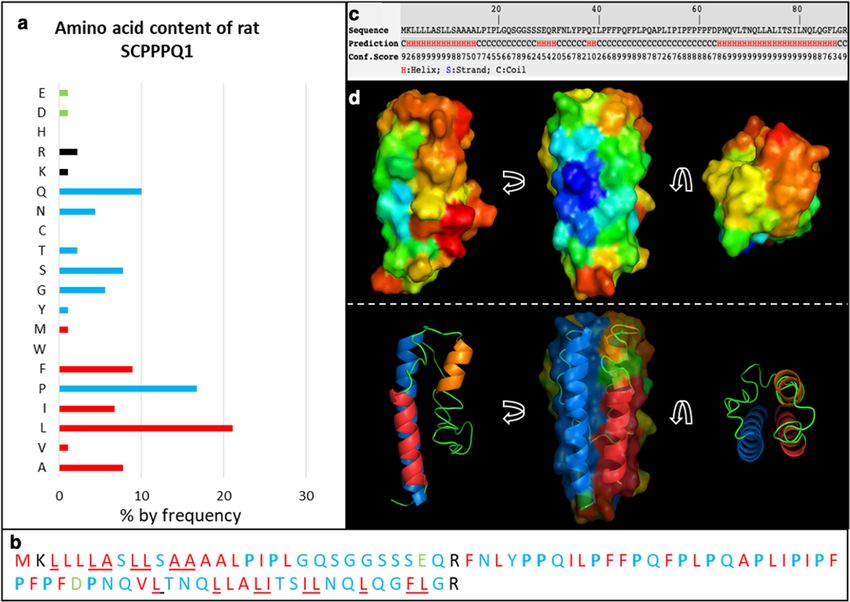
www.nature.com/scientificreports/
Throughout evolution, prokaryotic and eukaryotic organisms have developed host defense mechanisms
against microbial infections. Among these, naturally derived antimicrobial peptides (AMPs) are particularly
important as they may provide a new alternative to antibiotics14. Indeed, small AMPs act indirectly by creating
intra-bacterial defects, generally by interacting with key proteins or RNA15, while the largest AMPs directly attack
the bacterial outer membrane leading to important membrane disruptions. Small cationic molecules (< 10 kDa)
comprising an important proportion of hydrophobic residues (> 30%) form a category of AMPs16. Also, proline-
rich AMPs (PrAMPs) are well-known subset of AMPs that disrupt bacterial i ntegrity17. In the context of PD, a
number of small peptides of variable composition and origin (mammalian, eukaryotic, or purely synthetic), have
also been found to be effective at controlling the growth of P. gingivalis and/or biofilm formation (reviewed i n18).
SCPPPQ1 is a small 9 kDa protein encoded by the scpppq1 gene that localizes to the secretory calcium-binding
phosphoprotein (SCPP) gene cluster and is rich in hydrophobic residues such as proline (18%), leucine (16%)
and phenylalanine (10%)19. Sequence alignment reveals a high homology and conservation of SCPPPQ1 among
species20. The SCPP gene cluster also encodes for a number of proteins possessing antimicrobial properties21,
including some well-known AMPs such as H istatin114. This link to the SCPP gene cluster, its proline rich nature,
small size, and unique resistance to proteases13 suggest that SCPPPQ1 may have antimicrobial capacity. To
implicitly eliminate the null hypothesis (H0) that SCPPPQ1 has no antibacterial activity against P. gingivalis we
have used a two-pronged approach that is, look for structural alterations and test for statistical difference between
treatment parameters. Our specific objective was to apply molecular and biophysical techniques to define in silico
and in vitro the impact of rat SCPPPQ1 protein on this aggressive bacterium implicated in PD.
Results
In silico analysis predicts that SCPPPQ1 may possess antimicrobial properties. The software
APD322 was used to determine the general and antimicrobial properties of the rat SCPPPQ1 sequence. The
analysis predicted that the protein possesses antimicrobial properties and may interact with membranes (see
output in Fig. S1). The amino acid distribution of the sequence further highlighted the high hydrophobicity of
the protein (40%), the presence of 14 prolines and a total net charge of + 1 (Figs. 1a, S1). It also suggested that
the protein may form ⍺-helices comprising at least nine residues on the same hydrophobic surface (Fig. S1).
iTASSER23 and Quark24 were used to predict the 3D structure of SCPPPQ1. The results were consistent with the
APD3 prediction and assigned the hydrophobic amino acids to stable regions at the surface of the model and the
propensity to form ⍺-helices (Fig. 1b–d). Comparisons of alignments with peptides sequences from the APD3
database were further used to determine the percentage of similarities with other known AMPs. The results
showed that rat SCPPPQ1 has few similarities with other proteins with the exception of Bactenecin 7 (Bac-7)
(sequence alignment shown in Fig. S2), a well-known PrAMP produced by bovine n eutrophils25.
Incubation with rat SCPPPQ1 stops bacterial growth. Fluorescence imaging was used to follow the
growth of the bacteria in solution over two-hours in the presence or in the absence of purified SCPPPQ1. Super-
resolution imaging of P. gingivalis stained with Syto9 and propidium iodide revealed no change in fluorescence
and a significant increase (p < 0.001) of bacterial numbers (almost doubling) under control conditions (Fig. 2).
When incubated with SCPPPQ1, bacteria growth stalled and there was an overall decrease in fluorescence over
time (Figs. 2, S3). As a bacterium dies, the Syto9 fluorophore will interact less with nucleic acid resulting in a
decrease of the overall fluorescence of cells. Analysis of the gray value (fluorescence intensity) of the super-
resolution images indicated a significant (p < 0.01) decrease of the intensity by ~ 30% in the first 30 min, ~ 50%
after 1 h and up to 70% after 2 h (Figs. 2, S3). Also, the fact that we did not observed red stained bacteria with
propidium iodide indicates that the dying bacteria were not completely permeable. These results suggest that
incubation with SCPPPQ1 has a bacteriostatic effect under the conditions tested.
Rat SCPPPQ1 decreases the adherence capacity of P. gingivalis. Scanning electron microscopy
(SEM) was used to evaluate the number of bacteria adhering to surface of the titanium sample support follow-
ing exposure to SCPPPQ1, or its sister protein ODAM and buffer only as controls (Fig. 3a–c). The design of this
type of experiment differs slightly from the one presented above in Fig. 2. Rather than continuously monitor-
ing bacterial growth over a 2 h period, it is a snapshot of those bacteria that are still capable of adhering to the
titanium surface after exposure. After 2 h of incubation with SCPPPQ1, as compared with ODAM or the buffer
only, there was a statistically significant (p < 0.0001) decrease (~ 75%) (Fig. 3d) in the number of bacteria adher-
ing to the SEM support.
Rat SCPPPQ1 disrupts the cell envelope of P. gingivalis. Three different electron microscopy
approaches were then used to determine the effect of SCPPPQ1 on the bacterial membrane structure and integ-
rity. First, high-resolution SEM imaging suggested that the outer membrane of P. gingivalis was affected after
incubation with SCPPPQ1 (Fig. 4). Compared to control (Fig. 4a,b), we observed interruptions of the continu-
ity of the cell envelope and abundant blebbing (Fig. 4c,d). We then analyzed thin sections of the bacteria by
transmission electron microscopy (TEM). The results clearly confirmed that the outer membrane was affected
by SCPPPQ1 (Fig. 5c,d) as compared to the control (Fig. 5a,b). The space between the double-membranes of
the bacteria varied (Fig. 5c) and several bacteria showed important outer membrane breaches that are bound
to disrupt the integrity of the bacterial cells (Fig. 5d). TEM images revealed that some of the blebbing observed
by SEM (Fig. 4) correspond to outer membrane vesicles26 (Fig. 5e). There were residues between the cells that
may correspond to membrane fragments (Fig. 5c,d). In the presence of SCPPPQ1, some of the bacteria also
exhibited accumulation of dark bodies in the cytoplasm (Fig. 5c). Further three-dimensional reconstructions of
P. gingivalis incubated with SCPPPQ1 obtained by ‘focused ion beam (FIB) and view’ imaging permitted to visu-
Scientific Reports | (2021) 11:2353 | https://doi.org/10.1038/s41598-021-81791-7 2
Vol:.(1234567890)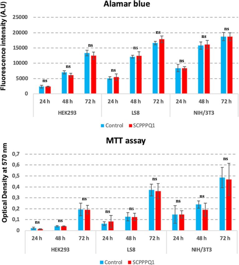
www.nature.com/scientificreports/
Figure 1. In silico analysis of the rat SCPPPQ1 sequence. (a) Analysis of the amino acid content reveals
the high hydrophobicity of the protein (40%) and a total net charge of + 1. (red = hydrophobic amino acids;
black = positively charged amino acids; green = negatively charged amino acids; blue = other amino acids)
(APD322, http://aps.unmc.edu/AP/). (b) SCPPPQ1 possesses a sequence rich in proline residues (highlighted
in blue and bold). The hydrophobic residues believed to be on a same 3D surface are underlined. (c) iTASSER23
(https://zhanglab.ccmb.med.umich.edu/I-TASSER/) analysis predicts the position of helices on SCPPPQ1. (d)
The Quark24 (https://zhanglab.ccmb.med.umich.edu/QUARK/) model further predicts that the red ⍺-helix
corresponds to signal peptide (residues 2 to 15), the orange ⍺-helix to residues 28 to 32, and the blue ⍺-helix to
residues 64 to 87, which is consistent with the iTASSER prediction.
alize membrane alterations along the entire bacterial surface (Movies S1, S2). These imaging techniques clearly
confirm that SCPPPQ1 can profoundly affect the bacterial membranes.
Rat SCPPPQ1 is not toxic to mammalian cells. We wanted to make sure that the concentration of the
protein used to elicit antibacterial activity does not initiate plasma membrane rupture and has no toxic effects on
mammalian cells. There was no significant effect of the presence of SCPPPQ1 on cell proliferation of HEK293,
LS8, and NIH/3T3, as compared to the buffer (control) (Fig. 6). We have also sampled the culture media of LS8
cells for up to 24 h and the aliquots were processed for Western blot analysis. The intensity of the band cor-
responding to SCPPPQ1 did not significantly change throughout the sampling period (Fig. 7d). LS8 cells were
also processed for immunofluorescence to highlight the actin cytoskeleton and the nucleus. No readily apparent
changes in the cell morphology and number were observed (Fig. 7a–c). These results indicate that cells were not
affected by the presence of SCPPPQ1 in the medium during the 24 h culture interval. They also indicate that
SCPPPQ1 levels in the media persisted stably throughout the incubation period, suggesting no apparent binding
to, or capture by, the mammalian cells tested.
Discussion
The objective of our work was to better characterize the antibacterial potential of rat SCPPPQ1 protein, a struc-
tural matrix molecule produced by the JE that is resistant to bacterial degradation13. We have applied fluorescence
and electron microscopy to evaluate whether SCPPPQ1 also has antimicrobial activity. The severe structural
alterations of the cell membrane of P. gingivalis observed when exposed to SCPPPQ1 and the resulting highly
significant reduction in population size exclude the null hypothesis and support the alternate hypothesis. This
leads us to the conclusion that SCPPPQ1 has a bactericidal effect on this bacterium. Because P. gingivalis is a
Scientific Reports | (2021) 11:2353 | https://doi.org/10.1038/s41598-021-81791-7 3
Vol.:(0123456789)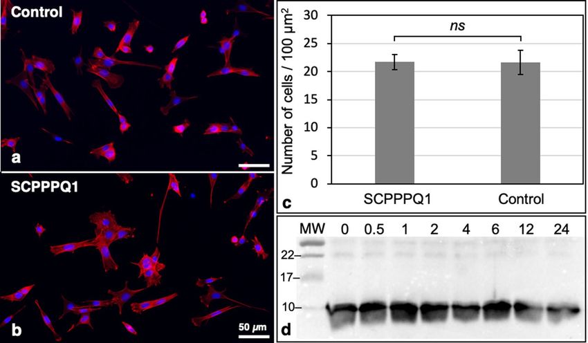
www.nature.com/scientificreports/
Figure 2. Super-resolution live-and-dead fluorescence imaging of P. gingivalis incubated with buffer only
(control) and SCPPPQ1 (20 µM). Both qualitative visual evaluation (insets) and quantitative evaluation of the
fluorescence (also see Fig. S3), show that there are no significant changes in fluorescence intensity throughout
the incubation interval for the control, while there is a major drop when cells are incubated with SCPPPQ1.
Quantification of the bacteria (upper right panel) shows that between the 0 and 120 min interval the number
of bacteria almost doubles whereas with SCPPPQ1 it remains static. In all cases, very few dead cells (red) are
evident. Green = Syto9 labelling; Red = Propidium iodide. Data represent the means ± SD (n = 3).
keystone periodontopathogen during the onset and progression of P D4, and because it has been linked to a
7
number of systemic a fflictions , SCPPPQ1 may also be part of the complex strategies devised by the body to
control and prevent bacterial dissemination27.
SCPPPQ1 belongs to the SCPP genes c luster19 that also encodes for some well-known salivary A MPs14,28 and
some milk-AMPs21. SCPPPQ1 can be classified as a cationic AMP29 and shares similarities with some extensively
studied PrAMPs30, noteworthy with Bac-7, an AMP which disrupts Gram-negative bacteria31. Proteins from both
of these classes adopt conformations that favor interaction with the negatively-charged bacterial m embranes32
33
and cause membrane b reaches . As we show here, both SEM and TEM support the concept that the primary
mechanism of action of SCPPPQ1 is a direct alteration of the bacterial outer membrane, a capacity consistent
with the mode of action of both PrAMPs and cationic AMPs. Its resistance to P. gingivalis however highlights
the unique nature of SCPPPQ1 and places it in a distinctive subset of AMPs. Like for smaller AMPs, in addition
to the destruction of the outer bacterial membrane, it could also be envisaged that the smaller active portion of
SCPPPQ1 could directly penetrate the cells to enhance the efficacy of the anti-bacterial activity34.
While keratinocytes produce AMPs to prevent infections of skin w ounds35, only cells of the JE produce
SCPPPQ120. The JE deposits a unique extracellular matrix at the interface with the tooth36. The proteins consti-
tuting this matrix interact to form a supramolecular network in which they are differentially distributed, with
SCPPPQ1 tending towards the tooth s urface12. As PD set in, bacteria such as P. gingivalis, could cleave/degrade
AMTN, ODAM and Lam33213. This is supported by clinical research showing that digested portions of ODAM
and Lam332 are released into the gingival space above the JE in patients with advanced periodontitis37,38. Such
a degradation is bound to directly expose SCPPPQ1 to the bacteria, a situation comparable to AMPs that accu-
mulate at infected sites, such as defensins in the skin27. However, SCPPPQ1 is not continuously produced by the
cells of the JE under healthy conditions20. Therefore, its production may not be sufficient during the progressive
phase of PD, during which the ever-increasing number of bacteria of the dental biofilms, and not only P. gingi-
valis, could overwhelm the available SCPPPQ1. As it has been shown for the sister protein AMTN39 that has no
antibacterial capacity13, inflammatory factors produced during disease could stimulate production of SCPPPQ1
and thereby increase its available concentration. Under this circumstance, adding the protein or derived peptides
at therapeutic doses may ultimately be the best strategy to both prevent and counteract PD progression. Despite
the fact that P. gingivalis is a keystone bacterium in PD, it will be necessary to further characterize the effect of
SCPPPQ1 on a larger spectrum of bacteria, and even on fungi such as Candida albicans, which can all have a
role in PD. Indeed, the dental plaque is a polymicrobial environment comprising several kinds of bacteria more
or less aggressive. Our study is thus a first step to address the broader antimicrobial potential of SCPPPQ1.
Conventional antibiotic treatments have shown the capacity to temporarily slow down PD progression, but
their beneficial effects are not sustained over time40. There are other molecules such as chloromethane compounds
that are very efficient against P. gingivalis in vitro41, but these damage mammalian cells41 and are therefore not
optimal for in vivo therapy. Interestingly, SCPPPQ1 does not affect mammalian cells (Figs. 6, 7). The lack of cell
Scientific Reports | (2021) 11:2353 | https://doi.org/10.1038/s41598-021-81791-7 4
Vol:.(1234567890)www.nature.com/scientificreports/
Figure 3. Scanning electron microscopy (SEM) imaging of P. gingivalis. Cultures of P. gingivalis were incubated
in (a) the absence (buffer only, control), or presence of (b) ODAM (20 µM) or (c) SCPPPQ1 (20 µM) for 2 h.
Bacteria were then deposited on titanium disks, allowed to adhere for 30 min and processed for SEM imaging.
Both qualitative (a–c) and quantitative (d) analysis reveal an important decrease in the number of P. gingivalis
when incubated with SCPPPQ1 compared to ODAM or buffer as controls. Representative pictures from
one experiment are depicted. T-test analysis was used to determine the significance of the quantification in
comparison to the control (means ± SD; n = 5). ns not significant; ****p < 0.0001.
toxicity is actually expected given the fact that SCPPPQ1 is naturally-produced and present in the JE epithelium
of all individuals. Even if further studies are needed, the inherent exposure of oral bacteria to SCPPPQ1 is likely
to limit any bacterial resistance resulting from its therapeutic use. It must be mentioned, however, that a number
of short peptides with sequences distinct from SCPPPQ1 have been found to be effective at killing P. gingivalis
and/or preventing biofilm formation18. For instance, Pac-525 (KWRRWVRWI) is a tryptophane-rich peptide-
derivative of the bovine neutrophil granule indolicidin42. It was shown to inhibit the growth of P. gingivalis with
a minimal effective concentration of 62.5 μg/ml, and to prevent adhesion to titanium surfaces at 500 μg/ml. The
P-113 peptide (AKRHHGYKRKFH), derived from the saliva protein Histatin 5, showed bactericidal activity
on P. gingivalis at 320 μg/ml in the planktonic state, or at 1280 μg/ml when used on biofilms34. Lastly, two other
peptides derived from the streptococcal proteins SspB (LEAAPKKVQDLLKKANITVKGAFQLFS)43 and ArcA
(NIFKKNVGFKK)44 were found effective at 4 μg/ml and 32 μg/ml, respectively, to kill and reduce virulence of
P. gingivalis.
While periodontopathogens are typically associated with oral manifestations such as PD, they are now being
increasingly implicated in major systemic complications6. Conventional antibiotic therapies have not proven
efficient and there is still no treatment for the control of both periodontal and associated systemic afflictions. In
the past 10 years, several studies have linked PD, and more specifically P. gingivalis, with Alzheimer disease8,9,45.
Dominy et al.8 suggested that “gingipains were neurotoxic in vivo and in vitro, exerting detrimental effects on
tau” and thus exacerbating amyloid plaque formation, a key factor of Alzheimer disease development. While they
reported exciting outcomes concerning the inhibition of g ingipains46, a group of cysteine proteinases, controlling
them in the brain could be a challenging endeavor and may require continuous treatments. Furthermore, the
origin of gingipains in human brain needs further clarification. They could originate locally in the brain from
invading bacteria or they could be translocated via membrane vesicles from the periodontal space. Irrespectively
of the origin of the vesicles, it would seem that, in both cases, controlling the source of the problem, that is the
bacteria itself, might ultimately represent a desirable prospect. While Alzheimer is a complex disease, if the link
to P. gingivalis bears through, a better control of PD is certainly expected to translate into a concurrent impact
at the brain level.
We have shown that SCPPPQ1 possesses antimicrobial activity, yet there are still several issues that need to
be explored in future studies. First, it remains to be determined if the whole SCPPPQ1 protein is necessary to
exhibit its activity. Alternatively, the activity could reside within a short peptide segment of the protein, whether
Scientific Reports | (2021) 11:2353 | https://doi.org/10.1038/s41598-021-81791-7 5
Vol.:(0123456789)www.nature.com/scientificreports/
Figure 4. Scanning electron microscopy (SEM) imaging of the bacterial surface. Cultures of P. gingivalis were
incubated in (a,b) the absence (buffer only, control) or (c,d) presence of SCPPPQ1 (20 µM) for 2 h. Bacteria
were then deposited on titanium disks, allowed to adhere for 30 min and processed for high-resolution SEM
imaging. P. gingivalis, incubated with buffer only (a,b) show intact membranes, while incubation with SCPPPQ1
(c,d) results in important surface breaches (arrows) and abundant blebbing.
it be the central proline-rich domain, or the alpha-helix located in the C-terminus. The identification of the active
portion will also perhaps allow to further explain the molecular mechanism by which it can disrupt the bacterial
membrane, and be compared to established scenarios put forward for other AMPs47–49. This will further guide
the optimization of more effective peptides for controlling bacterial pathogens, whereby smaller active peptides
could be more potent. While the concentration used in the current study (20 μM; 160 μg/ml) has proven very
efficient to repress bacterial growth, a direct experimental comparison of the antimicrobial effects between
SCPPPQ1 and known AMPs will eventually need to be conducted to determine whether it is superior or not. It
is difficult, however, to relate the concentration used in vitro with that produced locally by cells of the JE in vivo,
and whether SCPPPQ1 could resist attacks by P. gingivalis in the complex physiological environment of the oral
cavity. It can also be questioned if SCPPPQ1 would have the same antibacterial potential when tested on mixtures
of pathogens present in dental plaques. The development of a mouse model lacking the scpppq1 gene (under way
in our laboratory) will be key to confirm its antimicrobial role in vivo. Likewise, the model will also provide an
opportunity to validate if exogenously administered SCPPPQ1 protein or peptides may be able to slow down
breakdown of the gingival seal, the onset of PD, and associated systemic complications.
In conclusion, our results show for the first time that a small, oral keratinocyte-derived protein, SCPPPQ1,
has antibacterial activity against P. gingivalis. Because of its endogenous origin and its non-toxicity on mamma-
lian cells, it represents an excellent candidate for limiting the progression of PD, but also to deal with systemic
complications related to P. gingivalis. Small AMPs disseminate better, are generally more efficient and stable, and
are less prone to degradation50, all major assets in the fight against bacteria. While SCPPPQ1 is a small protein,
identification of its active portions will allow optimization of its effectiveness and of its delivery, particularly
across the blood brain barrier.
Methods
In silico analysis. The AMP database APD322 (http://aps.unmc.edu/AP/, University of Nebraska, Omaha,
NE, USA) was used to determine the antimicrobial potential of SCPPPQ1. The rat SCPPPQ1 sequence was ana-
lyzed to evaluate the peptide length, its net charge, its amino acid composition, the secondary structure and its
sequence similarity with other AMPs. Quark24 and iTasser23 (https://zhanglab.ccmb.med.umich.edu/, University
of Michigan, Ann Arbor, MI, USA) were used individually to predict the tridimensional (3D) conformation.
MacPyMOL (The PyMol Molecular Graphics System, Version 2.0 Schrödinger, LLC, New York, NY, USA) was
finally used to analyze the model and to highlight the position of the hydrophobic residues.
Scientific Reports | (2021) 11:2353 | https://doi.org/10.1038/s41598-021-81791-7 6
Vol:.(1234567890)www.nature.com/scientificreports/
Figure 5. Transmission electron microscopy (TEM) visualization of P. gingivalis. Cultures of P. gingivalis were
incubated in (a,b) the absence (buffer only, control) or (c–e) presence of SCPPPQ1 (20 µM) for 2 h. Cultures
were fixed and processed for TEM imaging. Comparison P. gingivalis incubated with (a,b) buffer or (c–e)
with SCPPPQ1 reveals that the presence of SCPPPQ1 causes membrane disruptions (arrows), accumulation
of dark bodies in the cytoplasm (asterisks) and increases the space between the inner and outer membranes
(arrowheads). Debris were also frequently present interspersed between the bacteria in the presence of
SCPPPQ1. (e) High magnification image showing that bacteria incubated with SCPPPQ1 frequently have outer
membrane vesicles on their surface.
Cloning procedures. The sister protein O DAM51, also belonging to the SCPP cluster, was used as control.
Truncated versions of SCPPPQ1 and ODAM genes lacking regions encoding the predicted N-terminal signal
sequence were PCR-amplified from rat cDNA sequences using primers as previously described12. PCR products
were cloned into the vector pHT for purification s tudies35. The recombinant pHT plasmids allow to produce
recombinant proteins with an in-frame N-terminal hexahistidyl-tag (His-tag) and TEV protease cleavage site.
Escherichia coli strain XL-1 Blue were used as hosts for cloning52.
Protein overexpression and purification. The protein overexpression protocol is similar to the ones
used in Fouillen et al.12,13. Briefly, BL21(DE3)-star cells containing either pHT-SCPPPQ1 or pHT-hODAM12 were
Scientific Reports | (2021) 11:2353 | https://doi.org/10.1038/s41598-021-81791-7 7
Vol.:(0123456789)www.nature.com/scientificreports/
Figure 6. Analysis of the effect of SCPPPQ1 on mammalian cells. HEK293, LS8 and NIH/3T3 cells were
incubated with SCPPPQ1 (20 µM) or only the buffer (control) for 72 h. Each 24 h, some cells were processed for
both Alamar Blue and MTT assays. The data reveal no significant difference in cell proliferation in the presence
or absence of SCPPPQ1. Data represent the means ± SD (n = 3). A.U. arbitrary unit, ns not significant.
grown at 37 °C and 250 rpm to an optical density at 660 nm (OD660) of around 0.6, and protein expression was
induced with 0.1 mM isopropyl-ß-d-thiogalactoside (Thermo Fisher Scientific, Waltham, MA, USA) overnight
at 30 °C and 250 rpm. For SCPPPQ1, bacterial cells were harvested, suspended in equilibration buffer (50 mM
Na2HPO4 (Thermo Fisher Scientific, Waltham, MA, USA), 150 mM NaCl (Thermo Fisher Scientific, Waltham,
MA, USA), 10 mM imidazole (Sigma-Aldrich, Saint-Louis, MO, USA), 8 M urea (Thermo Fisher Scientific,
Waltham, MA, USA), pH 7) at 4 °C, and sonicated 6 × 15 s with 15 s ice incubations in between. Lysates were
centrifuged at 13,400 × g and the 6His-tagged protein in the supernatant was bound on nickel-nitriloacetic acid
(Ni–NTA)-agarose affinity resin (Qiagen, Hilden, Germany) at room temperature. After washing the resin with
10 volumes of binding buffer (50 mM Na2HPO4 (Thermo Fisher Scientific, Waltham, MA, USA), 300 mM NaCl
(Thermo Fisher Scientific, Waltham, MA, USA), 20 mM imidazole (Sigma-Aldrich, Saint-Louis, MO, USA), 8 M
Urea (Thermo Fisher Scientific, Waltham, MA, USA), pH 7), proteins were eluted with elution buffer (50 mM
Na2HPO4 (Thermo Fisher Scientific, Waltham, MA, USA), 300 mM NaCl (Thermo Fisher Scientific, Waltham,
MA, USA), 300 mM imidazole (Sigma-Aldrich, Saint-Louis, MO, USA), 8 M Urea (Thermo Fisher Scientific,
Waltham, MA, USA), pH 7). Concentration of the collected fractions were assessed using a BioDrop (Montreal
Biotech, Montréal, Canada) and fractions were then analyzed by sodium dodecyl sulfate–polyacrylamide gel
electrophoresis (SDS–PAGE) and Coomassie blue staining. Proteins were finally dialyzed into 50 mM Na2HPO4
(Thermo Fisher Scientific, Waltham, MA, USA), 8 M Urea (Thermo Fisher Scientific, Waltham, MA, USA) (pH
7.2) and stored at 4 °C. ODAM was purified in the same conditions as SCPPPQ1 but in a buffer without Urea.
Western blot gels were acquired using a Bio-Rad ChemiDoc imager and the software Image Lab version 6.1 (Bio-
Rad, Hercules, CA, USA). Exposure time for the acquisition were between 1 and 2 s.
Scientific Reports | (2021) 11:2353 | https://doi.org/10.1038/s41598-021-81791-7 8
Vol:.(1234567890)www.nature.com/scientificreports/
Figure 7. Evaluation of the effect of SCPPPQ1 on LS8 cells. Ameloblast-like LS8 cells were seeded on
coverslips, grown overnight, and incubated for 24 h in (a) the absence (control) or (b) presence of SCPPPQ1
(20 µM) in the media. (a,b) Cells were fixed and stained with rhodamine phalloidin (red) and Hoechst (blue),
and (c) images analyzed to assess cell density. Data represent the means ± SD (n = 3). ns non-significant. (d) The
media of cells incubated with SCPPPQ1 was sampled over the 24 h incubation period and analyzed by Western
blotting to detect SCPPPQ1. Numbers at top indicate the time in hours and molecular weight (MW) markers
(kDa) are labeled at left.
Bacterial culture and assays. Porphyromonas gingivalis ATCC 33277 (ATCC, Manassas, VA, USA) were
grown anaerobically for at least 24 h (80% N 2, 10% CO2, 10% H2) at 37 °C in Todd-Hewitt broth (THB) (Thermo
Fisher Scientific, Waltham, MA, USA) supplemented with 0.001% hemin (Thermo Fisher Scientific, Waltham,
MA, USA) and 0.0001% vitamin K (Thermo Fisher Scientific, Waltham, MA, USA). 20 µM of each of the purified
proteins (SCPPPQ1 and ODAM) were exposed in a test tube to 800 µl of a suspension of bacteria at a OD660 of 1
(CFU = 109 cells/ml13) for 2 h at 37 °C. This concentration of P. gingivalis was previously used to experimentally
ice38. The protein concentration of 20 μM (160 μg/ml) was selected based on our own
induce periodontitis in m
prior work showing resistance of SCPPPQ1 to degradation by P. gingivalis13. Also, it represented a concentration
within the range used in other studies looking at the effects of antimicrobial effects of p eptides17,18,34,42,43. The
mixture was then sampled for fluorescence microscopy, SEM and TEM characterization. As a negative control,
buffer without the proteins was incubated with P. gingivalis under the same conditions.
Scanning electron microscopy. The SEM protocol is similar to the one described in Fouillen et al.13. Fol-
lowing 2 h incubations in a 1.5 ml tube, bacterial samples were applied to polished grade II titanium supports for
30 min. The bacteria that adhered to the support were fixed for 1 h at 4 °C with 4% paraformaldehyde (Thermo
Fisher Scientific, Waltham, MA, USA) and 0.1% glutaraldehyde (Electron Microscopy Sciences, Hatfield, PA,
USA) in 0.1 M phosphate buffer (PB, pH 7.3), and subsequently rinsed three times with PB. The samples were
then post-fixed for 1 h with 1% osmium tetroxide (Electron Microscopy Sciences, Hatfield, PA, USA), dehy-
drated through an ethanol series (30%, 50%, 70%, 90%, 95% and two times 100%) followed by drying using a
Critical Point Drier CPD300 (Leica Biosystems, Concord, ON, Canada). A high-resolution field emission (FE)-
SEM Regulus 8230 (Hitachi High-Tech Corporation, Tokyo, Japan), operated at 0.8 kV, was used for imaging
the uncoated samples. For each condition, counting of at least 50 representative images, from four independent
experiments were analyzed using ImageJ version 1.50i (NIH, Bethesda, MD, USA).
Immunofluorescence studies. Some bacteria were stained using the Live/Dead BacLight kit (Thermo
Fisher Scientific, Waltham, MA, USA) before incubation with SCPPPQ1. Briefly, 1.5 µl of Syto9 and 1.5 µl of
propidium iodide were added to 800 µl of bacteria at an O D660 of 1. Two hundred µl of protein were added
to the mixture in order to obtain a final concentration of protein of 20 µM. As a control, only the buffer was
added to the bacterial suspension. During 2 h of incubation, the suspensions were sampled at 15, 30, 60 and
120 min. After each sampling, the mixture was fixed using 1% glutaraldehyde (Electron Microscopy Sciences,
Hatfield, PA, USA) to obtain a final concentration of 2.5%. Ten µl of each suspension were finally examined
with an Elyra PS1 microscope (Carl Zeiss Microscopy, Oberkochen, Germany) equipped with 63× oil objective
(numerical aperture 1.4) and an EMCCD iXon3 DU-885K camera (Andor, Connecticut, USA). Z-stack volumes
were acquired using the Structural Illumination Method (SIM) and reconstructed using the Zen Black software
version 2.1 (Carl Zeiss Microscopy, Oberkochen, Germany). For each condition, counting and intensity analysis
Scientific Reports | (2021) 11:2353 | https://doi.org/10.1038/s41598-021-81791-7 9
Vol.:(0123456789)www.nature.com/scientificreports/
of at least 50 bacteria for each of 20 representative images (5 samples per conditions) from three independent
experiments were analyzed using ImageJ version 1.50i (NIH, Bethesda, MD, USA).
Embedding procedures. Some bacteria were fixed as above, post-fixed with potassium ferrocyanide-
reduced osmium tetroxide (Electron Microscopy Sciences, Hatfield, PA, USA) and then processed for embed-
ding in Epon resin (Electron Microscopy Sciences, Hatfield, PA, USA). Ultrathin sections of 80–100 nm were
cut with a diamond knife on an Ultracut EM UC6 ultramicrotome (Leica Biosystems, Concord, ON, Canada)
and transferred onto Formvar-coated 200-mesh nickel grids for TEM imaging. The grid-mounted sections were
examined in a Tecnai 12 TEM (FEI (now Thermo Fisher Scientific), Eindhoven, Netherlands) operating at 80 kV.
Some embedded samples were used tomographic imaging using a Crossbeam 550 Focused Ion Beam (FIB)-SEM
(Carl Zeiss Microscopy, Oberkochen, Germany). Samples were serially milled at 4 nm thickness using a probe
current of 1.5 nA/30 kV. Exposed surfaces were observed at 1.5 kV using Secondary Electron and Energy Selec-
tive Backscatter detectors. The Dragonfly software 2020.1 (Object Research Systems, Montréal, Canada) was
used for further alignment and 3D reconstruction of the FIB-SEM image stacks.
Cell analysis. Although SCPPPQ1 is naturally-produced and present in the JE epithelium of all individuals,
we wanted to make sure that the protein has no effect against eukaryotic cells. To target the two cell lineages
found in the oral cavity (epithelial, fibroblastic) that can be in direct contact with SCPPPQ1, we have used as rep-
resentative cells LS8 ameloblast-derived epithelial cells53 (ATCC, Manassas, VA, USA), and NIH/3T3 fibroblast
cells54 (ATCC, Manassas, VA, USA). More conventional human embryonic kidney (HEK293) epithelial c ells55
(ATCC, Manassas, VA, USA) were also used. Cells were cultured in DMEM supplemented with 10% fetal bovine
serum (FBS, Thermo Fisher Scientific, Waltham, MA, USA) at 37 °C in a 5% CO2 atmosphere. 5000 cells were
placed in each well of 96-well plates and grown overnight. The culture medium was then exchanged with fresh
one containing either SCPPPQ1 at a final concentration of 20 µM or the equivalent volume of buffer as control.
Cells were then grown up to 72 h. After 24 h, 48 h and 72 h of incubation, some cells were processed for an
Alamar Blue assay (Thermo Fisher Scientific, Waltham, MA, USA) and others for a MTT assay (3-(4,5-Dimeth-
ylthiazol-2-yl)-2,5-Diphenyltetrazolium Bromide) (Thermo Fisher Scientific, Waltham, MA, USA) following
company instructions for respective assays. Briefly, for Alamar Blue, the cell media was replaced by 90 µl of
DMEM without phenol red (Thermo Fisher Scientific, Waltham, MA, USA) and 10 µl of the Alamar Blue HS
solution and placed for 4 h at 37 °C in a 5% C O2 atmosphere. Fluorescence of the 96-wells plates were then read
at a wavelength of 570 nm. For MTT, the cell media were replaced by 100 µl of DMEM without phenol red and
10 µl of a PBS solution containing 12 mM of MTT. The 96-well plates were then placed for 4 h at 37 °C in a 5%
CO2 atmosphere, before adding 100 µl of a SDS-HCl solution to each well and to incubate the microplate for
an additional 4 h at 37 °C in a 5% C O2 atmosphere. Finally, the samples were mixed a last time before reading
their absorbance at 570 nm with a 2104 EnVision Multilabel Plate Reader (PerkinElmer, Waltham, MA, USA).
LS8 cells immunofluorescence staining and western blotting. Ameloblast-like cells LS8 were cul-
tured in DMEM supplemented with 10% FBS at 37 °C in a 5% C O2 atmosphere. 250,000 cells were placed on
coverslips and grown overnight. SCPPPQ1 was added to the cell media at a final concentration of 20 µM. The
same volume of buffer was added as control to some coverslips. After 24 h of incubation, the cells were stained
with rhodamine phalloidin and Hoechst (1:500; 1:2000 respectively) (Thermo Fisher Scientific, Waltham,
MA, USA) to highlight actin and nuclei. Samples were acquired on an Axio Imager (Carl Zeiss Microscopy,
Oberkochen, Germany) using the Zen software version 2.1 (Carl Zeiss Microscopy, Oberkochen, Germany) and
the images were analyzed using ImageJ version 1.50i (NIH, Bethesda, MD, USA). Cell media of LS8 cells were
collected multiple times during a 24 h culture period and then aliquots were analyzed using SDS-PAGE followed
by Western Blot using anti-SCPPPQ1 antibodies (1:1000). Western blot gels were acquired at an exposure times
between 1 and 2 s using a ChemiDoc imager (Bio-Rad, Hercules, CA, USA).
Statistical analysis. For fluorescence, SEM, and cell analysis, values and standard deviations were cal-
culated from at least three independent experiments, and the p values were obtained by t test analysis of each
condition from data in Excel (Microsoft Windows, Albuquerque, NM, USA). Statistical significance was defined
as ns, p > 0.05; *p < 0.05; **p < 0.01; ***p < 0.001; and ****p < 0.0001.
Data availability
The datasets and/or analyses generated during the current study are available from the corresponding author,
Dr. Antonio Nanci, upon reasonable request.
Received: 9 July 2020; Accepted: 8 January 2021
References
1. Kilian, M. et al. The oral microbiome—An update for oral healthcare professionals. Br. Dent. J. 221, 657–666. https://doi.
org/10.1038/sj.bdj.2016.865 (2016).
2. Costalonga, M. & Herzberg, M. C. The oral microbiome and the immunobiology of periodontal disease and caries. Immunol. Lett.
162, 22–38. https://doi.org/10.1016/j.imlet.2014.08.017 (2014).
3. Schytte-Bix, I. J., Hars, R., Preus, H. R. & Helgeland, K. Entrance of Actinobacillus actinomycetemcomitans into HEp-2 cells in vitro.
J. Periodontal. 63, 723–728 (1992).
4. Potempa, J. & Travis, J. Porphyromonas gingivalis proteinases in periodontitis, a review. Acta Biochim. Pol. 43, 455–465 (1996).
5. Bosshardt, D. D. & Lang, N. P. The junctional epithelium: From health to disease. J. Dent. Res. 84, 9–20 (2005).
Scientific Reports | (2021) 11:2353 | https://doi.org/10.1038/s41598-021-81791-7 10
Vol:.(1234567890)www.nature.com/scientificreports/
6. Bui, F. Q. et al. Association between periodontal pathogens and systemic disease. Biomed. J. 42, 27–35. https://doi.org/10.1016/j.
bj.2018.12.001 (2019).
7. Nazir, M. A. Prevalence of periodontal disease, its association with systemic diseases and prevention. Int. J. Health Sci. 11, 72
(2017).
8. Dominy, S. S. et al. Porphyromonas gingivalis in Alzheimer’s disease brains: Evidence for disease causation and treatment with
small-molecule inhibitors. Sci. Adv. 5, 3333 (2019).
9. Kamer, A. R., Craig, R. G., Niederman, R., Fortea, J. & de Leon, M. J. Periodontal disease as a possible cause for Alzheimer’s disease.
Periodontology 2000(83), 242–271. https://doi.org/10.1111/prd.12327 (2020).
10. Teles, R., Teles, F., Frias-Lopez, J., Paster, B. & Haffajee, A. Lessons learned and unlearned in periodontal microbiology. Periodon-
tology 2000(62), 95–162 (2013).
11. Schroeder, H. E. The junctional epithelium: Origin, structure, and significance. Acta Med. Dent. Helv. 1, 155–167 (1996).
12. Fouillen, A. et al. Interactions of AMTN, ODAM and SCPPPQ1 proteins of a specialized basal lamina that attaches epithelial cells
to tooth mineral. Sci. Rep. 7, 46683. https://doi.org/10.1038/srep46683 (2017).
13. Fouillen, A. et al. Selective bacterial degradation of the extracellular matrix attaching the gingiva to the tooth. Eur. J. Oral Sci. https
://doi.org/10.1111/eos.12623(2019).
14. Khurshid, Z. et al. Oral antimicrobial peptides: Types and role in the oral cavity. Saudi Pharm. J. 24, 515–524. https://doi.
org/10.1016/j.jsps.2015.02.015 (2016).
15. Dale, B. A. & Fredericks, L. P. Antimicrobial peptides in the oral environment: Expression and function in health and disease.
Curr. Issues Mol. Biol. 7, 119–133 (2005).
16. Fan, L. et al. DRAMP: A comprehensive data repository of antimicrobial peptides. Sci. Rep. 6, 24482. https: //doi.org/10.1038/srep2
4482 (2016).
17. Holfeld, L., Knappe, D. & Hoffmann, R. Proline-rich antimicrobial peptides show a long-lasting post-antibiotic effect on Entero-
bacteriaceae and Pseudomonas aeruginosa. J. Antimicrob. Chemother. 73, 933–941. https://doi.org/10.1093/jac/dkx482 (2018).
18. Gerits, E., Verstraeten, N. & Michiels, J. New approaches to combat Porphyromonas gingivalis biofilms. J. Oral Microbiol. 9, 1300366.
https://doi.org/10.1080/20002297.2017.1300366 (2017).
19. Kawasaki, K. The SCPP gene family and the complexity of hard tissues in vertebrates. Cells Tissues Organs 194, 108–112 (2011).
20. Moffatt, P., Wazen, R. M., Dos Santos Neves, J. & Nanci, A. Characterisation of secretory calcium-binding phosphoprotein-proline-
glutamine-rich 1: A novel basal lamina component expressed at cell-tooth interfaces. Cell Tissue Res. 358, 843–855. https://doi.
org/10.1007/s00441-014-1989-3 (2014).
21. Kawasaki, K., Lafont, A. G. & Sire, J. Y. The evolution of milk casein genes from tooth genes before the origin of mammals. Mol.
Biol. Evol. 28, 2053–2061 (2011).
22. Wang, G., Li, X. & Wang, Z. APD3: The antimicrobial peptide database as a tool for research and education. Nucleic Acids Res. 44,
D1087-1093. https://doi.org/10.1093/nar/gkv1278 (2016).
23. Yang, J. & Zhang, Y. Protein structure and function prediction using I-TASSER. Curr. Protoc. Bioinform. 52, 1–15. https://doi.
org/10.1002/0471250953.bi0508s52 (2015).
24. Zhang, C., Mortuza, S. M., He, B., Wang, Y. & Zhang, Y. Template-based and free modeling of I-TASSER and QUARK pipelines
using predicted contact maps in CASP12. Proteins 86(Suppl 1), 136–151. https://doi.org/10.1002/prot.25414 (2018).
25. Scocchi, M., Romeo, D. & Zanetti, M. Molecular cloning of Bac7, a proline- and arginine-rich antimicrobial peptide from bovine
neutrophils. FEBS Lett. 352, 197–200. https://doi.org/10.1016/0014-5793(94)00954-6 (1994).
26. Sato, K. et al. Identification of Porphyromonas gingivalis proteins secreted by the Por secretion system. FEMS Microbiol. Lett. 338,
68–76. https://doi.org/10.1111/1574-6968.12028 (2013).
27. Bernard, J. J. & Gallo, R. L. Protecting the boundary: The sentinel role of host defense peptides in the skin. Cell. Mol. Life Sci. 68,
2189–2199. https://doi.org/10.1007/s00018-011-0712-8 (2011).
28. van ‘t Hof, W., Veerman, E. C., Nieuw-Amerongen, A. V. & Ligtenberg, A. J. Antimicrobial defense systems in saliva. Monogr. Oral
Sci. 24, 40–51. https://doi.org/10.1159/000358783 (2014).
29. Hancock, R. E. W. Cationic peptides: Effectors in innate immunity and novel antimicrobials. Lancet Infect. Dis. 1, 156–164. https
://doi.org/10.1016/s1473-3099(01)00092-5 (2001).
30. Li, W. et al. Proline-rich antimicrobial peptides: Potential therapeutics against antibiotic-resistant bacteria. Amino Acids 46,
2287–2294. https://doi.org/10.1007/s00726-014-1820-1 (2014).
31. Mardirossian, M. et al. The host antimicrobial peptide Bac71-35 binds to bacterial ribosomal proteins and inhibits protein synthesis.
Chem. Biol. 21, 1639–1647. https://doi.org/10.1016/j.chembiol.2014.10.009 (2014).
32. Kuppusamy, R., Willcox, M., Black, D. S. & Kumar, N. Short cationic peptidomimetic antimicrobials. Antibiotics (Basel). https://
doi.org/10.3390/antibiotics8020044 (2019).
33. Hartmann, M. et al. Damage of the bacterial cell envelope by antimicrobial peptides gramicidin S and PGLa as revealed by trans-
mission and scanning electron microscopy. Antimicrob. Agents Chemother. 54, 3132–3142. https://doi.org/10.1128/AAC.00124
-10 (2010).
34. Wang, H. Y. et al. Efficacy of a novel antimicrobial peptide against periodontal pathogens in both planktonic and polymicrobial
biofilm states. Acta Biomater. 25, 150–161. https://doi.org/10.1016/j.actbio.2015.07.031 (2015).
35. Pfalzgraff, A., Brandenburg, K. & Weindl, G. Antimicrobial peptides and their therapeutic potential for bacterial skin infections
and wounds. Front. Pharmacol. 9, 281. https://doi.org/10.3389/fphar.2018.00281 (2018).
36. Nanci, A. T. Cate’s Oral Histology: Development, Structure, and Function (Mosby Elsevier, Hoboken, 2017).
37. Emingil, G., Kuula, H., Pirila, E., Atilla, G. & Sorsa, T. Gingival crevicular fluid laminin-5 gamma2-chain levels in periodontal
disease. J. Clin. Periodontol. 33, 462–468. https://doi.org/10.1111/j.1600-051X.2006.00933.x (2006).
38. Lee, H. K. et al. Odontogenic ameloblast-associated protein (ODAM) in gingival crevicular fluid for site-specific diagnostic value
of periodontitis: A pilot study. BMC Oral Health 18, 148. https://doi.org/10.1186/s12903-018-0609-0 (2018).
39. Nakayama, Y. et al. C/EBPbeta and YY1 bind and interact with Smad3 to modulate lipopolysaccharide-induced amelotin gene
transcription in mouse gingival epithelial cells. FEBS Open Bio. 9, 276–290. https://doi.org/10.1002/2211-5463.12566 (2019).
40. Haque, M., Sartelli, M. & Haque, S. Z. Dental infection and resistance-global health consequences. Dent. J. (Basel). https://doi.
org/10.3390/dj7010022 (2019).
41. Olsen, I. & Potempa, J. Strategies for the inhibition of gingipains for the potential treatment of periodontitis and associated systemic
diseases. J. Oral Microbiol. https://doi.org/10.3402/jom.v6.24800 (2014).
42. Li, J. Y. et al. High in vitro antibacterial activity of Pac-525 against Porphyromonas gingivalis biofilms cultured on titanium. Biomed.
Res. Int. 2015, 909870. https://doi.org/10.1155/2015/909870 (2015).
43. Daep, C. A., James, D. M., Lamont, R. J. & Demuth, D. R. Structural characterization of peptide-mediated inhibition of Porphy-
romonas gingivalis biofilm formation. Infect. Immunol. 74, 5756–5762. https://doi.org/10.1128/IAI.00813-06 (2006).
44. Ho, M. H., Lamont, R. J. & Xie, H. A novel peptidic inhibitor derived from Streptococcus cristatus ArcA attenuates virulence
potential of Porphyromonas gingivalis. Sci. Rep. 7, 16217. https://doi.org/10.1038/s41598-017-16522-y (2017).
45. Zeng, F. et al. Receptor for advanced glycation end products up-regulation in cerebral endothelial cells mediates cerebrovascular-
related amyloid beta accumulation after Porphyromonas gingivalis infection. J. Neurochem. https: //doi.org/10.1111/jnc.15096 (2020).
46. Ryder, M. I. Porphyromonas gingivalis and Alzheimer’s disease: Recent findings and potential therapies. J. Periodontol. https://doi.
org/10.1002/JPER.20-0104 (2020).
Scientific Reports | (2021) 11:2353 | https://doi.org/10.1038/s41598-021-81791-7 11
Vol.:(0123456789)www.nature.com/scientificreports/
47. Pasupuleti, M., Schmidtchen, A. & Malmsten, M. Antimicrobial peptides: Key components of the innate immune system. Crit.
Rev. Biotechnol. 32, 143–171. https://doi.org/10.3109/07388551.2011.594423 (2012).
48. Rothstein, D. M. et al. Anticandida activity is retained in P-113, a 12-amino-acid fragment of histatin 5. Antimicrob. Agents Chem-
other. 45, 1367–1373. https://doi.org/10.1128/AAC.45.5.1367-1373.2001 (2001).
49. Strömstedt, A. A., Ringstad, L., Schmidtchen, A. & Malmsten, M. Interaction between amphiphilic peptides and phospholipid
membranes. Curr. Opin. Colloid Interface Sci. 15, 467–478. https://doi.org/10.1016/j.cocis.2010.05.006 (2010).
50. Lau, J. L. & Dunn, M. K. Therapeutic peptides: Historical perspectives, current development trends, and future directions. Bioorg.
Med. Chem. 26, 2700–2707. https://doi.org/10.1016/j.bmc.2017.06.052 (2018).
51. Moffatt, P., Smith, C. E., Sooknanan, R., St-Arnaud, R. & Nanci, A. Identification of secreted and membrane proteins in the rat
incisor enamel organ using a signal-trap screening approach. Eur. J. Oral Sci. 114, 139–146 (2006).
52. Brown, D. Antibiotic resistance breakers: Can repurposed drugs fill the antibiotic discovery void?. Nat. Rev. Drug Discov. 14,
821–833 (2015).
53. Chen, L. S., Couwenhoven, R. I., Hsu, D., Luo, W. & Snead, M. L. Maintenance of amelogenin gene expression by transformed
epithelial cells of mouse enamel organ. Arch. Oral Biol. 37, 771–778 (1992).
54. Negmadjanov, U. et al. TGF-B1-mediated differentiation of fibroblasts is associated with increased mitochondrial content and
cellular respiration. PLoS ONE. https://doi.org/10.1371/journal.pone.0123046 (2015).
55. Stepanenko, A. A. & Dmitrenko, V. V. HEK293 in cell biology and cancer research: Phenotype, karyotype, tumorigenicity, and
stress-induced genome-phenotype evolution. Gene 569, 182–190. https://doi.org/10.1016/j.gene.2015.05.065 (2015).
Acknowledgements
We extend our thanks to Hitachi High-Tech Corporation for time allocation on their FE-SEM platform in Clarks-
burg (MD, USA) and to Zeiss in Pleasanton (CA, USA) for FIB and View imaging of samples. This work was
supported by the Electron Imaging Facility, (Faculty of Dental Medicine, Université de Montréal, QC, Canada).
We thank Drs. Christian Baron and Rima M. Wazen for their critical reading of the manuscript and comments.
This study was supported by the Canadian Institutes of Health Research [A.N. (CIHR MOP-110972), the Network
for Oral and Bone Health Research (FRQ-S). A.N. is recipient of a Canada Research Chair in Calcified tissues,
Biomaterials and Structural Imaging.
Author contributions
A.F., C.M., P.M. and A.N. participated in the design of the experiments and in interpretation of results. A.F. and
A.N. wrote the manuscript and P.M. participated in the revision. A.F. performed most of the experimental work
and data analysis. C.M. carried out bacterial growth and SEM preparations. K.J.P. prepared the TEM sections.
All authors have validated the manuscript. The project was under the supervision of A.N.
Competing interests
The authors declare no competing interests.
Additional information
Supplementary Information The online version contains supplementary material available at https://doi.
org/10.1038/s41598-021-81791-7.
Correspondence and requests for materials should be addressed to A.N.
Reprints and permissions information is available at www.nature.com/reprints.
Publisher’s note Springer Nature remains neutral with regard to jurisdictional claims in published maps and
institutional affiliations.
Open Access This article is licensed under a Creative Commons Attribution 4.0 International
License, which permits use, sharing, adaptation, distribution and reproduction in any medium or
format, as long as you give appropriate credit to the original author(s) and the source, provide a link to the
Creative Commons licence, and indicate if changes were made. The images or other third party material in this
article are included in the article’s Creative Commons licence, unless indicated otherwise in a credit line to the
material. If material is not included in the article’s Creative Commons licence and your intended use is not
permitted by statutory regulation or exceeds the permitted use, you will need to obtain permission directly from
the copyright holder. To view a copy of this licence, visit http://creativecommons.org/licenses/by/4.0/.
© The Author(s) 2021
Scientific Reports | (2021) 11:2353 | https://doi.org/10.1038/s41598-021-81791-7 12
Vol:.(1234567890)You can also read