Effects of Conserved Wedge Domain Residues on DNA Binding Activity of Deinococcus radiodurans RecG Helicase - Frontiers
←
→
Page content transcription
If your browser does not render page correctly, please read the page content below
ORIGINAL RESEARCH
                                                                                                                                             published: 04 February 2021
                                                                                                                                       doi: 10.3389/fgene.2021.634615

                                              Effects of Conserved Wedge Domain
                                              Residues on DNA Binding Activity of
                                              Deinococcus radiodurans RecG
                                              Helicase
                                              Sun-Wook Jeong 1,2†, Min-Kyu Kim 1†, Lei Zhao 1, Seul-Ki Yang 1, Jong-Hyun Jung 1,3,
                        Edited by:            Heon-Man Lim 2 and Sangyong Lim 1,3*
                      Hari S. Misra,
     Bhabha Atomic Research Centre            1
                                                Radiation Research Division, Korea Atomic Energy Research Institute, Jeongeup, South Korea, 2 Department of Biological
                      (BARC), India           Sciences, College of Biological Sciences and Biotechnology, Chungnam National University, Daejeon, South Korea,
                                              3
                                                Department of Radiation Science and Technology, University of Science and Technology, Daejeon, South Korea
                       Reviewed by:
                        Issay Narumi,
               Toyo University, Japan
                                              Deinococcus radiodurans is extremely resistant to ionizing radiation and has an exceptional
                           Huiming Lu,
    University of Texas Southwestern          ability to repair DNA damage caused by various DNA-damaging agents. D. radiodurans
       Medical Center, United States          uses the same DNA-repair strategies as other prokaryotes, but certain proteins involved
                     Joanna Timmins,
       UMR5075 Institut de Biologie           in the classical DNA repair machinery have characteristics different from their counterparts.
             Structurale (IBS), France        RecG helicase, which unwinds a variety of branched DNA molecules, such as Holliday
                    *Correspondence:          junctions (HJ) and D-loops, plays important roles in DNA repair, recombination, and
                         Sangyong Lim
                                              replication. Primary sequence analysis of RecG from a number of bacterial species revealed
                      saylim@kaeri.re.kr
                                              that three amino acids (QPW) in the DNA-binding wedge domain (WD) are well-conserved
     †
         These authors have contributed
                    equally to this work      across the Deinococcus RecG proteins. Interactions involving these conserved residues
                                              and DNA substrates were predicted in modeled domain structures of D. radiodurans
                     Specialty section:
                                              RecG (DrRecG). Compared to the WD of Escherichia coli RecG protein (EcRecG) containing
           This article was submitted to
         Genetics of Common and Rare          FSA amino acids corresponding to QPW in DrRecG, the HJ binding activity of DrRecG-WD
                                Diseases,     was higher than that of EcRecG-WD. Reciprocal substitution of FSA and QPW increased
                 a section of the journal
                    Frontiers in Genetics     and decreased the HJ binding activity of the mutant WDs, EcRecG-WDQPW, and DrRecG-
         Received: 06 December 2020           WDFSA, respectively. Following γ-irradiation treatment, the reduced survival rate of DrRecG
          Accepted: 18 January 2021           mutants (ΔrecG) was fully restored by the expression of DrRecG, but not by that of
         Published: 04 February 2021
                                              EcRecG. EcRecGQPW also enhanced γ-radioresistance of ΔrecG, whereas DrRecGFSA did
                             Citation:
                                              not. ΔrecG cells complemented in trans by DrRecG and EcRecGQPW reconstituted an
       Jeong S-W, Kim M-K, Zhao L,
   Yang S-K, Jung J-H, Lim H-M and            intact genome within 3 h post-irradiation, as did the wild-type strain, but ΔrecG with
   Lim S (2021) Effects of Conserved          EcRecG and DrRecGFSA exhibited a delay in assembly of chromosomal fragments induced
    Wedge Domain Residues on DNA
      Binding Activity of Deinococcus         by γ-irradiation. These results suggested that the QPW residues facilitate the association
          radiodurans RecG Helicase.          of DrRecG with DNA junctions, thereby enhancing the DNA repair efficiency of DrRecG.
            Front. Genet. 12:634615.
    doi: 10.3389/fgene.2021.634615            Keywords: Deinococcus radiodurans, radiation resistance, DNA repair, RecG helicase, wedge domain

Frontiers in Genetics | www.frontiersin.org                                          1                                         February 2021 | Volume 12 | Article 634615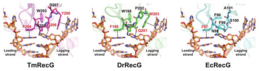
Jeong et al.                                                                                            Role of Conserved Residues in DrRecG

INTRODUCTION                                                          by RecJ; however, in D. radiodurans, RecQ is dispensable
                                                                      (Bentchikou et al., 2010). Different results have been reported
Deinococcus radiodurans is well known for its extreme                 with respect to recQ mutant phenotypes: a recQ mutant
resistance to lethal doses of ionizing radiation (IR) and             strain was reported to be sensitive to γ-irradiation, UV,
many other DNA damaging agents, including mitomycin C                 H2O2, and MMC (Huang et al., 2007), whereas another study
(MMC), UV-C radiation, and desiccation (Slade and Radman,             showed that recQ mutant cells display wild-type resistance
2011). This remarkable resistance is thought to be attributed         to γ-irradiation (Bentchikou et al., 2010). Recently, RecD2
to its highly efficient DNA repair capacity and various anti-         and RecQ proteins from D. radiodurans were reported to
oxidative systems (Lim et al., 2019). In D. radiodurans,              be able to unwind guanine quadruplex (G4) DNA structures
extensive IR-induced DNA double-strand breaks (DSBs),                 (Khairnar et al., 2019; Xue et al., 2020). Instead of RecQ,
which are the most lethal form of DNA damage, can                     in D. radiodurans, the UvrD helicase, which can unwind
be mended within a few hours (Zahradka et al., 2006). The             duplex DNA in both the 3'–5' and 5'–3' directions (Stelter
rapid reconstruction of an intact genome from hundreds                et al., 2013), plays a critical role in DSB repair and
of chromosomal fragments is achieved through extended                 reconstitution of the genome following IR exposure
synthesis-dependent strand annealing (ESDSA), followed by             (Bentchikou et al., 2010). However, the uvrD mutant still
homologous recombination (HR; Zahradka et al., 2006).                 retained significant radio-resistance as compared to a repair-
    ATP-dependent duplex DNA unwinding enzymes, termed                deficient recA mutant strain, suggesting that the redundant
DNA helicases, are prevalent in all kingdoms of life and play         activity of other helicase(s) is responsible for the residual
important roles in the processes of DNA replication, repair,          DNA repair capacity observed (Bentchikou et al., 2010).
recombination, etc. (Brosh and Matson, 2020). The human                   The RecFOR complex loads RecA onto ssDNA substrates,
genome encodes for 31 nonredundant DNA helicases (Umate               thereby resulting in the formation of a RecA nucleoprotein
et al., 2011). Given their fundamental roles in DNA metabolism,       filament that searches for homology and then invades the
mutations in some of these genes are associated with certain          double-stranded homologous DNA (Bentchikou et al., 2010).
human diseases characterized by premature aging and cancer,           RecA-mediated strand invasion creates a D-loop, and primes
including Xeroderma Pigmentosum, Cockayne Syndrome, and               DNA polymerase III (Pol III)- and/or Pol I-dependent DNA
Werner Syndrome (Uchiumi et al., 2015). Since DNA helicase            synthesis (Slade et al., 2009). DNA synthesis proceeds via a
was first discovered in the model bacterium Escherichia coli          migrating D-loop, in which the unwinding of the dsDNA
(Brosh and Matson, 2020), the E. coli helicases have been             template may be mediated by UvrD, RecD2, RecQ, RuvAB,
intensively studied, and their function has been compared             and/or other helicases (Slade and Radman, 2011). The RuvABC
to that of counterparts identified in different organisms. In         system can displace and resolve a four-way DNA intermediate
E. coli, HR initiation follows the RecBCD pathway. The RecBCD         named the Holliday junction (HJ). The RuvAB and RuvC
complex binds to double-stranded DNA (dsDNA) ends and                 proteins catalyze branch migration and the resolution of HJ
unwinds and degrades the DNA by using a combination of                recombination intermediates, respectively (Rocha et al., 2005).
helicase and nuclease activities, which promotes the repair           The D. radiodurans ruvB mutant is modestly sensitive to UV
of DSB (Rocha et al., 2005). D. radiodurans is devoid of              light, γ-irradiation, and MMC (Kitayama et al., 1997). However,
RecB and RecC proteins but possesses a RecD homolog named             the fact that inactivation of both ruv and recG resulted in a
RecD2 (Wang and Julin, 2004). Since RecD2 is present in               more dramatic increase in the sensitivity of cells to DNA
recBC-minus organisms, it is not associated with RecBC                damaging-agents has led to the suggestion that RecG and
(Montague et al., 2009). The D. radiodurans RecD2 protein             RuvABC are part of two overlapping pathways for processing
is a DNA helicase with 5'-3' polarity and low processivity            intermediates in HR and DNA repair (Lloyd and Rudolph,
(Wang and Julin, 2004). recD2 mutants are more sensitive              2016). The deletion of recG resulted in a growth delay and a
than wild type cells to the cytotoxic effect of γ-irradiation,        decrease in the resistance of D. radiodurans to γ-irradiation
UV light, and hydrogen peroxide (H2O2; Servinsky and Julin,           and H2O2 (Wu et al., 2009).
2007; Zhou et al., 2007), but recD2 mutations does not alter              RecG, a monomeric dsDNA translocase that unwinds a
the sensitivity of D. radiodurans to treatment with mitomycin C       variety of branched DNA molecules, such as replication forks,
(MMC), methyl methanesulfonate, and hydroxyurea (Servinsky            HJs, D-, and R-loops (Lloyd and Rudolph, 2016), consists of
and Julin, 2007; Montague et al., 2009). D. radiodurans lacks         an N-terminal wedge domain (WD), two RecA-like helicase
not only RecBC, but also exonuclease I (SbcB), hence DSBs             domains, and a C-terminal translocation in RecG (TRG) motif
in D. radiodurans are repaired by the RecFOR pathway, which           (Fairman-Williams et al., 2010). The WD, which is not found
is significantly more common than RecBCD in bacterial                 in other DNA helicases, provides specificity for binding branched
genomes (Rocha et al., 2005). RecFOR-dependent DSB                    DNA structures (Rudolph et al., 2010). Amino acid sequence
repair is initiated by unwinding duplex DNA, followed by              analysis of WDs of RecGs from Deinococcus species revealed
DNA end resection that degrades the broken ends in the                that a “Gln(Q)-Pro(P)-Trp(W)” residue motif is highly conserved
5'–3' direction to obtain 3' single-stranded DNA (ssDNA)              in Deinococus RecG proteins. In this study, we found that
tails (Rocha et al., 2005).                                           QPW residues contributed to strong binding of RecG to HJ
    In E. coli, RecQ is the major helicase implicated in the          and, consequently, enhanced the ability of RecG to repair DNA
RecFOR pathway and in nucleolytic degradation catalyzed               damage in D. radiodurans.

Frontiers in Genetics | www.frontiersin.org                       2                                  February 2021 | Volume 12 | Article 634615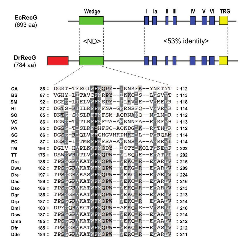
Jeong et al.                                                                                              Role of Conserved Residues in DrRecG

MATERIALS AND METHODS                                                   Construction and Purification of RecG
                                                                        Wedge Domain
Bacterial Strains and Culture Conditions                                A maltose-binding protein (MBP)-RecG-WD fusion was
Deinococus radiodurans R1 (ATCC13939) and its isogenic recG             constructed using the pMAL-c2x vector, in which the E. coli
mutant strains (ΔrecG), which had been previously constructed           malE gene encoding MBP was expressed via an IPTG-inducible
(Jeong et al., 2016), were cultivated at 30°C in TGY broth              promoter. The WD regions of recG genes were amplified
(0.5% tryptone, 0.1% glucose, and 0.3% yeast extract) with              using the DrWD-F/-R and EcWD-F/-R primer sets
aeration or on TGY plates supplemented with 1.5% Bacto-agar.            (Supplementary Table S1) by using D. radiodurans and
The E. coli strain DH5α was used for routine cloning experiments.       E. coli genomic DNA as template, respectively. The PCR products
Escherichia coli strains were grown at 37°C in Luria-Bertani            were digested with EcoRI and HindIII and cloned into pMAL-c2x
(LB) medium or on LB plates solidified with 1.5% Bacto-agar.            for purification of the wild-type WDs, named DrRecG-WDWT
Antibiotics were added to the medium if necessary: ampicillin,          and EcRrecG-WDWT, respectively. To generate mutant RecG-
100 μg/ml (E. coli), and chloramphenicol, 3 μg/ml (D.                   WDs, DrRecG-WDFSA, and EcRecG-WDQPW, the WD regions
radiodurans).                                                           were amplified from plasmids pDrRecGFSA and pEcRecGQPw,
                                                                        respectively, and then cloned into pMAL-c2x as described
                                                                        above. The resulting plasmids were transformed into E. coli
Plasmid Construction                                                    strain BL21 (DE3) for purification of the RecG-WDs fused to
The pRADZ3 shuttle vector, which functions in E. coli as                MBP. Cells were grown in 500 ml of LB broth at 37°C. IPTG
well as in D. radiodurans, contains the groEL promoter for              (0.1 mM) was added to the culture when the cells reached
constitutive gene expression (Jeong et al., 2016). Complete             an optical density at 600 nm (OD600) of 0.5 and were further
recG coding sequences were PCR-amplified from genomic                   grown at 16°C overnight. The cells were harvested by
DNA of D. radiodurans R1 and E. coli MG1655 by using                    centrifugation and resuspended in buffer A (20 mM Tris-HCl,
DR1916F/R and B3652F/R primer pairs, respectively, carrying             pH 7.5; 0.2 M NaCl; and 1 mM EDTA, pH 8.0). Following
the SpeI and BamHI restriction sites. The PCR products                  sonication on ice, cell debris was removed by centrifugation,
were cloned into pRADZ3 at the SpeI and BamHI sites to                  and the supernatant was loaded onto a 5 ml amylose column.
generate the plasmids pDrRecG and pEcRecG, respectively.                The column was washed with five column volumes of buffer A.
These plasmids were transformed into ΔrecG for                          The MBP-RecG-WD fusion protein was eluted with 5 ml of
complementation studies. For transformation, D. radiodurans             buffer B (buffer A containing 10 mM maltose). Following 10%
cells from exponentially growing cultures were collected by             SDS PAGE, fractions containing the approximately 51-kDa
centrifugation and concentrated 50-fold in TGY supplemented             fusion protein were collected and concentrated by ultrafiltration
with 30 mM CaCl2. The cell mixture (100 μl) containing                  using Amicon® Ultra Filters (Merck Millipore, Darmstadt,
the constructed plasmid DNAs was held on ice for 30 min                 Germany). Protein concentrations were determined using the
and then incubated at 32°C for 90 min. The transformation               Bradford protein assay with bovine serum albumin as
mixture was diluted 10-fold with TGY broth and incubated                the standard.
at 30°C for 5 h with aeration, prior to being plated on
drug-selective agar. The partial nucleotide sequence of recG
encoding DrRecG lacking the N-terminal region (residues                 DNA Binding Assay
1–99) was PCR-amplified using DR1916-ΔNF/-ΔNR primer                    To create the HJ DNA substrate, four complementary
pairs and cloned into pRADZ3 to generate pDrRecGΔN99 as                 oligonucleotides (oligo 1, 5'-GTCGGATCCTCTAGACAGCT
described above. To produce RecG mutant proteins, EcRecG                CCATGATCACTGGCACTGGTAGAATTCGGC-3'; oligo 2,
with QPW instead of 99FSA101 and DrRecG with FSA instead                5'-CAACGTCATAGACGATT ACATTGCTACATGGAGCTGTC
of 201QPW203, mutagenesis was carried out using fusion PCR              TAGAGGATCCGA-3'; oligo 3, 5'-TGCCGAATTCTACCAGT
techniques. Complementary primer pairs, EcQPW-F/-R and                  GCCAGTGATGGACATCTTTGCCACGTTGACCC-3'; oligo 4,
DrFSA-F/-R, were designed to contain the desired mutations              5'-TGGGTCAACGTGGGCAAGATGTCCTAGCAATGTAATC
in the middle of the primers. To introduce the triple point             GTCTATGACGT-3') were synthesized as described previously
mutation “QPW” into EcRecG, in the first step, two fragments            (McGlynn et al., 2000). The oligonucleotides were denatured
were amplified using the primer pairs B3652F/EcQPW-R                    for 15 min at 95°C and allowed to anneal at 25°C for 45 min.
and EcQPW-F/B3652R, respectively. In the second step, the               To examine the DNA binding activity of RecG-WD, an
two PCR products were annealed at their overlapping                     electrophoretic mobility shift assay kit (Invitrogen, Carlsbad,
homologous regions and were amplified by the 3652F/R                    CA, United States) was used. Purified recombinant RecG-WD
primer pair. The fusion PCR product was cloned into pRADZ3              proteins and 100 nM of HJ DNA substrates were mixed in
to generate pEcRecGQPW as described above. The plasmid                  binding buffer containing 50 mM Tris-HCl (pH 8.0), 5 mM
pDrRecGFSA was constructed using primer pairs DR1916F/                  EDTA, 1 mM dithiothreitol, 100 μg/ml bovine serum albumin,
DrFSA-R and DrFSA-F/DR1916R. The plasmid constructs                     and 6% glycerol (v/v) and incubated on ice for 15 min as
were transformed into ΔrecG. The plasmids were verified                 described previously (McGlynn et al., 1997; Briggs et al., 2005).
by DNA sequencing. Primers used in this study are listed                The DNA binding reaction was terminated by the addition of
in Supplementary Table S1.                                              2 μl of 2× loading dye. Samples were loaded onto 6%

Frontiers in Genetics | www.frontiersin.org                         3                                  February 2021 | Volume 12 | Article 634615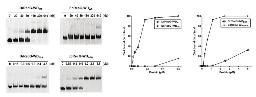
Jeong et al.                                                                                              Role of Conserved Residues in DrRecG

polyacrylamide gels in low-ionic strength buffer at 120 V for            RESULTS
35 min at room temperature, and the electrophoresed
DNA-protein complexes were visualized using a ChemiDoc™                  QPW Residues Are Highly Conserved in
Touch imaging system (Bio-Rad, Hercules, CA, United States).             the WD of Deinococcal RecG Proteins
Reaction products were quantified using Image Lab Software               In general, RecG proteins largely consist of an N-terminal
(Bio-Rad).                                                               WD critical to DNA binding, helicase core domains, including
                                                                         seven motifs common to DNA helicases, and the signature
Growth and Survival Assays                                               RecG TRG motif (Fairman-Williams et al., 2010). Compared
A stationary-phase culture that had grown overnight was used             to the E. coli RecG protein (EcRecG), the D. radiodurans
as a seed culture. The seed culture was inoculated into 25 ml            RecG protein (DrRecG) has an extended region with 99 amino
of TGY broth at 1:100 dilution, and cell growth was monitored            acids at the N-terminus (Figure 1A). When the C-terminal
at OD600 by using a spectrophotometer. For survival studies,             regions containing the helicase domain and TRG motif are
cells grown to log phase (OD600 ≈ 1.0, corresponding to                  considered, sequence identity between the two proteins
~108 CFU/ml) in TGY broth were adjusted to OD600 ≈ 0.1                   approaches 53%, whereas there is no significant similarity in
with TGY. After γ-irradiation treatment, cells were serially             the WD (Figure 1A; Supplementary Figure S1). The crystal
diluted 10-fold in saline (0.85% NaCl), spotted onto TGY plates,         structure of RecG from Thermatoga maritima (TmRecG)
and incubated at 30°C for 3 days to allow colony formation.              revealed that Phe204 and Tyr208 in its WD are important for
                                                                         the binding of branched DNA molecules (Singleton et al.,
                                                                         2001). Multiple sequence alignment of the WD showed that
Quantitative Real-Time PCR Assay
                                                                         the phenylalanine residue was highly conserved, and the
A 5 ml culture grown to mid-log phase was harvested by
                                                                         tyrosine residue was replaced with another aromatic amino
centrifugation. RNA preparation and cDNA synthesis were
                                                                         acid, tryptophan, in all deinococcal RecG proteins (Figure 1B;
performed as described previously (Jeong et al., 2016). The
                                                                         Supplementary Figure S2). Interestingly, glutamine (Q), proline
quantitative real-time PCR (qRT-PCR) was performed on a
                                                                         (P), and tryptophan (W) at positions 201, 202, and 203,
CFX Connect real-time PCR system (Bio-Rad) using SYBR
                                                                         respectively (D. radiodurans numbering) were found in all
Premix Ex Taq (Takara Bio Inc., Otsu, Japan). PCR reactions
                                                                         deinococcal RecGs (Supplementary Figure S2).
were performed as follows one cycle of 95°C for 5 min and
then 40 cycles of 95°C for 10 s and 60°C for 15 s. The
housekeeping gene dr1343 encoding glyceraldehyde-3-phosphate             Comparative Modeling of RecG Proteins
dehydrogenase was used as an internal control. Primers used              To gain insight into the forked-DNA binding mode of
in qRT-PCR are denoted with the prefix “RT-” in                          DrRecG, we generated structural models of DrRecG and
Supplementary Table S1.                                                  EcRecG with SWISS-MODEL (Biasini et al., 2014) by using
                                                                         the crystal structure of TmRecG (PDB id: 1gm5) as a template.
Pulsed Field Gel Electrophoresis                                         TmRecG (780 amino acids) shares 36.5 and 32.3% sequence
Irradiated (6 kGy) and unirradiated cells were diluted in 50 ml          identities with DrRecG (784 amino acids) and EcRecG (693
TGY broth to an OD600 of 0.25, and incubated at 30°C. At                 amino acids), respectively. We predicted structural models
the indicated times, 1.5, 3, 4.5, 6, and 8 h after re-inoculation,       for DrRecG comprising of 735 amino acid residues (10–744)
cells (~ 5 × 10⁸ CFU/ml) were harvested and washed with                  and EcRecG comprising of 635 amino acids (6–660),
10 mM phosphate buffer (pH 7.0). The cells were resuspended              respectively. The modeled structures were validated by using
in 0.125 M EDTA (pH 8.0) and mixed with low-melt agarose                 QMEAN and ProSA (Supplementary Figure S3) and were
(Bio-Rad) to obtain a final concentration of 0.8% agarose. The           superimposed onto the TmRecG-DNA complex structure.
DNA plugs were incubated overnight at 37°C in 0.05 M EDTA                Our analyses suggested that Phe199 and Trp203 in DrRecG,
(pH 7.5) containing 1 mg/ml lysozyme, placed in proteinase               which are likely to be equivalent to Phe204 and Tyr208,
K solution at 50°C for 16 h, and washed once with 1 ×                    respectively, in TmRecG, may have roles in the stabilization
washing buffer containing 1 mM phenylmethylsulfonyl fluoride             of the leading and lagging strand templates by base stacking
(PMSF) and then three times with 1× washing buffer. The                  interactions between nucleotide bases and aromatic side
prepared DNA plugs were digested with 30 U of NotI restriction           chains, whereas Gln201 corresponding to Gln206 in TmRecG
enzyme overnight at 37°C and then subjected to pulsed field              could interact with nucleotide bases by electrostatic
gel electrophoresis (PFGE). DNA fragments were separated on              interactions, thereby participating in the binding of DrRecG
1% (wt/vol) agarose gels in 0.5 × TBE. Electrophoresis was               to DNA (Figure 2). In the case of EcRecG, only Phe97,
performed using a CHEF-Mapper apparatus (Bio-Rad) under                  corresponding to Phe204 in TmRecG, seemed to directly
the following conditions: pulse ramping from 10 to 60 s, angle           interact with a DNA base (Figure 2). Phe96 in EcRecG is
of 120°, current of 6 V/cm, and 22 h run time at 12°C. Gels              also required for interaction with branched DNA molecules
were stained with 0.5 × TBE containing 0.5 μg/ml ethidium                because it may have a stabilizing effect similar to that of
bromide for 30 min, destained for 10 min in deionized water,             Trp203 in TmRecG, which is partially buried in WD hydrophobic
and then visualized using a ChemiDoc™ Touch imaging system               core and preserves WD conformation as a whole (Briggs
(Bio-Rad).                                                               et al., 2005). Phe96 in EcRecG, Trp203 in TmRecG, and Trp198

Frontiers in Genetics | www.frontiersin.org                          4                                 February 2021 | Volume 12 | Article 634615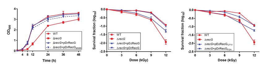
Jeong et al.                                                                                                                          Role of Conserved Residues in DrRecG

                                         A

                                         B

  FIGURE 1 | Alignment of Escherichia coli RecG (EcRecG) and Deinococcus radiodurans RecG (DrRecG). (A) Schematic comparison of the domain structures of EcRecG
  and DrRecG. EcRecG and DrRecG are aligned via wedge (green), helicase (blue), and TRG (yellow) motifs. The N-terminal extended region of DrRecG is shown as a red
  box. The percentages of conservation of amino acid sequences between EcRecG and DrRecG are indicated for wedge domain (ND, not determined) and helicase and
  TRG motifs (53% identity). (B) Multiple alignment of amino acid sequences containing the conserved phenylalanine (F) residue within the wedge domain. The program
  Genedoc (www.psc.edu/biomed/genedoc) was used to visualize the alignment in quantify mode, which highlights residues most-frequently found in each column of the
  alignment. Gaps introduced to maximize alignment are indicated by dash. Black and white letters on gray shading represent ≥60 and ≥80% identity, respectively. White
  letters on black shading represent 100% identity. Wedge domain sequences were obtained from RecGs of Clostridium acetobutylicum, CA; Bacillus subtilis, BS;
  Streptobacillus moniliformis, SM; Haemophilus influenza, HI; Shewanella oneidensis, SO; Vibrio cholera, VC; Pseudomonas aeruginosa, PA; Streptomyces griseus, SG;
  Escherichia coli, EC; Thermatoga maritima, TM; Thermus thermophilus, TT; D. radiodurans, Dra; D. wulumuqiensis, Dwu; D. reticulitermitis, Dre; D. actinosclerus, Dac; D.
  soli, Dso; D. grandis, Dgr; D. radiopugnans, Drp; D. maricopensis, Dmi; D. swuensis, Dsw; D. marmoris, Dma; D. frigensis, Dfr; and D. deserti, Dde.

  FIGURE 2 | Structural model of RecG wedge domains in complex with a partial replication fork. The modeled wedge domains of RecG from Thermatoga maritima
  (TmRecG), DrRecG, and EcRecG are shown in purple, green, and cyan, respectively. DNA molecules are displayed in an orange stick model. Nitrogen, oxygen, and
  phosphorous atoms are colored in blue, red, and orange, respectively.

in DrRecG, which is equivalent to Trp203 in TmRecG, are                                   fit well with the crystal structure of TmRecG. It can thus
located in the same position (Figure 2). Taken together,                                  be assumed that QPW residues may play important roles
our homology models obtained for DrRecG and EcRecG                                        in the DNA binding of deinococcal RecGs.

Frontiers in Genetics | www.frontiersin.org                                           5                                           February 2021 | Volume 12 | Article 634615
Jeong et al. Role of Conserved Residues in DrRecG
QPW Residues Enhance RecG DNA cell growth defects (Wu et al., 2009). Thus, we first monitored
Binding Activity the growth of the wild-type D. radiodurans strain (WT) and
The isolated WD can bind to HJ structures, although its affinity ΔrecG by measuring their OD600 values over time. ΔrecG exhibited
is lower than that of full-length RecG (Briggs et al., 2005). To delayed growth relative to WT, and expression of the native
test whether the QPW residues could affect the DNA binding DrRecG protein (pDrRecG) was able to complement the growth
activity of RecG, we cloned partial recG gene fragments encoding defects. Interestingly, the growth of ΔrecG harboring pDrRecGΔN99
the WD into the expression vector pMAL-c2x to produce an was comparable to that of ΔrecG harboring pDrRecG (Figure 4A).
MBP-RecG-WD fusion protein. The constructs encompassed In addition, the resistance of ΔrecG to γ-irradiation was fully
residues 59–135 of EcRecG, and 158–235 of DrRecG restored to the levels of WT by DrRecGΔN99 provided in trans
(Supplementary Figure S1). We also constructed mutant versions (data not shown). These observations indicate that the N-terminal
of RecG-WD by replacing the QPW of DrRecG with FSA of extended region is not involved in the ΔrecG phenotypes, growth
EcRecG corresponding to QPW in DrRecG (Figure 1B), and defect, and increased γ-irradiation sensitivity, under the experimental
vice versa. DNA binding assays involving the four different kinds conditions tested here.
of WDs, EcRecG-WDWT, EcRecG-WDQPW, DrRecG-WDWT, and
DrRecG-WDFSA, were performed using HJ structures formed by The QPW Motif Increases RecG DNA
annealing four complementary oligonucleotides, as previously Repair Capacity
described (McGlynn et al., 2000). As shown in Figure 3, DrRecG- Because the N-terminal extended region was not essential for
WDWT almost reached a saturation of the total DNA binding DrRecG activity (Figure 4A), and EcRecG displays a high
at 160 nM protein, whereas EcRecG-WDWT did not even at degree of sequence conservation with DrRecG in the helicase
higher concentrations (to 640 nM). The substitution of QPW and TRG motifs (Supplementary Figure S1), we introduced
with FSA (DrRecG-WDFSA) dramatically reduced binding ability, the pEcRecG plasmid carrying the E. coli recG gene into ΔrecG
but EcRecG-WDQPW exhibited a significant increase compared cells and measured cell survival rates following γ-irradiation
to EcRecG-WDWT (Figure 3). These results clearly indicated to determine if EcRecG can functionally replace DrRecG.
that the QPW signature motif in deinococcal RecGs plays a Compared to WT, ΔrecG showed approximately 0.25-, 0.5-,
role in enhancing the DNA-binding affinity of RecG. and 1-log reductions in survival at 6, 9, and 12 kGy of
γ-irradiation, respectively, and these reductions were completely
N-Terminal Extended Region Deletion restored by DrRecG. However, EcRecG was able to partially
Does Not Affect DrRecG Activity restore the γ-irradiation-sensitive phenotype of ΔrecG only at
Deinococus radiodurans RecG has an extended region, with 99 12 kGy of γ-irradiation (Figure 4B). To test the functional
amino acids in the N-terminus compared to EcRecG (Figure 1A). role of the QPW residues, we constructed pEcRecGQPW encoding
We constructed plasmid pDrRecGΔN99 expressing the N-terminal an EcRecG mutant in which 99FSA101 was substituted with
deletion mutant (residues 100–784) of DrRecG, with Ala100 converted QPW, and pDrRecGFSA encoding a DrRecG mutant in which
to methionine, and transformed it into a DrRecG mutant strain 201
QPW203 was substituted with FSA. The resulting plasmids
(ΔrecG). Disruption of recG is known to result in D. radiodurans were transformed into ΔrecG cells, which were then exposed
A B
FIGURE 3 | DNA binding activity of RecG wedge domains (RecG-WDs). (A) Binding affinities of RecG-WDs as measured in band shift assays with Holiday junction
(HJ) substrate. Native RecG-WDs, DrRecG-WDWT and EcRecG-WDWT from D. radiodurans and E. coli, respectively, and their mutants, DrRecG-WDFSA and EcRecG-
WDQPW were used in this assay. Reactions contained 100 nM HJ DNA and native RecG-WDs or mutant RecG-WDs at as shown. (B) Quantification of the binding
activity of RecG-WDs. The formation of RecG-WD complexes with HJ in (A) was quantified and plotted as a function of increasing concentration of RecG-WDs.
Frontiers in Genetics | www.frontiersin.org 6 February 2021 | Volume 12 | Article 634615Jeong et al. Role of Conserved Residues in DrRecG
A B C
FIGURE 4 | Growth and survival assays of the D. radiodurans recG mutant strain (ΔrecG). (A) Growth curves of D. radiodurans strains. Optical density (OD600)
measurements were employed to estimate the growth of D. radiodurans R1 (WT), ΔrecG, and ΔrecG harboring the plasmids pDrRecG and pDrRecGΔN99, which encode
the full-length DrRecG and the N-terminal truncation mutant of DrRecG, respectively. Survival curves for ΔrecG with pEcRecG and pDrRecG (B) and with pEcRecGQPW and
pDrRecGFSA (C). Cells grown to log phase were exposed to γ-irradiation and spotted onto TGY plates. The survival fraction was calculated by dividing the colony-forming
units (CFUs) of γ-irradiation-treated cells by the CFUs of unirradiated cells. The error bars represent the SD of three independent experiments conducted in duplicate (n = 3).
to γ-irradiation. There was no significant difference in recG uracil from DNA molecules, formed as a result of cytosine
mRNA levels between ΔrecG harboring pDrRecG and pDrRecGFSA deamination, and represents part of the base-excision repair
(Supplementary Figure S4), but resistance to γ-irradiation was pathway. The uracil-DNA N-glycosylase DrUNG (DR_0689),
not recovered by DrRecGFSA (Figure 4C). In contrast, EcRecGQPW the main UNG in D. radiodurans, possesses a much higher
restored the ΔrecG survival in a similar way as DrRecG, catalytic efficiency than human UNG, which is attributed to
although the restoration levels were somewhat different at the high substrate affinity caused by an increased number of
9 kGy (Figures 4B,C). These results showed that substituting positively charged residues close to the DNA binding site
the three amino acids in the WD between EcRecG and DrRecG (Pedersen et al., 2015). Mismatch-specific UNG (MUG) also
is sufficient to exchange their abilities to complement the loss- removes uracil from DNA. The overall structure of DrMUG
of-function phenotype of ΔrecG after γ-irradiation. is similar to that of EcMUG, but DrMUG possesses a novel
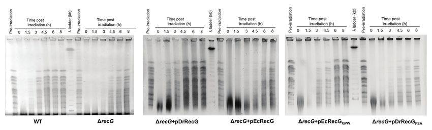
To investigate the role of DrRecG in DSB, the DNA repair catalytic residue (Asp-93) that can provide DrMUG with broad
kinetics of cells subjected to γ-irradiation were examined using substrate specificity (Moe et al., 2006). The novel catalytic
PFGE. DNA fragmented by γ-irradiation was reconstructed within residue identified in DrMUG is conserved in other deinococcal
3 h in WT, whereas ΔrecG was able to compete the repair of MUG proteins (Lim et al., 2019). The D. radiodurans X family
shattered genomes 4.5 h after γ-irradiation (Figure 5). ΔrecG DNA polymerase (DrPolX), which not only has polymerase
harboring pDrRecG and pEcRecG showed repair kinetics similar activity, but also exerts strong Mn2+-dependent 3'→5' exonuclease
to those observed in WT and ΔrecG, respectively. Compared activity affects DSB repair efficiency in D. radiodurans (Blasius
to ΔrecG with pEcRecGQPW, the process of genome reassembly et al., 2006). At the active site of the polymerase catalytic
was delayed by 1.5 h in ΔrecG with pDrRecGFSA (Figure 5). domain, the “DXD” motif conserved in almost all pol X-family
The similar patterns of results shown in survival assays and members is replaced by an “AAE” motif in D. radiodurans
PFGE analysis, which are caused by the reciprocal swapping of (Leulliot et al., 2009; Bienstock et al., 2014). RecA is a critical
the three amino acid motifs between DrRecG and EcRecG, enzyme in HR for DSB repair. Unlike canonical RecAs, DrRecA
strongly suggest that the QPW residues enhance DNA-binding first forms a filament on dsDNA and then takes up a homologous
activity, thereby leading to efficient DNA repair in D. radiodurans. single strand to initiate DNA strand exchange, which is the
exact inverse of the major pathway seen with other proteins
of the RecA family (Kim and Cox, 2002). Although the overall
DISCUSSION fold of DrRecA is similar to EcRecA, there are a few key
amino acid changes in the DrRecA C-terminal domain that
Analysis of the genome sequence of D. radiodurans identifies interacts with dsDNA. One of the key differences, Phe303 in
the typical complement of prokaryotic DNA repair proteins, DrRecA, equivalent to Trp290 in EcRecA, would seem to be a
suggesting the possibility that D. radiodurans uses the same likely candidate in dictating a possible specificity for binding
DNA-repair strategies as other prokaryotes, but it does so in to dsDNA (Rajan and Bell, 2004). It has also been observed
a manner that is somehow much more effective than that that a single amino acid mutation at the C-terminus of EcRecA
observed in other species to retain the extraordinary tolerance alters its activity. The RecA variants found in radioresistant
to DNA damage (Battista et al., 1999; White et al., 1999). A E. coli strain CB2000 have mutations at residue 276 (D276A
series of biochemical and structural analyses of D. radiodurans and D276N), which increase the rates of filament nucleation
proteins involved in DNA repair systems have revealed unusual on DNA, and promote DNA strand exchange more efficiently
features distinguishing them from their counterparts in other than wild-type RecA proteins (Piechura et al., 2015). These
prokaryotic species. Uracil-DNA glycosylase (UNG) removes studies imply that DNA repair proteins with altered substrate
Frontiers in Genetics | www.frontiersin.org 7 February 2021 | Volume 12 | Article 634615Jeong et al. Role of Conserved Residues in DrRecG FIGURE 5 | DNA repair in ΔrecG and ΔrecG cells harboring plasmids encoding the native (pDrRecG and pEcRecG) and mutant (pDrRecGFSA and pEcRecGQPW) RecG proteins. Exponentially growing cells were exposed to 6 kGy of γ-irradiation and then recovered in TGY liquid media. At the indicated times (0–8 h), samples were removed, and genomic DNA was isolated. The amount of DNA double-strand breaks (DSBs) was analyzed by pulsed-field gel electrophoresis (PFGE). Pre-irradiation indicates unirradiated cells. specificity and enhanced catalytic activity, which might and EcRecG models suggested that the conserved QPW be attributed to unique amino acid residues, likely contribute residues in the WD confer greater RecG-DNA interaction to the improved DNA repair capacity of D. radiodurans to compared to FSA because of the additional charge interactions survive DNA damage caused by γ-irradiation. In this aspect, and base packing through Gln and Trp, respectively (Figure 2; the highly conserved QPW residues in the WD can be assumed Supplementary Figure S5). Indeed, DrRecG-WDWT and to affect the binding activity of deinococcal RecGs. EcRecG-WDQPW exhibited enhanced affinities for HJ compared The WD found in the N-terminal region of RecG is a to EcRecG-WDWT and DrRecG-WDFSA (Figure 3). Full-length DNA-binding domain that has an oligonucleotide/ DrRecG and EcRecGQPW restored resistance to γ-irradiation oligosaccharide binding-fold (OB-fold) motif ranging between and DNA repair capacity of ΔrecG to that of WT (Figures 4, 5). 70 and 150 amino acids in length (Theobald et al., 2003). Also noteworthy is the N-terminal extended region of DrRecG The phenylalanine residue, which is critical for RecG binding (Figure 1A). This feature is shared with RecG proteins from of branched DNA molecules, is conserved in the WDs (Briggs thermophilic bacteria, Thermotoga maritima and Aquifex aeolicus, et al., 2005; Figure 1B). This residue is regarded to stabilize and a number of cyanobacteria, and is longer than in many the orphan base of the leading strand template by base other RecG proteins such as EcRecG (Wen et al., 2005). RecG stacking, effectively capping the parental duplex (Singleton from the extreme thermophile A. aeolicus unwinds DNA well et al., 2001; Figure 2). In Mycobacterium tuberculosis RecG, at high temperature (60°C), whereas the N-terminal extension substitution of Phe99 corresponding to Phe204 in TmRecG present in this protein is dispensable for activity and thermo- to Ala was seen to significantly lower the DNA binding stability (Wen et al., 2005). In D. radiodurans, the N-/C-terminal activity compared to that of wild-type RecG protein (Zegeye extension (or variation) is found not only in RecG, but also et al., 2014). In this study, protein structure modeling analysis in other helicases, including RecD2 and RecQ. DrRecD2 contains showed that two other amino acids, Gln206 and Tyr208 in an additional N-terminal region of approximately 200 amino TmRecG and Gln201 and Trp203 in DrRecG, interacted with acids, which is longer than the corresponding feature in E. coli the nucleotide bases of the leading and lagging forked-DNA RecD protein (RecD1; Montague et al., 2009). In contrast to strands, together with the key residues, Phe204 in TmRecG, most other RecQ proteins composed of the helicase, RecQ-C- and Phe199 in DrRecG, respectively (Figure 2). The aromatic terminal (RQC), and helicase-and-RNaseD-like-C terminal side-chain of Tyr participates in DNA binding of the WD (HRDC) domains, DrRecQ contains three tandem HRDC domains through hydrophobic interactions such as base stacking in the C-terminus (Killoran and Keck, 2006). Full-length DrRecD2 (Singleton et al., 2001). Tyr was not observed in DrRecG, restored the H2O2-resistant phenotype of recD2 mutants, whereas but it was substituted with an equivalent aromatic residue, the N-terminal truncation mutant of DrRecD2 could not, Trp (Figure 2), suggesting that Trp203 may play a similar suggesting that the N-terminal domain is necessary for the role role in the stabilization of the lagging DNA strand duplex. of DrRecD2 in antioxidant pathways (Zhou et al., 2007). The Of the 79 deinococcal RecGs analyzed in this study, only three tandem HRDC domains increase the efficiency of DNA two species Deinococcus ruber and Deinococcus aquiradiocola, unwinding and ATPase activities of DrRecQ (Huang et al., 2007), have QAW instead of the signature QPW residues and could work together with other functional motifs to enhance (Supplementary Figure S2). However, it is noteworthy that DrRecQ interactions with DNA (Liu et al., 2013). Considering Q and W are strictly conserved. EcRecG does not have a that ΔrecG cells are sensitive to γ-irradiation and H2O2 (Wu signature motif with an aromatic residue in the corresponding et al., 2009; Figure 4), and DrRecG is also involved in catalase position (99FSA101 in EcRecG). Comparisons of the DrRecG gene katE1 regulation (Jeong et al., 2016), further research is Frontiers in Genetics | www.frontiersin.org 8 February 2021 | Volume 12 | Article 634615
Jeong et al.                                                                                                                          Role of Conserved Residues in DrRecG

needed to determine the role of the additional DrRecG N-terminal                           Thermus RecG proteins have either QPW or QTW
region, although it seems not to be implicated in DNA repair                               (Supplementary Figure S6), and the dimeric SSBs like DrSSB
under the experimental conditions tested in this study.                                    are discovered in the Deinococcus-Thermus group (Zhang
    RecG provides a more general defense against pathological                              et al., 2014), supporting that RecG and SSB might
DNA replication, e.g., the rescue of stalled or damaged                                    be evolutionarily related. T. thermophilus is a thermophile,
replication forks, and is associated with a number of additional                           which is relatively sensitive to IR, whereas D. radiodurans is
cellular processes (Rudolph et al., 2010; Azeroglu et al., 2016;                           a mesophile, which is highly IR-resistant (Omelchenko et al.,
Lloyd and Rudolph, 2016; Romero et al., 2020). Recently, it                                2005). However, considering that the mechanisms employed
has become increasingly clear that RecG is an enzyme                                       by thermophiles to overcome DNA damage by high temperature
responsible for regression of stalled DNA replication forks                                may also be employed in repair of damage caused by IR
that can be induced by various types of lesions, including                                 (Ranawat and Rawat, 2017), the coevolution between RecG
single-strand breaks and DSB. RecG is directly loaded onto                                 and SSB, which likely occurred in the Deinococcus-Thermus
the forks by binding to ssDNA-binding protein (SSB) and                                    group, may contribute to the enhanced abilities of the
reverses the stalled forks, leading to the formation of HJ-type                            Deinococcus and Thermus lineages to survive different kinds
structures. PriA helicase then targets the repaired fork to                                of environmental stresses. Further research is warranted to
reload and assemble a DNA replication machinery (the                                       investigate the effects of QPW on RecG-SSB interactions.
replisome complex), enabling DNA replication to restart (Lloyd
and Rudolph, 2016; Bianco and Lyubchenko, 2017). During
HR for DSB repair, RecG is proposed to re-model branched                                   DATA AVAILABILITY STATEMENT
intermediates of recombination to direct the correct binding
of PriA and subsequent DNA synthesis (Azeroglu et al., 2016).                              The original contributions presented in the study are included
These studies indicate that SSB-mediated RecG loading onto                                 in the article/Supplementary Material, further inquiries can
DNA plays an important role in facilitating stalled replication                            be directed to the corresponding author.
fork rescue. D. radiodurans SSB (DrSSB) is different from
that of the prototype E. coli SSB (EcSSB), in that DrSSB
(301 amino acids) contains two OB folds per monomer, and                                   AUTHOR CONTRIBUTIONS
functions as homodimers, in contrast to the homotetrameric
EcSSB (178 amino acids), with each monomer encoding a                                      S-WJ performed the experiments and wrote a first draft. M-KK
single OB fold (Bernstein et al., 2004). In E. coli, SSB-RecG                              performed the comparative protein structure modeling and
interactions occur via the WD of RecG and the PXXP motifs                                  revised the first draft. LZ and S-KY constructed the plasmids
within the SSB linker domain. In particular, the SSB binding                               and performed the survival assays. M-KK, J-HJ, and H-ML
site on RecG overlaps the residues of the binding site for                                 guided the experiments and interpreted the results. SL conceived
the leading strand arm of the fork in RecG (Ding et al.,                                   the study and was in charge of overall direction and planning.
2020). Thus, it is likely that these different features do not                             All authors reviewed and edited the manuscript, and approved
enable DrSSB to deliver EcRecG to the fork with an efficiency                              the final version.
equal to that of DrRecG, which may explain why EcRecG
does not confer resistance to ΔrecG (Figure 4B). EcRecG
also partially complements Helicobacter pylori recG mutants                                FUNDING
but not to the same extent as the H. pylori RecG protein,
suggesting that the host context appears to be critical in                                 This research was supported by the Nuclear R&D program of
defining the function of RecG (Kang et al., 2004). In this                                 the Ministry of Science and ICT (MSIT), South Korea.
aspect, the QPW residues found in the WD of DrRecG might be
interpreted as the result of evolutionary adaptation to cooperate
with the unique DrSSB. D. radiodurans is most closely related                              SUPPLEMENTARY MATERIAL
to Thermus thermophilus, and the two genera Deinococcus
and Thermus belong to a distinct bacterial clade called the                                The Supplementary Material for this article can be found online
Deinococcus-Thermus group (Omelchenko et al., 2005).                                       at: https://www.frontiersin.org/articles/10.3389/fgene.2021.6346
Regarding RecG and SSB, they share common features.                                        15/full#supplementary-material

REFERENCES                                                                                 Bentchikou, E., Servant, P., Coste, G., and Sommer, S. (2010). A major role of the
                                                                                              RecFOR pathway in DNA double-strand-break repair through ESDSA in Deinococcus
Azeroglu, B., Mawer, J. S., Cockram, C. A., White, M. A., Hasan, A. M.,                       radiodurans. PLoS Genet. 6:e1000774. doi: 10.1371/journal.pgen.1000774
   Filatenkova, M., et al. (2016). RecG directs DNA synthesis during double-               Bernstein, D. A., Eggington, J. M., Killoran, M. P., Misic, A. M., Cox, M. M.,
   strand break repair. PLoS Genet. 12:e1005799. doi: 10.1371/journal.pgen.1005799            and Keck, J. L. (2004). Crystal structure of the Deinococcus radiodurans
Battista, J. R., Earl, A. M., and Park, M. J. (1999). Why is Deinococcus radiodurans          single-stranded DNA-binding protein suggests a mechanism for coping with
   so resistant to ionizing radiation? Trends Microbiol. 7, 362–365. doi: 10.1016/            DNA damage. Proc. Natl. Acad. Sci. U. S. A. 101, 8575–8580. doi: 10.1073/
   S0966-842X(99)01566-8                                                                      pnas.0401331101

Frontiers in Genetics | www.frontiersin.org                                            9                                           February 2021 | Volume 12 | Article 634615
Jeong et al. Role of Conserved Residues in DrRecG
Bianco, P. R., and Lyubchenko, Y. L. (2017). SSB and the RecG DNA helicase: Moe, E., Leiros, I., Smalås, A. O., and McSweeney, S. (2006). The crystal
an intimate association to rescue a stalled replication fork. Protein Sci. 26, structure of mismatch-specific uracil-DNA glycosylase (MUG) from Deinococcus
638–649. doi: 10.1002/pro.3114 radiodurans reveals a novel catalytic residue and broad substrate specificity.
Biasini, M., Bienert, S., Waterhouse, A., Arnold, K., Studer, G., Schmidt, T., J. Biol. Chem. 281, 569–577. doi: 10.1074/jbc.M508032200
et al. (2014). SWISS-MODEL: modelling protein tertiary and quaternary Montague, M., Barnes, D., Smith, H. O., Chuang, R. -Y., and Vashee, S. (2009).
structure using evolutionary information. Nucleic Acids Res. 42, W252–W258. The evolution of RecD outside of the RecBCD complex. J. Mol. Evol. 69,
doi: 10.1093/nar/gku340 360–371. doi: 10.1007/s00239-009-9290-x
Bienstock, R. J., Beard, W. A., and Wilson, S. H. (2014). Phylogenetic analysis Omelchenko, M. V., Wolf, Y. I., Gaidamakova, E. K., Matrosova, V. Y., Vasilenko, A.,
and evolutionary origins of DNA polymerase X-family members. DNA Repair Zhai, M., et al. (2005). Comparative genomics of Thermus thermophilus and
22, 77–88. doi: 10.1016/j.dnarep.2014.07.003 Deinococcus radiodurans: divergent routes of adaptation to thermophily and
Blasius, M., Shevelev, I., Jolivet, E., Sommer, S., and Hübscher, U. (2006). DNA radiation resistance. BMC Evol. Biol. 5:57. doi: 10.1186/1471-2148-5-57
polymerase X from Deinococcus radiodurans possesses a structure-modulated Pedersen, H. L., Johnson, K. A., McVey, C. E., Leiros, I., and Moe, E. (2015).
3′→5′ exonuclease activity involved in radioresistance. Mol. Microbiol. 60, Structure determination of uracil-DNA N-glycosylase from Deinococcus
165–176. doi: 10.1111/j.1365-2958.2006.05077.x radiodurans in complex with DNA. Acta Crystallogr. D Biol. Crystallogr. 71,
Briggs, G. S., Mahdi, A. A., Wen, Q., and Lloyd, R. G. (2005). DNA binding 2137–2149. doi: 10.1107/S1399004715014157
by the substrate specificity (wedge) domain of RecG helicase suggests a role Piechura, J. R., Tseng, T. L., Hsu, H. F., Byrne, R. T., Windgassen, T. A.,
in processivity. J. Biol. Chem. 280, 13921–13927. doi: 10.1074/jbc.M412054200 Chitteni-Pattu, S., et al. (2015). Biochemical characterization of RecA variants
Brosh, R. M. Jr., and Matson, S. W. (2020). History of DNA helicases. Gene that contribute to extreme resistance to ionizing radiation. DNA Repair 26,
11:255. doi: 10.3390/genes11030255 30–43. doi: 10.1016/j.dnarep.2014.12.001
Ding, W., Tan, H. Y., Zhang, J. X., Wilczek, L. A., Hsieh, K. R., Mulkin, J. A., Rajan, R., and Bell, C. E. (2004). Crystal structure of RecA from Deinococcus
et al. (2020). The mechanism of single strand binding protein-RecG binding: radiodurans: insights into the structural basis of extreme radioresistance.
implications for SSB interactome function. Protein Sci. 29, 1211–1227. doi: J. Mol. Biol. 344, 951–963. doi: 10.1016/j.jmb.2004.09.087
10.1002/pro.3855 Ranawat, P., and Rawat, S. (2017). Radiation resistance in thermophiles:
Fairman-Williams, M. E., Guenther, U. P., and Jankowsky, E. (2010). SF1 and mechanisms and applications. World J. Microbiol. Biotechnol. 33:112. doi:
SF2 helicases: family matters. Curr. Opin. Struct. Biol. 20, 313–324. doi: 10.1007/s11274-017-2279-5
10.1016/j.sbi.2010.03.011 Rocha, E. P., Cornet, E., and Michel, B. (2005). Comparative and evolutionary
Huang, L., Hua, X., Lu, H., Gao, G., Tian, B., Shen, B., et al. (2007). Three tandem analysis of the bacterial homologous recombination systems. PLoS Genet.
HRDC domains have synergistic effect on the RecQ functions in Deinococcus 1:e15. doi: 10.1371/journal.pgen.0010015
radiodurans. DNA Repair 6, 167–176. doi: 10.1016/j.dnarep.2006.09.006 Romero, Z. J., Chen, S. H., Armstrong, T., Wood, E. A., van Oijen, A.,
Jeong, S. W., Seo, H. S., Kim, M. K., Choi, J. I., Lim, H. M., and Lim, S. Robinson, A., et al. (2020). Resolving toxic DNA repair intermediates in
(2016). PprM is necessary for up-regulation of katE1, encoding the major every E. coli replication cycle: critical roles for RecG, Uup and RadD. Nucleic
catalase of Deinococcus radiodurans, under unstressed culture conditions. Acids Res. 48, 8445–8460. doi: 10.1093/nar/gkaa579
J. Microbiol. 54, 426–431. doi: 10.1007/s12275-016-6175-8 Rudolph, C. J., Upton, A. L., Briggs, G. S., and Lloyd, R. G. (2010). Is RecG
Kang, J., Tavakoli, D., Tschumi, A., Aras, R. A., and Blaser, M. J. (2004). Effect a general guardian of the bacterial genome? DNA Repair 9, 210–223. doi:
of host species on recG phenotypes in Helicobacter pylori and Escherichia 10.1016/j.dnarep.2009.12.014
coli. J. Bacteriol. 186, 7704–7713. doi: 10.1128/JB.186.22.7704-7713.2004 Servinsky, M. D., and Julin, D. A. (2007). Effect of a recD mutation on DNA
Khairnar, N. P., Maurya, G. K., Pandey, N., Das, A., and Misra, H. S. (2019). damage resistance and transformation in Deinococcus radiodurans. J. Bacteriol.
DrRecQ regulates guanine quadruplex DNA structure dynamics and its 189, 5101–5107. doi: 10.1128/JB.00409-07
impact on radioresistance in Deinococcus radiodurans. Mol. Microbiol. 112, Singleton, M. R., Scaife, S., and Wigley, D. B. (2001). Structural analysis of
854–865. doi: 10.1111/mmi.14321 DNA replication fork reversal by RecG. Cell 107, 79–89. doi: 10.1016/
Killoran, M. P., and Keck, J. L. (2006). Three HRDC domains differentially S0092-8674(01)00501-3
modulate Deinococcus radiodurans RecQ DNA helicase biochemical activity. Slade, D., Lindner, A. B., Paul, G., and Radman, M. (2009). Recombination
J. Biol. Chem. 281, 12849–12857. doi: 10.1074/jbc.M600097200 and replication in DNA repair of heavily irradiated Deinococcus radiodurans.
Kim, J. I., and Cox, M. M. (2002). The RecA proteins of Deinococcus radiodurans Cell 136, 1044–1055. doi: 10.1016/j.cell.2009.01.018
and Escherichia coli promote DNA strand exchange via inverse pathways. Slade, D., and Radman, M. (2011). Oxidative stress resistance in Deinococcus
Proc. Natl. Acad. Sci. U. S. A. 99, 7917–7921. doi: 10.1073/pnas.122218499 radiodurans. Microbiol. Mol. Biol. Rev. 75, 133–191. doi: 10.1128/MMBR.00015-10
Kitayama, S., Kohoroku, M., Takagi, A., and Itoh, H. (1997). Mutation of D. Stelter, M., Acajjaoui, S., McSweeney, S., and Timmins, J. (2013). Structural
radiodurans in a gene homologous to ruvB of E. coli. Mutat. Res. 385, and mechanistic insight into DNA unwinding by Deinococcus radiodurans
151–157. doi: 10.1016/s0921-8777(97)00048-7 UvrD. PLoS One 8:e77364. doi: 10.1371/journal.pone.0077364
Leulliot, N., Cladière, L., Lecointe, F., Durand, D., Hübscher, U., and van Theobald, D. L., Mitton-Fry, R. M., and Wuttke, D. S. (2003). Nucleic acid
Tilbeurgh, H. (2009). The family X DNA polymerase from Deinococcus recognition by OB-fold proteins. Annu. Rev. Biophys. Biomol. Struct. 32,
radiodurans adopts a non-standard extended conformation. J. Biol. Chem. 115–133. doi: 10.1146/annurev.biophys.32.110601.142506
284, 11992–11999. doi: 10.1074/jbc.M809342200 Uchiumi, F., Seki, M., and Furuichi, Y. (2015). Helicases and human diseases.
Lim, S., Jung, J. H., Blanchard, L., and de Groot, A. (2019). Conservation and Front. Genet. 6:39. doi: 10.3389/fgene.2015.00039
diversity of radiation and oxidative stress resistance mechanisms in Deinococcus Umate, P., Tuteja, N., and Tuteja, R. (2011). Genome-wide comprehensive
species. FEMS Microbiol. Rev. 43, 19–52. doi: 10.1093/femsre/fuy037 analysis of human helicases. Commun. Integr. Biol. 4, 118–137. doi: 10.4161/
Liu, S., Zhang, W., Gao, Z., Ming, Q., Hou, H., Lan, W., et al. (2013). NMR cib.4.1.13844
structure of the N-terminal-most HRDC1 domain of RecQ helicase from Wang, J., and Julin, D. A. (2004). DNA helicase activity of the RecD protein
Deinococcus radiodurans. FEBS Lett. 587, 2635–2642. doi: 10.1016/j. from Deinococcus radiodurans. J. Biol. Chem. 279, 52024–52032. doi: 10.1074/
febslet.2013.06.048 jbc.M408645200
Lloyd, R. G., and Rudolph, C. J. (2016). 25 years on and no end in sight: a Wen, Q., Mahdi, A. A., Briggs, G. S., Sharples, G. J., and Lloyd, R. G. (2005).
perspective on the role of RecG protein. Curr. Genet. 62, 827–840. doi: Conservation of RecG activity from pathogens to hyperthermophiles. DNA
10.1007/s00294-016-0589-z Repair 4, 23–31. doi: 10.1016/j.dnarep.2004.07.008
McGlynn, P., Al-Deib, A. A., Liu, J., Marians, K. J., and Lloyd, R. G. (1997). White, O., Eisen, J. A., Heidelberg, J. F., Hickey, E. K., Peterson, J. D., Dodson, R. J.,
The DNA replication protein PriA and the recombination protein RecG et al. (1999). Genome sequence of the radioresistant bacterium Deinococcus
bind D-loops. J. Mol. Biol. 270, 212–221. doi: 10.1006/jmbi.1997.1120 radiodurans R1. Science 286, 1571–1577. doi: 10.1126/science.286.5444.1571
McGlynn, P., Mahdi, A. A., and Lloyd, R. G. (2000). Characterisation of the Wu, Y., Chen, W., Zhao, Y., Xu, H., and Hua, Y. (2009). Involvement of RecG
catalytically active form of RecG helicase. Nucleic Acids Res. 28, 2324–2332. in H2O2-induced damage repair in Deinococcus radiodurans. Can. J. Microbiol.
doi: 10.1093/nar/28.12.2324 55, 841–848. doi: 10.1139/W09-028
Frontiers in Genetics | www.frontiersin.org 10 February 2021 | Volume 12 | Article 634615Jeong et al. Role of Conserved Residues in DrRecG Xue, Z. Y., Wu, W. Q., Zhao, X. C., Kumar, A., Ran, X., Zhang, X. H., et al. Zhou, Q., Zhang, X., Xu, H., Xu, B., and Hua, Y. (2007). A new role of (2020). Single-molecule probing the duplex and G4 unwinding patterns of Deinococcus radiodurans RecD in antioxidant pathway. FEMS Microbiol. Lett. a RecD family helicase. Int. J. Biol. Macromol. 164, 902–910. doi: 10.1016/j. 271, 118–125. doi: 10.1111/j.1574-6968.2007.00703.x ijbiomac.2020.07.158 Zahradka, K., Slade, D., Bailone, A., Sommer, S., Averbeck, D., Petranovic, M., Conflict of Interest: The authors declare that the research was conducted in et al. (2006). Reassembly of shattered chromosomes in Deinococcus radiodurans. the absence of any commercial or financial relationships that could be construed Nature 443, 569–573. doi: 10.1038/nature05160 as a potential conflict of interest. Zegeye, E. D., Balasingham, S. V., Laerdahl, J. K., Homberset, H., Kristiansen, P. E., and Tonjum, T. (2014). Effects of conserved residues and naturally occurring Copyright © 2021 Jeong, Kim, Zhao, Yang, Jung, Lim and Lim. This is an open-access mutations on Mycobacterium tuberculosis RecG helicase activity. Microbiology article distributed under the terms of the Creative Commons Attribution License 160, 217–227. doi: 10.1099/mic.0.072140-0 (CC BY). The use, distribution or reproduction in other forums is permitted, provided Zhang, J., Zhou, R., Inoue, J., Mikawa, T., and Ha, T. (2014). Single molecule the original author(s) and the copyright owner(s) are credited and that the original analysis of Thermus thermophilus SSB protein dynamics on single-stranded publication in this journal is cited, in accordance with accepted academic practice. DNA. Nucleic Acids Res. 42, 3821–3832. doi: 10.1093/nar/gkt1316 No use, distribution or reproduction is permitted which does not comply with these terms. Frontiers in Genetics | www.frontiersin.org 11 February 2021 | Volume 12 | Article 634615
You can also read