Etiology-Discriminative Multimodal Imaging of Left Ventricular Hypertrophy and Synchrotron-Based Assessment of Microstructural Tissue Remodeling
←
→
Page content transcription
If your browser does not render page correctly, please read the page content below
ORIGINAL RESEARCH
                                                                                                                                                     published: 25 May 2021
                                                                                                                                            doi: 10.3389/fcvm.2021.670734

                                              Etiology-Discriminative Multimodal
                                              Imaging of Left Ventricular
                                              Hypertrophy and Synchrotron-Based
                                              Assessment of Microstructural
                                              Tissue Remodeling
                                              Filip Loncaric 1*† , Patricia Garcia-Canadilla 1† , Ana Garcia-Alvarez 1,2 , Laura Sanchis 2 ,
                                              Susana Prat 2 , Adelina Doltra 2 , Eduard Quintana 2 , Daniel Pereda 2 , Hector Dejea 3,4 ,
                                              Anne Bonnin 3 , Marta Sitges 1,2,5 and Bart Bijnens 1,6
                                              1
                                                Institut d’Investigacions Biomèdiques August Pi i Sunyer, Barcelona, Spain, 2 Cardiovascular Institute, Hospital Clínic and
                                              Universitat de Barcelona, Barcelona, Spain, 3 Photon Science Department, Paul Scherrer Institut, Villigen, Switzerland,
                                              4
                                                Eidgenössische Technische Hochschule Zurich, Zurich, Switzerland, 5 Centro de Investigación en Red de Enfermedades
                                              Cardiovasculares (CERCA), Madrid, Spain, 6 Institució Catalana de Recerca i Estudis Avançats, Barcelona, Spain
                           Edited by:
                       Savvas Loizos,
               Hygeia Hospital, Greece
                                              Background: Distinguishing the etiology of left ventricular hypertrophy (LVH) is
                      Reviewed by:
                                              clinically relevant due to patient outcomes and management. Easily obtained,
                            Ali Yilmaz,
University Hospital Münster, Germany          echocardiography-based myocardial deformation patterns may improve standard
              Emmanuel Androulakis,           non-invasive phenotyping, however, the relationship between deformation phenotypes
    Royal Brompton & Harefield NHS
   Foundation Trust, United Kingdom
                                              and etiology-related, microstructural cardiac remodeling has not been reported.
                    *Correspondence:
                                              Synchrotron radiation-based X-ray phase-contrast imaging (X-PCI) can provide high
                             Filip Loncaric   resolution, three-dimensional (3D) information on myocardial microstructure. The aim of
                loncaric.filip@gmail.com
                                              this pilot study is to apply a multiscale, multimodality protocol in LVH patients undergoing
     † These   authors have contributed       septal myectomy to visualize in vivo and ex vivo myocardial tissue and relate non-invasive
                    equally to this work
                                              LVH imaging phenotypes to the underlying synchrotron-assessed microstructure.
                    Specialty section:        Methods and findings: Three patients (P1-3) undergoing septal myectomy were
          This article was submitted to       comprehensively studied. Medical history was collected, and patients were imaged
               Cardiovascular Imaging,
                a section of the journal      with echocardiography/cardiac magnetic resonance prior to the procedure. Myocardial
  Frontiers in Cardiovascular Medicine        tissue samples obtained during the myectomy were imaged with X-PCI generating high
         Received: 22 February 2021           spatial resolution images (0.65 µm) to assess myocyte organization, 3D connective
            Accepted: 07 April 2021
                                              tissue distribution and vasculature remodeling. Etiology-centered non-invasive imaging
            Published: 25 May 2021
                                              phenotypes, based on findings of hypertrophy and late gadolinium enhancement (LGE)
                             Citation:
       Loncaric F, Garcia-Canadilla P,        distribution, and enriched by speckle-tracking and tissue Doppler echocardiography
 Garcia-Alvarez A, Sanchis L, Prat S,         deformation patterns, identified a clear phenotype of hypertensive heart disease
      Doltra A, Quintana E, Pereda D,
    Dejea H, Bonnin A, Sitges M and
                                              (HTN) in P1, and hypertrophic cardiomyopathy (HCM) in P2/P3. X-PCI showed
                     Bijnens B (2021)         extensive interstitial fibrosis with normal 3D myocyte and collagen organization in
   Etiology-Discriminative Multimodal
                                              P1. In comparison, in P2/P3, X-PCI showed 3D myocyte and collagen disarray,
            Imaging of Left Ventricular
Hypertrophy and Synchrotron-Based             as well as arterial wall hypertrophy with increased perivascular collagen, compatible
Assessment of Microstructural Tissue          with sarcomere-mutation HCM in both patients. The results of this pilot study
                         Remodeling.
  Front. Cardiovasc. Med. 8:670734.
                                              suggest the association of non-invasive deformation phenotypes with etiology-related
    doi: 10.3389/fcvm.2021.670734             myocyte and connective tissue matrix disorganization. A larger patient cohort could

Frontiers in Cardiovascular Medicine | www.frontiersin.org                            1                                                May 2021 | Volume 8 | Article 670734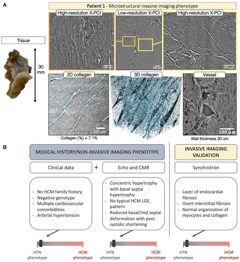
Loncaric et al. Novel Imaging of Myocardial Hypertrophy
enable statistical analysis of group characteristics and the assessment of deformation
pattern reproducibility.
Conclusion: High-resolution, 3D X-PCI provides novel ways to visualize myocardial
remodeling in LVH, and illustrates the correspondence of macrostructural and functional
non-invasive phenotypes with invasive microstructural phenotypes, suggesting the
potential clinical utility of non-invasive myocardial deformation patterns in phenotyping
LVH in everyday clinical practice.
Keywords: hypertrophic cardiomyopathy, hypertension, myocardial disarray, fibrosis, remodeling, synchrotron,
speckle tracking, cardiac magnet resonance
INTRODUCTION The 5-year HCM risk score was evaluated (8). Upper arm
cuff blood pressure measurement in the sitting position was
Distinguishing sarcomere protein gene mutation hypertrophic performed directly preceding the echo examination. Genomic
cardiomyopathy (HCM) from other etiologies of left ventricular DNA was obtained from peripheral blood and analyzed using
hypertrophy (LVH) is clinically relevant, given the association NGS technology (MiSeq, Illumina). The most prevalent gene
with elevated risk for sudden death, familiar inheritance, and mutations involved in cardiomyopathies were scanned (i.e.,
different pharmacological management. The contemporary hypertrophic cardiomyopathy (CM), laminopathies, Danon
approach to diagnosing LVH is based on non-invasive disease, dilatative CM, arrhythmogenic CM, non-compaction
multimodality imaging [echocardiography and magnetic CM, Marfan syndrome, long QT syndrome, Holt-Oram, Ehlers-
resonance (CMR)] – where LV chamber size/shape remodeling, Danlos, and Brrugada syndrome). A generational family pedigree
distribution of hypertrophy/fibrosis, and functional alterations was assessed to explore the genetic origin of disease.
form the basis for diagnosis (1, 2). Cardiac mechanics
are influenced by the structural and pathophysiological Echocardiography
processes underlying the LV remodeling. Therefore, easily All participants underwent a comprehensive transthoracic
obtained, echocardiography-based myocardial deformation echocardiographic examination, prior to the surgical myectomy,
patterns, reflecting cardiac mechanics and suggested as performed on a commercially available E95 system (GE, Vingmed
highly characteristic for specific etiologies (3), may improve Ultrasound, Horten, Norway) equipped with a 4Vc transthoracic
phenotyping when added to the traditional data integration. transducer. In addition to full two-dimensional and Doppler
Nevertheless, the relationship between deformation phenotypes echocardiography, additional parasternal short-axis, and 4-
and etiology-related microstructural cardiac remodeling has chamber, 2-chamber, and 3-chamber apical acquisitions with
not been reported. While relatively novel in the field of cardiac appropriate frame rates were obtained for speckle-tracking and
imaging, synchrotron radiation-based X-ray phase-contrast Tissue Doppler analysis, respectively.
imaging (X-PCI) can provide high resolution, three-dimensional LV and LA volumes were assessed in the apical 4- and 2-
(3D) histological information on myocardial microstructure ex chamber views. LV ejection fraction was calculated by using the
vivo non-destructively (4–7) – providing a novel approach to biplane Simpson method. LA volumes were indexed to the BSA.
visualize complex structural changes related to remodeling in Cardiac dimensions were measured in appropriate 2D views, as
different disease etiologies. according to current guidelines (9). LV mass was calculated using
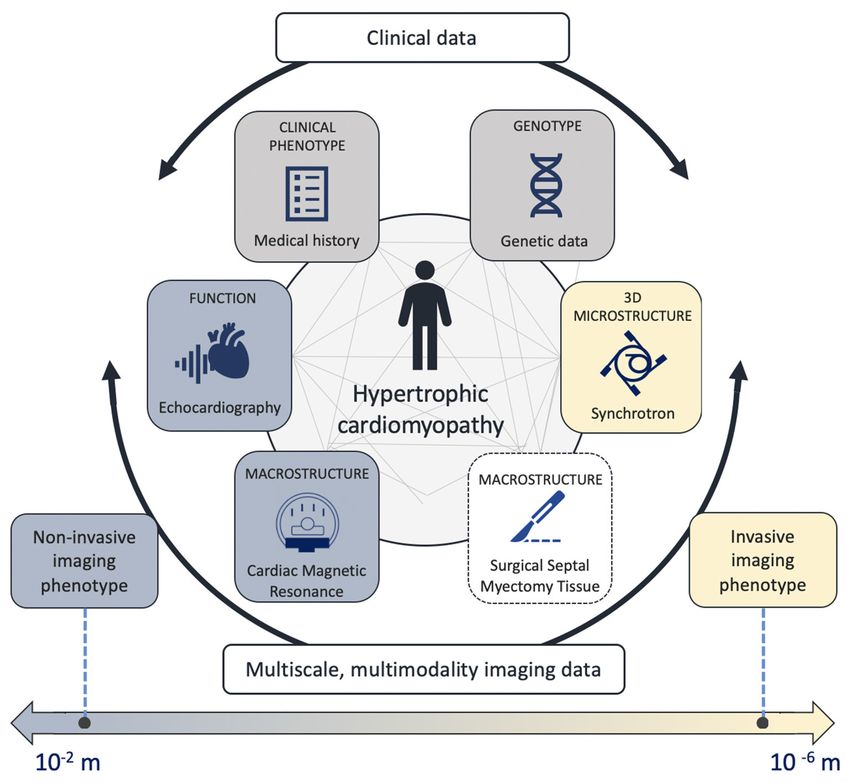
The aim of this pilot study is to apply a multiscale, the linear method and normalized by body surface area, whereas
multimodality protocol in LVH patients undergoing septal hypertrophy was defined as >88 g/m2 in females and >102 g/m2
myectomy to visualize in vivo and ex vivo myocardial tissue in males (9). Relative wall thickness (RWT) was calculated by
with non-invasive and X-PCI imaging, respectively, and relate dividing the doubled value of the end-diastolic posterior wall
non-invasive LVH imaging phenotypes to the underlying thickness with the end-diastolic internal diameter of the LV. The
synchrotron-assessed microstructure (Figure 1). type of LV remodeling was determined based on the RWT and
indexed LV mass.
Resting LVOT peak velocity was measured by using
METHODS continuous-wave Doppler echocardiography, and the LVOT
pressure gradient peak estimated by using a simplified Bernoulli
Patients Under Study equation. The maximal LVOT gradient was defined as the highest
The population under study were three patients (P1-3) with recorded gradient, either in rest of during Valsalva maneuver.
obstructive LVH referred for septal myectomy from the familiar Pulsed-wave Doppler was performed in the apical 4-chamber
cardiomyopathy outpatient clinic. At the moment of inclusion, a view by placing the sample volume at the level of the leaflet
clinical interview was performed. Medical history was reviewed tips to obtain mitral inflow velocities. Peak velocity of early (E)
and data on demographic characteristics, cardiovascular risk and late (A) diastolic filling, E velocity deceleration time and
factors, comorbidities, pharmacological treatment was collected. A wave duration were measured, and the E/A ratio calculated.
Frontiers in Cardiovascular Medicine | www.frontiersin.org 2 May 2021 | Volume 8 | Article 670734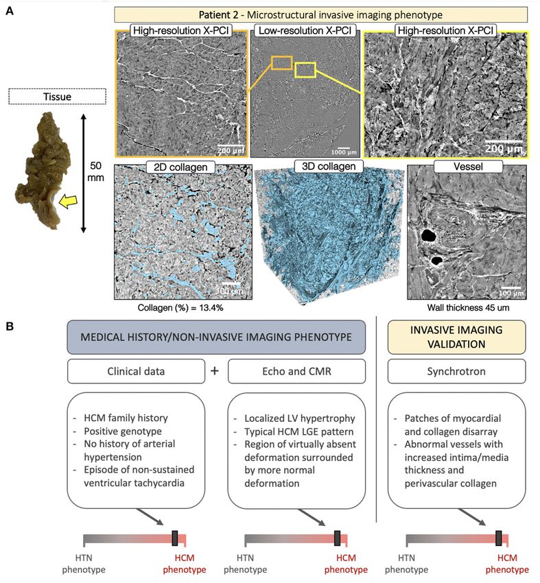
Loncaric et al.                                                                                                                Novel Imaging of Myocardial Hypertrophy

 FIGURE 1 | Central figure – The multiscale, multimodality analysis evaluating genetic information, microstructure, macrostructure and cardiac function.

Isovolumic relaxation time (IVRT) was measured as the time                             septum. TDI-derived deformation was assessed using color-
difference between aortic valve closure and mitral valve opening                       coded maps of myocardial deformation and by determining ROIs
as assessed in the five-chamber view using continuous-wave                             to generate regional TDI-derived longitudinal strain curves of
Doppler of the LV outflow tract. Tissue Doppler was used to                            myocardial areas within the adjacent ventricular segment.
measure early and late diastolic mitral annular velocity at the
septal (e’ and a’ septal) and lateral (e’ and a’ lateral) annular sites.               Cardiac Magnetic Resonance Imaging
Myocardial deformation of the left ventricle (LV) was assessed                         All cardiac magnetic resonance (CMR) exams were performed in
with speckle tracking echocardiography (STE) on 2D grayscale                           a 3.0 Tesla scanner (Signa Architect, General Electric Healthcare),
images obtained from the three apical cardiac views and with                           equipped with a 32-channel chest coil. All images were ECG-
tissue Doppler deformation imaging (TDI) in the 4-chamber                              triggered and obtained in apnea. Standard short axis SSFP
view – both using GE Echopac software (GE Medical Systems,                             cines were obtained (slice thickness 6–8 mm, 2–4 mm gap)
version 202.41.0). The endocardial border was manually marked                          in order to calculate left and right ventricular volumes and
at end-systole of the LV. A region of interest with six segments                       function. Additionally, 4-, 3-, and 2-chamber SSFP cines were
was automatically generated. If needed, manual adjustments                             acquired. A standard phase contrast in-plane flow sequence in
were performed to achieve optimal tracking. Longitudinal strain                        a 3-chamber view orientation (or a similar orientation with
curves were generated and end-systolic strain, defined by the                          a good visualization of the LV outflow tract) was acquired
aortic valve closure time, measured. The LV global longitudinal                        in order to assess for the presence of flow obstruction. An
strain was calculated by averaging values of the 18 segments. The                      additional through-plane phase contrast image was obtained
focus of analysis was the basal and mid septal region, as this                         at the point of maximal turbulence; VENC was appropriately
is where myocardial tissue is removed during septal myectomy.                          adjusted in each case to avoid aliasing. A perfusion sequence
Regional deformation was therefore explored by placing the                             in three standard short axis orientations (basal, midventricular
region of interest (ROI) in different parts of the interventricular                    and apical) was obtained after administration of a single bolus

Frontiers in Cardiovascular Medicine | www.frontiersin.org                         3                                               May 2021 | Volume 8 | Article 670734

Loncaric et al.                                                                                           Novel Imaging of Myocardial Hypertrophy

of gadolinium-based contrast (0.15 mmol/kg). Seven min after             method developed by Paganin was applied (12, 13) using specific
contrast administration a standard Look-Locker sequence was              ring correction (14). Several overlapping scans were acquired
acquired and the optimal inversion time selected for each                to cover the full sample in LR or the full ROI in HR. These
patient. Immediately after, late gadolinium enhancement imaging          scans were later stitched in order to obtain full sample/ROI
(inversion recovery gradient echo and PSIR sequences) was                datasets. HR images enabled assessment of individual myocyte
acquired using the same imaging planes, slice thickness and              organization, vessels morphology and collagen distribution.
spacing as the cine images; inversion time was adjusted during               X-PCI datasets were visualized and analyzed with Fiji
the acquisition, if necessary.                                           (stitching/cropping) (15) and the open-source software Ilastik
                                                                         (collagen segmentation) (16) with the aim of reproducible
Surgical Myectomy and Tissue Handling                                    segmentations of the different microstructural components.
Septal tissue samples were obtained as a part of the planned             Specifically, the pixel classification workflow of Ilastik was used
surgical procedure performed at the center. Patients underwent           for collagen segmentation. Finally, 3D slicer was used to generate
myectomy as previously described (10). No additional invasive            the 3D volume renders of segmented collagen (17).
procedure was performed beyond the ones indicated for the
management of the patient’s clinical condition. The tissue that
was removed by the expert judgement of the cardiac surgeon.              RESULTS
The number and size of tissue specimens were dependent
on the predisposing patient anatomy. The tissue was initially            Non-Invasive Imaging Phenotypes –
placed in a vile with a heparin solution, and afterwards fixed           Macrostructure and Function
and stored in a formalin solution at room temperature in a               Information on family history of HCM, demographic data
standard formalin container regularly used in the clinical setting       and medical history is shown in Table 1, and additional
(DiaPath SafeCapsule 31.7 ml). Tissue samples were measured              echocardiographic measurements in Supplementary Table 1. All
and photographed for reference.                                          patients had severe symptomatic LV outflow tract obstruction
                                                                         with systolic anterior motion (SAM) of the mitral valve. The
Synchrotron Imaging and Data Analysis                                    non-invasive imaging for P1-3 is illustrated in Figures 2–4.
Samples were imported to the TOMCAT beamline at Swiss Light                  P1 had normal LV cavity dimensions, showing concentric
Source (Villigen, Switzerland) according to the participating            LVH combined with pronounced basal anteroseptal hypertrophy
institution’s ethical recommendations and the governing                  (19 mm) (Figure 2), while LGE showed traces of septal
international legal requirements. Due to limited available               intramyocardial and pronounced endocardial fibrosis (Figure 2,
beamtime, only the largest removed pieces of tissue were selected        first row, yellow arrow). Global longitudinal strain was reduced,
for scanning. The tissue was imaged using X-PCI. A multiscale            whereas regional STE deformation analysis revealed impaired
protocol combining a low-resolution (LR) and a high-resolution           deformation at both the basal, basal/mid and mid septum
(HR) setup (5.8 and 0.65 µm pixel size, respectively) was used0          (Figure 2, yellow, pink and blue curves), associated with post-
(7). Briefly, the tissue sample was introduced in a tube with            systolic shortening (PSS) (Figure 2, yellow arrows). Upon further
deionized degassed water in order to minimally affect the                exploration with TDI, areas with reduced deformation and PSS
tissue structural conditions and avoid bubble formation. After           were identified in the basal septum.
positioning the sample on the rotation stage, image acquisition              P2 had normal LV dimensions and an asymmetric LVH
was performed using a 20 keV parallel synchrotron X-ray beam.            localized in the inferoseptal region (17 mm) (Figure 3), with
The sample was first imaged at LR (5,001 projections, exposure           focal intramyocardial enhancement in the basal and mid inferior
time = 30 ms, field of view (FoV) = 11.83 × 3.29 mm2 , 360◦              septum and both right ventricular insertion points (Figure 3,
rotation) with a sample-detector distance of 333 cm. X-rays              first row, yellow arrows). While global longitudinal strain was
were converted to visible light through a LuAG:Ce 300 µm                 only slightly reduced, septal regional STE deformation analysis
scintillator and detected by a sCMOS camera (PCO Edge 4.2).              showed a heterogeneous deformation pattern: reduced, but
LR scans correspond well to traditional histology (11), enabling         normally profiled deformation in the basal segment (Figure 3,
a non-destructive evaluation of the overall morphology of the            yellow curve), virtually completely absent deformation on the
septal tissue – allowing visualization of the endocardium, an            transition from basal to mid region (Figure 3, pink curve),
overview of myocyte organization to locate regional disruptions,         and normalizing deformation toward the apex (Figure 3, blue
and the identification of patches of replacement fibrosis or areas       and green curves). Exploration with color-coded TDI and TDI
with interstitial fibrosis. Furthermore, LR scans were also used         deformation curves confirmed these findings visualizing an
to select several ROIs to be imaged at HR (2,501 projections,            isolated area with very abnormal deformation in the transition
exposure time = 220 ms, FoV 1.64 × 1.38 mm2 , 180◦ rotation),            from basal to mid septum (Figure 3, blue septal region in the
with a sample-detector distance of 20 cm, a LuAG:Ce 20 µm                color-coded TDI, blue strain curve).
scintillator and a PCO.Edge 5.5 CMOS detector. Additionally,                 P3 had a slightly dilated LV, with an extreme, asymmetric
50 pre-flat, 50 post-flat and 50 dark images were acquired for           LVH, localized throughout the whole septum (37 mm), and
flat-field and dark-field corrections of each acquisition. The           paired with severe enhancement in the septum and inferior
acquired projections were reconstructed using the Gridrec                interventricular junction (Figure 4). While ejection fraction was
algorithm. In the case of HR, the single distance phase retrieval        supranormal, global longitudinal strain and septal e’ velocity

Frontiers in Cardiovascular Medicine | www.frontiersin.org           4                                       May 2021 | Volume 8 | Article 670734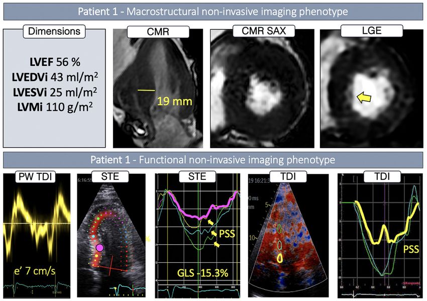
Loncaric et al.                                                                                                                    Novel Imaging of Myocardial Hypertrophy

TABLE 1 | Clinical data and medical history.

                                                        Patient 1                          Patient 2                                               Patient 3

Age (years)                                             64                                 50                                                      32
Gender                                                  Male                               Male                                                    Female
Body mass index (kg/m2 )                                32.0                               26.5                                                    22.8
Arterial hypertension                                   Yes                                No                                                      No
Diabetes mellitus                                       Yes                                No                                                      No
Hyperlipidemia                                          Yes                                No                                                      No
Sleep apnea                                             Yes                                No                                                      No
Atrial fibrillation                                     Paroxysmal                         No                                                      No
New York Heart Association Functional Classification    III                                III                                                     III
Known family history of HCM                             –                                  Brother had obstructive HCM and underwent               –
                                                                                           septal myectomy
Family history of SCD                                   No                                 No                                                      No
Cardiac sarcomere protein gene mutations                Negative for tested variants       MYH7                                                    Negative for tested variants
History of unexplained presyncope or syncope            No                                 No                                                      Yes
History of arrhythmia                                   No                                 Non-sustained ventricular tachycardia (2017)            No
5-year HCM risk (%)                                     2.09                               6.25                                                    4.88

  FIGURE 2 | Non-invasive imaging phenotype of P1.

was severely reduced. Septal regional STE deformation analysis                              basal to mid septum showed completely abnormal deformation
showed completely abnormal deformation throughout the basal                                 (Figure 4, blue curve), slowly recovering toward the apical region
and mid region (Figure 4, yellow, pink and blue curves), returning                          (Figure 4, green curve).
to normal values in the apex (Figure 4, green curve). Color-
coded TDI and TDI deformation curves, based on smaller, more                                Invasive Imaging Phenotypes –
focused regions of interest, revealed an underlying heterogeneous                           Microstructure
deformation pattern. Deformation in the basal region was, in                                After surgical removal in P1, the septal tissue showed a
fact, normal (Figure 4, yellow curve), whereas the transition from                          smooth endocardial fibrotic layer which could be visualized

Frontiers in Cardiovascular Medicine | www.frontiersin.org                             5                                                  May 2021 | Volume 8 | Article 670734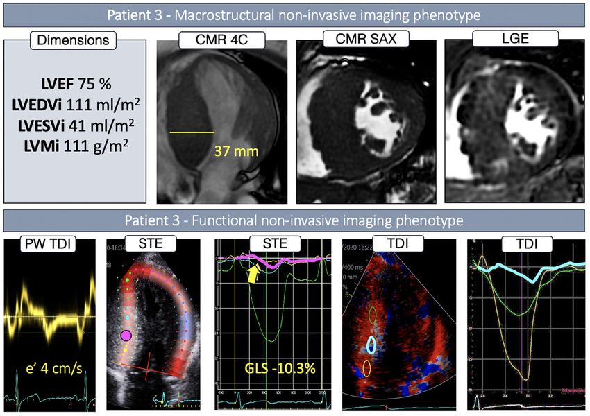
Loncaric et al.                                                                                       Novel Imaging of Myocardial Hypertrophy

 FIGURE 3 | Non-invasive imaging phenotype of P2.

 FIGURE 4 | Non-invasive imaging phenotype of P3.

with LR, and the underlying collagen organization with HR              arranged myocytes (Figure 5A, yellow frame). The segmented
X-PCI (Figure 5A, orange frame, Supplementary Video 1, 2).             3D collagen distribution was visualized (Figure 5A, second row,
HR revealed extensive interstitial fibrosis surrounding normally       collagen shown in blue, Supplementary Video 3), demonstrating

Frontiers in Cardiovascular Medicine | www.frontiersin.org         6                                     May 2021 | Volume 8 | Article 670734
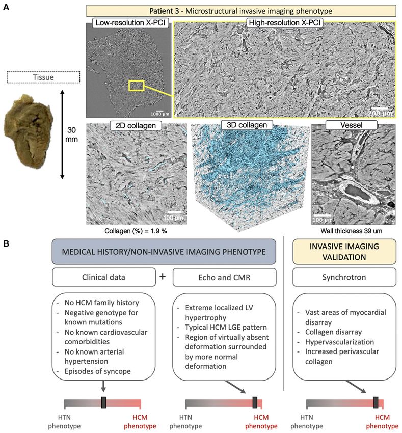
Loncaric et al.                                                                                                              Novel Imaging of Myocardial Hypertrophy

 FIGURE 5 | (A) Invasive imaging phenotype of P1. (B) Clinical data described a patient with no family history of HCM, negative genotype, and a burden of
 cardiovascular comorbidities, including arterial hypertension. The non-invasive imaging concurred with the HTN clinical phenotype showing basal septal hypertrophy
 with no typical HCM LGE pattern and a characteristic hypertensive spatiotemporal deformation pattern in the basal and mid septum coupled with post-systolic
 deformation. The invasive imaging confirmed the non-invasive-imaging-based HTN phenotype with findings of normal myocyte, collagen and blood vessel
 organization.

extensive (quantified at 7.1% in the selected volume), but spatially                  increased fibrosis (13.4%), highly disorganized, except in the
normally organized, fibrosis. A blood vessel is shown with                            smaller regions of normal organization (Figure 6A, yellow arrow,
normal dimensions, normal perivascular collagen, and mild wall                        Supplementary Video 5). The vasculature revealed abnormal
hypertrophy (20 µm). The integration of clinical data for P1 is                       vessels with intima/media hypertrophy (45 µm) and increased
shown in Figure 5B.                                                                   perivascular collagen. The integration of data for P2 is shown in
   In P2 the septal tissue revealed a localized (likely related                       Figure 6B.
to the SAM impact) fibrotic patch on the endocardial side                                Septal myocardial samples from P3 showed no pronounced
(Figure 6A, yellow arrow). LR revealed regions of normal                              fibrotic patch visible on the endocardial side. LR, further
myocardial organization (Figure 6A, orange frame) alternated                          enhanced with HR, revealed hypertrophied myocytes with
with patches of myocyte disarray (Figure 6A, yellow frame,                            overt myocyte disarray, interlaced with small patches of normal
Supplementary Video 4). Disarray was further explored                                 organization (Figure 7A, yellow frame, Supplementary Video 6).
with HR. Visualization of the 3D collagen distribution                                In the acquired tissue sample, collagen was not increased
(Figure 6A, second row, collagen shown in blue) showed                                (1.9%), but showed spatial disarray (Supplementary Video 7).

Frontiers in Cardiovascular Medicine | www.frontiersin.org                        7                                              May 2021 | Volume 8 | Article 670734
Loncaric et al.                                                                                                                Novel Imaging of Myocardial Hypertrophy

 FIGURE 6 | (A) Invasive imaging phenotype of P2. (B) Clinical data described a patient with family history of obstructive HCM, history of arrhythmic episodes, and a
 positive genotype. Here, non-invasive imaging confirmed the clinical HCM phenotype. Although localized basal septal hypertrophy can also be seen in hypertension,
 the deformation pattern was characteristic of HCM, and the LGE findings concurred. Invasive imaging validated the non-invasive-imaging-based HCM remodeling
 phenotype with findings of myocardial disarray, abnormal collagen organization and hypertrophied blood vessels.

Pronounced hypervascularization was noted throughout                                   of myocyte and connective tissue matrix spatial disorganization
the sample with an increased number of blood vessels,                                  and myocardial deformation – thus suggesting the potential
combined with wall hypertrophy (39 µm) and increased                                   clinical utility of deformation patterns in phenotyping left
perivascular collagen.                                                                 ventricular hypertrophy.

DISCUSSION                                                                             The Functional Consequences of
                                                                                       Myoarchitectural Abnormalities
In this pilot study we demonstrate a unique multiscale,                                X-PCI has proven as a powerful tool for visualizing cardiac
multimodality protocol to visualize in vivo and ex vivo                                microstructure in cardiac biopsies of rat models, human
myocardial tissue with non-invasive and X-PCI imaging. We                              fetal hearts, as well as in endomyocardial biopsies of heart
report a novel approach to visualize 3D cardiac microstructure                         transplantation patients. The modality enables imaging up to
in LVH and relate the findings to the etiology-discriminative                          the scale of an individual myocyte, demonstrating feasibility
interpretation of non-invasive imaging data. The findings are                          of quantification of fiber orientation, vessels and collagen
hypothesis-generating, providing insights about the relationship                       from multiscale 3D datasets; enabling multiresolution, 3D,

Frontiers in Cardiovascular Medicine | www.frontiersin.org                         8                                              May 2021 | Volume 8 | Article 670734
Loncaric et al. Novel Imaging of Myocardial Hypertrophy
FIGURE 7 | (A) Invasive imaging phenotype of P3. (B) Clinical data was inconclusive, revealing negative family history of HCM, lack of cardiovascular comorbidities, a
history presyncope, and a genotype negative for the most common mutations associated with HCM. Here, non-invasive imaging played a crucial role in revealing the
etiology – showing extreme wall thickening in the septal region, with a characteristic HCM deformation pattern, and severe LGE in the septum. Invasive imaging
confirmed the non-invasive-imaging HCM remodeling phenotype with findings of vast myocardial disarray, collagen disarray, hypervascular tissue and hypertrophied
blood vessels.
quantitative ex vivo analysis of cardiac microstructure, without regions of myocardial hypertrophy (23). In human hearts non-
the need for artifact prone slice-processing that strains invasive imaging, histology, and in vitro experiments suggested
histological/microscopic reconstruction (4, 5, 7, 18). In our study, an association between disarray, fibrosis, active contraction in
X-PCI and the subsequent analysis - applying machine-learning vitro, and STE-derived deformation in vivo (24, 25). Similarly,
solutions to provide automated, 3D segmentations of myocardial diffusion tensor CMR and CMR-derived strain rate imaging
structure - enabled a novel way to visualize and quantify the inferred intramural disarray correlated with both active and
complex microstructural abnormalities that inherently influence passive myocardial function (26). The intrinsic contractile
cardiac mechanics in LVH. dysfunction can be recognized with deformation imaging
The histological findings of myocyte (19, 20) and connective even at the early disease stages, before onset of LVH (27).
tissue matrix (21, 22) disorganization in HCM has evoked This described relationship between abnormal myoarchitecture
research relating abnormal myocardial architecture to LV and cardiac mechanics in HCM is captured by characteristic
function. A transgenic HCM mouse model demonstrated deformation patterns, easily obtained in everyday workflow, and
decreased sarcomere length and impaired systolic shortening in potentially clinically useful in the process of distinguishing LVH
Frontiers in Cardiovascular Medicine | www.frontiersin.org 9 May 2021 | Volume 8 | Article 670734Loncaric et al. Novel Imaging of Myocardial Hypertrophy
etiologies, especially when integrated with remaining clinical and compensatory hypertrophy leads to high variability in regional
imaging findings. myocardial wall thickness and characteristic regional contractile
heterogeneity (37–40). The structural finding of basal septal
hypertrophy in HCM commonly overlaps with that seen in
HTN (e.g., P1 vs. P2), however, with clearly different patterns
The Challenge of Distinguishing Disease of deformation. In P2 and P3, we noted a heterogeneous
Etiologies septal deformation pattern, with localized parts of the septum
Clinical practice relies on interpretation of available clinical showing virtually absent deformation, unlike that seen in HTN,
information for diagnosis in LVH – medical history and physical while surrounding regions show normal deformation pattern
examination, genetic analysis for the most frequent mutations, (with/without reduced amplitude).
and insights gained from non-invasive cardiac imaging (28). Regional longitudinal strain is still burdened by
The challenge for a genetic diagnosis is the considerable genetic reproducibility and inter-vendor variability (41–43).
heterogeneity of HCM, where the underlying genetic cause is Nevertheless, regional spatiotemporal strain patterns contain
only found in a percentage of patients fitting the phenotype important diagnostic information (3, 41), and remain consistent
(29). In our analysis, P3 had a “textbook” microstructural, despite underlying variability in inter-observer segmentations
macrostructural, and functional HCM phenotype, however, and regional peak strain values (44). The results of this pilot study
the genotyping approach was unsuccessful in identifying the are hypothesis-generating, suggesting non-invasive deformation
causative genetic mutation strongly suggested to be present. phenotypes are associated with etiology-related myocyte and
Differentiating HCM from hypertensive heart disease is a connective tissue matrix 3D disorganization, thus inferring the
process based on integrating findings from clinical history potential clinical value of deformation patterns in everyday
and multimodal non-invasive imaging - the distribution of clinical analysis and phenotyping LV hypertrophy.
LVH and LGE, frequency of LV outflow tract obstruction,
severity of longitudinal dysfunction, functional asynchrony Scientific and Clinical Implications
and deformation heterogeneity (2). Such integration is shown The ability to explore microstructural organization is essential
in Figures 5B, 6B, 7B. In this analysis, echo-based regional for understanding myocardial mechanics and resolving the
deformation patterns, reflective of the influence of structural etiology of non-invasive imaging phenotypes in LV hypertrophy.
and pathophysiological processes in LV remodeling on cardiac In contemporary translational research, electro-mechanical
mechanics, are easily accessible and of potential clinical use (3). computational models of the heart integrate multiscale and
The echocardiographic finding of basal septal hypertrophy multimodal imaging, and apply novel methods of data extraction
has been shown to be a morphological marker of increased from large datasets and across different resolutions, with the
afterload in arterial hypertension (30, 31) Here intra-ventricular aim toward deciphering mechanistic descriptors of personalized
heterogeneity is the consequence of heterogeneous wall stress structure and function (45). Detailed information on fiber
distribution in response to elevated blood pressure. In an average orientation and fibrosis organization can be integrated to
heart, the septum has a greater radius of curvature compared these models (46), a concept particularly relevant in HCM
to the free wall (32, 33), leading to a disproportionately higher (47). Previously, fiber structure information has been extracted
wall stress in the basal parts in the setting of high systemic from diffusion tensor imaging (at low resolution) or localized
pressure (33). This results with an imbalance between locally microscopy (48), but this may be advanced with high resolution,
developed force and wall stress, and, consequently, decreased 3D X-PCI data. Derived information about disease-specific
local deformation and PSS. Thus, in HTN, prolonged exposure patterns could help relate structural and dynamic features
to increased afterload can result in compensatory localized basal measured in vivo with high-resolution characterization of
septal hypertrophy in an attempt to normalize wall stress and microstructure ex vivo, enabling personalized modeling of
maintain deformation (3), and ultimately lead to LV outflow cardiac biomechanics, potentially bringing incremental insights
obstruction (34). Localized hypertrophy in HTN, as seen in to disease pathophysiology and tailoring risk assessment (49).
P1 and opposed to P2, was associated with microstructurally In a more clinical perspective, continuing evidence point
described overt interstitial fibrosis (not clearly inferred by LGE), out the inconsistencies of the single sarcomere gene hypothesis
showing organized 3D structure of the collagen surrounding in HCM, suggesting the need to incorporate the influences of
normally arranged myocytes. These microstructural findings numerous disease modifiers, each exerting a small effect on
support the hypertensive etiology of the hypertrophy, especially phenotype expression (50–52). Here, the opportunity to explore
when combined with the HTN-related deformation pattern - the structure-function relationship, through the insights of
reduction in peak systolic strain (30, 35) and PSS occurring in combined in vivo and ex vivo imaging is highly relevant. Applying
the basal and mid-septum (36). this methodology enables quantification of myocardial structure
Another type of intra-ventricular heterogeneity can be abnormalities potentially associated to sarcomere protein gene
seen in HCM, where, as compared to the heterogeneity mutations and clinical risk, even in patients with no signs
of loading in HTN, the heterogeneity is in the tissue of macrostructural remodeling when assessed with traditional
structure itself. In our study, X-PCI of HCM myocardial non-invasive imaging. In comparison to X-PCI, histology is a
tissue revealed both myocyte and connective tissue matrix destructive imaging method, relying on tissue preparation –
3D disorganization or disarray. The heterogeneous tissue and slicing and staining, and of limited (2D) analysis. Multiscale
Frontiers in Cardiovascular Medicine | www.frontiersin.org 10 May 2021 | Volume 8 | Article 670734Loncaric et al. Novel Imaging of Myocardial Hypertrophy
analysis with CMR may provide better soft tissue contrast, ETHICS STATEMENT
however, at much lower resolutions and/or prolonged scan
time. Furthermore, CMR-based techniques rely on indirect The studies involving human participants were reviewed and
measures of myocardial structure, through the use of contrast approved by Ethics Committee of the Hospital Clinic of
agents or diffusion tensor imaging, whereas with X-PCI we Barcelona. The patients/participants provided their written
can directly measure myocardial structure based on changes informed consent to participate in this study.
in X-ray intensity and phase. On an important note, X-PCI is
currently a research methodology, and as such it is linked to AUTHOR CONTRIBUTIONS
synchrotron facilities with limited accessibility. Integration of
X-PCI technology in traditional hospital CT machines is for FL: conception, design, analysis and interpretation of data, and
now still not feasible (53), but the developments in compact drafting of the manuscript. PG-C: analysis and interpretation
synchrotrons (54) and grating based X-PCI technology (55) may of data, drafting of the manuscript, revising for important
lead to a translation of this methodology towardz clinical, ex intellectual content, and final approval of the manuscript
vivo use – particularly for imaging tissue biopsies. Application submitted. AG-A, HD, and AB: conception and design, revising
of these tools has already been suggested – for clinically relevant for important intellectual content, and final approval of the
topics such as the assessment of ex vivo endomyocardial tissue in manuscript submitted. LS, SP, AD, EQ, and DP: revising
heart transplantation patients to assess graft rejection with more for important intellectual content and final approval of the
reproducibility (56), or assessment in vivo to clarify ambiguous manuscript submitted. MS and BB: conception and design,
findings in traditional mammography (57). interpretation of data, revising for important intellectual content,
and final approval of the manuscript submitted. All authors
contributed to the article and approved the submitted version.
Limitations
Our pilot study consisted of a detailed, multi-modality in vivo FUNDING
and ex vivo analysis of a small sample, with the goal of applying
existing X-PCI technology to the field of cardiac imaging to This work was supported by the Horizon 2020 European
provide a novel visualization of microstructural organization Commission Project H2020-MSCA-ITN (764738) and the
in LVH, and to generate a hypothesis of structure/function Clinical Research in Cardiology of the Spanish Foundation of
relations by linking these invasive findings to non-invasive the Heart grant from the Spanish Cardiac Society (SEC/FEC-
imaging phenotypes. Nonetheless, no causation can be claimed INV-CLI 20/028). MS holds a grant from Fundacio La Marató
based on these initial results. The results motivate larger patient de TV3 (040310, Exp 2015.40.30). HC has received funding
cohorts to enable statistical analysis/group comparison, as well as from the Strategic Focal Area Personalized Health and Related
the assessment of reproducibility of deformation patterns (44). Technologies (PHRT) of the ETH Domain (2017-303). PG-C has
All imaged tissue samples were derived from surgical received funding from the post-doctoral fellowships programme
myectomy; therefore, the sample size was limited and potentially Beatriu de Pinos (2018-BP-00201) and the Horizon 2020
not representative of the heart as a whole. However, septal tissue European Commission Project H2020-MSCA-ITN (801370).
has consistently demonstrated structural abnormalities in prior
studies (22, 24, 25, 52). ACKNOWLEDGMENTS
We acknowledge the Paul Scherrer Institut, Villigen, Switzerland
CONCLUSIONS for provision of synchrotron radiation beamtime at beamline
TOMCAT of the SLS.
High-resolution, 3D X-PCI provides novel ways to visualize
myocardial remodeling in excised myectomy tissue, and SUPPLEMENTARY MATERIAL
illustrates the correspondence of macrostructural and functional
non-invasive phenotypes with invasive microstructural The Supplementary Material for this article can be found
phenotypes, suggesting the potential clinical utility of non- online at: https://www.frontiersin.org/articles/10.3389/fcvm.
invasive myocardial deformation patterns in phenotyping left 2021.670734/full#supplementary-material
ventricular hypertrophy. A larger patient cohort could enable Supplementary Video 1 | Patient 1 - Top to bottom scan of a subvolume of the
statistical analysis of established differences and the assessment HR X-PCI Paganin reconstructed dataset.
of the reproducibility of deformation patterns. Supplementary Video 2 | Patient 1 - Top to bottom scan of a subvolume of the
HR X-PCI Paganin reconstructed dataset.
Supplementary Video 3 | Patient 1 – A 3D visualization of the
collagen organization.
DATA AVAILABILITY STATEMENT
Supplementary Video 4 | Patient 2 - Top to bottom scan of a subvolume of the
The data presented in the study are deposited in the PSI Public HR X-PCI Paganin reconstructed dataset.
Data Repository, accession number http://doi.psi.ch/detail/10. Supplementary Video 5 | Patient 2 – A 3D visualization of the
16907%2Fb97cdb87-83be-4176-87f4-2e89679ff333. collagen organization.
Frontiers in Cardiovascular Medicine | www.frontiersin.org 11 May 2021 | Volume 8 | Article 670734Loncaric et al. Novel Imaging of Myocardial Hypertrophy
Supplementary Video 6 | Patient 3 - Top to bottom scan of a subvolume of the Supplementary Figure 1 | Photographs of tissue collected after surgical
HR X-PCI Paganin reconstructed dataset. myectomy.
Supplementary Video 7 | Patient 3 – A 3D visualization of the Supplementary Table 1 | Echocardiographic measurements.
collagen organization. Supplementary Table 2 | Measurements of the septal myectomy tissue samples.
REFERENCES 16. Berg S, Kutra D, Kroeger T, Straehle CN, Kausler BX, Haubold C, et al.
ilastik: interactive machine learning for (bio)image analysis. Nat Meth. (2019)
1. Tower-Rader A, Kramer CM, Neubauer S, Nagueh SF, Desai MY. 16:1226–32. doi: 10.1038/s41592-019-0582-9
Multimodality imaging in hypertrophic cardiomyopathy for risk stratification. 17. Fedorov A, Beichel R, Kalpathy-Cramer J, Finet J, Fillion-Robin J-C, Pujol S,
Circulation. (2020) 13:e009026. doi: 10.1161/CIRCIMAGING.119.0 et al. 3D slicer as an image computing platform for the quantitative imaging
09026 network. Mag Res Imaging. (2012) 30:1323–41. doi: 10.1016/j.mri.2012.05.001
2. Cardim N, Galderisi M, Edvardsen T, Plein S, Popescu BA, D’Andrea A, 18. Jouk P-S, Mourad A, Milisic V, Michalowicz G, Raoult A, Caillerie D, et al.
et al. Role of multimodality cardiac imaging in the management of patients Analysis of the fiber architecture of the heart by quantitative polarized light
with hypertrophic cardiomyopathy: an expert consensus of the European microscopy. Accuracy, limitations and contribution to the study of the fiber
Association of cardiovascular imaging endorsed by the saudi heart association. architecture of the ventricles during fetal and neonatal life. Eur J Cardiothorac
Eur Heart J Cardiov Imaging. (2015) 16:280. doi: 10.1093/ehjci/jeu291 Surg. (2007) 31:915–21. doi: 10.1016/j.ejcts.2006.12.040
3. Cikes M, Sutherland GR, Anderson LJ, Bijnens BH. The role of 19. Hughes SE. The pathology of hypertrophic cardiomyopathy. Histopathology.
echocardiographic deformation imaging in hypertrophic myopathies. Nat Rev (2004) 44:412–27. doi: 10.1111/j.1365-2559.2004.01835.x
Cardiol. (2010) 7:384–96. doi: 10.1038/nrcardio.2010.56 20. Maron BJ, Roberts WC. Quantitative analysis of cardiac muscle
4. Gonzalez-Tendero A, Zhang C, Balicevic V, Cárdenes R, Loncaric S, Butakoff cell disorganization in the ventricular septum of patients
C, et al. Whole heart detailed and quantitative anatomy, myofibre structure with hypertrophic cardiomyopathy. Circulation. (1979) 59:689–
and vasculature from X-ray phase-contrast synchrotron radiation-based 706. doi: 10.1161/01.CIR.59.4.689
micro computed tomography. Eur Heart J Cardiov Imaging. (2017) 18:732– 21. Shirani J, Pick R, Roberts WC, Maron BJ. Morphology and significance of
41. doi: 10.1093/ehjci/jew314 the left ventricular collagen network in young patients with hypertrophic
5. Garcia-Canadilla P, Dejea H, Bonnin A, Balicevic V, Loncaric S, Zhang cardiomyopathy and sudden cardiac death. J Am Coll Cardiol. (2000) 35:36–
C, et al. Complex congenital heart disease associated with disordered 44. doi: 10.1016/S0735-1097(99)00492-1
myocardial architecture in a midtrimester human fetus. Circulation. (2018) 22. Factor SM, Butany J, Sole MJ, Wigle ED, Williams WC, Rojkind M. Pathologic
11:e007753. doi: 10.1161/CIRCIMAGING.118.007753 fibrosis and matrix connective tissue in the subaortic myocardium of patients
6. Garcia-Canadilla P, Cook AC, Mohun TJ, Oji O, Schlossarek S, Carrier L, et al. with hypertrophic cardiomyopathy. J Am Coll Cardiol. (1991) 17:1343–
Myoarchitectural disarray of hypertrophic cardiomyopathy begins pre-birth. 51. doi: 10.1016/S0735-1097(10)80145-7
J Anatomy. (2019) 235:962–76. doi: 10.1111/joa.13058 23. Karlon WJ, McCulloch AD, Covell JW, Hunter JJ, Omens JH. Regional
7. Dejea H, Garcia-Canadilla P, Cook AC, Guasch E, Zamora M, Crispi dysfunction correlates with myofiber disarray in transgenic mice with
F, et al. Comprehensive analysis of animal models of cardiovascular ventricular expression of ras. Am J Physiol Heart Circulator Physiol. (2000)
disease using multiscale X-ray phase contrast tomography. Sci Rep. (2019) 278:H898–906. doi: 10.1152/ajpheart.2000.278.3.H898
9:6669. doi: 10.1038/s41598-019-54945-x 24. Kobayashi T, Popovic Z, Bhonsale A, Smedira NG, Tan C, Rodriguez ER, et al.
8. O’Mahony C, Jichi F, Pavlou M, Monserrat L, Anastasakis A, Rapezzi C, Association between septal strain rate and histopathology in symptomatic
Biagini E et al. Hypertrophic cardiomyopathy outcomes investigators. A hypertrophic cardiomyopathy patients undergoing septal myectomy. Am
novel clinical risk prediction model for sudden cardiac death in hypertrophic Heart J. (2013) 166:503–11. doi: 10.1016/j.ahj.2013.06.011
cardiomyopathy (HCM risk-SCD). Eur Heart J. (2014) 35:2010–20. 25. Dhillon A, Sweet W, Popovic ZB, Smedira NG, Thamilarasan M,
9. Lang RM, Badano LP, Mor-Avi V, Afilalo J, Armstrong A, Ernande L, et al. Lytle BW, et al. Association of noninvasively measured left ventricular
Recommendations for cardiac chamber quantification by echocardiography mechanics with in vitro muscle contractile performance: a prospective
in adults: an update from the American Society of Echocardiography and study in hypertrophic cardiomyopathy patients. J Am Heart Assoc. (2014)
the European Association of Cardiovascular Imaging. Eur Heart J Cardiovasc 3:e001269. doi: 10.1161/JAHA.114.001269
Imaging. (2015) 16:233-71. 26. Tseng W-YI, Dou J, Reese TG, Wedeen VJ. Imaging myocardial fiber disarray
10. Hang D, Nguyen A, Schaff HV. Surgical treatment for hypertrophic and intramural strain hypokinesis in hypertrophic cardiomyopathy with MRI.
cardiomyopathy: a historical perspective. Ann Cardiothorac Surg. (2017) J Mag Res Imaging. (2006) 23:1–8. doi: 10.1002/jmri.20473
6:318–28. doi: 10.21037/acs.2017.04.03 27. Baudry G, Mansencal N, Reynaud A, Richard P, Dubourg O, Komajda M,
11. Mirea I, Varray F, Zhu YM, Fanton L, Langer M, Jouk PS, et al. Very high- et al. Global and regional echocardiographic strain to assess the early phase
resolution imaging of post-mortem human cardiac tissue using X-ray phase of hypertrophic cardiomyopathy due to sarcomeric mutations. Eur Heart J
contrast tomography. In: van Assen H, Bovendeerd P, Delhaas T, editors. Cardiovasc Imaging. (2020) 21:291–8. doi: 10.1093/ehjci/jez084
Functional Imaging and Modeling of the Heart. FIMH 2015, Vol. 9126. Cham: 28. Allen RD, Edwards WD, Tazelaar HD, Danielson GK. Surgical pathology of
Springer (2015). subaortic septal myectomy not associated with hypertrophic cardiomyopathy:
12. Marone F, Stampanoni M. Regridding reconstruction algorithm for a study of 98 cases (1996-2000). Cardiovasc Pathol. (2003) 12:207–
real-time tomographic imaging. J Synchrotron Rad. (2012) 19:1029– 15. doi: 10.1016/S1054-8807(03)00057-7
37. doi: 10.1107/S0909049512032864 29. Charron P, Arad M, Arbustini E, Basso C, Bilinska Z, Elliott P et al.
13. Paganin D, Mayo SC, Gureyev TE, Miller PR, Wilkins SW. Genetic counselling and testing in cardiomyopathies: a position statement
Simultaneous phase and amplitude extraction from a single of the European society of cardiology working group on myocardial
defocused image of a homogeneous object. J Micro. (2002) and pericardial. Eur Heart J. (2010) 31:2715–26. doi: 10.1093/eurheartj/
206:33–40. doi: 10.1046/j.1365-2818.2002.01010.x ehq271
14. Vo NT, Atwood RC, Drakopoulos M. Superior techniques for eliminating 30. Gaudron PD, Liu D, Scholz F, Hu K, Florescu C, Herrmann S, et al. The septal
ring artifacts in X-ray micro-tomography. Opt Express. (2018) 26:28396– bulge—an early echocardiographic sign in hypertensive heart disease. J Am
412. doi: 10.1364/OE.26.028396 Soc Hyper. (2016) 10:70–80. doi: 10.1016/j.jash.2015.11.006
15. Schindelin J, Arganda-Carreras I, Frise E, Kaynig V, Longair M, Pietzsch T, 31. Loncaric F, Nunno L, Mimbrero M, Marciniak M, Fernandes JF, Tirapu L, et al.
et al. Fiji: an open-source platform for biological-image analysis. Nat Meth. Basal ventricular septal hypertrophy in systemic hypertension. Am J Cardiol.
(2012) 9:676–82. doi: 10.1038/nmeth.2019 (2020) 125:1339–46. doi: 10.1016/j.amjcard.2020.01.045
Frontiers in Cardiovascular Medicine | www.frontiersin.org 12 May 2021 | Volume 8 | Article 670734Loncaric et al. Novel Imaging of Myocardial Hypertrophy
32. Bogaert J, Rademakers FE. Regional nonuniformity of normal adult 46. Land S, Niederer SA, Louch WE, Sejersted OM, Smith NP. Integrating multi-
human left ventricle. Am J Physiol Heart Circul Physiol. (2001) 280:H610– scale data to create a virtual physiological mouse heart. Int Focus. (2013)
20. doi: 10.1152/ajpheart.2001.280.2.H610 3:20120076. doi: 10.1098/rsfs.2012.0076
33. Heng MK, Janz RF, Jobin J. Estimation of regional stress in the left 47. Lyon A, Bueno-Orovio A, Zacur E, Ariga R, Grau V, Neubauer S,
ventricular septum and free wall: an echocardiographic study suggesting a et al. Electrocardiogram phenotypes in hypertrophic cardiomyopathy
mechanism for asymmetric septal hypertrophy. Am Heart J. (1985) 110:84– caused by distinct mechanisms: apico-basal repolarization gradients vs.
90. doi: 10.1016/0002-8703(85)90519-8 Purkinje-myocardial coupling abnormalities. EP Europace. (2018) 20:iii102–
34. Yalçin F, Yigit F, Erol T, Baltali M, Korkmaz ME, Müderrisoglu H. Effect 12. doi: 10.1093/europace/euy226
of dobutamine stress on basal septal tissue dynamics in hypertensive 48. Romero D, Sebastian R, Bijnens BH, Zimmerman V, Boyle PM, Vigmond
patients with basal septal hypertrophy. J Human Hypert. (2006) 20:628– EJ, et al. Effects of the purkinje system and cardiac geometry on
30. doi: 10.1038/sj.jhh.1002041 biventricular pacing: a model study. Ann Biomed Eng. (2010) 38:1388–
35. Baltabaeva A, Marciniak M, Bijnens B, Moggridge J, He F, Antonios T, 98. doi: 10.1007/s10439-010-9926-4
et al. Regional left ventricular deformation and geometry analysis provides 49. Krishnamurthy A, Villongco CT, Chuang J, Frank LR, Nigam V, Belezzuoli
insights in myocardial remodelling in mild to moderate hypertension. Eur J E, et al. Patient-specific models of cardiac biomechanics. J Comput Physics.
Echocardio. (2008) 9:501–8. doi: 10.1016/j.euje.2007.08.004 (2013) 244:4–21. doi: 10.1016/j.jcp.2012.09.015
36. Bijnens B, Claus P, Weidemann F, Strotmann J, Sutherland GR. 50. McLeod CJ, Bos JM, Theis JL, Edwards WD, Gersh BJ, Ommen SR,
Investigating cardiac function using motion and deformation et al. Histologic characterization of hypertrophic cardiomyopathy with
analysis in the setting of coronary artery disease. Circulation. (2007) and without myofilament mutations. Am Heart J. (2009) 158:799–
116:2453–64. doi: 10.1161/CIRCULATIONAHA.106.684357 805. doi: 10.1016/j.ahj.2009.09.006
37. Piella G, De Craene M, Bijnens BH, Tobon-Gómez C, Huguet M, Avegliano G, 51. Maron BJ, Maron MS, Maron BA, Loscalzo J. Moving beyond the sarcomere
et al. Characterizing myocardial deformation in patients with left ventricular to explain heterogeneity in hypertrophic cardiomyopathy. J Am Coll Cardiol.
hypertrophy of different etiologies using the strain distribution obtained by (2019) 73:1978–86. doi: 10.1016/j.jacc.2019.01.061
magnetic resonance imaging. Revista Española Cardiología. (2010) 63:1281– 52. Marian AJ, Braunwald E. Hypertrophic cardiomyopathy: genetics,
91. doi: 10.1016/S1885-5857(10)70253-X pathogenesis, clinical manifestations, diagnosis, and therapy. Cir Res.
38. Kobayashi T, Dhillon A, Popovic Z, Bhonsale A, Smedira NG, (2017) 121:749–70. doi: 10.1161/CIRCRESAHA.117.311059
Thamilarasan M, et al. Differences in global and regional left 53. Hetterich H, Willner M, Fill S, Herzen J, Bamberg F, Hipp A, et al.
ventricular myocardial mechanics in various morphologic subtypes Phase-contrast CT: qualitative and quantitative evaluation of atherosclerotic
of patients with obstructive hypertrophic cardiomyopathy referred carotid artery plaque. Radiology. (2014) 271:870–8. doi: 10.1148/radiol.141
for ventricular septal myotomy/myectomy. Am J Cardiol. (2014) 31554
113:1879–85. doi: 10.1016/j.amjcard.2014.03.020 54. Eggl E, Schleede S, Bech M, Achterhold K, Loewen R, Ruth RD, et al. X-ray
39. Sun J, Xu T, Ni X, Yang X, Hu J, Wang S, et al. Echocardiographic strain in phase-contrast tomography with a compact laser-driven synchrotron source.
hypertrophic cardiomyopathy and hypertensive left ventricular hypertrophy. PNAS. (2015) 112:5567–72. doi: 10.1073/pnas.1500938112
Echocardiography. (2019) 36:257–65. doi: 10.1111/echo.14222 55. Viermetz MP, Birnbacher LJB, Fehringer A, Willner M, Noel PB, Pfeiffer F,
40. Afonso L, Kondur A, Simegn M, Niraj A, Hari P, Kaur R, et al. Two- et al. High resolution laboratory grating-based x-ray phase-contrast CT. Sci
dimensional strain profiles in patients with physiological and pathological Rep. (2018) 8:15884. doi: 10.1038/s41598-018-33997-5
hypertrophy and preserved left ventricular systolic function: a comparative 56. Planinc I, Ilic I, Garcia-canadilla I, Dejea H, Skoric B, Jurin H, et al. X-
analyses. BMJ Open. (2012) 2:e001390. doi: 10.1136/bmjopen-2012-001390 ray Phase Contrast Imaging of Endo-Myocardial Biopsies Following Heart
41. Sperry BW, Sato K, Phelan D, Grimm R, Desai MY, Hanna M, et al. Transplantation - Agreement With Classical Histology. Eur Heart J. (2020)
Regional variability in longitudinal strain across vendors in patients with 22(Suppl S1):413.
cardiomyopathy due to increased left ventricular wall thickness. Circulation. 57. Castelli E, Tonutti M, Arfelli F, Longo R, Quaia E, Rigon L,
(2019) 12:e008973. doi: 10.1161/CIRCIMAGING.119.008973 et al. Mammography with synchrotron radiation: first clinical
42. Mirea O, Pagourelias ED, Duchenne J, Bogaert J, Thomas JD, Badano LP, et al. experience with phase-detection technique. Radiology. (2011)
Variability and reproducibility of segmental longitudinal strain measurement: 259:684–94. doi: 10.1148/radiol.11100745
a report from the EACVI-ASE strain standardization task force. JACC
Cardiovasc Imaging. (2018) 11:15–24. doi: 10.1016/j.jcmg.2017.01.027 Conflict of Interest: The authors declare that the research was conducted in the
43. Mirea O, Pagourelias ED, Duchenne J, Bogaert J, Thomas JD, Badano LP, absence of any commercial or financial relationships that could be construed as a
et al. Intervendor differences in the accuracy of detecting regional functional potential conflict of interest.
abnormalities: a report from the EACVI-ASE strain standardization task force.
JACC Cardiovasc Imaging. (2018) 11:25–34. doi: 10.1016/j.jcmg.2017.02.014 Copyright © 2021 Loncaric, Garcia-Canadilla, Garcia-Alvarez, Sanchis, Prat,
44. Duchateau N, Loncaric F, Cikes M, Doltra A, Sitges M, Bijnens Doltra, Quintana, Pereda, Dejea, Bonnin, Sitges and Bijnens. This is an open-access
B. Variability in the assessment of myocardial strain patterns: article distributed under the terms of the Creative Commons Attribution License (CC
implications for adequate interpretation. Ultraso Med Biol. (2020) BY). The use, distribution or reproduction in other forums is permitted, provided
46:244–54. doi: 10.1016/j.ultrasmedbio.2019.10.013 the original author(s) and the copyright owner(s) are credited and that the original
45. Lamata P, Casero R, Carapella V, Niederer SA, Bishop MJ, Schneider JE, et al. publication in this journal is cited, in accordance with accepted academic practice.
Images as drivers of progress in cardiac computational modelling. Pro Biop No use, distribution or reproduction is permitted which does not comply with these
Mol Biol. (2014) 115:198–212. doi: 10.1016/j.pbiomolbio.2014.08.005 terms.
Frontiers in Cardiovascular Medicine | www.frontiersin.org 13 May 2021 | Volume 8 | Article 670734You can also read