Hydrologic Alteration and Enhanced Microbial Reductive Dissolution of Fe(III) (hydr)oxides Under Flow Conditions in Fe(III)-Rich Rocks: ...
←
→
Page content transcription
If your browser does not render page correctly, please read the page content below
ORIGINAL RESEARCH
                                                                                                                                                published: 14 July 2021
                                                                                                                                      doi: 10.3389/fmicb.2021.696534

                                             Hydrologic Alteration and Enhanced
                                             Microbial Reductive Dissolution of
                                             Fe(III) (hydr)oxides Under Flow
                                             Conditions in Fe(III)-Rich Rocks:
                                             Contribution to Cave-Forming
                                             Processes
                                             Kayla A. Calapa 1 , Melissa K. Mulford 2 , Tyler D. Rieman 3 , John M. Senko 1,2,3 ,
                                             Augusto S. Auler 4 , Ceth W. Parker 5 and Hazel A. Barton 1,2,3*
                                             1
                                              Department of Biology, University of Akron, Akron, OH, United States, 2 Integrated Bioscience, University of Akron, Akron,
                                             OH, United States, 3 Department of Geosciences, University of Akron, Akron, OH, United States, 4 Instituto do Carste, Belo
                                             Horizonte, Brazil, 5 Planetary Protection Center of Excellence, NASA Jet Propulsion Laboratory, Pasadena, CA, United States

                            Edited by:
                               Sujun Li,     Previous work demonstrated that microbial Fe(III)-reduction contributes to void
      Indiana University, United States      formation, and potentially cave formation within Fe(III)-rich rocks, such as banded iron
                      Reviewed by:           formation (BIF), iron ore and canga (a surficial duricrust), based on field observations
                    James F. Holden,
University of Massachusetts Amherst,         and static batch cultures. Microbiological Fe(III) reduction is often limited when biogenic
                        United States        Fe(II) passivates further Fe(III) reduction, although subsurface groundwater flow and the
                      Xiaolong Liang,
                                             export of biogenic Fe(II) could alleviate this passivation process, and thus accelerate
   Washington University in St. Louis,
                        United States        cave formation. Given that static batch cultures are unlikely to reflect the dynamics of
                  *Correspondence:           groundwater flow conditions in situ, we carried out comparative batch and column
                      Hazel A. Barton        experiments to extend our understanding of the mass transport of iron and other
                 bartonh@uakron.edu
                                             solutes under flow conditions, and its effect on community structure dynamics and
                   Specialty section:        Fe(III)-reduction. A solution with chemistry approximating cave-associated porewater
         This article was submitted to       was amended with 5.0 mM lactate as a carbon source and added to columns packed
            Microbiological Chemistry
                and Geomicrobiology,         with canga and inoculated with an assemblage of microorganisms associated with
               a section of the journal      the interior of cave walls. Under anaerobic conditions, microbial Fe(III) reduction was
             Frontiers in Microbiology
                                             enhanced in flow-through column incubations, compared to static batch incubations.
             Received: 16 April 2021
            Accepted: 21 June 2021
                                             During incubation, the microbial community profile in both batch culture and columns
            Published: 14 July 2021          shifted from a Proteobacterial dominance to the Firmicutes, including Clostridiaceae,
                              Citation:      Peptococcaceae, and Veillonellaceae, the latter of which has not previously been shown
                Calapa KA, Mulford MK,       to reduce Fe(III). The bacterial Fe(III) reduction altered the advective properties of canga-
    Rieman TD, Senko JM, Auler AS,
    Parker CW and Barton HA (2021)           packed columns and enhanced permeability. Our results demonstrate that removing
  Hydrologic Alteration and Enhanced         inhibitory Fe(II) via mimicking hydrologic flow of groundwater increases reduction rates
      Microbial Reductive Dissolution
     of Fe(III) (hydr)oxides Under Flow
                                             and overall Fe-oxide dissolution, which in turn alters the hydrology of the Fe(III)-rich
      Conditions in Fe(III)-Rich Rocks:      rocks. Our results also suggest that reductive weathering of Fe(III)-rich rocks such as
        Contribution to Cave-Forming         canga, BIF, and iron ores may be more substantial than previously understood.
                             Processes.
         Front. Microbiol. 12:696534.        Keywords: cave (speleogenic) and alluvial deposits (formations), iron reduction bacteria, hydrology and water,
    doi: 10.3389/fmicb.2021.696534           Desulfosporosinus, Veillonella

Frontiers in Microbiology | www.frontiersin.org                                     1                                            July 2021 | Volume 12 | Article 696534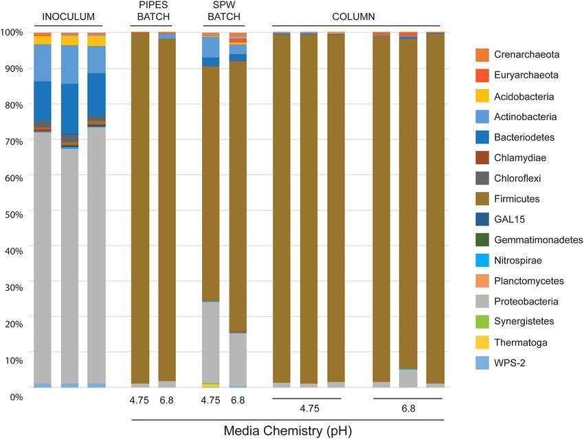
Calapa et al.                                                                                                 Microbial Fe(III) Reduction Under Flow

INTRODUCTION                                                                  Prior enrichment of canga-associated microorganisms
                                                                           from IFCs demonstrated that the microbial communities
The Southern Espinhaço Mountain Range (SE) of southeastern                 present were capable of Fe(III) reduction to extents that
Brazil contains commercially important, high-grade iron ore                could contribute to IFC formation (Parker et al., 2018), but
hosted by the Serra da Serpentina Group, a stratigraphic unit              Fe(II) that accumulates during Fe(III) (hydr)oxide reduction
which includes the iron-rich Serra do Sapo Formation (Auler                can adsorb to Fe(III) phase surfaces and induce mineral
et al., 2019). These sedimentary units were formed by the                  (trans)formations (Roden et al., 2000; Benner et al., 2002;
precipitation of Fe(III) and Si phases from solution during                Hansel et al., 2003, 2005; Gonzalez-Gil et al., 2005). These
the Proterozoic Eon (Weber et al., 2006; Rosière et al., 2019;             consequences of Fe(III) reduction could self-limit further
Silveira Braga et al., 2021). Iron ores can include magnetite              Fe(III) (hydr)oxide reduction, although subsurface water
(Fe3 O4 ), hematite (α-Fe2 O3 ), or a ferric oxyhydroxide like             flow could help overcome these limitations by advective
goethite (α-FeOOH) or limonite (FeO(OH)·n(H2 O), and high-                 transport of Fe(II) (Gonzalez-Gil et al., 2005; Minyard and
grade ore averages between 60 and 67% Fe (Dorr, 1964; Beukes               Burgos, 2007; Wefer-Roehl and Kübeck, 2014). Additionally,
et al., 2003; Rolim et al., 2016). The SE and Quadrilátero Ferrífero       it remained unclear if the extents of microbiological Fe(III)
(Iron Quadrangle; QF) located ∼150 km south of SE, contain                 (hydr)oxide dissolution observed were sufficient to induce
abundant Fe(III)-rich minerals, which can be found in intact               hydrologic alterations that would culminate in cave formation.
banded iron formation (BIF), Si-depleted ore, and canga rock               To understand whether this hydrologic flow could influence
(Beukes et al., 2003; Souza et al., 2015; Auler et al., 2019). BIF         Fe-reduction rates and enhance IFC formation we compared
contains alternating bands of quartz (SiO2 ) and either magnetite          batch cultures [where Fe(II) will accumulate] to columns
or hematite that range from a few millimeters to a few centimeters         [where Fe(II) is removed via flow] to evaluate how water
in thickness, and averages between 15 and 38% iron (Smith, 2015;           flow influenced microbiologically mediated Fe(III) reduction,
Rolim et al., 2016). Canga is a brecciated duricrust containing            and whether such activity could influence the hydraulic
clasts of iron oxide (usually BIF) with an iron-oxide cement               properties of canga.
matrix that averages between 57 and 62% iron (Dorr, 1964;
Spier et al., 2007; Gagen et al., 2019). Canga contains the most
poorly crystalline Fe(III) of the three major phases (i.e. canga,
                                                                           MATERIALS AND METHODS
BIF, and ore), with goethite the most prominent mineral phase
(Parker et al., 2013).
    Canga and BIF are generally considered highly resistant to
                                                                           Sample Collection and Preparation for
both mechanical and chemical weathering at pH ≥ 3 (Dorr,                   Batch Incubations and Column
1964; Johnson et al., 2012; Auler et al., 2014; Spier et al.,              Experiments
2018; Gagen et al., 2019), and canga covers the slopes and                 Five IFCs were sampled in the SE region of Brazil in December
valleys of the SE region. Yet this area is also associated with            2018. The cave designations are: CSS-0009, CSS-0080, CSS-
hundreds of caves (iron formation caves; IFCs) that form                   0010, CSS-0107, and CSS-0074. Authorization for sampling
mostly at the BIF/canga boundary (Auler et al., 2019). The                 during the destruction of these caves through mining had
identification of these caves suggests that processes leading              been approved by the Brazilian environmental agency, and the
to Fe(III) weathering and removal increase porosity at the                 sampling was part of a final recovery effort prior to mining.
canga/BIF interface, despite the resistance of both types of               Canga was collected from the interior of a cave that was
rocks to dissolution. At circumneutral pH, Fe solubility can               forming at a BIF-canga interface. Large (0.5–5.5 kg) chunks
be enhanced by microbially mediated reductive dissolution of               of canga were removed from cave walls using an electric
Fe(III) phases to relatively soluble Fe(II). This activity may             demolition hammer during the final sampling effort prior to
facilitate the mass transport necessary for the increased porosity         cave destruction and placed in plastic bags. The soft, microbial-
and the formation of the observed IFCs (Parker et al., 2013,               rich material behind the walls (sub muros) was collected by
2018). In support of this hypothesis, while the walls of the               first removing the rigid oxide layer and then using a sterile
IFCs are lined with a hard, oxidized layer of canga, the interior          garden shovel to place this material in sterile glass jars and
(approximately 3 cm behind) of the wall surface contains a                 sealed with plastic tape to maintain anaerobic/microaerobic
soft, gooey, water-saturated material that contains abundant               conditions. Samples were placed in a refrigerator upon return
microbial cells (Parker et al., 2018). Given the inhibition of             from the field. Collected canga was pulverized for column
Fe-reduction by oxygen, we wondered whether this material                  experiments by cutting large chunks into smaller pieces with
was involved in promoting Fe-reduction and increased porosity,             a water-cooled trim saw with a 25 cm blade, which were
leading to formation of the IFCs. Canga is a rather porous                 then processed through a ball mill (SPEX Industries, Inc.,
media, and active vertical percolation of water occurs during              Metuchen, NJ, United States) until all the material passed
rainfall, the patterns of which can be irregular, depending on             through a 1.44 mm sieve. This pulverized canga was sterilized by
season (Mesquita et al., 2017; Parker et al., 2018). As such,              autoclaving at 121◦ C for 15 min, allowing a 1 h recovery, and
intermittent periods of extensive water circulation around and             then autoclaved again to assure deactivation of spore-forming
within caves and their hosting rocks can occur, followed by water          bacteria. This sterilized canga was then dried at 65◦ C for 14–
stagnation or dry periods.                                                 16 h in an oven.

Frontiers in Microbiology | www.frontiersin.org                        2                                     July 2021 | Volume 12 | Article 696534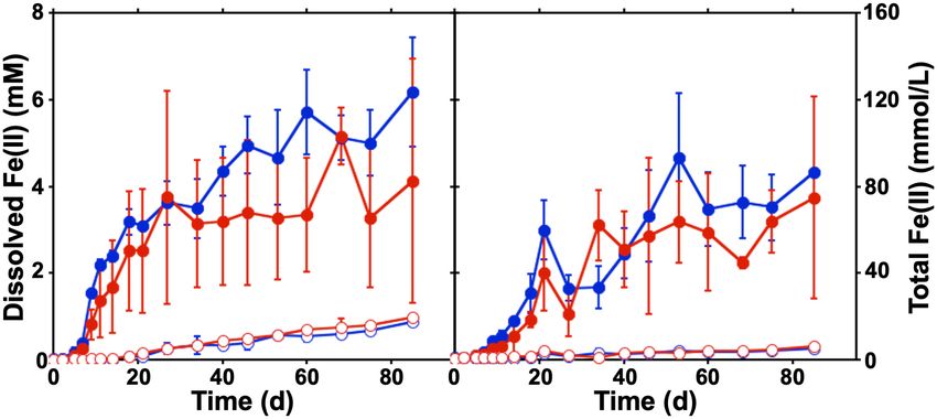
Calapa et al.                                                                                                Microbial Fe(III) Reduction Under Flow

Batch Incubations                                                         incubated statically for 7 days at room temperature between
Preparation of batch incubations was carried out in a Coy                 sampling events, whereupon 3–5 column void volumes of
anaerobic glove bag (Coy Laboratory Products, Inc., Grass Lake,           SPW were passed through at a flow rate of 0.2 mL/min,
MI, United States) filled with 3-5% H2 , balance N2 . Synthetic           and during this period samples were collected from each
porewater (SPW) composition was based on characterization of              column volume for measurement of pH, sulfate concentration,
IFC porewaters, and contained 5 mM CaCl2 , 0.1 mM Na2 SO4 ,               and dissolved Fe(II) concentration (described below). At the
and 0.1 mM KH2 PO4 . Sodium lactate (5.0 mM) was included                 completion of the experiments, breakthrough curves were
as an electron donor and carbon source, and the pH of the                 determined with 1 mM NaBr-amended (Acros Organics, Morris,
SPW was adjusted to either pH 4.75 or pH 6.8 with HCl or                  NJ, United States) SPW at a flow rate of 0.2 mL/min, and samples
NaOH, respectively, which were chosen to approximate the pH               were periodically collected for bromide quantification (described
of porewaters we have observed in IFCs (Parker et al., 2018).             below). At the conclusion of the experiments, columns were
O2 was removed from the SPW by bringing the liquid to nearly              deconstructed and sediments were removed for analysis of the
boiling, then cooling under a stream of N2 gas for 45 min. Once           microbial communities and quantification of total Fe(II) and
cooled, SPW-containing bottles were sealed, transferred to the            microbial cells.
anaerobic glove bag, and filter-sterilized with a 0.2 µm PES filter
(VWR, Radnor, PA, United States) until use in incubation bottles          Sample Processing and Analytical
or column experiments. Batch sediment incubations contained               Techniques
20 g of pulverized canga (equivalent to approximately 120 mmol            Column effluent samples for dissolved Fe(II) quantification
Fe(III)) and 60 mL of SPW in 160 mL serum bottles that were               were preserved in 0.5 M HCl, while those intended for
sealed with butyl rubber stoppers held in place with aluminum             measurement of pH, sulfate, and bromide were untreated.
crimp seals. Where appropriate, 5 g of sub muros material and             Solids were removed from the samples by centrifugation at
associated microbial community was used as inoculum for non-              12,100 × g for 5 min. To measure solid-associated Fe(II) in
sterile incubations. Incubations were carried out in triplicate.          column sediments, approximately 0.5 g of material was placed in
                                                                          microcentrifuge tubes with 1 mL 0.5 M HCl (Lovley and Phillips,
Column Assembly and Operation                                             1987) and solids were then separated from the solution by
All materials used for packing columns were acid-washed and               centrifugation before measurement of Fe(II) in the supernatant.
sterilized prior to use, and all columns (10 cm × 1 cm Econo-             Fe(II) was quantified by ferrozine assay (Stookey, 1970). pH was
Columns; Bio-Rad Laboratories, Hercules, CA, United States)               measured using a SevenGo Pro pH/Ion meter (Mettler Toledo,
were packed and operated in a Coy anaerobic glove bag as                  Columbus, OH, United States). Sulfate, bromide, phosphate,
described. Approximately 2 g of 3 and 2 mm diameter glass                 nitrate, and chloride were measured by ion chromatography
beads were placed at the bottom of the column, respectively,              with a Dionex DX-120 System with an IonPac AS22 Column
to prevent sediment clogging. The uninoculated columns were               and conductivity detection (Thermo Fisher Scientific, Waltham,
packed with pulverized sterile canga using a “lift” technique: a          MA, United States). To analyze the mineralogy of the
2 g lift of canga was added, followed by the injection of 200–            column at the end of the experiment, approximately 1 mL
500 µL SPW (described above) through bottom of the column.                of column contents were dried in a closed container with
After the addition of each lift, the columns were tapped to remove        CaCl2 (Acros Organics, Morris, NJ, United States) as a drying
air bubbles, and sediments were allowed to settle for 1–2 min.            agent under anaerobic conditions. Mineral characterization of
A suspension of sub muros was prepared with 8 g pulverized                the homogenized rock was determined by X-Ray Diffraction
canga and 8 g sub muros in 6 mL SPW, which was added 250 µL               (XRD) with a Rigaku Ultima IV (Rigaku, Woodlands, TX,
at a time after each lift of canga/SPW during column packing.             United States). Samples were analyzed with a 2θ between
The final amount of sub muros added was ∼2 mL, which resulted             5◦ and 70◦ and diffraction patterns were compared to those
in ∼9.2 × 10⁷ cells/g column material (as determined by direct            standards available on the American Mineralogist Crystal
cell counting; see below). All columns received 4 g of 2 mm               Structure Database (Downs and Hall-Wallace, 2003).
beads followed by 2 g of 3 mm beads added to on top of the
sediments to prevent clogging. After packing, columns contained           Microbial Community Analysis
a total of 16 g of solids and 7 mL liquid (either SPW only                Microbial cells in the sub muros inoculum, batch incubations,
in uninoculated columns or SPW and microbial suspension in                and in the columns at the conclusion of the experiments were
inoculated columns). All column incubations were carried out              enumerated by direct cell counting. Briefly, ∼1 g samples were
in triplicate in the anaerobic glove bag for 14 days prior to             collected while the columns remained in the anaerobic glove
the first sampling.                                                       bag and mixed with 1 mL of filter-sterilized Dulbecco pH
   Synthetic porewater was delivered to columns in an upward              7.4 phosphate buffered saline (PBS) (Thermo Fisher Scientific,
flow during sampling using a Masterflex L/S Precision Variable-           Waltham, MA, United States) and vigorously shaken by hand to
Speed Console peristaltic pump with Masterflex L/S 8-Channel              mix the contents. One hundred microliter of this mixture was
Pump Head (Cole-Parmer, Vernon Hills, IL, United States),                 then added to 9.9 mL sterile PBS and 1X thiazole green DNA
and 1.6 mm inner diameter Tygon tubing (Fischer Scientific,               stain (Biotium, Fremont, CA, United States). This mixture was
Pittsburgh, PA, United States). The columns were subsequently             incubated in the dark for 30 min at room temperature and then

Frontiers in Microbiology | www.frontiersin.org                       3                                     July 2021 | Volume 12 | Article 696534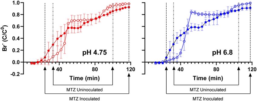
Calapa et al.                                                                                                      Microbial Fe(III) Reduction Under Flow

filtered onto a 0.2 µm GTBP Isopore polycarbonate membrane                     Gagen et al., 2019). The drivers of this enchanced reduction
filter via a Millipore stainless steel filtration unit (Millipore Sigma,       remain unclear at this time and represent a good target for
Darmstadt, Germany). The filter was removed and placed on a                    future research.
glass slide with 25 µL SlowFade Gold Antifade Mountant with                        In previous cultures using various Fe(III) mineral phases
DAPI (Thermo Fisher Scientific, Waltham, MA, United States)                    (including canga; Parker et al., 2018) we used a PIPES-buffered
and cells were counted using a BX53 fluorescent microscope                     (also at pH 4.75 and 6.8) mineral salts medium for growth. In
(Olympus America Inc., Center Valley, PA, United States). The                  these cultures we saw a shift in microbial community structure
total cell number was calculated based on 100 fields-of-view                   from the Proteobacteria-dominated sub muros to one dominated
(FOV) at 1,000× magnification, with the average number of cells                by the Firmicutes, representing >97% of sequences (Figure 2).
per FOV multiplied by the total dilution factor, area of the filter            In these previous experiments, we had assumed that the shift in
membrane, and standardized against the absolute weight of the                  community structure had been driven in part by the high amount
material/g (Hershey et al., 2018).                                             of organic carbon, while the closed nature of the experiment
    For DNA analyses, genomic DNA was extracted from samples                   allowed H2 to accumulate and drive fermentative Fe-reduction
using either then DNeasy PowerLyzer PowerSoil or PowerBiofilm                  by members of the Clostridia (Shah et al., 2014; Parker et al.,
Kit (Qiagen, Germantown, MD, United States). Subsamples                        2018). We tested this hypothesis in this study, using a basal
from the triplicate batch incubations were pooled before DNA                   medium (SPW) and 5 mM lactate as a carbon source. Analysis
extraction. PCR amplification of the 16S rRNA V3 and V4 regions                of partial 16S rRNA gene sequences in the batch incubations after
using the primers 806R (50 -GGA CTA CHV GGG TWT CTA AT-                        85 days revealed a similar dominance by fermentative Firmicutes
30 ) and 515F (50 -GTG CCA GCM GCC GCG GTA A-30 ), with                        (Figure 2). Nonetheless, using SPW/lactate, the Proteobacteria
samples identified using unique barcodes along with Illumina                   remained abundant, comprising 23 and 15% of the sequences
adapter sequences (Integrated DNA Technologies, Coralville, IA,                recovered from pH 4.75 and 6.8 incubations, respectively. We
United States). Amplification was using a Mastercycler Nexus                   also saw a small, but significant population of Actinobacteria (6%
Gradient (Eppendorf, Enfield, CT, United States), including a                  at pH 4.75 and 3% at pH 6.8) and Bacteriodetes (2.5% only at pH
3 min 94◦ C hot start, followed by 30 cycles of: denaturing at                 6.8) that had not been observed previously (Figure 2). The pH
94◦ C for 45 s, annealing at 50◦ C for 60 s, and then a 72◦ C                  of the uninoculated controls averaged 5.44, regardless of whether
extension for 90 s, followed by a final extension step at 72◦ C                the pH 4.75 or 6.8 SPW was used to initiate the experiment,
for 10 min. The PCR products were gel purified, and quantified                 suggesting that canga buffered the pH; however, in the inoculated
using a Qubit dsDNA HS Assay Kit (Life Technologies, Waltham,                  batch cultures, the SPW/lactate pH 4.75 culture increased to pH
MA, United States). Samples were then sequenced on an Illumina                 6.10, while the SPW pH 6.8 culture remained reasonably constant
MiSeq and de-multiplexed in QIIME2 (version 2020.2; Bolyen                     at pH 6.67. There was no dramatic change in pH of the cultures
et al., 2019) using cutadapt demux-paired and a quality check was              following the addition of sub muros at day 0. This suggests Fe(III)
carried out using q2-deblur-denoise-16S and quality-filter-q-score.            reduction through microbial activity likely raises the pH (Eq 1):
OTU picking and taxonomic assignments were completed using
the feature-classifier-classify-sklearn (Bolyen et al., 2019).                            Fe (OH)3 + 3H+ + e− → Fe2+ + 3H2 O                         (1)

                                                                               The similarity in pH of the final culture conditions may explain
RESULTS AND DISCUSSION                                                         the similarity of the final observed community profiles (Figure 2).
                                                                                  At the genus level within the dominant Firmicutes (Figure 3),
Fe(III) Reduction in Static Incubations                                        the SPW/lactate batch cultures displayed a different structural
To evaluate the canga-Fe(III) reducing activities of the microbial             diversity to our previous work. Previously, at pH 4.75 the batch
communities in the sub muric material, static batch incubations                cultures were dominated by members of the genus Clostridium
were conducted. Canga was provided as the Fe(III) source with                  (Family Clostridiales; 71%), with a small but significant
SPW at a pH of 4.75 or 6.8, which matched the measured pH                      representation by the Desulfosporosinus (Family Peptococcaceae;
values in situ (Parker et al., 2018). Minimal Fe(II) was generated             6%), while at pH 6.8, the PIPES batch cultures were dominated by
in uninoculated incubations, but accumulated in the sub muros-                 both the Desulfosporosinus (38%) and Clostridium (36%) (Parker
inoculated incubations at both pH 4.75 and 6.8 (Figure 1A).                    et al., 2018). Both cultures also contained minor populations
The concentration of dissolved Fe(II) that accumulated in the                  of the Paenibacilli (Family Bacillales; 3% at both pH 4.75 and
sub muros-inoculated batch incubations (approximately 5 mM)                    6.8). In the batch cultures presented here, we saw a similar
exceeded previous batch incubation work in which Shewanella                    dominance by members of the Clostridia (31% at pH 4.75 and
oneidensis MR-1 was used to catalyze canga-Fe(III) reduction                   47% at pH 6.8), and Desulfosporosinus (20 and 24% at pH 4.75
(less than 0.6 mM; Parker et al., 2013). Indeed, mean total Fe(II)             and 6.8, respectively). The Desulfosporosinus sp. are normally
concentration of sub muros-inoculated incubations exceeded                     associated with sulfate reduction, but have also been shown
80 mmol/L (Figure 1B). In previous work, a maximum of 3%                       to reduce Fe(III) enzymatically (Senko et al., 2009; Sato et al.,
of canga-Fe(III) could be reduced by S. oneidensis MR-1 (Parker                2019). If not enzymatic, the production of a minor amount
et al., 2013); however, greater extents of Fe(III) reduction have              of sulfide could be sufficient to enable Fe(III) reduction via S
been observed by fermentative enrichments and isolates from                    as an electron shuttle (Hansel et al., 2015). Members of the
canga by ourselves and other researchers (Parker et al., 2018;                 Paenibacilli, which have recently been demonstrated to play an

Frontiers in Microbiology | www.frontiersin.org                            4                                      July 2021 | Volume 12 | Article 696534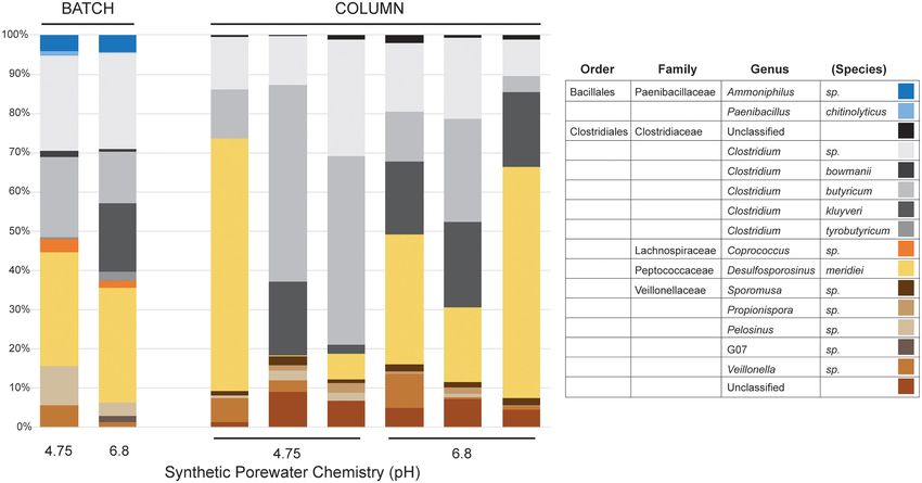
Calapa et al.                                                                                                                      Microbial Fe(III) Reduction Under Flow

  FIGURE 1 | Batch cultures of Fe(III) reduction in SPW canga. The concentration of dissolved Fe(II) (A) and total Fe(II) (B) were measured under static conditions over
  3 months. Comparisons were made between sterile canga (open circles) or canga inoculated with sub muros material (closed circles), with a basal SPW medium pH
  of 4.75 (red) or pH 6.8 (blue). Error bars represent the standard deviation of triplicate incubations.

  FIGURE 2 | Illumina sequencing results of phylum-level community diversity in batch and column cultures. Illumina sequencing of sub muros inoculated samples at
  day 0 are shown (inoculum). The diversity in our previous batch culture experiments, where the basal media was buffered with PIPES is shown (indicated as PIPES
  BATCH; Parker et al., 2018), followed by the cultures presented here using SPW with lactate (SPW BATCH). Illumina data is also provided for each of the individual
  columns in the flow-through experiments (COLUMN). The basal pH of each media formulation at day 0 is shown (Media Chemistry pH).

important role in iron oxide weathering in soils (including in                          as to why members of this genus would be enriched under the
Brazil; Loyaux-Lawniczak et al., 2019) were also represented                            batch culture conditions; however, their growth is stimulated by
at both pH 4.75 and 6.8 (4% of total diversity; Figure 3).                              fermentable carbohydrates, suggesting that the use of lactate may
Interestingly, we saw a higher percentage of members of the                             have enhanced their growth (Cotta and Forster, 2006; Rainey,
Coprococcus (Family Lachnospiraceae; 2%) at both pH 4.75                                2009). Members of this genus have not been associated with Fe-
and 6.8. The genus Coprococcus includes strict anaerobes that                           reduction, or isolated from iron-rich environments, although the
play an important role in carbohydrate fermentation in the                              production of H2 during fermentation may contribute to the
mammalian rumen, including lactate (Rainey, 2009). It is unclear                        overall culture Fe-reduction conditions (Cotta and Forster, 2006;

Frontiers in Microbiology | www.frontiersin.org                                     5                                              July 2021 | Volume 12 | Article 696534
Calapa et al.                                                                                                                       Microbial Fe(III) Reduction Under Flow

  FIGURE 3 | Illumina sequencing results of genus-level community diversity within the Firmicutes from the batch and column cultures. Only the SPW/lactate results
  are shown. The distribution of genera in the batch cultures (BATCH) and individual columns (COLUMN) are shown, along with the basal pH of the SPW at day 0 is
  shown. Given the myriad of Family- and Genera-level distributions within the Firmicutes, the Order/Family/Genus classification is provided for each identified species.

Parker et al., 2018). We also observed a significant representation                     the canga is highly impermeable with values as low as 10−8 m/s
by members of the Family Veillonellaceae, with 11% at pH                                (Mesquita et al., 2017). Thus, rainfall infiltrates quickly through
4.75 and 5% at pH 6.8 (Figure 3). Recently, genera within the                           the canga towards the caves and then drains rapidly toward
Veillonellaceae, such as Sporomusa spp. and Propionispora spp.,                         the surface, with very little retention of water, except in a
have been shown to carry out Fe(III) reduction (Sass et al.,                            few shallow internal ponds. Despite the robust Fe(III) reducing
2004; Kato et al., 2015); however, rather using respiratory Fe(III)                     activity observed in the batch incubations (Figure 1; Parker et al.,
reduction, the Sporomusa appear to use Fe(III) as an electron sink                      2018), they do not mimic the hydrologic flow associated with
in acetogenesis (Igarashi and Kato, 2021).                                              the rocks of the SE or QF in which cave formation occurs with
                                                                                        Fe(II) accumulating in the cultures. To mimic flow conditions
Fe(III) Reduction in Column Incubations                                                 in a laboratory setting, we packed canga into columns under
The underlying hypothesis of our work is that microbiological                           conditions analogous to the batch incubations, and introduced
Fe(III) reducing activities are sufficient to induce porosity                           flow into the system. This approach allowed us to answer the
generation within the host rocks (i.e., canga, BIF, and iron                            two major questions of this work: (1) does advective removal
ore); however, we have not observed hydrologic alterations of                           of biogenic Fe(II) enhance further canga-Fe(III) reduction and
cave hosting rocks. Biogenic Fe(II) can limit the extent of                             (2) are the Fe(III) reducing microbial activities associated with
Fe(III) (hydr)oxide reduction (Roden and Zachara, 1996; Urrutia                         the sub muric material sufficient to induce hydrologic alterations
et al., 1999; Roden and Urrutia, 2002; Roden, 2004, 2006),                              to the host rock.
and induce mineralogical changes that would otherwise limit                                 The columns were packed with crushed canga alone, or with
further Fe(III) reduction or limit the export of soluble Fe(II)                         crushed canga mixed with sub muric material. The columns were
(i.e., the formation of secondary minerals; Benner et al., 2002;                        incubated statically for 14 days, allowing Fe(III) reduction to
Hansel et al., 2003, 2005). Nonetheless, the advective removal of                       initiate, before four column volumes of SPW were then passed
biogenic Fe(II) as water flows through Fe(III) (hydr)oxide-rich                         through the column and collected separately for analysis of
rocks could enhance their reduction (Roden and Urrutia, 1999;                           effluent chemistry (Figure 4). This process of static incubation
Roden et al., 2000; Royer et al., 2004; Minyard and Burgos, 2007).                      followed by introduced flow was then repeated at 7 day
For example, 95% of Fe(III) coating on sand was reduced over                            intervals. Minimal dissolved Fe(II) was detected in the effluent
six months by Shewanella putrefaciens CN32 in flow-through                              of uninoculated control columns throughout the incubation
columns, compared to 13% of the Fe(III) in batch incubations                            (Figure 4), and effluent pH was ∼4.5–5.0, regardless of influent
(Roden et al., 2000).                                                                   SPW pH. This is slightly lower than the values obtained in
    The climate regime in the SE area is highly seasonal, with over                     the batch experiments shown in Figure 1. In the inoculated
80% of the ∼1,400 mm/year rainfall concentrated in November-                            columns, progressively higher concentrations of dissolved Fe(II)
March. Canga is a highly porous rock, with values between 24                            accumulated over the course of the incubation, with maximum
and 29% (Costa and Sá, 2018), while the friable ore underneath                          Fe(II) concentrations of approximately 3 mM Fe(II) detected

Frontiers in Microbiology | www.frontiersin.org                                     6                                              July 2021 | Volume 12 | Article 696534
Calapa et al. Microbial Fe(III) Reduction Under Flow
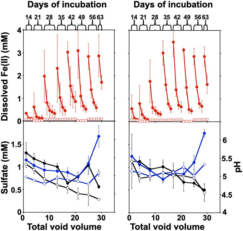
FIGURE 4 | Fe(III) reduction and changes in sulfate and pH in the column experiments. (A,B) Columns were operated semi-continuously, and sulfate and pH were
measured in each pore volume (four volumes) recovered after each static incubation. The column was disassembled after two column volumes at day 63 for post
mortem analysis. Error bars represent one standard deviation of triplicate columns. The concentration of dissolved Fe(II) is shown at pH 4.75 (A) and 6.8 (B). Sulfate
concentrations (black) and pH (blue) in column effluents are shown in panels (C) (pH 4.75) and (D) (pH 6.8). The values for uninoculated columns are shown with
open circles, with the sub muros-inoculated columns represented by closed circles.
after the fifth round of static incubation at both pH 4.75 and 6.8 of Fe(OH)3 were reductively dissolved and exported as Fe(II)
(Figures 4A,B). from the packing material of inoculated columns, with minimal
The concentration of total Fe(II) (dissolved and solid- export of Fe from uninoculated columns (Table 1). In the batch
associated) produced in the columns incubated at either pH 4.75
or 6.8 SPW are shown in Table 1. A two-sample t-test assuming
unequal variances suggested that there was no significant
TABLE 1 | Post mortem analysis of column contents.
difference (P = 0.97) in total dissolved Fe(II) accumulation in the
columns receiving SPW with pH 4.75 and 6.8 (Figure 4). The pH pH 4.75 with pH 4.75 pH 6.8 with pH 6.8
of the column effluent suggests that there was an increase in pH sub muros uninoculated sub muros uninoculated
above 6.0 (Figure 4C), similar to the batch cultures. The increase
Total Fe(II) 60 ± 15 4.4 ± 0.2 89 ± 19 4.3 ± 0.1
in Fe(III) reduction in the pH 4.75 column as the experiment (µmol/g)
progressed may reflect a change in column community structure Cell 9.3 × 10⁷ ± N/D 9.1 × 10⁷ N/D
as the cultures move toward similar pH conditions. The increase abundances 2.3 × 10⁵ ± 1.8 × 10⁵
in pH conditions is correlated with the increasing observation t = 0 (cell/g wet)
of Clostridium kluyveri (Figure 3) and may suggest either the Cell 4.0 × 10⁸ ± N/D 4.2 × 10⁸ ± N/D
selection of this species und these pH conditions, or a role in abundances 8.1 × 10⁷ 4.7 × 10⁷
t = 63 (cell/g
driving Fe(III)-reduction. wet)
While canga is composed mostly of goethite and poorly Fe(OH)3 38 ± 18 2.3 ± 0.2 40 ± 3.5 2.3 ± 0.01
crystalline Fe(III) (hydr)oxides (Parker et al., 2013), if we assume removed as
dissolved Fe(II) is derived from Fe(OH)3 , approximately 40 mg Fe2+ (mg)
Frontiers in Microbiology | www.frontiersin.org 7 July 2021 | Volume 12 | Article 696534Calapa et al. Microbial Fe(III) Reduction Under Flow
incubations, only ∼30 mg of Fe(OH)3 were reductively dissolved Veillonella, which represented ≥90% of the identified partial
(Figure 1). After two porewater replacement events (21 days), 16S rRNA gene sequences (Figure 3); however, members of the
the dissolved Fe(II) concentration in effluent from sub muros- Paenibacilli were not observed. There was some inter-column
inoculated columns exceeded 6 mM in total in pH 6.8 columns variability under each of the pH conditions, particularly in regard
and over 5 mM total in pH 4.75 columns; iron reduction levels to the dominance of Clostridium relative to Desulfosporosinus
which only accumulated after 60 days continuous culture in batch (Figure 3). In the Desulfosporosinus-dominated columns, we
incubations (Figures 1A, 4A,B). These results indicate that water saw a darkening of the column material, which could indicate
flow enhances the reductive solubilization of Fe from canga and sulfidogenesis, but there was no decrease in the effluent sulfate
separation of the Fe(II) products from solid phases. concentration over the course of the incubations (Figures 4C,D).
This suggests that while members of the Desulfosporosinus are
accumulating in these columns, they may be functioning as
Microbial Communities in Column Fe(III) reducers. Indeed, members of this genus have been
Incubations shown to be the primary Fe(III) reducers under oligotrophic
The microbial community composition in the batch incubations conditions (Nixon et al., 2017; Bomberg et al., 2019). Fe(III)
suggested that non-respiratory Fe(III) reduction could play a reduction is widespread among the Clostridia, including a strain
role in the observed iron reduction (Figure 3). To determine of Clostridium beijerinckii (Dobbin et al., 1999; Lehours et al.,
the extent of growth during column operation, we counted cells 2010; Shah et al., 2014; List et al., 2019). Indeed, in our
associated with the sub muros inoculum and at the conclusion of previous batch cultures were capable of extensive (in some cases,
the column experiments. All the columns seeded with sub muros complete) Fe(III) reduction (Parker et al., 2018), and Lentini et al.
were initially inoculated at ∼9.2 × 10⁷ cells/g. At 63 days, the (2012) have demonstrated that Clostridium-enriched cultures are
population had increased in the columns at pH 4.75 by 4.3×, with capable of extensive reduction of goethite- and hematite-Fe(III).
the cell number in the pH 6.8 column increasing 4.6×. These
data suggested an increase in microbial growth, and indeed the
higher cell number is the pH 6.8 columns matches a higher- Microbially Induced Hydrologic
level of Fe(III) reduction. No microbial cells were detected in the Alterations of Canga Columns
uninoculated controls (Table 1). Based on dissolved Fe(II) in column effluents, approximately
DNA extraction from the inoculated columns produced 40 mg of Fe(OH)3 were removed from the columns due to
sufficient DNA for Illumina sequencing, but repeated attempts to microbiological Fe(III) reduction (Table 1). To determine if
extract DNA from the uninoculated columns failed, matching the this export of mass impacted the hydraulic properties of the
observations by direct cell counting. Illumina sequencing of the columns, we pumped bromide-amended SPW through the
microbial communities in the columns matched our observations columns. Bromide breakthrough in the sub muros-inoculated
in batch culture (Figures 2, 3); there had been a shift from columns preceded that of the uninoculated columns, and
dominance by the Proteobacteria, to dominance by members of breakthrough was spread out in comparison to that of the
the Firmicutes. At the genus level, the columns were similarly uninoculated columns, which had a sharper curve (Figure 5).
dominated by members of Clostridium, Desulfosporosinus, and These observations indicate that flow through the uninoculated
FIGURE 5 | Bromide breakthrough curves of sub muros-inoculated (closed shapes) and uninoculated (open shapes) columns after 63 days of operation. The
columns that received the basal SPW pH 4.75 media are in red, with the SPW pH 6.8 in blue. Mass transfer zone (MTZ) lines represent initial breakthrough point
where bromide-amended SPW is mixing with bromide-free SPW and adsorption exhaustion point where column is saturated with bromide-amended SPW. SPW
was fed to columns at a rate of 0.2 mL/min. Error bars represent one standard deviation of triplicate columns.
Frontiers in Microbiology | www.frontiersin.org 8 July 2021 | Volume 12 | Article 696534Calapa et al. Microbial Fe(III) Reduction Under Flow
columns did not experience the same mass transfer resistance rocks in the SE and QF by microbiological weathering via
seen in the columns in which microbiological Fe(III) reduction microbial Fe(III) reduction (either through respiratory activity
occurred (Lassabatere et al., 2004; Koestel et al., 2011; Safadoust or as an electron sink) and separation, which can be enhanced
et al., 2016). The porosity that allowed earlier bromide by groundwater flow. These results should be applicable to other
breakthrough is due to reductive dissolution of Fe(III) phases and iron formation areas in Brazil and help explain why caves are
export of dissolved Fe(II). In similar column experiments, Liang larger in the iron deposits of Carajás, in the wetter Amazon
et al. (2019) found that bioreduction of sediment-associated Basin (Auler et al., 2019). A positive feedback mechanism, in
Fe(III) led to the structural breakdown of particles in the which fast infiltration water would lead to increased porosity
columns and led to the earlier breakthrough of poorly-diffusible and thus even faster water percolation could operate, enhancing
2,6-difluorobenzoate. No change in more diffusible bromide the mass transfer mechanisms required to mobilize Fe(II). Our
breakthrough was observed after Fe(III) bioreduction (Liang observations indicate that microorganisms associated with these
et al., 2019). In the work presented here, Fe(III) bioreduction systems are capable of robust Fe(III) reducing activity, which
was more extensive, with maximal effluent Fe(II) concentrations could induce sufficient reductive dissolution of Fe(III) phases to
of approximately 3 mM, in comparison to the maximal Fe(II) form a cave. The numerous caves of the SE and QF (>3,000; Auler
concentration of 0.3 mM observed by Liang et al. (2019). Taken et al., 2019) indicate that the activity is extensive and continuously
together, the extensive Fe(III) bioreduction observed in these occurring. Indeed, we have observed remarkably high dissolved
column experiments induced changes to the water flow paths in Fe concentrations in water circulating around caves in the
the packed canga. QF (Parker et al., 2018). This extensive weathering of SE and
QF Fe(III) phases may represent a previously underappreciated
component of regional, and perhaps global Fe budgets.
Biogeochemical Implications
The results of our experiments indicate that the Fe(III) reducing
activities of microorganisms associated with IFCs can induce
reductive dissolution of Fe(III) phases, resulting in the transport
DATA AVAILABILITY STATEMENT
of dissolved Fe(II) and hydrologic changes that are consistent The datasets presented in this study can be found in
with cave formation. While the Fe(III)-rich rocks of this region online repositories. The names of the repository/repositories
were generally considered to be resistant to weathering (Schuster and accession number(s) can be found in the article/
et al., 2012; Monteiro et al., 2014), it is becoming increasingly supplementary material.
clear that microbiological activities may induce extensive
transformations to these rocks, especially canga (Parker et al.,
2013, 2018; Levett et al., 2016, 2020; Gagen et al., 2018, 2019; Paz AUTHOR CONTRIBUTIONS
et al., 2020). Previous work has focused on the transformations
of canga-Fe as a mechanism of canga permanence, whereby KC, MM, and TR carried out the lab work. JS, AA, CP, and HB
the weathering resistance of canga is owed to the alternating carried out the fieldwork. All authors were involved in the design
reductive dissolution of Fe(III) (hydr)oxides and abiotic or of the experiments and contributed to the manuscript.
microbiological reoxidation of Fe(II) back to Fe(III) (Levett et al.,
2016, 2020; Gagen et al., 2018, 2019, 2020; Paz et al., 2020).
In this way, canga appears to be continuously weathering and FUNDING
reforming. The work here indicates that the Fe(III) rich phases
could be more extensively weathered and removed from the This research was funded by the NSF grant #1645180
systems, driven by the increased rates of Fe-reduction induced from NSF Geobiology and Low-temperature Geochemistry
by water flow. Thus, Fe may be extensively mobilized from program to HB and JS.
REFERENCES Bolyen, E., Rideout, J. R., Dillon, M. R., Bokulich, N. A., Abnet, C. C., Al-
Ghalith, G. A., et al. (2019). Reproducible, interactive, scalable and extensible
Auler, A. S., Parker, C. W., Barton, H. A., and Soares, G. A. (2019). “Iron formation microbiome data science using QIIME 2. Nat. Biotech. 37, 852–857. doi: 10.
caves: genesis and ecology,” in Encyclopedia of Caves, eds W. B. White, D. C. 1038/s41587-019-0209-9
Culver, and T. Pipan (London: Academic Press), 559–566. Bomberg, M., Claesson, L. L., Lamminmäki, T., and Kontula, A.
Auler, A. S., Piló, L. B., Parker, C. W., Senko, J. M., Sasowsky, I. D., and Barton, (2019). Highly diverse aquatic microbial communities separated
H. A. (2014). “Hypogene cave patterns in iron ore caves: convergence of forms by permafrost in Greenland show distinct features according to
or processes,” in Hypogene Cave Morphologies, eds K. Klimchou, I. D. Sasowsky, environmental niches. Front. Microbiol. 10:1583. doi: 10.3389/fmicb.2019.
J. Mylroie, and A. S. Engel (Lewisburg, PA: Karst Waters Institute), 15–19. 01583
Benner, S. G., Hansel, C. M., Wielinga, B. W., Barber, T. M., and Fendorf, S. (2002). Costa, T., and Sá, G. (2018). “Soft iron ores: geotechnical characteristics,” in
Reductive dissolution and biomineralization of iron hydroxide under dynamic Guidelines for Open Pit Slope Design in Weak Rocks, eds D. Martin, and P. Stacey
flow conditions. Environ. Sci. Technol. 36, 1705–1711. doi: 10.1021/es01 (Clayton South, VIC: CSIRO Publishing), 273–285.
56441 Cotta, M., and Forster, R. (2006). “The family Lachnospiraceae, including the
Beukes, N. J., Gutzmer, J., and Mukhopadhyay, J. (2003). The geology and genesis genera Butyrivibrio, Lachnospira and Roseburia,” in The Prokaryotes, Vol. 4,
of high-grade hematite iron ore deposits. Appl. Earth Sci. 112, 18–25. doi: eds M. Dworkin, S. Falkow, E. Rosenberg, K. H. Schleifer, and E. Stackebrandt
10.1179/037174503225011243 (New York, NY: Springer), 1002–1021.
Frontiers in Microbiology | www.frontiersin.org 9 July 2021 | Volume 12 | Article 696534Calapa et al. Microbial Fe(III) Reduction Under Flow Dobbin, P. S., Carter, J. P., San Juan, C. G.-S., von Hobe, M., Powell, A. K., non-diffusible nanoparticles in soils. Chemosphere 220, 391–402. doi: 10.1016/ and Richardson, D. J. (1999). Dissimilatory Fe(III) reduction by Clostridium j.chemosphere.2018.12.165 beijerinckii isolated from freshwater sediment using Fe(III) maltol enrichment. List, C., Hosseini, Z., Meibom, K. L., Hatzimanikatis, V., and Bernier-Latmani, FEMS Microbiol. Lett. 176, 131–138. doi: 10.1111/j.1574-6968.1999.tb13653.x R. (2019). Impact of iron reduction on the metabolism of Clostridium Dorr, J. V. N. (1964). Supergene iron ores of minas Gerais, Brazil. Econ. Geol. 59, acetobutylicum. Environ. Microbiol. 21, 3548–3563. doi: 10.1111/1462-2920. 1203–1240. doi: 10.2113/gsecongeo.59.7.1203 14640 Downs, R. T., and Hall-Wallace, M. (2003). The American Mineralogist crystal Lovley, D. R., and Phillips, E. J. P. (1987). Rapid assay for microbially reducible structure database. Am. Mineral. 88, 247–250. ferric iron in aquatic sediments. Appl. Envir. Microbiol. 53, 1536–1540. doi: Gagen, E. J., Levett, A., Paz, A., Bostlemann, H., da Silva Valadares, R. B., 10.1128/aem.53.7.1536-1540.1987 Bitencourt, J. A. P., et al. (2020). Accelerating microbial iron cycling promotes Loyaux-Lawniczak, S., Vuilleumier, S., and Geoffroy, V. A. (2019). Efficient re-cementation of surface crusts in iron ore regions. Microb. Biotechnol. 13, reduction of iron oxides by Paenibacillus spp. strains isolated from tropical soils. 1960–1971. doi: 10.1111/1751-7915.13646 Geomicrobiol. J. 36, 423–432. doi: 10.1080/01490451.2019.1566415 Gagen, E. J., Levett, A., Paz, A., Gustauer, M., Caldeira, C. F., da Silva Valadares, Mesquita, D. C., Dantas, J. C. M., Paula, R. S., and Guerra, K. J. (2017). Estudo dos R. B., et al. (2019). Biogeochemical processe in canga ecosystems: armoring of parâmetros hidrodinâmicos obtidos em ensaios de campo em itabiritos brandos iron ore against erosion and importance in iron duricrust restoration in Brazil. da porção sudoeste do Quadrilátero Ferrífero, MG. Geonomos 25, 12–19. Ore Geol. Rev. 107, 573–586. doi: 10.1016/j.oregeorev.2019.03.013 Minyard, M. L., and Burgos, W. D. (2007). Hydrologic flow controls on biologic Gagen, E. J., Levett, A., Shuster, J., Fortin, D., Vasconcelos, P. M., and Southam, iron(III) reduction in natural sediments. Environ. Sci. Technol. 41, 1218–1224. G. (2018). Microbial diversity in actively forming iron oxides from weathered doi: 10.1021/es0619657 banded iron formation systems. Microbes Environ. 33, 385–393. doi: 10.1264/ Monteiro, H. S., Vasconcelos, P. M., Farley, K. A., Spier, C. A., and Mello, C. L. jsme2.ME18019 (2014). (U-Th)/He geochronology of goethite and the origin and evolution of Gonzalez-Gil, G., Amonette, J. E., Romine, M. F., Gorby, Y. A., and Geesey, cangas. Geochim. Cosmochim. Acta 131, 267–289. doi: 10.1016/j.gca.2014.01. G. G. (2005). Bioreduction of natural specular hematite under flow conditions. 036 Geochem. Cosmochim. Acta 69, 1145–1155. doi: 10.1016/j.gca.2004.08.014 Nixon, S. L., Telling, J. P., Wadham, J. L., and Cockell, C. S. (2017). Viable cold- Hansel, C. M., Benner, S. G., and Fendorf, S. (2005). Competing Fe(II)-induced tolerant iron-reducing microorganisms in geographically diverse subglacial mineralization pathways of ferrihydrite. Environ. Sci. Technol. 29, 7147–7153. environments. Biogeosciences 14, 1445–1455. doi: 10.5194/bg-14-1445-2017 doi: 10.1021/es050666z Parker, C. W., Auler, A. S., Barton, M. D., Sasowsky, I. D., Senko, J. M., and Barton, Hansel, C. M., Benner, S. G., Neiss, J., Dohnalkova, A., Kukkadapu, R. K., and H. A. (2018). Fe (III) reducing microorganisms from iron ore caves demonstrate Fendrof, S. (2003). Secondary mineralization pathways induced by dissimilatory fermentative Fe (III) reduction and promote cave formation. Geomicrobiol. J. iron reduction of ferrihydrite under advective flow. Geochem. Cosmochim. Acta 35, 311–322. doi: 10.1080/01490451.2017.1368741 67, 2977–2992. doi: 10.1016/S0016-7037(03)00276-X Parker, C. W., Wolf, J. A., Auler, A. S., Barton, H. A., and Senko, J. M. (2013). Hansel, C. M., Lentini, C. J., Tang, Y., Johnston, D. T., Wankel, S. D., and Jardine, Microbial reducibility of Fe (III) phases associated with the genesis of iron P. M. (2015). Dominance of sulfur-fueled iron oxide reduction in low-sulfate ore caves in the Iron Quadrangle, Minas Gerais, Brazil. Minerals 3, 395–411. freshwater sediments. ISME J. 9, 2400–2412. doi: 10.1038/ismej.2015.50 doi: 10.3390/min3040395 Hershey, O. S., Kallmeyer, J., Wallace, A., Barton, M. D., and Barton, H. A. (2018). Paz, A., Gagen, E. J., Levett, A., Zhao, Y., Kopittke, P. M., and Southam, G. (2020). High microbial diversity despite extremely low biomass in a deep karst aquifer. Biogeochemical cycling of iron oxides in the rhizosphere of plants grown on Front. Microbiol. 9:2823. doi: 10.3389/fmicb.2018.02823 ferruginous duricrust (canga). Sci. Total Environ. 713:136637. doi: 10.1016/j. Igarashi, K., and Kato, S. (2021). Reductive transformation of Fe(III) scitotenv.2020.136637 (oxyhydr)oxides by mesophilic momoacetogens in the genus Sporomusa. Rainey, F. A. (2009). “Family V. Lachnospiraceae fam. nov,” in Bergey’s Manual of Front. Microbiol. 12:600808. doi: 10.3389/fmicb.2021.600808 Systematic Bacteriology, Vol. 3, eds P. De Vos, G. M. Garrity, D. Jones, N. R. Johnson, D. B., Kanao, T., and Hedrich, S. (2012). Redox transformations of iron Krieg, W. Ludwig, F. A. Rainey, et al. (Dordrecht: Springer), 921–968. at extremely low pH: fundamental and applied aspects. Front. Microbiol. 3:96. Roden, E. E. (2004). Analysis of long-term bacterial vs. chemical Fe(III) oxide doi: 10.3389/fmicb.2012.00096 reduction kinetics. Geochem. Cosmochim. Acta 68, 3205–3216. doi: 10.1016/j. Kato, S., Yumoto, I., and Kamagata, Y. (2015). Isolation of acetogenic bacteria that gca.2004.03.028 induce biocorrosion by utilizing metallic iron as the sole electron donor. Appl. Roden, E. E. (2006). Geochemical and microbiological controls on dissimilatory Environ. Microbiol. 81, 67–73. doi: 10.1128/AEM.02767-14 iron reduction. C. R. Geosci. 338, 456–467. doi: 10.1016/j.crte.2006.04.009 Koestel, J. K., Moeys, J., and Jarvis, N. J. (2011). Meta-analysis of the effects of Roden, E. E., and Urrutia, M. M. (1999). Ferrous iron removal promotes microbial soil properties, site factors and experimental conditions on preferential solute reduction of crystalline iron(III) oxides. Environ. Sci. Technol. 33, 1847–1853. transport. Hydrol. Earth Syst. Sci. Discuss. 8, 10007–10052. doi: 10.5194/hessd- doi: 10.1021/es9809859 8-10007-2011 Roden, E. E., and Urrutia, M. M. (2002). Influence of biogenic Fe(II) on bacterial Lassabatere, L., Winiarski, T., and Galvez-Cloutier, R. (2004). Retention of three crystalline Fe(III) oxide reduction. Geomicrobiol. J. 19, 209–251. doi: 10.1080/ heavy metals (Zn, Pb, and Cd) in a calcareous soil controlled by the modification 01490450252864280 of flow with geotextiles. Environ. Sci. Technol. 38, 4215–4221. doi: 10.1021/ Roden, E. E., Urrutia, M. M., and Mann, C. J. (2000). Bacterial reductive dissolution es035029s of crystalline Fe(III) oxide in continuous-flow column reactors. Appl. Environ. Lehours, A.-C., Rabiet, M., Morel-Desrosiers, N., Morel, J.-P., Jouve, L., Arbeille, Microbiol. 66, 1062–1065. doi: 10.1128/AEM.66.3.1062-1065.2000 B., et al. (2010). Ferric iron reduction by fermentative strain BS2 isolated from Roden, E. E., and Zachara, J. M. (1996). Microbial reduction of crystalline iron(III) an iron-rich anoxic environment (Lake Pavin, France). Geomicrobiol. J. 27, oxides: influence of oxide surface area and potential for cell growth. Environ. 714–722. doi: 10.1080/01490451003597663 Sci. Technol. 30, 1618–1628. doi: 10.1021/es9506216 Lentini, C. J., Wankel, S. D., and Hansel, C. M. (2012). Enriched iron(III)- Rolim, V. K., Rosière, C. A., Santos, J. O. S., and McNaughton, N. J. (2016). The reducing bacterial communities are shaped by carbon substrate and iron oxide Orosirian-Statherian banded iron formation-bearing sequences of the southern mineralogy. Front. Microbiol. 3:404. doi: 10.3389/fmicb.2012.00404 border of the Espinhaço Range, Southeast Brazil. J. S. Am. Earth Sci. 65, 43–66. Levett, A., Gagen, E., Shuster, J., Rintoul, L., Tobin, M., Vongsvivut, J., et al. (2016). doi: 10.1016/j.jsames.2015.11.003 Evidence of biogeochemical processes in iron duricrust formation. J. S. Am. Rosière, C., Bekker, A., Rolim, V., and Santos, J. (2019). Post-Great Oxidation Earth Sci. 71, 131–142. doi: 10.1016/j.jsames.2016.06.016 Event Orosirian–Statherian iron formations on the São Francisco Levett, A., Vasconcelos, P. M., Gagen, E. J., Rintoul, L., Spier, C., Guagliardo, P., craton: geotectonic implications. I. Arc 28:e12300. doi: 10.1111/iar. et al. (2020). Microbial weathering signatures in lateritic ferrginous duricrusts. 12300 Earth Planet. Sci. Lett. 538:116209. doi: 10.1016/j.epsl.2020.116209 Royer, R. A., Dempsey, B. A., Jeon, B.-H., and Burgos, W. D. (2004). Inhibition Liang, X., Radosevich, M., Löffler, F., Schaeffer, S. M., and Zhuang, J. (2019). Impact of biological reductive dissolution of hematite by ferrous iron. Environ. Sci. of microbial iron oxide reduction on the transport of diffusible tracers and Technol. 38, 187–193. doi: 10.1021/es026466u Frontiers in Microbiology | www.frontiersin.org 10 July 2021 | Volume 12 | Article 696534
Calapa et al.                                                                                                                               Microbial Fe(III) Reduction Under Flow

Safadoust, A., Khaboushan, E. A., Mahboubi, A. A., Gharabaghi, B.,                           Souza, A., Figueiredo e Silva, R., Rosière, C., Dias, G., and Morais, F. (2015).
    Mosaddeghi, M. H., Ahrens, B., et al. (2016). Comparison of three                           Estudos geoquímicos de iabiritos da Serra do Sapo, espinhaço meridional,
    models describing bromide transport affected by different soil structure                    Minas Gerais. Rev. Geonomos 22, 1–17. doi: 10.18285/geonomos.v22i2.313
    types. Arch. Agron. Soil Sci. 62, 674–687. doi: 10.1080/03650340.2015.10                 Spier, C. A., de Oliveira, S. M. B., Sial, A. N., and Rios, F. J. (2007). Geochemistry
    74184                                                                                       and genesis of the banded iron formations of the Cauê Formation, Quadrilátero
Sass, H., Overmann, J., Rütters, H., Babenzien, H.-D., and Cypionka, H. (2004).                 Ferrífero, Minas Gerais, Brazil. Precambrian Res. 152, 170–206. doi: 10.1016/j.
    Desulfosporomusa polytropa gen. nov., sp. nov., a novel sulfate-reducing                    precamres.2006.10.003
    bacterium from sediments of an oligotrophic lake. Arch. Microbiol. 182, 204–             Spier, C. A., Levett, A., and Rosière, C. A. (2018). Geochemistry of canga (ferricrete)
    211. doi: 10.1007/s00203-004-0703-3                                                         and evolution of the weathering profile developed on itabirite and iron ore in
Sato, Y., Hamai, T., Hori, T., Aoyagi, T., Inaba, T., Kobayashi, M., et al. (2019).             the Quadrilátero Ferrífero, Minas Gerais, Brazil. Miner. Depos. 54, 983–1010.
    Desulfosporosinus spp. were the most predominant sulfate-reducing bacteria in               doi: 10.1007/s00126-018-0856-7
    pilot- and laboratory-scale passive bioreactors for acid mine drainage treatment.        Stookey, L. L. (1970). Ferrozine—a new spectrophotometric reagent for iron. Anal.
    Appl. Microbiol. Biotechnol. 103, 7783–7793. doi: 10.1007/s00253-019-10                     Chem. 42, 779–781.
    063-2                                                                                    Urrutia, M. M., Roden, E. E., and Zachara, J. M. (1999). Influence of aqueous and
Schuster, D. L., Farley, K. A., Vasconcelos, P. M., Balco, G., Monteiro, H. S.,                 solid-phase Fe(II) complexants on microbial reduction of crystalline iron(III)
    Waltenberg, K., et al. (2012). Cosmogenic 3 He in hematite and goetite                      oxides. Environ. Sci. Technol. 33, 4022–4028. doi: 10.1021/es990447b
    from Brazilian canga duricrust demonstrates the extreme stability of these               Weber, K. A., Achenbach, L. A., and Coates, J. D. (2006). Microorganisms pumping
    surfaces. Earth Planet. Sci. Lett. 329–330, 41–50. doi: 10.1016/j.epsl.2012.                iron: anaerobic microbial iron oxidation and reduction. Nat. Rev. Microbiol. 4,
    02.017                                                                                      752–764. doi: 10.1038/nrmicro1490
Senko, J. M., Zhang, G., McDonough, J. T., Bruns, M. A., and Burgos, W. D.                   Wefer-Roehl, A., and Kübeck, C. (2014). Guidelining Protocol for Soil-Column
    (2009). Metal reduction at low pH by a Desulfosporosinus species: implications              Experiments Assessing Fate and Transport of Trace Organics. 308339.
    for the biological treatment of acidic mine drainage. Geomicrobiol. J. 26, 71–82.           Available online at: http://demeau-fp7.eu/sites/files/D123a Guidelines Column
    doi: 10.1080/01490450802660193                                                              experiments.pdf (accessed June 14, 2018).
Shah, M., Lin, C.-C., Kukkadapu, R., Engelhard, M. H., Zhao, X., Wang, Y., et al.
    (2014). Syntrophic effects in a subsurface Clostridial consortium on Fe(III)-            Conflict of Interest: The authors declare that the research was conducted in the
    (oxyhydr)oxide reduction and secondary mineralization. Geomicrobiol. J. 31,              absence of any commercial or financial relationships that could be construed as a
    101–115. doi: 10.1080/01490451.213.806601                                                potential conflict of interest.
Silveira Braga, F. C., Rosière, C. A., Schneider Santos, J. O., Hagemann, S. G.,
    Danyushevsky, L., and Valle Salles, P. (2021). Geochemical and tectonic                  Copyright © 2021 Calapa, Mulford, Rieman, Senko, Auler, Parker and Barton.
    constraints on the genesis of iron formation-hosted magnetite-hematite                   This is an open-access article distributed under the terms of the Creative Commons
    deposits at the Guanhães Block (Brazil) by contact metasomatism with                     Attribution License (CC BY). The use, distribution or reproduction in other forums
    pegmatite intrusions. Ore Geol. Rev. 129:103931. doi: 10.1016/j.oregeorev.2020.          is permitted, provided the original author(s) and the copyright owner(s) are credited
    103931                                                                                   and that the original publication in this journal is cited, in accordance with accepted
Smith, A. J. B. (2015). The life and times of banded iron formations. Geology 43,            academic practice. No use, distribution or reproduction is permitted which does not
    1111–1112. doi: 10.1130/focus122015.1                                                    comply with these terms.

Frontiers in Microbiology | www.frontiersin.org                                         11                                                 July 2021 | Volume 12 | Article 696534
You can also read