The RNAi mechanism regulates a new exonuclease gene in- volved in the virulence of Mucorales - Preprints.org
←
→
Page content transcription
If your browser does not render page correctly, please read the page content below
Preprints (www.preprints.org) | NOT PEER-REVIEWED | Posted: 14 January 2021                                       doi:10.20944/preprints202101.0277.v1

     Article

     The RNAi mechanism regulates a new exonuclease gene in-
     volved in the virulence of Mucorales
     Carlos Pérez-Arques1, María Isabel Navarro-Mendoza1, Laura Murcia1, Eusebio Navarro1, Victoriano Garre1,*, Fran-
     cisco Esteban Nicolás1,*

                                             1   Departmento de Genética y Microbiología, Facultad de Biología, Universidad de Murcia, Spain.

                                             * Correspondence: fnicolas@um.es; vgarre@um.es

                                             Abstract: Mucormycosis is a lethal disease caused by Mucorales, which are emerging as human

                                             causes that explain the high mortality for this disease. Consequently, the research community is
                                             searching for virulence determinants that could be repurposed as targets to develop new treatments
                                             against mucormycosis. Our work explores an RNA interference (RNAi)-based approach to find tar-
                                             gets involved in the virulence of Mucorales. A transcriptome-wide analysis compared sRNAs and
                                             their target mRNAs in two Mucor lusitanicus different pathotypes, virulent and avirulent, generating
                                             a list of 75 loci selected by their differential sRNA accumulation in these strains. As a proof of con-
                                             cept and validity, an experimental approach characterized two loci showing opposite behavior, con-
                                             firming that RNAi activity causes their differential expression in the two pathotypes. We generated
                                             deletion mutants for two loci and a knockin-strain overexpressing for one these loci. Their functional
                                             analysis in murine virulence assays identified the gene wex1, a putative DEDDy exonuclease with
                                             RNase domains, as an essential factor for virulence. The identification of wex1 showed the potential
     Citation: Lastname, F.; Lastname, F.;    of our approach to discover virulence factors not only in Mucorales but also in any other fungal
     Lastname, F. Title. Int. J. Mol. Sci.    model with an active RNAi machinery. But, more importantly, it adds a new layer to the biological
     2021, 22, x.                             processes controlled by RNAi in M. lusitanicus, confirming that the Dicer-dependent RNAi path-
     https://doi.org/10.3390/xxxxx            way can silence gene expression to promote virulence.

                                             Keywords: mucormycosis; Mucor lusitanicus; Mucorales; RNAi; exonuclease; wex1 gene; virulence;
                                             virulence factor.

                                             1. Introduction
                                                   Pathogens and hosts are continuously adapting their defense mechanisms to succeed
                                             and survive during their interactions. Among human fungal pathogens, Mucorales are
                                             emerging as a source of new adaptations such as their capability of infecting immuno-
                                             competent hosts, general resistance to most of the current antifungal compounds, and
                                             aggressive and virulent behavior during the infection known as mucormycosis [1–4].
                                             These potentiated virulence features can explain our current lack of effective treatments,
                                             which is the direct cause behind the alarming mortality rates that exceed 90% in dissem-
                                             inated infections [5–7]. In this context, we are a step behind in the fight against these
                                             emerging pathogens, evidencing the imperious demand for studies designed to discover
                                             new molecular targets that can serve to develop effective antifungal compounds.
                                                   The main reason explaining the lack of targets for effective antifungal compounds is
                                             the disparity between Mucorales and other distant fungi like Ascomycetes and Basidio-
                                             mycetes [8,9]. Thus, our arsenal of antifungal compounds was initially selected for their
                                             high efficiency against other fungal pathogens such as Aspergillus and Candida spp., ne-
                                             glecting the study of mucormycosis for being considered a rare infection only affecting

               © 2021 by the author(s). Distributed under a Creative Commons CC BY license.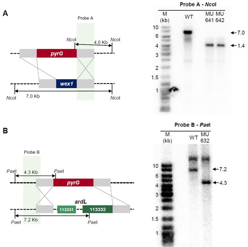
Preprints (www.preprints.org) | NOT PEER-REVIEWED | Posted: 14 January 2021                 doi:10.20944/preprints202101.0277.v1

                                 immunocompromised patient until very recently [3,10]. Mucoralean pathogens possess
                                 specific attributes like non-septated hyphae and a cell wall containing the polysaccharide
                                 chitosan, an N-deacetylated version of chitin [8]. Recently, new studies have found other
                                 molecular divergences as their atypical centromeric chromatin, which have lost two es-
                                 sential components of the kinetochore –CENP-A and CENP-C– resulting in a mosaic of
                                 point and regional centromeres [11]. Regarding their intrinsic antifungal drug resistance,
                                 the dissection of the enzymes involved in the synthesis of ergosterol has unveiled another
                                 deviation only conserved in these fungi. Thus, the lanosterol 14α-demethylase CYP51 F5
                                 of Mucorales has two amino acid substitution compared to other fungi, which has been
                                 related to their resistance to short-tailed azole compounds [4]. However, there is a molec-
                                 ular process that has paramount importance in regulating the biology of these fungi: their
                                 RNAi mechanism. In Mucorales, RNAi has been related to their antifungal resistance and
                                 virulence, but also to their general physiology [12,13]. Up to three different RNAi path-
                                 ways have been described and involved in the gene regulation of the fungus Mucor lusi-
                                 tanicus [14,15]. The first one produces small RNAs from exonic regions (ex-siRNAs, exonic
                                 small interfering RNAs) that regulate hundreds of mRNAs [16]. Mutants in essential
                                 genes of this pathway show defects in general processes such as growth, morphology,
                                 asexual sporulation, and sexual reproduction [13]. The second pathway, known as the
                                 epimutation pathway, is directly involved in an exclusive antifungal resistance mecha-
                                 nism of Mucorales [12,17,18]. This pathway generates temporarily resistant strains to an-
                                 tifungal compounds like FK506 and 5-fluoroorotic acid (5-FOA). This transient resistance
                                 is lost as soon as the antifungal compound is removed from the media. The mechanism
                                 behind this resistance is RNAi, which explicitly degrades the mRNA of the genes that are
                                 targets of the antifungal compounds. Thus, the RNAi mechanism specifically degrades
                                 the mRNAs of pyrimidine synthesis genes –pyrG or pyrF– in the presence of 5-FOA,
                                 whereas the fkbA mRNA is degraded in the presence of FK506. The third RNAi pathway
                                 is the most recently described in Mucorales and also one of their unique features as it is
                                 only conserved in this group of fungi [19]. It is a non-canonical RNAi pathway (NCRIP)
                                 because its machinery does not depend on Dicer and Argonaute proteins to degrade tran-
                                 scripts. Instead, NCRIP relies on RNA-dependent RNA polymerases (RdRPs) and a novel
                                 ribonuclease III-like named R3B2. NCRIP is involved in the regulation of virulence in Mu-
                                 corales. Thus, mutants in this pathway show reduced virulence in a murine model, likely
                                 because NCRIP regulates the expression of hundreds of genes during the interaction of
                                 macrophages and fungal spores [20–24].
                                       The studies dissecting the RNAi pathways of Mucorales have unveiled the regulation
                                 of thousands of genes by this mechanism. Although many of these genes must be involved
                                 in virulence, the broad regulatory control of RNAi over other physiological processes hin-
                                 ders identifying unknown virulence factors. Previous works demonstrated the validity of
                                 a strategy based on the comparative genomic analysis between virulent and avirulent
                                 strains of Mucor lusitanicus [25]. Focusing on these different pathotypes, a comparative
                                 transcriptional analysis during macrophage phagocytosis revealed an intricate gene net-
                                 work activated in the virulent strain to survive the immune response [24]. Here, we pro-
                                 pose a similar approach, but comparing the loci regulated by RNAi in the virulent and
                                 avirulent pathotypes. The result of this strategy revealed a population of differentially
                                 regulated loci accumulating sRNAs that could be involved in the virulence differences
                                 observed in these strains. Selected candidate genes from this population have been exper-
                                 imentally evaluated, demonstrating that our strategy can identify new virulence factors
                                 regulated by RNAi.

                                 2. Results
                                 2.1. Comparative analysis of sRNAs transcriptomic profiles between virulent and avirulent
                                 pathotypes
                                     Previous studies showed differential phenotypes regarding virulence between two
                                 opposite mating type strains of Mucor lusitanicus. Thus, the mating-type (-) CBS277.49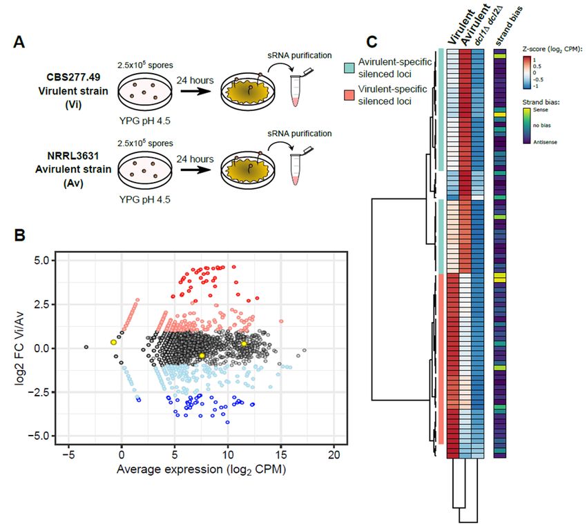
Preprints (www.preprints.org) | NOT PEER-REVIEWED | Posted: 14 January 2021               doi:10.20944/preprints202101.0277.v1

                                 and its derivative strains (hereinafter Vi) have an increased virulence potential in immu-
                                 nosuppressed mice, manifested by high mortality rates. In opposition, the mating-type (+)
                                 NRRL3631 (hereinafter Av) is harmless at the same doses [25]. Also, several differences at
                                 a genomic and transcriptomic level were related to the opposite virulent behavior of these
                                 two strains [25]. However, the complexity of the pathogenic potential and the profuse
                                 regulatory functions of the RNAi mechanism in M. lusitanicus suggested an extra regula-
                                 tory layer at the transcriptomic level [21]. To test this hypothesis and explore the role of
                                 the RNAi mechanism in the virulence of Mucorales, sRNA samples from both strains were
                                 deeply sequenced and compared (Figure 1, A). sRNA samples were obtained from myce-
                                 lia grown for 24h at 26ºC in rich medium YPG. After sRNA sequencing, sRNA reads were
                                 mapped to the genome of M. lusitanicus (Muccir1_3), to compare the endogenous short
                                 RNA (esRNA) accumulation patterns between the two pathotypes. The results of this
                                 analysis revealed a total of 843 loci showing differential esRNA accumulation (log2 FC ≥ 1
                                 and ≤ -1) (Figure 1B) (Table S1). The amount of sRNA overaccumulating loci is evenly
                                 distributed among Vi and Av. There are 454 sRNA overproducing loci in the Vi pathotype
                                 as opposed to 350 loci in the Av pathotype. Among these loci, we selected a total of 84
                                 showing high significance (log2 FC ≥ 2.5 and ≤ -2.5, and p-value ≤ 0.05) as the most likely
                                 regions regulated by the RNAi pathway (Figure 1B and 1C) (Table S2). M. lusitanicus har-
                                 bors several RNAi pathways, both Dicer-dependent and independent. To discriminate
                                 between the two, the transcriptomic sRNA data of a double Dicer mutant (dcl1Δ dcl2Δ)
                                 was also analyzed. Loci producing Dicer-dependent sRNAs in the Vi or Av pathotype
                                 would show a substantial decrease in sRNA production in the dcl1Δ dcl2Δ mutant, con-
                                 firming that Dicer activity is needed to generate those sRNAs. A total of 75 loci from the
                                 previous 84 show a decrease of sRNAs in this double mutant (Figure 1C, third column)
                                 [26,27], suggesting that Dicer-dependent pathways are critical for the differences observed
                                 among the two pathotypes. In M. lusitanicus, Dicer generates both sense and preferentially
                                 antisense sRNAs to promote active silencing. Hence, the average ratio of sense:antisense
                                 sRNAs in the two pathotypes was analyzed, confirming that these loci produced mainly
                                 antisense sRNAs. To further delimitate the role of the RNAi pathway in the two strains,
                                 the 75 loci were grouped in two different clusters: 35 of them showing low accumulation
                                 in the Vi strain, and 40 showing low accumulation in the Av strain (Figure 1C, first and
                                 second columns, respectively). Considering the different virulence potentials observed
                                 between both pathotypes, the sRNA profiles analyzed here led us to identify a specifically
                                 delimitated list of candidates to find new genes involved in the pathogenesis of Mu-
                                 corales.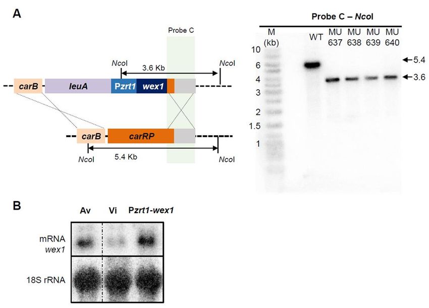
Preprints (www.preprints.org) | NOT PEER-REVIEWED | Posted: 14 January 2021 doi:10.20944/preprints202101.0277.v1
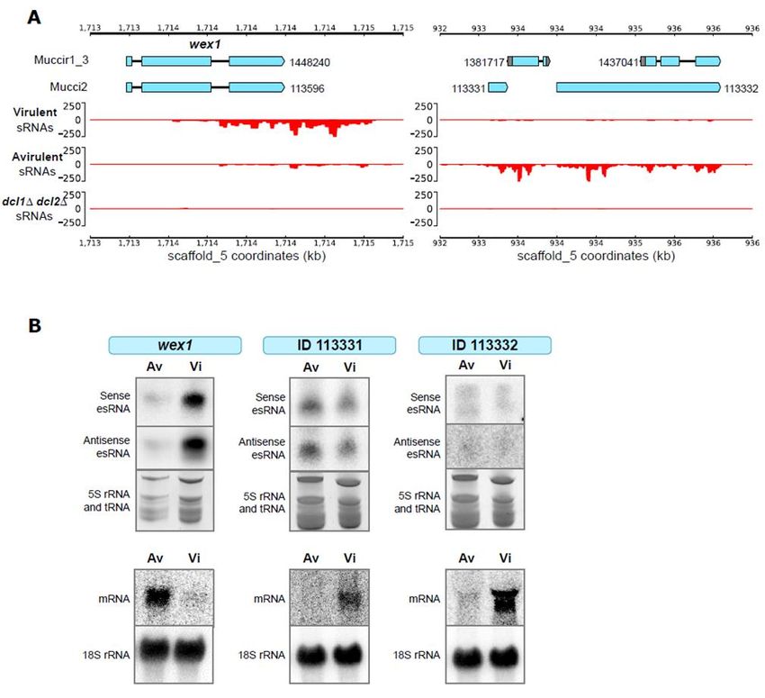
Figure 1. RNAi machinery targets different subsets of genes in each pathotype. (A) Experimental design for sRNA purifi-
cation from two different pathotypes of M. lusitanicus: strains CBS277.49 and NRRL3631, respectively. (B) Scatterplot
showing differences in sRNA accumulation in both pathotypes, virulent (Vi) and avirulent (Av). Each dot represents a
protein-coding locus of the annotated M. lusitanicus genome (Muccir1_3), showing the log2 fold-change (FC) between Vi
and Av (y-axis) and the average expression in both pathotypes (x-axis, in log2 CPM). Each gene is color-coded to depict
non-significant FC (black), upregulation (pink, log2 FC ≥ 1) or downregulation (light blue, log2 FC ≤ -1), and significant
upregulation (red, log2 FC ≥ 2.5 and p-value ≤ 0.05) or downregulation (blue, log2 FC ≥ -2.5 and p-value ≤ 0.05). Three
housekeeping genes are marked as yellow to assure a proper normalization among samples. (C) Heatmap showing the
accumulation of sRNAs (calculated as the Z-score of log2 CPM) in 84 protein-coding loci that showed significant log2 FC
differences between Vi and Av. Equivalent values in an RNAi-deficient dcl1 and dcl2 double deletion mutant (dcl1Δ dcl2Δ)
are shown. Values are clustered by similarity in protein-coding loci (rows) and strains (columns), identifying two major
subsets of protein-coding loci. Strand bias, i.e., the proportion of antisense: sense sRNAs are shown in the rightmost col-
umn.
2.2. Validation of target genes that are differentially regulated by RNAi
The next step in our strategy was to validate the results of the transcriptomic analysis
(previous section) by an alternative approach. We randomly selected two loci, one show-
ing low levels specifically in the Vi strain and another one in the Av strain (Figure 2A).
The locus selected from the Vi strain contained one gene (ID 148240) encoding a protein
with nuclease features. In particular, this gene showed similarity with the Werner Syn-
drome-like exonuclease (WEX) (a DEDDy exonuclease, part of the DnaQ-like exonuclease
superfamily), denominating the gene as wex1 in M. lusitanicus [28]. The alignment of the
sRNAs showed that an important amount of them were generated from the 3’-UTR re-
gion, suggesting a regulatory role of this region. Surprisingly, the locus selected from thePreprints (www.preprints.org) | NOT PEER-REVIEWED | Posted: 14 January 2021 doi:10.20944/preprints202101.0277.v1
Av strain contained several putative genes, interspersed in the same 3-kb esRNA-produc-
ing region, indicating that they share the same regulation via the RNAi pathway. These
genes are unique in Mucorales, and their function is unknown. This region was analyzed
as a whole, denominating the locus as Avirulent-RNAi-Dependent-Locus (ARDL). To
confirm RNAi activity, we measured the esRNAs and the mRNA levels in the two patho-
types by Northern blots using specific probes from the gene wex1 and the locus ARDL.
The blots showed elevated production of wex1 esRNAs and reduced levels of the corre-
sponding mRNA in the Vi strain (Figure 2B) and, conversely, elevated accumulation of
ARDL esRNAs and reduced levels of the corresponding mRNA in the Av strain (Figure
2B). These results, along with the lack of sRNAs in the double Dicer mutant profiles, con-
firmed the differential behavior of RNAi in the Vi and Av pathotypes for the two studied
loci.
Figure 2. A putative DEDDy exonuclease and a 3-kb genomic region are differentially targeted by RNAi in each pathotype.
(A) Genomic plots of two selected candidate regions: one containing the wex1 gene that is actively silenced in the Vi patho-
type, and the other spanning a 3-kb sequence that is targeted in the Av pathotype. Genome annotation and gene IDs from
two different models (Muccir1_3 filtered models and Mucci2) are shown. Below, each track shows the accumulation of
sense (y-axis, positive values) and antisense sRNAs (y-axis, negative values) as CPM values across the genomic coordi-
nates (x-axis). (B) The sRNA Northern blots (above) show sense and antisense sRNA accumulation in each strain depicted
for three analyzed loci: wex1, 113331, and 113332 (from Mucci2). Loading controls consisting of tRNAs and 5S rRNA
stained with ethidium bromide are shown. The mRNA Northern blots (below) show the transcription of each analyzed
loci in the depicted strains, confirming the inverse correlation between sRNA accumulation and mRNA levels. The ex-
pression level of 18S rRNA is shown as a loading control.Preprints (www.preprints.org) | NOT PEER-REVIEWED | Posted: 14 January 2021 doi:10.20944/preprints202101.0277.v1
2.3. Generation of mutant strains with altered expression of the candidate genes
The RNAi-based functional transcriptomic approach applied to the identification of
new virulence factors resulted in a narrowed list of 75 candidates. The validation of this
approach to identify virulence factors required the generation of different mutants that
mimicked the behavior of the Av pathotype. The wex1 gene was being targeted by the
RNAi degrading mechanism in the Vi pathotype, as opposed to its upregulation in the Av
pathotype. Accordingly, we generated a strain that overexpressed wex1 in the Vi patho-
type genetic background to study its role in M. lusitanicus virulence. To overexpress this
gene, we constructed a knockin vector containing an engineered cassette with adjacent
regions of the carRP gene, the selectable marker pyrG gene, and the wex1 gene fused to the
previously described strong promoter Pzrt1 (Figure 3A, left)[29,30] The integration into
the locus carRP facilitates the screening for homokaryotic target mutants, as they show an
albino phenotype [30]. The proper insertion of this cassette was confirmed in five inde-
pendent mutants by Southern blot (Figure 3A, right). Analysis of wex1 expression by
Northern blot (Figure 3B)showed an increase of expression of this gene in the knockin
strain, showing similar levels to the Av strain. We also generated a deletion mutant for
the wex1 gene, as a control strain, by using a knockout vector designed to replace wex1
with the pyrG gene after homologous recombination. This knockout vector contained an
engineered cassette with adjacent regions of the wex1 gene flanking the pyrG gene (Figure
4A, left). The correct insertion of this cassette was also validated in two independent mu-
tants by Southern blot (Figure 4A, right). On the contrary, the locus ARDL was upregu-
lated in Vi compared to the specific RNAi-based degradation and downregulation in the
Av strain. To mimic this regulation found in the Av pathotype, we generated a knockout
mutant of ARDL in the Vi strain. Following a similar approach to the wex1 knockout
strain, we generated a cassette with the adjacent regions of ARDL flanking the pyrG gene
(Figure 3B, left), and the insertion of this cassette was confirmed in two independent mu-
tants by Southern blot (Figure 3B, right).Preprints (www.preprints.org) | NOT PEER-REVIEWED | Posted: 14 January 2021 doi:10.20944/preprints202101.0277.v1
Figure 3. Overexpression of wex1 overcomes RNAi silencing. (A, B) Southern blot depicting the generation of the wex1
overexpressing mutant Pzrt1-wex1. A schematic representation of the wild-type and mutant loci after homologous recom-
bination with the designed overexpression fragment targeting the carRP locus (depicted as crossing lines) is shown to the
left. Fragments digested with NcoI and their expected sizes are indicated. The position of the radioactive probe and its
complementary sequence is shown. To the right, the blots showing the digested DNA fragments hybridizing to the probe
C of each mutant and wild-type strains allow for size discrimination using a DNA ladder (M). (B) The transcription of
wex1 is shown in the Pzrt1-wex1 mutant and compared to both Av and Vi pathotypes, measured by Northern blot. 18S
rRNA is shown as a loading control. Samples are rearranged for consistency (shown as a discontinuous line).
Figure 4. Deletion mutants in wex1 and ardL. (A, B) Southern blots are illustrating the generation of (A) the wex1 and
(B) the ardL deletion mutants. A schematic representation of the wild-type and mutant loci after homologous recombina-
tion with the designed disruption fragment (depicted as crossing lines) is shown to the left. Fragments digested with (A)
NcoI or (B) PaeI and their expected sizes are indicated. Radioactive probes and their complementary sequences are shown
in each locus. To the right, the blots showing the digested DNA fragments of each mutant and wild-type strains hybridiz-
ing to the corresponding probes allow for size discrimination using a DNA ladder (M).Preprints (www.preprints.org) | NOT PEER-REVIEWED | Posted: 14 January 2021 doi:10.20944/preprints202101.0277.v1
2.4. Functional analysis of the genes regulated by RNAi and their role in virulence
The virulence of fungal pathogens is an intricate feature depending on multiple cel-
lular processes, mechanisms, and gene pathways [31]. The final aim of this work was to
find new target genes both regulated by RNAi and related to pathogenesis in Mucorales.
Thus, the wex1 knockin and ARDL deletion mutants were studied in a virulence assay
employing a mouse model previously validated in M. lusitanicus [32,33]. As control
strains, the wex1 deletion mutant and the Vi and Av pathotypes were also injected in mice..
The overexpression of wex1 in the knockin mutant resulted in an avirulent phenotype like
the Av pathotype, whereas the virulence potential of the mutant wex1Δ was not affected
(Figure 5). These results indicate that RNAi silencing of wex1 in the Vi pathotype is essen-
tial to uphold its virulence potential. The overexpression of wex1 probably compensates
the RNAi degradation, mimicking the transcription levels of the Av strain. The deletion
of ARDL in Vi simulated the silencing of this locus observed in the Av strain; however,
this mutation did not alter the virulence. These results suggest that the ARDL region has
a function non-related to virulence, although it might be involved in other cellular pro-
cesses that differ between the Vi and Av pathotypes [25]. Overall, we demonstrated the
validity of the functional sRNA transcriptomic approach employed in this work to find
new virulence determinants in Mucorales.
Figure 5. Silencing wex1 is critical for virulence. Virulence assays in immunosuppressed mice in-
jected with 1x10⁶ spores of the wex1Δ (purple) and Pzrt1-wex1 (blue) mutants. As infection controls,
the mice were also injected with spores from the recipient transformation strain R7B that derives
from the Vi pathotype (red) and the Av pathotype (green). The survival rate of the mutants was
compared to the Vi control strain and statistically analyzed by a Mantel-Cox test.
3. Discussion
The RNAi mechanism plays an essential role in the defense against invasive nucleic
acids and the regulation of endogenous transcripts [15,16,34,35]. The evolutionary conser-
vation of these roles throughout the eukaryotic domain suggests a selective advantage to
retain the mechanism of RNAi [36]. This observation is particularly highlighted in the case
of Mucorales, a group of early-diverging fungi that not only have conserved the canonical
RNAi machinery, but also have evolved a Dicer and Argonaute-independent non-canon-
ical RNA mechanism and a special pathway involved in antifungal resistance not found
in other organisms [12,19,34]. The RNAi mechanism regulates different pathways and cel-
lular processes in M. lusitanicus [13]. Among these processes, virulence has been recentlyPreprints (www.preprints.org) | NOT PEER-REVIEWED | Posted: 14 January 2021 doi:10.20944/preprints202101.0277.v1
related to the RNAi mechanism in diverse regulatory pathways [21]. Here, we have de-
veloped an experimental approach to identify genes related to virulence and regulated by
RNAi, showing as a proof of concept the identification and characterization of the gene
wex1. Previous studies successfully tested a functional transcriptomic approach using
samples from host-pathogen interactions, which resulted in long lists of candidate genes
with putative roles in virulence [21,24]. That approach was pivotal to demonstrate the
essential role of the RNAi pathways in virulence, controlling broad gene-networks during
the host-pathogen interactions.
On the other hand, another study compared the Vi and Av pathotypes to find the
differences that could explain their opposite virulence potential at a genomic level [25].
This genomic approach found 773 modified loci in the Av strain, which is also a long list
of candidates to find virulence factors. In this regard, our hypothesis contemplated com-
paring Vi and Av pathotypes but restricting the transcriptomic profiles to only those reg-
ulated by RNAi. To our knowledge, this work is the first application of a functional tran-
scriptomic approach based on the differential production of sRNAs in virulent and avir-
ulent strains.
Our specific transcriptomic analysis produced an approachable list of 75 candidate
genes for roles in virulence and regulation by RNAi. The experimental validation of two
randomly selected candidate genes demonstrated the reliability of this gene set. Moreo-
ver, the construction of avirulent-mimicking strains and their phenotypic study regarding
virulence unveiled the essential role of the wex1 gene in maintaining the virulence poten-
tial. The Vi strain ceases to be pathogenic when wex1 is overpressed, showing a similar
phenotype to the Av pathotype. Thus, the RNAi-dependant specific degradation of wex1
mRNA levels in the Vi pathotype is required to ensure the virulence potential of this
strain. The wex1 gene encodes a putative DnaQ-like exonuclease, a superfamily of en-
zymes that catalyze nucleoside monophosphate excision at DNA or RNA ends in the 3'-5'
direction. These types of enzymes contain RNases domains such as RNase D and Rrp6p.
A fine-tuned balance of exonuclease activity is critical for developmental processes and
fungal virulence [37,38]. Assuming the hypothesis that wex1 could be involved in the pro-
cess of degradation and recycling of defective and aged mRNAs, the overexpression of
this gene might reduce the levels of substrate mRNAs available for the non-canonical
RNAi pathway (NCRIP) in M. lusitanicus. The competition for the target mRNAs among
the different RNA degrading pathways has been proposed in several studies [19,20].
Thus, the overexpression of wex1 would leave NCRIP without target substrate mRNAs,
producing an avirulent strain similar to mutants lacking NCRIP activity [21]. This hypoth-
esis could explain the same avirulent phenotype previously observed in NCRIP mutants
and the strain overexpressing wex1 generated in this study. However, other roles of wex1
in unknown pathways related to virulence cannot be excluded at this point.
The identification of wex1 suggests that many others may be discovered by further
dissection of our genes list. However, the functional characterization of the ARDL showed
that this list also contains genes unrelated to virulence. Besides virulence, there are several
differences between the Vi and Av pathotypes, such as mating type, heat stress tolerance,
chitosan content, and toxic compound susceptibility [25]. These differential phenotypes
and their related cellular processes also define a close functional area in the fungal cell
physiology complexity, which will facilitate the functional study of those candidates
showing a phenotype unrelated to virulence.
In summary, this work developed a successful approach to identify new virulence
determinants regulated by the RNAi machinery. The sRNA transcriptome-wide analysis
of strains with opposite virulence potentials was functionally validated, presenting a nar-
rowed list of 75 candidate genes that are both regulated by the RNAi machinery and re-
lated to the phenotypic differences between the two pathotypes. Among these candidate
genes, the characterization of wex1 and its essential role in virulence served as proof of
concept for the entire approach. Moreover, the putative exonuclease activity of this gene
might suggest a function in the complex interplay of the RNAi pathways in M. lusitanicus
and their role in virulence. An additional advantage of the approach presented here is itsPreprints (www.preprints.org) | NOT PEER-REVIEWED | Posted: 14 January 2021 doi:10.20944/preprints202101.0277.v1
exportability to any pathogen accounting with a conserved RNAi machinery and strains
showing different pathogenic potential. Similarly, other model organisms having those
two features can use our approach to investigate cellular processes unrelated to virulence.
Further dissection of the genes identified here and the pathways in which they are in-
volved will represent a new source of specific targets to design improved treatments
against mucormycosis.
4. Materials and Methods
4.1. Fungal strains
In summary, Two different M. lusitanicus pathotypes were used as wild-type strains
throughout the study: the avirulent pathotype NRRL3631 (hereinafter Av) and the viru-
lent pathotype CBS277.49 (hereinafter Vi) [24,25,32]. Strain MU402 [26] derives from Vi
and is a leucine and uracil double auxotroph that served as a recipient strain for all genetic
transformations. As a result, mutant strains wex1∆ (MU641 and MU642), ARDL∆ (MU632),
and Pzrt1-wex1 (MU637, MU638, MU639, and MU640) were generated. Double mutant
strain in dcl1 and dcl2 was MU411 [27]. Unless otherwise stated, MU641, MU632, and
MU640 were employed to conduct the experiments described in the manuscript.
4.2. RNA isolation
2.5x10⁵ spores from Av and Vi strains were plated in solid rich YPG medium with
adjusted pH of 4.5 and incubated at a constant temperature of 26 °C for 24 hours. Before
spore inoculation, plates were covered with a thin layer of cellophane film to facilitate
media-free mycelium harvesting after incubation. 100-mg strips of mycelium were har-
vested and used for RNA isolation. Sequencing sRNA samples were obtained with a mir-
Vana miRNA isolation kit (Ambion), whereas sRNA samples for Northern blotting were
isolated employing Trizol and a polyethylene glycol-based differential precipitation of
high molecular weight RNAs procedure described in previous studies [39]. The RNeasy
plant Minikit (Qiagen) was used to isolate mRNA following the supplier recommenda-
tions for fungal RNA.
4.3. RNA-sequencing and analysis for small RNA production
The sRNA samples from Av and Vi were sent to BaseClear sequencing facility. Li-
braries were prepared using TruSeq Small RNA Library Prep Kit and sequenced with a
HiSeq 2500 to produce single-end, 50-bp reads. Raw sRNA reads from Av, Vi, and pub-
licly available dcl1∆ dcl2∆ double mutant strain (see Data Availability) were quality-
checked with FASTQC v0.11.8, and adapter or contaminant sequences overlapping ≥2 ba-
ses at the 3’-end were removed with Trim Galore! v.0.6.2 (available at http://www.bioin-
formatics.babraham.ac.uk/projects/). Reads with Q ≤20 and total length ≤13 or ≥29 nt were
excluded. Processed sRNA reads were aligned to the M. lusitanicus MU402 genome (from
now on Muccir1_3, available at the JGI portal: https://mycocosm.jgi.doe.gov/Muc-
cir1_3/Muccir1_3.info.html) using HISAT2 v2.1.0 [40]. The differential sRNA production
between Vi and Av across all protein-coding loci was analyzed using DESeq2 v1.18.1 [41]
and Muccir1_3 gene annotation. The resulting log2 fold-change (FC) and average log2
Counts per million (CPM) for sRNA production values were correlated in a scatter plot,
highlighting three housekeeping genes that have a stable sRNA production among sam-
ples [EF-1 (Muccir1_3 ID: 1382517), TFIIIC (Muccir1_3 ID: 1386549), V-ATPase (Muccir1_3
ID: 1377858)]. The z-score of log2 CPM values for Av, Vi, and dcl1∆ dcl2∆ samples (calcu-
lated as the number of standard deviation units a value differs from the mean) was plotted
using the pheatmap v.1.0.12 R package. The sRNA production values were clustered ac-
cording to their similarity both at each protein-coding loci and sample using Euclidean
distance and Ward’s clustering method. The average strand bias, i.e. the proportion of
sense and antisense reads in all samples, was calculated for each loci plotted in the
heatmap as the ratio comparing the difference of sense and antisense reads with all reads
[(sense reads-antisense reads)/(sense reads+antisense reads)]. Values close to 1 indicate aPreprints (www.preprints.org) | NOT PEER-REVIEWED | Posted: 14 January 2021 doi:10.20944/preprints202101.0277.v1
strong bias towards sense reads, whereas -1 implies a strong bias towards antisense reads;
intermediate values (≈ 0) means an equal proportion of sense and antisense reads.
To generate sense and antisense sRNA genomic plots, aligned sRNA reads were split
into forward and reverse strand reads by filtering through their Binary Alignment/Map
(BAM) FLAG field (-F 16 for forwarding reads and -f 16 for reverse reads) with SAMtools
v1.10-2 [42]. This allowed the identification of sense and antisense sRNAs as follows: for-
ward or reverse reads aligning to same-sense protein-coding loci were considered sense
sRNAs. Those aligning to opposite-sense protein-coding loci were considered antisense
sRNAs. Aligned reads were normalized to bins per million reads (BPM) in 25-bp bins us-
ing deepTools v3.2.1 [43] bamCoverage function. Genomic plots were visualized using
the deepTools pyGenomeTracks module.
4.4. Northern blot analyses for mRNA and sRNA
After sRNA isolation, samples were separated by denaturing urea polyacrylamide
electrophoresis [44]. sRNAs with lengths ranging from 10 to 100 bp were transferred to a
neutral Hybond-NX nylon membrane (Amersham) and chemically fixed as described in
other studies [39]. PCR-amplified fragments containing the target locus and the pT7 pro-
moter sequence at their 5’ or 3’-end were used as DNA templates to transcribe in vitro
specific sense or antisense riboprobes, respectively, using a MAXIscript™ T7 Transcrip-
tion Kit (Ambion). The [α-32P]UTP riboprobes were fragmented with an alkali solution,
filtered to remove individual nucleotides, and hybridized to the transferred sRNAs as de-
scribed in previous works [39]. The gel portion containing sRNAs with lengths ≥ 100 bp
(5S rRNA and tRNAs) was stained with ethidium bromide as a loading control.
For mRNA Northern blotting, samples were separated by denaturing formaldehyde
agarose electrophoresis and then transferred to a positively charged Hybond-XL nylon
membrane (Amersham). Specific [α-32P]dCTP-labeled probes were generated with the
Ready-To-Go (GE Healthcare Life Science) and used for hybridization. 18S rDNA [α-
32P]dCTP-labeled probes were hybridized as loading controls. Both sRNA and mRNA
membranes were developed using a phosphor screen (Fujifilm) and a Personal Molecular
Imager system (Bio-Rad) after a time exposure.
4.5. Genetic transformation
Disruption fragments contained either a 2.0-kb fragment of the selectable marker
pyrG for Ura+ or a 3.6-kb fragment of the selectable marker leuA for Leu+ selection,
flanked by 1-kb upstream and downstream sequences of the target locus to facilitate gene
replacement by homologous recombination. These fragments were PCR-amplified sepa-
rately and then fused by overlapping PCR using specific primers for each deleted gene.
MU402 was grown in solid rich YPG medium with adjusted pH 4.5 during 5-6 days for
optimal growth and sporulation conditions. Spores were harvested to produce proto-
plasts as described in previous studies [39]. Protoplasts were transferred to 0.2cm-cuvettes
and electroporated with a field strength of 4kV/cm (800V), the capacitance of 25 µF, and a
resistance of 400 Ω to allow the introduction of 5µg of disruption DNA fragments into the
cells. After the pulse, the protoplasts were recovered in liquid YPG medium and plated
into the selective medium. Protoplasts were plated in either Minimal medium with
Casamino acids (MMC) with adjusted pH of 3.2 to select Ura+ transformants, or Yeast
Nitrogen Base (YNB) minimal medium with adjusted pH of 3,2 to select Leu+ trans-
formants. The transformants underwent 10 passages of vegetative sporulation, single-col-
ony harvesting, and streaking in a selective medium to favor the selection of homokary-
otic spores. Then, homokaryosis was confirmed by Southern blotting. Briefly, genomic
DNA was purified as described in previous works [39], digested with specific restriction
enzymes, and separated by electrophoresis. DNA fragments were transferred to a posi-
tively charged Hybond-XL nylon membrane (Amersham) and hybridized to specific [α-
32P]dCTP-labeled probes, which were generated following the Ready-To-Go kit proce-Preprints (www.preprints.org) | NOT PEER-REVIEWED | Posted: 14 January 2021 doi:10.20944/preprints202101.0277.v1
dure (GE Healthcare Life Science). After stringent hybridization, the membranes were de-
veloped to discriminate mutant from wild-type DNA alleles, as described for Northern
blotting.
4.6. Virulence assays
Male OF-1 mice weighing 30g (Charles River, Barcelona, Spain) were used as host
models for virulence assays for their reliability in previous Mucoralean virulence assays
[21,45]. The mice were immunosuppressed by intraperitoneal injection of cyclophospha-
mide (200 mg/kg of body weight), 2 days before the infection and once every 5 days there-
inafter. Groups of ten were challenged intravenously by injecting 1x10⁶ spores in the
retroorbital venous sinus [18]. To ensure animal comfort, the mice were anesthetized with
isoflurane via inhalation and monitored until they recovered from the anesthesia. Vi and
Av strains were also injected as a positive and negative virulence control, respectively.
Mice were housed under established conditions with free food and autoclaved water. An-
imal welfare was monitored twice a day for 20 days, and those mice following the criteria
for discomfort were euthanized by CO2 inhalation. Survival rates during a time were plot-
ted in a Kaplan-Meier curve, and differences were considered statistically significant with
a p-value ≤ 0.05 in a Mantel-Cox test (Graph Pad Prism).
Supplementary Materials: The following are available online at www.mdpi.com/xxx/s1, Table S1,
Table S2.
Author Contributions: Conceptualization, V.G., and F.E.N.; data curation, C.P-A.; methodology,
C.P-A., M.I.N-M., and L.M.; validation, V.G., F.E.N., and C.P-A.; formal analysis, F.E.N., C.P-A.,
and M.I.N-M.; investigation, C.P-A., M.I.N-M., F.E.N., and L.M.; resources, V.G., F.E.N., and E.N.;
visualization, C.P-A., and M.I.N-M.; writing—original draft preparation, F.E.N.; writing—review
and editing, F.E.N., C.P-A., M.I.N-M., and V.G. All authors have read and agreed to the published
version of the manuscript.
Funding: This investigation was supported by the Ministerio de Economía y Competitividad,
Spain (BFU2015-65501-P, co-financed by FEDER, and RYC-2014-15844), the Ministerio de Cien-
cia, Innovación y Universidades, Spain (PGC2018-097452-B-I00, co-financed by FEDER), and the
Fundación Séneca-Agencia de Ciencia y Tecnología de la Región de Murcia, Spain (19339/PI/14).
C.P.A. and M.I.N.M. were supported by predoctoral fellowships from the Ministerio de Edu-
cación, Cultura y Deporte, Spain (FPU14/01983 and FPU14/01832, respectively).
Institutional Review Board Statement: To guarantee the welfare of the animals and the ethics of
any procedure related to animal experimentation, all the experiments performed in this work com-
plied with the Guidelines of the European Union Council (Directive 2010/63/EU) and the Spanish
RD 53/2013. Experiments and procedures were supervised and approved by the University of Mur-
cia Animal Welfare and Ethics Committee and the Council of Water, Agriculture, Farming, Fishing
and Environment of Murcia (Consejería de Agua, Agricultura, Ganadería, Pesca y Medio Ambiente
de la CARM), Spain (authorization number REGA ES300305440012).
Data Availability Statement: The sRNA raw data files generated by this study are deposited at the
National Center for Biotechnology Information Sequence Read Archive (NCBI SRA) and are pub-
licly available through the project accession number PRJNA674566. These data were compared to a
double dcl1∆ dcl2∆ mutant strain (accession number SRR039128) [13]. The M. lusitanicus genome
Muccir1_3 [14] and annotation files can be accessed at the Joint Genome Institute (JGI) website
(http://genome.jgi.doe.gov/) and used under the JGI Data Usage Policy.
Acknowledgments: This investigation was supported by the Ministerio de Economía y Competi-
tividad, Spain (BFU2015-65501-P, co-financed by FEDER, and RYC-2014-15844) and the Ministerio
de Ciencia, Innovación y Universidades, Spain (PGC2018-097452-B-I00, co-financed by FEDER).
C.P.-A. and M.I.N.-M. were supported by predoctoral fellowships from the Ministerio de Edu-
cación, Cultura y Deporte, Spain (FPU14/01983 and FPU14/01832, respectively).
Conflicts of Interest: The authors declare no conflict of interest. The funders had no role in the
design of the study; in the collection, analyses, or interpretation of data; in the writing of the manu-
script, or in the decision to publish the results.Preprints (www.preprints.org) | NOT PEER-REVIEWED | Posted: 14 January 2021 doi:10.20944/preprints202101.0277.v1
References
1. Dannaoui, E. Antifungal resistance in mucorales. Int J Antimicrob Agents 2017, 50, 617–621,
doi:10.1016/j.ijantimicag.2017.08.010.
2. Kontoyiannis, D.P.; Yang, H.; Song, J.; Kelkar, S.S.; Yang, X.; Azie, N.; Harrington, R.; Fan, A.; Lee, E.; Spalding, J.R.
Prevalence, clinical and economic burden of mucormycosis-related hospitalizations in the United States: a retrospective
study. BMC Infect. Dis. 2016, 16, 730, doi:10.1186/s12879-016-2023-z.
3. Prakash, H.; Chakrabarti, A. Global epidemiology of mucormycosis. J. Fungi 2019, 5, doi:10.3390/jof5010026.
4. Caramalho, R.; Tyndall, J.D.A.; Monk, B.C.; Larentis, T.; Lass-Flörl, C.; Lackner, M. Intrinsic short-tailed azole resistance in
mucormycetes is due to an evolutionary conserved aminoacid substitution of the lanosterol 14α-demethylase. Sci. Rep. 2017,
7, 15898, doi:10.1038/s41598-017-16123-9.
5. Li, C.H.; Cervantes, M.; Springer, D.J.; Boekhout, T.; Ruiz-Vazquez, R.M.; Torres-Martinez, S.R.; Heitman, J.; Lee, S.C.
Sporangiospore size dimorphism is linked to virulence of Mucor circinelloides. PLoS Pathog 2011, 7, e1002086,
doi:10.1371/journal.ppat.1002086.
6. Katragkou, A.; Walsh, T.J.; Roilides, E. Why is mucormycosis more difficult to cure than more common mycoses? Clin.
Microbiol. Infect. 2014, 20, 74–81, doi:10.1111/1469-0691.12466.
7. Nishimoto, A.T.; Sharma, C.; Rogers, P.D. Molecular and genetic basis of azole antifungal resistance in the opportunistic
pathogenic fungus Candida albicans. J. Antimicrob. Chemother. 2019, doi:10.1093/jac/dkz400.
8. Naranjo-Ortiz, M.A.; Gabaldón, T. Fungal evolution: diversity, taxonomy and phylogeny of the Fungi. Biol. Rev. 2019, 94,
2101–2137, doi:10.1111/brv.12550.
9. Corrochano, L.M.; Kuo, A.; Marcet-Houben, M.; Polaino, S.; Salamov, A.; Villalobos-Escobedo, J.M.; Grimwood, J.; Álvarez,
M.I.; Avalos, J.; Bauer, D.; et al. Expansion of Signal Transduction Pathways in Fungi by Extensive Genome Duplication. Curr.
Biol. 2016, 26, 1577–1584, doi:10.1016/j.cub.2016.04.038.
10. Ribes, J.A.; Vanover-Sams, C.L.; Baker, D.J. Zygomycetes in human disease. Clin. Microbiol. Rev. 2000, 13, 236–301.
11. Navarro-Mendoza, M.I.; Pérez-Arques, C.; Panchal, S.; Nicolás, F.E.; Mondo, S.J.; Ganguly, P.; Pangilinan, J.; Grigoriev, I. V.;
Heitman, J.; Sanyal, K.; et al. Early Diverging Fungus Mucor circinelloides Lacks Centromeric Histone CENP-A and Displays
a Mosaic of Point and Regional Centromeres. Curr. Biol. 2019, 29, 3791-3802.e6, doi:10.1016/j.cub.2019.09.024.
12. Calo, S.; Shertz-Wall, C.; Lee, S.C.; Bastidas, R.J.; Nicolás, F.E.; Granek, J.A.; Mieczkowski, P.; Torres-Martínez, S.; Ruiz-
Vázquez, R.M.; Cardenas, M.E.; et al. Antifungal drug resistance evoked via RNAi-dependent epimutations. Nature 2014,
513, 555–558, doi:10.1038/nature13575.
13. Nicolás, F.E.; Vila, A.; Moxon, S.; Cascales, M.D.; Torres-Martínez, S.; Ruiz-Vázquez, R.M.; Garre, V. The RNAi machinery
controls distinct responses to environmental signals in the basal fungus Mucor circinelloides. BMC Genomics 2015, 16, 237,
doi:10.1186/s12864-015-1443-2.
14. Lax, C.; Pérez-arques, C.; Navarro-mendoza, M.I.; Cánovas-márquez, J.T.; Tahiri, G.; Pérez-ruiz, J.A.; Osorio-concepción, M.;
Murcia-flores, L.; Navarro, E.; Garre, V.; et al. Genes, pathways, and mechanisms involved in the virulence of mucorales.
Genes (Basel). 2020, 11.
15. Nicolás, F.; Ruiz-Vázquez, R. Functional Diversity of RNAi-Associated sRNAs in Fungi. Int. J. Mol. Sci. 2013, 14, 15348–15360,
doi:10.3390/ijms140815348.
16. Nicolas, F.E.; Moxon, S.; de Haro, J.P.; Calo, S.; Grigoriev, I. V; Torres-Martinez, S.; Moulton, V.; Ruiz-Vazquez, R.M.; Dalmay,
T. Endogenous short RNAs generated by Dicer 2 and RNA-dependent RNA polymerase 1 regulate mRNAs in the basal
fungus Mucor circinelloides. Nucleic Acids Res 2010, 38, 5535–5541, doi:10.1093/nar/gkq301.
17. Chang, Z.; Billmyre, R.B.; Lee, S.C.; Heitman, J. Broad antifungal resistance mediated by RNAi-dependent epimutation in the
basal human fungal pathogen Mucor circinelloides. PLoS Genet. 2019, 15, doi:10.1371/journal.pgen.1007957.
18. Chang, Z.; Heitman, J. Drug-resistant epimutants exhibit organ-specific stability and induction during murine infectionsPreprints (www.preprints.org) | NOT PEER-REVIEWED | Posted: 14 January 2021 doi:10.20944/preprints202101.0277.v1
caused by the human fungal pathogen Mucor circinelloides. MBio 2019, 10, doi:10.1128/mBio.02579-19.
19. Trieu, T.A.; Calo, S.; Nicolás, F.E.; Vila, A.; Moxon, S.; Dalmay, T.; Torres-Martínez, S.; Garre, V.; Ruiz-Vázquez, R.M. A Non-
canonical RNA Silencing Pathway Promotes mRNA Degradation in Basal Fungi. PLOS Genet. 2015, 11, e1005168,
doi:10.1371/journal.pgen.1005168.
20. Calo, S.; Nicolas, F.E.; Lee, S.C.; Vila, A.; Cervantes, M.; Torres-Martinez, S.; Ruiz-Vazquez, R.M.; Cardenas, M.E.; Heitman,
J. A non-canonical RNA degradation pathway suppresses RNAi-dependent epimutations in the human fungal pathogen
Mucor circinelloides. PLoS Genet 2017, 13, e1006686, doi:10.1371/journal.pgen.1006686.
21. Pérez-Arques, C.; Navarro-Mendoza, M.I.; Murcia, L.; Navarro, E.; Garre, V.; Nicolás, F.E. A non-canonical RNAi pathway
controls virulence and genome stability in Mucorales. PLoS Genet. 2020, 16, doi:10.1371/journal.pgen.1008611.
22. Calo, S.; Nicolás, F.E.; Lee, S.C.; Vila, A.; Cervantes, M.; Torres-Martinez, S.; Ruiz-Vazquez, R.M.; Cardenas, M.E.; Heitman,
J. A non-canonical RNA degradation pathway suppresses RNAi-dependent epimutations in the human fungal pathogen
Mucor circinelloides. PLoS Genet. 2017, 13, doi:10.1371/journal.pgen.1006686.
23. Nicolás, F.E.; Murcia, L.; Navarro, E.; Navarro-Mendoza, M.I.; Pérez-Arques, C.; Garre, V. Mucorales species and
macrophages. J. Fungi 2020, 6, 1–12.
24. Pérez-Arques, C.; Navarro-Mendoza, M.I.; Murcia, L.; Lax, C.; Martínez-García, P.; Heitman, J.; Nicolás, F.E.; Garre, V. Mucor
circinelloides Thrives inside the Phagosome through an Atf-Mediated Germination Pathway. MBio 2019, 10,
doi:10.1128/mBio.02765-18.
25. López-Fernández, L.; Sanchis, M.; Navarro-Rodríguez, P.; Nicolás, F.E.; Silva-Franco, F.; Guarro, J.; Garre, V.; Navarro-
Mendoza, M.I.; Pérez-Arques, C.; Capilla, J. Understanding Mucor circinelloides pathogenesis by comparative genomics and
phenotypical studies. Virulence 2018, 00–00, doi:10.1080/21505594.2018.1435249.
26. Nicolas, F.E.; de Haro, J.P.; Torres-Martinez, S.; Ruiz-Vazquez, R.M. Mutants defective in a Mucor circinelloides dicer-like
gene are not compromised in siRNA silencing but display developmental defects. Fungal Genet Biol 2007, 44, 504–516,
doi:10.1016/j.fgb.2006.09.003.
27. de Haro, J.P.; Calo, S.; Cervantes, M.; Nicolas, F.E.; Torres-Martinez, S.; Ruiz-Vazquez, R.M. A single dicer gene is required
for efficient gene silencing associated with two classes of small antisense RNAs in Mucor circinelloides. Eukaryot Cell 2009, 8,
1486–1497, doi:10.1128/EC.00191-09.
28. Zuo, Y.; Deutscher, M.P. Exoribonuclease superfamilies: Structural analysis and phylogenetic distribution. Nucleic Acids Res.
2001, 29, 1017–1026, doi:10.1093/nar/29.5.1017.
29. Rodríguez-Frómeta, R.A.; Gutiérrez, A.; Torres-Martínez, S.; Garre, V. Malic enzyme activity is not the only bottleneck for
lipid accumulation in the oleaginous fungus Mucor circinelloides. Appl. Microbiol. Biotechnol. 2013, 97, 3063–3072,
doi:10.1007/s00253-012-4432-2.
30. Binder, U.; Navarro-Mendoza, M.I.; Naschberger, V.; Bauer, I.; Nicolas, F.E.; Pallua, J.D.; Lass-Flörl, C.; Garre, V. Generation
of a mucor circinelloides reporter strain—A promising new tool to study antifungal drug efficacy and mucormycosis. Genes
(Basel). 2018, 9, doi:10.3390/genes9120613.
31. Casadevall, A. Determinants of virulence in the pathogenic fungi. Fungal Biol. Rev. 2007, 21, 130–132.
32. Li, C.H.; Cervantes, M.; Springer, D.J.; Boekhout, T.; Ruiz-Vazquez, R.M.; Torres-Martinez, S.R.; Heitman, J.; Lee, S.C.
Sporangiospore size dimorphism is linked to virulence of Mucor circinelloides. PLoS Pathog. 2011, 7, e1002086,
doi:10.1371/journal.ppat.1002086.
33. Trieu, T.A.; Navarro-Mendoza, M.I.; Perez-Arques, C.; Sanchis, M.; Capilla, J.; Navarro-Rodriguez, P.; Lopez-Fernandez, L.;
Torres-Martinez, S.; Garre, V.; Ruiz-Vazquez, R.M.; et al. RNAi-Based Functional Genomics Identifies New Virulence
Determinants in Mucormycosis. PLoS Pathog 2017, 13, e1006150, doi:10.1371/journal.ppat.1006150.
34. Nicolas, F.E.; Torres-Martínez, S.; Ruiz-Vázquez, R.M. Two classes of small antisense RNAs in fungal RNA silencing
triggered by non-integrative transgenes. EMBO J. 2003, 22, 3983–3991, doi:10.1093/emboj/cdg384.Preprints (www.preprints.org) | NOT PEER-REVIEWED | Posted: 14 January 2021 doi:10.20944/preprints202101.0277.v1
35. Lax, C.; Tahiri, G.; Patiño-Medina, J.A.; Cánovas-Márquez, J.T.; Pérez-Ruiz, J.A.; Osorio-Concepción, M.; Navarro, E.; Calo,
S. The Evolutionary Significance of RNAi in the Fungal Kingdom. Int. J. Mol. Sci. 2020, 21, 9348, doi:10.3390/ijms21249348.
36. Nicolás, F.E.; Torres-Martínez, S.; Ruiz-Vázquez, R.M. Loss and Retention of RNA Interference in Fungi and Parasites. PLoS
Pathog. 2013, 9, doi:10.1371/journal.ppat.1003089.
37. Stead, J.A.; Costello, J.L.; Livingstone, M.J.; Mitchell, P. The PMC2NT domain of the catalytic exosome subunit Rrp6p
provides the interface for binding with its cofactor Rrp47p, a nucleic acid-binding protein. Nucleic Acids Res. 2007, 35, 5556–
5567, doi:10.1093/nar/gkm614.
38. Wollschlaeger, C.; Trevijano-Contador, N.; Wang, X.; Legrand, M.; Zaragoza, O.; Heitman, J.; Janbon, G. Distinct and
redundant roles of exonucleases in Cryptococcus neoformans: Implications for virulence and mating. Fungal Genet. Biol. 2014,
73, 20–28, doi:10.1016/j.fgb.2014.09.007.
39. Nicolás, F.E.; Navarro-Mendoza, M.I.; Pérez-Arques, C.; López-García, S.; Navarro, E.; Torres-Martínez, S.; Garre, V.
Molecular tools for carotenogenesis analysis in the mucoral Mucor circinelloides. In Methods in Molecular Biology; Humana
Press Inc., 2018; Vol. 1852, pp. 221–237.
40. Kim, D.; Paggi, J.M.; Park, C.; Bennett, C.; Salzberg, S.L. Graph-based genome alignment and genotyping with HISAT2 and
HISAT-genotype. Nat. Biotechnol. 2019, 37, 907–915, doi:10.1038/s41587-019-0201-4.
41. Love, M.I.; Huber, W.; Anders, S. Moderated estimation of fold change and dispersion for RNA-seq data with DESeq2.
Genome Biol. 2014, 15, doi:10.1186/s13059-014-0550-8.
42. Li, H.; Handsaker, B.; Wysoker, A.; Fennell, T.; Ruan, J.; Homer, N.; Marth, G.; Abecasis, G.; Durbin, R. The Sequence
Alignment/Map format and SAMtools. Bioinformatics 2009, 25, 2078–2079, doi:10.1093/bioinformatics/btp352.
43. Ramírez, F.; Ryan, D.P.; Grüning, B.; Bhardwaj, V.; Kilpert, F.; Richter, A.S.; Heyne, S.; Dündar, F.; Manke, T. deepTools2: a
next generation web server for deep-sequencing data analysis. Nucleic Acids Res. 2016, 44, W160–W165,
doi:10.1093/nar/gkw257.
44. Lopez-Gomollon, S.; Nicolas, F.E. Purification of DNA oligos by denaturing polyacrylamide gel electrophoresis (PAGE). In
Methods in Enzymology; Academic Press Inc., 2013; Vol. 529, pp. 65–83 ISBN 9780124186873.
45. Navarro-Mendoza, M.I.; Pérez-Arques, C.; Murcia, L.; Martínez-García, P.; Lax, C.; Sanchis, M.; Capilla, J.; Nicolás, F.E.;
Garre, V. Components of a new gene family of ferroxidases involved in virulence are functionally specialized in fungal
dimorphism. Sci. Rep. 2018, 8, 7660, doi:10.1038/s41598-018-26051-x.You can also read