Friends With Benefits: Exploring the Phycosphere of the Marine Diatom Skeletonema marinoi
←
→
Page content transcription
If your browser does not render page correctly, please read the page content below

Friends With Benefits: Exploring the Phycosphere of the Marine
Diatom Skeletonema marinoi

Downloaded from: https://research.chalmers.se, 2021-09-29 19:22 UTC

     Citation for the original published paper (version of record):
     Johansson, O., Pinder, M., Ohlsson, F. et al (2019)
     Friends With Benefits: Exploring the Phycosphere of the Marine Diatom Skeletonema marinoi
     Frontiers in Microbiology, 10
     http://dx.doi.org/10.3389/fmicb.2019.01828

N.B. When citing this work, cite the original published paper.

research.chalmers.se offers the possibility of retrieving research publications produced at Chalmers University of Technology.
It covers all kind of research output: articles, dissertations, conference papers, reports etc. since 2004.
research.chalmers.se is administrated and maintained by Chalmers Library

                                           (article starts on next page)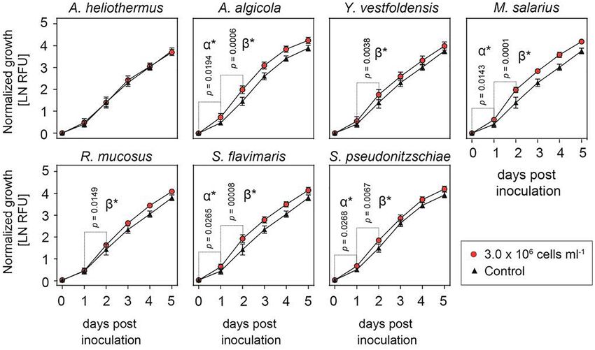
ORIGINAL RESEARCH
                                                                                                                                        published: 06 August 2019
                                                                                                                                   doi: 10.3389/fmicb.2019.01828

                                             Friends With Benefits: Exploring the
                                             Phycosphere of the Marine Diatom
                                             Skeletonema marinoi
                                             Oskar N. Johansson 1 , Matthew I. M. Pinder 2 , Fredrik Ohlsson 3 , Jenny Egardt 1 ,
                                             Mats Töpel 2,4 and Adrian K. Clarke 1*
                                             1
                                              Department of Biological and Environmental Sciences, University of Gothenburg, Gothenburg, Sweden, 2 Department
                                             of Marine Sciences, University of Gothenburg, Gothenburg, Sweden, 3 Department of Mathematical Sciences, Chalmers
                                             University of Technology, University of Gothenburg, Gothenburg, Sweden, 4 Gothenburg Global Biodiversity Centre,
                                             Gothenburg, Sweden

                                             Marine diatoms are the dominant phytoplankton in the temperate oceans and coastal
                                             regions, contributing to global photosynthesis, biogeochemical cycling of key nutrients
                                             and minerals and aquatic food chains. Integral to the success of marine diatoms is
                                             a diverse array of bacterial species that closely interact within the diffusive boundary
                                             layer, or phycosphere, surrounding the diatom partner. Recently, we isolated seven
                                             distinct bacterial species from cultures of Skeletonema marinoi, a chain-forming, centric
                                             diatom that dominates the coastal regions of the temperate oceans. Genomes of all
                         Edited by:          seven bacteria were sequenced revealing many unusual characteristics such as the
                         Zhiyong Li,
                                             existence of numerous plasmids of widely varying sizes. Here we have investigated
 Shanghai Jiao Tong University, China
                                             the characteristics of the bacterial interactions with S. marinoi, demonstrating that
                        Reviewed by:
                         Eric Fouilland,     several strains (Arenibacter algicola strain SMS7, Marinobacter salarius strain SMR5,
UMR 9190 Centre pour la Biodiversité         Sphingorhabdus flavimaris strain SMR4y, Sulfitobacter pseudonitzschiae strain SMR1,
             Marine, l’Exploitation et la
     Conservation (MARBEC), France           Yoonia vestfoldensis strain SMR4r and Roseovarius mucosus strain SMR3) stimulate
                       Pedro Cermeno,        growth of the diatom partner. Testing of many different environmental factors including
  Institute of Marine Sciences (CSIC),
                                             low iron concentration, high and low temperatures, and chemical signals showed
                                  Spain
                                             variable effects on this growth enhancement by each bacterial species, with the most
                  *Correspondence:
                      Adrian K. Clarke       significant being light quality in which green and blue but not red light enhanced the
           adrian.clarke@bioenv.gu.se        stimulatory effect on S. marinoi growth by all bacteria. Several of the bacteria also
                    Specialty section:
                                             inhibited growth of one or more of the other bacterial strains to different extents when
          This article was submitted to      mixed together. This study highlights the complex interactions between diatoms and
                  Microbial Symbioses,       their associated bacteria within the phycosphere, and that further studies are needed to
                a section of the journal
              Frontiers in Microbiology      resolve the underlying mechanisms for these relationships and how they might influence
            Received: 17 May 2019            the global success of marine diatoms.
            Accepted: 24 July 2019
                                             Keywords: bacteria, diatoms, microbiome, marine, symbiosis
          Published: 06 August 2019
                             Citation:
          Johansson ON, Pinder MIM,
    Ohlsson F, Egardt J, Töpel M and
                                            INTRODUCTION
        Clarke AK (2019) Friends With
  Benefits: Exploring the Phycosphere
                                            Silica-encrusted diatoms are ubiquitous marine primary producers that globally dominate the
   of the Marine Diatom Skeletonema         temperate oceans and coastlines. Together with other phytoplankton, they contribute around half
   marinoi. Front. Microbiol. 10:1828.      of the total CO2 fixation on earth (Field et al., 1998). The diatom hallmark, its silicated shell or
      doi: 10.3389/fmicb.2019.01828         frustule, is covered with secreted exopolymeric substances (EPS) causing them to aggregate and

Frontiers in Microbiology | www.frontiersin.org                                   1                                          August 2019 | Volume 10 | Article 1828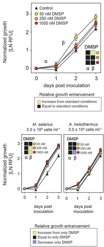
Johansson et al.                                                                                              Characterization of Diatom–Bacterial Interactions

sink to the ocean floor as “marine snow,” which is an integral             recently proposed S. marinoi as a new genetic model for chain-
part of the ocean cycling of carbon and other nutrients (Fowler            forming diatoms, having sequenced its genome and established
and Knauer, 1986; Passow and Alldredge, 1995; Simon et al.,                a collection of randomly mutagenized but identifiably tagged
2002). In close proximity to the cell surface of diatoms is a              strains suitable for large-scale phenotyping (Johansson et al.,
diffusion gradient, the phycosphere, where key nutrients are               2019). We have now also isolated several different bacteria from
exchanged between the diatom and the surrounding bacteria                  S. marinoi that has been in continuous culture for nearly a decade
(Bell and Mitchell, 1972; Amin et al., 2012). Formation of                 and determined the effect each individual bacterial species has on
this micro-environment beneficial for microbial interaction is             their diatom host.
promoted by EPS. It is known that co-culturing of diatoms
with specific marine bacteria accelerates the flocculation process
by increasing EPS production and changing its composition                  MATERIALS AND METHODS
(Grossart, 1999; Sonnenschein et al., 2011). This is especially
important since aggregation is a key event at the end of di-annual         Cell Isolation and Culture Conditions
diatom blooms (Alldredge and Gotschalk, 1989; Liu et al., 2008;            Skeletonema marinoi strains ST54 and R05AC were maintained
Park et al., 2010, 2015).                                                  in f/2 + Si medium under standard growth conditions (16◦ C,
    Ocean-living bacteria, like other life forms, are in constant          16 h photoperiod with an irradiance of 75 µmol photons m−2 s−1
need of organic carbon and are either passively or actively                from LED-light panels (Heliospectra, Sweden). Both strains are
pursuing sequestration of such compounds from photosynthetic               also available from the Gothenburg University algal culture
primary producers such as diatoms. The interaction between                 bank1 . To isolate any bacterial species growing in association
these organisms ranges from the purely symbiotic to parasitic              with S. marinoi in culture, aliquots were plated onto marine agar
and opportunistic, including passive acquaintances that only feed          2216 (BD, United States) and incubated at 16◦ C in darkness until
upon dead or dying diatom cells (Grossart, 1999; Grossart et al.,          colonies appeared. This procedure was performed on multiple
2005). This flux of organic carbon from phytoplankton to marine            occasions and the plates incubated over different time periods in
bacteria forms the microbial loop through which no less than               order to maximize the range of bacterial species isolated. Colonies
half of all fixed carbon flows through the bacteria (Azam et al.,          were then subjected to multiple iterations of serial dilution
1983). Microbes that proliferate in close proximity to the diatom          streaking until single bacterial isolates could be distinguished,
differ significantly from those that live outside of the diatom-           after which they were maintained on marine agar plates with
EPS biofilm (Delong et al., 1993). Those interacting bacteria are          regular re-streaking.
well adapted to a narrow range of compounds secreted by their
diatom host, forming an exclusiveness that contributes to their            DNA Extraction and Sequencing
specific association (Bell, 1984; Schafer et al., 2002; Sison-Mangus       Cultures of each bacterial strain (50 mL) were grown overnight
et al., 2014). Within the diatom phycosphere, there are examples           at 30◦ C in marine broth 2216 (BD, United States). Cells were
of symbiotic bacteria providing the host with vitamins and                 later pelleted and ground in liquid nitrogen, with genomic
organic acids, including amino acids and siderophores (Amin                DNA then extracted using Plant DNAzol (Thermo Fisher
et al., 2009). Diatoms in return provide the bacteria with easily          Scientific, United States) according to the manufacturer’s
accessible carbon- and sulfur sources (Seymour et al., 2017).              instructions. A region of the highly conserved 16S rRNA
    The diatom lineage has a complex evolutionary history with             gene was PCR amplified using a degenerate forward primer
extensive horizontal gene transfer between diatom and bacteria,            (16S_fwd, 50 -ATTAGITWGTTGGTRRGGTAA-30 ) and one
suggestive of a long-term association within the phycosphere               of the two different specific reverse primers (16S_rev1,
(Armbrust et al., 2004; Bowler et al., 2008; Mock et al., 2017).           50 -CGGCTGCTGGCACGGAGTTAG-30 ,                16S_rev2,    50 -
Despite this, the contribution of each interacting bacterial                                                    0
                                                                           CCACATGCTCCACCGCTTGTG-3 ) along with the AmpliTaq
species within the phycosphere and the level of functional                 DNA polymerase (Thermo Fisher Scientific, United States).
overlap between them remains unclear. Equally uncertain is the             Amplicons were purified using a Wizard PCR purification
underlying genetic basis for the host-microbiome interaction and           kit (Promega, United States) and Sanger sequenced (Eurofins
if this mechanism is conserved between pennate and centric                 Genomics, Germany).
diatoms. In this study, we have investigated the different bacteria
that constitute the microbiome of the centric marine diatom                Skeletonema marinoi Growth Assays
Skeletonema marinoi. Skeletonema is a prolific genus in the                Skeletonema marinoi isolate R05AC was pre-cultured in replicate
temperate oceans and coastal waters where diatoms dominate the             cultures (n = 8) for five d in enriched growth media, EGM
phytoplankton communities. In Scandinavian waters, S. marinoi              (Johansson et al., 2019). Cultures were centrifuged (1500 × g,
is a crucial primary producer that occurs throughout the year,             5 min), washed once in fresh EGM, and dissolved in 20 mL EGM.
especially during spring when it forms extensive blooms (de                Cell density of washed pre-cultures was measured (Gross et al.,
Vargas et al., 2015). S. marinoi also represents the obligate              2018) and then used to prepare experimental cultures with a
chain-forming diatoms that play a central role in the oceanic              starting concentration of 25 000 cells mL−1 . Bacterial cultures
biogeochemical cycles that sequester and mineralize carbon,                (5 mL) were grown in Difco marine broth (BD Biosciences,
nitrogen and silica, as well as forming resting stages that are
revivable even after a century (Härnstrom et al., 2011). We have           1
                                                                               https://marine.gu.se/english/research/marine-biology/algal-bank

Frontiers in Microbiology | www.frontiersin.org                        2                                               August 2019 | Volume 10 | Article 1828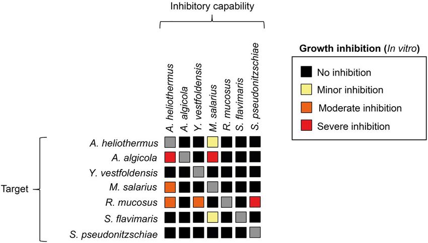
Johansson et al.                                                                                            Characterization of Diatom–Bacterial Interactions

United States) overnight with shaking a day before experiment.           et al., 2016) or Canu version 1.3 (Koren et al., 2017), and
Bacterial cells were pelleted (4000 × g, 7 min), washed in 10 mL         annotated using Prokka version 1.12 beta (Seemann, 2014).
EGM and again pelleted, then dissolved in 3 mL of EGM. OD600             A phylotaxonomic analysis was performed for each strain
was measured on a diluted aliquot and the cell concentration was         independently using PhyloPhlAn version 0.99 (Segata et al.,
based on OD600 = 1 that equates to 5 × 10⁸ cells mL−1 . Growth           2013). The taxonomic classification was also supported by marker
assays were performed under standard conditions in Falcon                gene comparisons, as well as by analysis of physical characteristics
48-well tissue culture plates (Corning, United States) unless            of the bacterial cells and colonies. The presence of prophages
otherwise stated and chlorophyll fluorescence was monitored              in each genome was determined using the phage search tool
daily. Average growth was measured as relative fluorescence units        PHASTER3 (Arndt et al., 2016). The presence of genes involved
and normalized at time zero, with the growth rates from time 0 to        in biotic-related interactions was ascertained using both Pathway
24 h (designated α) and 24 to 48 h (β) determined from the slope         Tools version 20.5/21.0 (Karp et al., 2002), and antiSMASH
of the growth curve. Low iron condition was set as 15% of EGM            version 3.0.54 with all optional analyses turned on (Weber et al.,
(49 to 7.2 µM). Light quality shifts were performed by covering          2015), as well as manual searches of each strain’s annotation.
the growth plates with plexiglass of the indicated color, which          The process is described in more detail in each strain’s respective
reduced the light intensity to 25–30 µmol photons m−2 s−1 .              genome announcement article (Töpel et al., 2017, 2018a,b,
Growth assays using various chemical additions were performed            2019a,b; and two additional papers in preparation).
as above, except for four replicate pre-cultures being used
instead of eight. The chemicals, their respective vendors, stock         Statistical Analysis
solvent and concentration are shown in Supplementary Table               Statistical analysis was performed in the software Prism 8
S1. Antibiotic sensitivity tests were performed by supplementing         (GraphPad Software Inc., United States).
S. marinoi cultures (n = 6) with the respective antibiotics at the
indicated concentrations prepared from stocks (Supplementary
Table S1), with growth measured by chlorophyll fluorescence              RESULTS
for four d.
                                                                         Bacterial Strains Cultured From
Bacterial Growth Assays and                                              S. marinoi
Characterization                                                         Non-axenic isolates of the diatom S. marinoi have been in
Antibiotic resistance and in vitro growth inhibition assays were         continuous culture in the Gothenburg University algal bank since
performed using the disc diffusion method (Davis and Stout,              their isolation in 2010. During the early stages of sequencing
1971). In short, filter paper discs (Ø = 5 mm) were sterilized           of the S. marinoi genome, DNA fragments from multiple
by autoclaving, submerged in either the antibiotic or respective         bacterial species were present within the genomic data (Töpel
bacterial suspension, and then placed on a marine agar plate             et al. in preparation). This, along with microscope observations
that had previously been covered with a thin layer of bacterial          led us to believe that several bacterial strains coexisted in
suspension (OD600 = 0.05). Plates were incubated at 30◦ C                the diatom cultures, even after almost a decade of laboratory
overnight, with growth inhibition then scored the following              culturing. To determine the composition of this microbiome,
day as none, minor, moderate or severe. Assessment of growth             two S. marinoi isolates (R05AC and ST54) were streaked onto
capability in terms of carbon sources was performed using                separate marine agar plates and incubated in darkness. Iterative
BIOLOG III (Bochner, 1989) plates in accordance with the                 dilution streaking produced identifiable single-strain bacterial
manufacturer’s instructions. Most strains were assayed on the            isolates, with the genus for each determined by sequencing
BIOLOG III plates using the standard IG-A solution, but due              partial 16S regions. Seven different genera were eventually
to the reductive nature of a subset of bacterial isolates IG-            isolated with multiple species present, one of which, from
B had to be used.                                                        each genus, was selected as a representative for further study
                                                                         (Supplementary Figure S1) including whole genome sequencing
Chlorophyll Fluorescence                                                 (Töpel et al., 2017, 2018a,b, 2019a,b).
Co-cultures (n = 8) of the respective bacteria and S. marinoi
R05AC were prepared in Falcon 48-well tissue culture plates from         Bacterial Stimulation of Diatom Growth
pre-cultures using the same procedure as for the growth assays.          Despite numerous attempts to establish axenic cultures of the
After growth for three d under standard conditions, the plate was        R05AC isolate of S. marinoi, all failed in part due to this
placed into an Imaging PAM (PSI, Czechia), dark-adapted for 1 h          diatom’s exceptional sensitivity to a wide variety of antibiotics
and photosynthetic performance as FV /FM was determined.                 and the inability of the antibiotic cocktails to remove all bacteria
                                                                         from the cultures (Supplementary Figures S2, S3). Given the
                                                                         likelihood that most if not all of the bacterial strains isolated
Bioinformatics Analysis                                                  from the S. marinoi cultures are in some form of symbiotic
The genomes of each bacterial strain were assembled using
                                                                         relationship, some of which could be important for the viability
either of the assembly programs Falcon version 1.7.52 (Gordon
                                                                         3
                                                                             http://phaster.ca
2                                                                        4
    https://github.com/PacificBiosciences/FALCON/                            https://antismash.secondarymetabolites.org/

Frontiers in Microbiology | www.frontiersin.org                      3                                               August 2019 | Volume 10 | Article 1828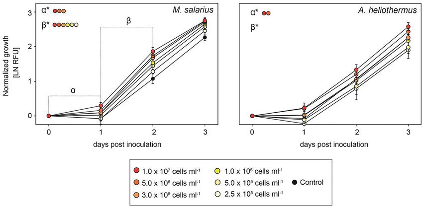
Johansson et al.                                                                                                        Characterization of Diatom–Bacterial Interactions

  FIGURE 1 | Growth-enhancing capacity of isolated bacterial cultures. Growth of Skeletonema marinoi cultures with (red circles) and without (black triangles) addition
  of the indicated bacterial species. Shown is average growth normalized at time zero and measured as relative fluorescence units, RFU (n = 8, ±SD).

  FIGURE 2 | The growth-promoting effect of added bacteria to Skeletonema marinoi cultures. Relative growth of S. marinoi after the addition of either M. salarius or
  A. heliothermus at different cell densities, with growth rates normalized at time zero and measured as RFU (n = 4, ±SD). Significance between slopes was calculated
  using multiple t-tests. P-values were adjusted for multiple testing using the Sidak–Bonferroni method, with asterisks indicating significant changes (adjusted
  p < 0.05).

of the diatom host, we tested if adding one bacterial species in                       0–24 h, and rate β, 24–48 h), whereas such growth stimulation
excess to the non-axenic culture affected growth of S. marinoi                         occurred only in the second half (rate β) after addition of Yoonia
under standard conditions (Figure 1). Equivalent abrupt shifts in                      vestfoldensis or Roseovarius mucosus.
microbiome composition would almost certainly occur in nature                             The growth-promoting effect of the added bacteria was
during diatom blooms or other sudden stochastic environmental                          positively correlated to the bacteria/diatom cell ratio, as
changes. As shown in Figure 1, the growth rate of S. marinoi was                       evidenced by the greater growth-promoting M. salarius and
significantly enhanced throughout the first 48 h after the addition                    lesser growth-promoting Antarctobacter heliothermus (Figure 2).
of Arenibacter algicola, Marinobacter salarius, Sphingorhabdus                         A threshold was specifically reached for M. salarius/S. marinoi
flavimaris or Sulfitobacter pseudonitzschiae (designated rate α,                       when adding more than 3 × 10⁶ bacterial cells per mL,

Frontiers in Microbiology | www.frontiersin.org                                    4                                             August 2019 | Volume 10 | Article 1828
Johansson et al.                                                                                                        Characterization of Diatom–Bacterial Interactions

  FIGURE 3 | The capacity of bacterial species to inhibit each other’s growth. Each bacterial specie was tested using the disc diffusion method. Bacterial
  cell-suspensions were dispersed to cover the entirety of a marine agar plate. Filter discs were submerged in a cell-suspension of each respective bacterial species,
  wiped and then one disc from each bacterial strain was put on each agar plate. The inhibitory capacity was scored as minor (yellow), moderate (orange) or severe
  (red) based on the zone of inhibition around the filter disc.

corresponding to a bacteria-to-diatom ratio of 100:1                                    commonly used for most marine diatoms as well as artificial sea
(Amin et al., 2015). At this ratio and higher, growth stimulation                       water did support the enhanced growth of S. marinoi after the
could be seen already after the first 24 h. A similar effect                            addition of M. salarius but not A. heliothermus (Supplementary
was evident also for A. heliothermus, which promoted                                    Figure S4), the further enriched media EGM as previously
growth within the first 24 h at a threshold concentration of                            described (Johansson et al., 2019) always resulted in a more
5 × 10⁶ bacterial cells per mL. It should be noted that no                              reliable effect, consistent with earlier findings that low nutrient
additional stimulation of S. marinoi growth occurred after 48 h                         conditions can attenuate symbiotic benefits (Gardes et al., 2012;
with any of the added bacteria. Also potentially complicating                           Meyer et al., 2016). In light of this, we examined if one or more of
the growth enhancement effects was the capability of certain                            the bacteria had a beneficial effect on S. marinoi growth under low
bacteria to inhibit the growth of other bacteria from the                               iron concentration (15% of that in standard f/2 + Si media), an
S. marinoi microbiome, pointing to a more intricate relationship                        often limiting factor in oceans for most phytoplankton (Martin
between the isolated bacteria within the phycosphere (Figure 3).                        and Gordon, 1988; Rue and Bruland, 1995). Interestingly, it
These complex interactions presumably re-establish inter-                               was only Y. vestfoldensis and S. pseudonitzschiae that had some
bacterial and diatom–bacterial homeostasis and resumption                               additional benefit under low iron concentrations during the β
of growth comparable to that of untreated S. marinoi post                               growth rate of S. marinoi (Figure 4A).
48 h of co-culture.                                                                         We next examined if other environmental variables
                                                                                        could influence the diatom–bacterial interactions. Shifting
                                                                                        temperatures are associated with ocean depth and the changing
Environmental Conditions Shape the                                                      seasons throughout the year. As shown in Figure 4B, the effect
Diatom–Bacterial Interactions                                                           of growth temperature on the interaction was strongest at the
The interaction between diatoms and their associated bacteria is                        low temperature (8◦ C), where the S. marinoi interactions with
influenced by the physical constraints of the marine environment,                       A. heliothermus and S. flavimaris were especially favorable.
including temperature and in particular nutrient availability                           In contrast, high temperature (24◦ C) had little effect on the
(Gardes et al., 2012; Meyer et al., 2016). The major nutrients                          interaction with the different bacteria compared to the standard
needed for diatom growth consist of nitrogen, phosphorus, silica                        growth temperature (16◦ C). Another environmental factor
and iron. Filtered and autoclaved deep ocean water with nutrient                        that varies with ocean depth is light quality, with blue light
and mineral enrichments has been the media of choice when                               penetrating far deeper than the increasingly longer visible
growing many marine phytoplankton (Guillard, 1975). While                               wavelengths of green and red. By illuminating the diatom under
S. marinoi grew well for limited periods in artificial seawater,                        co-culture with specific color ranges of the visible light spectrum,
it eventually proved unreliable as cultures would randomly die                          we next tested the effect of light quality on the interaction as
intermittently. Although the standard f/2 + Si growth media                             would be expected at different depths within the ocean water

Frontiers in Microbiology | www.frontiersin.org                                     5                                             August 2019 | Volume 10 | Article 1828
Johansson et al. Characterization of Diatom–Bacterial Interactions FIGURE 4 | Growth enhancement under variable iron, temperature and light conditions. Growth of Skeletonema marinoi cultures in co-culture with indicated bacterial species. Shown are growth rates normalized at time zero and measured as RFU (n = 8, ±SD) of the control (triangles) and co-cultures (circles) under different ecologically relevant low iron concentration (A), temperatures (B), and light qualities (C). Significance between slopes was calculated using multiple t-tests. p-values were adjusted for multiple testing using the Sidak–Bonferroni method. Significant increases in S. marinoi growth by the addition of a specific bacterial species (adjusted p < 0.05) are indicated by either α∗ (0–24 h) or β∗ (24–48 h). Square boxes display a comparison of growth rates during the α- and β-phases to the standard nutrient-replete growth conditions, with yellow indicating a significant enhancement, black unchanged and blue a significant decrease as determined by one-way ANOVA with corrections for multiple comparisons using the Bonferroni method. Frontiers in Microbiology | www.frontiersin.org 6 August 2019 | Volume 10 | Article 1828
Johansson et al.                                                                                                  Characterization of Diatom–Bacterial Interactions

                                                                                        column. As shown in Figure 4C, S. marinoi grew poorly under
                                                                                        red light and all bacteria when added had little or no stimulatory
                                                                                        effect upon the diatom growth rate. In contrast, S. marinoi grew
                                                                                        far better under either green or blue light than under red light
                                                                                        and the addition of each bacterial species enhanced the growth
                                                                                        rates, especially during the β-phase.

                                                                                        Mechanics of the Diatom–Bacteria
                                                                                        Interaction Do Not Resemble Those of
                                                                                        Plant Symbioses
                                                                                        Since a comprehensive model of the interaction of diatoms with
                                                                                        their associated bacteria is currently lacking, we compared this
                                                                                        interaction to those well-characterized ones between plants and
                                                                                        different microbial partners. For example, symbiotic interactions
                                                                                        between land plants and mycorrhiza-forming fungi are thought
                                                                                        to enhance photosynthetic performance (Schweiger et al., 2014),
                                                                                        but no such improvement in photosynthetic performance
                                                                                        of S. marinoi was observed after the addition of each of the
                                                                                        bacteria acting within the phycosphere under standard growth
                                                                                        conditions (data not shown). Certain important similarities
                                                                                        are known to exist between the diatom/bacteria interaction
                                                                                        in the phycosphere and that between plants and diazotrophic
                                                                                        bacteria of the rhizobium, such as the ability of the bacteria
                                                                                        to provide the host with iron (Carson et al., 1992). Another
                                                                                        is the use of plant hormones (Grunewald et al., 2009), with
                                                                                        evidence that auxins including indole-3-acetic acid (IAA)
                                                                                        and a structurally related compound, indole-3-acetonitrile
                                                                                        (IAN), are produced by bacteria within the phycosphere to
                                                                                        stimulate growth of their diatom partner (Amin et al., 2015).
                                                                                        Analysis of the S. marinoi reference genome does reveal genes
                                                                                        encoding most of the enzymes functioning within the auxin
                                                                                        biosynthetic pathway as defined in plants. Despite this, only
                                                                                        indole (IND), one of the early auxin precursors, had a growth
                                                                                        stimulatory effect on S. marinoi cultures, with no such effect
                                                                                        produced by any of the nine naturally occurring or synthetic
                                                                                        auxins, nor by the addition of the amino acids tyrosine (TYR)
                                                                                        and cysteine (CYS) or the phytoplankton-secreted molecule
                                                                                        taurine (TAU) (Supplementary Figure S5A). Moreover, the
  FIGURE 5 | Growth-enhancing capacities of DMSP. (A) Growth of                         addition of tryptophan (TRP), IAA or IAN had no effect on the
  Skeletonema marinoi cultures after the addition of different concentrations of        interactions between S. marinoi and either the growth-promoting
  DMSP (50 nM – yellow circles, 250 nM – orange circles and 1000 nM – red
                                                                                        M. salarius or non-growth-promoting A. heliothermus
  circles), with the control being S. marinoi with no added DMSP (black
  triangles). Shown are growth rates during the α- and β-phases normalized at
                                                                                        (Supplementary Figure S5B).
  time zero and measured as RFU (n = 4, ±SD). The box inserts display a                    Since the interaction between S. marinoi and most of its
  comparison of growth rates relative to the standard growth conditions                 bacterial partners is synergistic, we next examined the potential
  (control) during the respective time points as indicated by color. Significant        role of dimethylsulfoniopropionate (DMSP), a compound known
  changes (adjusted p < 0.05) in slopes were determined by multiple t-tests
                                                                                        to be produced by diatoms both as an intercellular osmolyte
  with Sidak–Bonferroni correction for multiple comparisons. (B) Change in
  growth rate of S. marinoi cultures after addition of both DMSP at indicated           and as a compound secreted into the phycosphere and used
  concentrations (50 nM – yellow, 250 nM – orange and 1000 nM – red circles)            by the surrounding bacteria (Moran et al., 2012; Buchan
  and either M. salarius or A. heliothermus, plus bacterial addition alone (green       et al., 2014; Amin et al., 2015). Growth enhancement of
  diamonds) compared to that of DMSP only. Growth rates during the α- and               the diatom provoked by the addition of DMSP would be
  β-phases were normalized at time zero and measured as RFU (n = 4, ±SD).
                                                                                        mediated by the various bacteria within the phycosphere,
  The box inserts display a comparison of growth rates relative to the standard
  growth conditions (control) during the respective time points as indicated by         suggesting that the diatom–bacteria interaction in standard
  color. Significant changes (adjusted p < 0.05) in slopes were determined by           non-axenic cultures is limited. To test if this was the case,
  multiple t-tests with Sidak–Bonferroni correction for multiple comparisons.           DMSP was added to cultures of S. marinoi at different
  Yellow shading indicates a significantly higher growth enhancement, blue a            concentrations in a range that was ecologically relevant (Aumont
  significantly reduced growth enhancement, and black no significant change.
                                                                                        et al., 2002). Interestingly, there was a stimulatory effect on

Frontiers in Microbiology | www.frontiersin.org                                     7                                      August 2019 | Volume 10 | Article 1828
Johansson et al. Characterization of Diatom–Bacterial Interactions
S. marinoi growth at the lowest concentration of DMSP used relatively scarce and is often the limiting factor for growth
(i.e., 50 nM) but not at higher concentrations (Figure 5A). of most phytoplankton (Martin and Gordon, 1988; Rue
We next combined the addition of DMSP with that of the and Bruland, 1995). Iron supplementation is also important
growth-promoting M. salarius and observed significant growth in certain terrestrial habitats such as for the symbiotic
enhancement of S. marinoi at the higher concentrations of interaction between plants and bacteria within the rhizobium
DMSP (i.e., ≥250 nM), whereas substituting M. salarius with the (Berraho et al., 1997). In the marine environment, certain
non-growth-promoting A. heliothermus attenuated the DMSP bacteria can also produce iron-chelating agents, including
growth-enhancing effect (Figure 5B). siderophores, which aid in iron assimilation. In general, the
absence of such chelating agents could pose potential problems
for the intrinsic relationship between diatoms and bacteria
DISCUSSION in a low iron environment. Interestingly, genes encoding
siderophores are present in the genomes of the M. salarius
Laboratory cultures of diatoms tend to keep many of and S. pseudonitzschiae (Töpel et al., 2019a) strains from our
their associated bacteria, especially those in some form of study (Supplementary Figure S5). Under low iron conditions,
symbiotic relationship if not treated with antibiotics. The addition of S. pseudonitzschiae did indeed enhance S. marinoi
relationship for many diatoms has proven so intertwined growth under otherwise standard growth conditions whereas
that even exhaustive attempts to remove them sometimes there was no additional effect on the already strong growth-
fail as was the case with S. marinoi in this study. We were promoting M. salarius, the lack of which suggests another
able to eventually isolate seven strains of bacteria from constraint being limiting. In contrast, co-culture of S. marinoi
stock cultures of S. marinoi that had been kept in culture with Y. vestfoldensis also improved growth under low iron
for almost a decade. By shifting the bacterial equilibrium conditions compared to standard conditions, despite the
within the S. marinoi cultures we were able to study the lack of recognizable genes encoding siderophores within the
effect of each individual bacterial species on the diatom Y. vestfoldensis genome, opening the possibility for other
host. Additions of each bacterium to the cultures revealed chelating agents being produced.
that all but one (A. heliothermus) strongly stimulated the Ocean water temperatures and light conditions shift
growth of S. marinoi. The growth enhancement effect was throughout the year and with water depth. Interestingly,
dependent on time and the diatom to bacteria ratio, with low temperature (8◦ C) enhanced the growth stimulation on
the two bacterial species that most consistently promoted S. marinoi cultures but only with the addition of A. heliothermus
growth under the different conditions tested being M. salarius and S. flavimaris, whereas no significant change was observed at
and A. algicola. high temperatures (24◦ C) relative to the standard temperature
Marinobacter sp. are commonly found associated with algal (16◦ C). At this stage, the reason for this beneficial effect of
field samples and remain even in decade-old cultures of A. heliothermus and S. flavimaris at low temperatures on
Skeletonema costatum (Jian et al., 2019). The growth-affecting S. marinoi growth remains unclear, with no obvious clues
properties (both positive and negative) of the Marinobacter genus from the genome comparisons between the different bacteria.
have been previously demonstrated by co-culturing with diatoms In comparison, changes in light quality did significantly
such as S. costatum and Thalassiosira pseudonana (Grossart alter the stimulatory response by each bacterial strain
et al., 2005, 2006; Grossart and Simon, 2007; Yang et al., 2013). on S. marinoi growth. The fact that green and blue, but
Interestingly, Marinobacter sp. either enhanced growth as in not red light produced the stimulatory response suggests
the case of T. pseudonana or reduced it as in S. costatum it can be limited by reduced photosynthetic activity of
compared to axenic cultures during the first days of co-culture. the diatom host. Given that the pigment composition of
In contrast, the M. salarius strain identified in our cultures diatoms consists of chlorophylls and various carotenoids that
significantly promoted the growth of S. marinoi, attenuated only together absorb higher proportions of blue/green light, the
by red light, suggesting that even at the species level there improved photosynthetic rates and thereby growth under
is exclusiveness within the phycosphere environment for each these light conditions would place fewer limitations on the
diatom species. Similarly, members of the Arenibacter genus are stimulatory effect the added bacteria have on S. marinoi
also known to stimulate growth of the diatoms T. pseudonana cultures. Conversely, lower light absorption and subsequent
and Phaeodactylum tricornutum (Zecher et al., 2015). This was reduced photosynthetic activity under red light would likely
indeed also true for A. algicola in this study, promoting the be rate-limiting on overall growth of S. marinoi, which would
growth of S. marinoi with similar characteristics to that by therefore mask any potential benefit from the addition of the
M. salarius, although again with the exception of red-light bacterial strains.
conditions. It should be noted that the relative lack of growth One group of signaling molecule that is known to influence the
stimulation of S. marinoi by these bacteria under red light is growth of certain diatoms and other eukaryotic phytoplankton
likely linked to limited light absorption of these visible light is the auxin IAA and its derivatives (Amin et al., 2015;
wavelengths and the resulting minimal photosynthetic activity Labeeuw et al., 2016). Auxins are best known as an important
and overall growth. group of plant hormones responsible for a wide range of
The low iron concentration experiment revealed specific growth and developmental changes such as cell expansion,
benefits of certain bacteria. Soluble iron (III) in oceans is tissue differentiation and fertility (Zhao, 2010). Besides plants,
Frontiers in Microbiology | www.frontiersin.org 8 August 2019 | Volume 10 | Article 1828Johansson et al. Characterization of Diatom–Bacterial Interactions
certain bacteria and fungi also synthesize auxins (Spaepen et al., several years, comprising hundreds of bacterial generations,
2007) although much of their function in these organisms has suggesting there remains a strong selective pressure for
been inferred only from the extensive studies done in plants. maintaining these different bacterial species within the
More recently, synthesis of IAA was shown in a Sulfitobacter diatom phycosphere.
sp. in response to an increase in tryptophan synthesis by its Although most of the bacterial species isolated from S. marinoi
diatom host, the pennate Pseudo-nitzschia multiseries, which in cultures had a positive effect on diatom growth, understanding
turn stimulated the growth of the diatom (Amin et al., 2015). the presence of A. heliothermus was more elusive. It would on
Interestingly, the lack of any growth stimulatory effect by IAA occasion, such as with low temperature and different light quality,
or other auxin-related compounds in S. marinoi cultures as improve S. marinoi growth to some extent but overall it had
shown in this study suggest a possible functional split between little significant effect. It should be noted, however, that this
centric- and pennate diatoms regarding the chemical interactions species was only found in the S. marinoi isolate ST54 and not
within their respective phycospheres. Indeed, the absence of a in the R05AC isolate used for much of this study. Although
recognizable plant-like auxin response mechanism mediated by the two isolates are very similar in terms of genome sequence,
ARF transcription factors in the genome of sequenced diatoms this difference in bacterial composition could indicate that the
further suggests that much remains unknown about the role of phycosphere in each S. marinoi isolate are more specialized and
auxins and other signaling compounds within the phycosphere fine-tuned than previously thought.
of different diatoms.
In contrast to auxins, DMSP did promote growth of
S. marinoi cultures within a certain concentration range, with DATA AVAILABILITY
the concentration dependence being explained by the addition
of either M. salarius or A. heliothermus together with DMSP. The datasets generated for this study are available on request to
DMSP is typically excreted by diatoms and taken up by associated the corresponding author.
bacteria, after which it is degraded to different organosulfur
compounds (Moran et al., 2012). Interestingly, the genes required
for DMSP degradation are present in the genomes of all but AUTHOR CONTRIBUTIONS
two of the seven bacteria isolated from S. marinoi cultures
(Supplementary Figure S6). In S. marinoi cultures with the OJ performed most of the experimental work, analyzed the
normal proportion of associated bacterial species, the lower experimental data, and wrote the manuscript. MT and MP
concentrations of DMSP were likely suitable for stimulating provided bioinformatic expertise and sequence analyses. JE
growth of the bacteria, which in turn further promoted growth helped with the growth assays. FO assisted in growth rate
of S. marinoi, with higher concentrations of DMSP possibly analyses. AC designed the project, analyzed the experimental
in excess and outside of the optimal range. In contrast, the data, and wrote the manuscript. All authors helped in
stimulatory effect of DMSP was only observed at the higher finalizing the manuscript.
concentrations after the addition of M. salarius, most likely
due to the greater proportion of bacterial cells within the
culture and the need for extra DMSP to promote their growth.
FUNDING
Besides DMSP, additional compounds are also likely secreted This work was supported by grants to AC from the Gordon and
by diatoms in order to stimulate growth of bacteria in the Betty Moore Foundation (No. 4967) and the Swedish Research
phycosphere. A range of carbohydrates and amino acids have Council VR (No. 2015-04286).
been suggested to be involved (Bruckner et al., 2008, 2011)
and most bacteria isolated from S. marinoi cultures could
utilize one or more forms of carbohydrates, although there did ACKNOWLEDGMENTS
appear a preference for amino acids and other organic acids
(Supplementary Figure S7). We thank the Linnéus Centre for Marine Evolutionary
It is well known that genomes of uncultivatable bacteria Biology (CeMEB, http://cemeb.science.gu.se/) for support. All
often occur in metagenomic data (Vartoukian et al., 2010), bioinformatics analyses were run on the Albiorix computer
suggesting that the phycosphere of S. marinoi likely includes cluster (http://albiorix.bioenv.gu.se/) at the Department of
more bacterial species than those isolated in this study. Marine Sciences, University of Gothenburg.
Nevertheless, our experiments provide ample evidence that
growth-enhancing properties remain among the associated
bacteria isolated from laboratory cultures even after a decade SUPPLEMENTARY MATERIAL
of being isolated from nature, and that these differ significantly
from those between plants and their symbionts. Indeed, The Supplementary Material for this article can be found
growth addition experiments have been performed with online at: https://www.frontiersin.org/articles/10.3389/fmicb.
similar results on numerous occasions over the course of 2019.01828/full#supplementary-material
Frontiers in Microbiology | www.frontiersin.org 9 August 2019 | Volume 10 | Article 1828Johansson et al. Characterization of Diatom–Bacterial Interactions
REFERENCES Fowler, S. W., and Knauer, G. A. (1986). Role of large particles in the transport
of elements and organic-compounds through the oceanic water column. Prog.
Alldredge, A. L., and Gotschalk, C. C. (1989). Direct observations of the mass Oceanogr. 16, 147–194. doi: 10.1016/0079-6611(86)90032-7
flocculation of diatom blooms - characteristics, settling velocities and formation Gardes, A., Ramaye, Y., Grossart, H. P., Passow, U., and Ullrich, M. S. (2012).
of diatom aggregates. Deep-Sea Res. Part a-Oceanogr. Res. Pap. 36, 159–171. Effects of Marinobacter adhaerens HP15 on polymer exudation by Thalassiosira
Amin, S. A., Green, D. H., Hart, M. C., Kupper, F. C., Sunda, W. G., and Carrano, weissflogii at different N:P ratios. Mar. Ecol. Prog. Ser. 461, 1–14. doi: 10.3354/
C. J. (2009). Photolysis of iron-siderophore chelates promotes bacterial-algal meps09894
mutualism. Proc. Natl. Acad. Sci. U.S.A. 106, 17071–17076. doi: 10.1073/pnas. Gordon, D., Huddleston, J., Chaisson, M. J., Hill, C. M., Kronenberg, Z. N.,
0905512106 Munson, K. M., et al. (2016). Long-read sequence assembly of the gorilla
Amin, S. A., Hmelo, L. R., van Tol, H. M., Durham, B. P., Carlson, L. T., Heal, K. R., genome. Science 352:aae0344. doi: 10.1126/science.aae0344
et al. (2015). Interaction and signalling between a cosmopolitan phytoplankton Gross, S., Kourtchenko, O., Rajala, T., Andersson, B., Fernandez, L., Blomberg,
and associated bacteria. Nature 522, 98–101. doi: 10.1038/nature14488 A., et al. (2018). Optimization of a high-throughput phenotyping method for
Amin, S. A., Parker, M. S., and Armbrust, E. V. (2012). Interactions between chain-forming phytoplankton species. Limnol. Oceanogr. Methods 16, 57–67.
diatoms and bacteria. Microbiol. Mol. Biol. Rev. 76, 667–684. doi: 10.1002/lom3.10226
Armbrust, E. V., Berges, J. A., Bowler, C., Green, B. R., Martinez, D., Putnam, N. H., Grossart, H. P. (1999). Interactions between marine bacteria and axenic diatoms
et al. (2004). The genome of the diatom Thalassiosira pseudonana: ecology, (Cylindrotheca fusiformis, Nitzschia laevis, and Thalassiosira weissflogii)
evolution, and metabolism. Science 306, 79–86. incubated under various conditions in the lab. Aquat. Microb. Ecol. 19, 1–11.
Arndt, D., Grant, J. R., Marcu, A., Sajed, T., Pon, A., Liang, Y., et al. (2016). doi: 10.3354/ame019001
PHASTER: a better, faster version of the PHAST phage search tool. Nucleic Acids Grossart, H. P., Czub, G., and Simon, M. (2006). Algae-bacteria interactions and
Res. 44, W16–W21. doi: 10.1093/nar/gkw387 their effects on aggregation and organic matter flux in the sea. Environ. Microb.
Aumont, O., Belviso, S., and Monfray, P. (2002). Dimethylsulfoniopropionate 8, 1074–1084. doi: 10.1111/j.1462-2920.2006.00999.x
(DMSP) and dimethylsulfide (DMS) sea surface distributions simulated from Grossart, H. P., Levold, F., Allgaier, M., Simon, M., and Brinkhoff, T. (2005).
a global three-dimensional ocean carbon cycle model. J. Geophys. Res. Oceans Marine diatom species harbour distinct bacterial communities. Environ.
107, 4–1. Microbiol. 7, 860–873. doi: 10.1111/j.1462-2920.2005.00759.x
Azam, F., Fenchel, T., Field, J. G., Gray, J. S., Meyerreil, L. A., and Thingstad, F. Grossart, H. P., and Simon, M. (2007). Interactions of planktonic algae and
(1983). The ecological role of water-column microbes in the sea. Mar. Ecol. Prog. bacteria: effects on algal growth and organic matter dynamics. Aquat. Microb.
Ser. 10, 257–263. doi: 10.3354/meps010257 Ecol. 47, 163–176. doi: 10.3354/ame047163
Bell, W., and Mitchell, R. (1972). Chemotactic and growth responses of marine Grunewald, W., van Noorden, G., van Isterdael, G., Beeckman, T., Gheysen, G.,
bacteria to algal extracellular products. Biol. Bull. 143, 265–277. and Mathesius, U. (2009). Manipulation of auxin transport in plant roots during
Bell, W. H. (1984). Bacterial adaptation to low-nutrient conditions as studied with Rhizobium symbiosis and nematode parasitism. Plant Cell 21, 2553–2562. doi:
algal extracellular products. Microb. Ecol. 10, 217–230. doi: 10.1007/Bf02010936 10.1105/tpc.109.069617
Berraho, E. L., Lesueur, D., Diem, H. G., and Sasson, A. (1997). Iron requirement Guillard, R. J. P. (1975). Culture of phytoplankton for feeding marine invertebrates.
and siderophore production in Rhizobium ciceri during growth on an iron- Cult. Mar. Invertebr. Anim. 755, 29–60.
deficient medium. World J. Microbiol. Biotechnol. 13, 501–510. doi: 10.1023/A: Härnstrom, K., Ellegaard, M., Andersen, T. J., and Godhe, A. (2011). Hundred years
1018553022960 of genetic structure in a sediment revived diatom population. Proc. Natl. Acad.
Bochner, B. R. (1989). Sleuthing out bacterial identities. Nature 339, 157–158. Sci. U.S.A. 108, 4252–4257. doi: 10.1073/pnas.1013528108
doi: 10.1038/339157a0 Jian, W., Zhaohui, W., and Jiangang, Z. (2019). Isolation, characterization
Bowler, C., Allen, A. E., Badger, J. H., Grimwood, J., Jabbari, K., Kuo, A., et al. and implications of the bacterial communities associated with established
(2008). The Phaeodactylum genome reveals the evolutionary history of diatom cultures of Chattonella marina (Raphidophyceae) and Skeletonema costatum
genomes. Nature 456, 239–244. doi: 10.1038/nature07410 (Bacillariophyceae). Acta Oceanol. Sin. 38, 128–135.
Bruckner, C. G., Bahulikar, R., Rahalkar, M., Schink, B., and Kroth, P. G. (2008). Johansson, O. N., Töpel, M., Pinder, M. I. M., Kourtchenko, O., Blomberg, A.,
Bacteria associated with benthic diatoms from lake constance: phylogeny Godhe, A., et al. (2019). Skeletonema marinoi as a new genetic model for marine
and influences on diatom growth and secretion of extracellular polymeric chain-forming diatoms. Sci. Rep. 9:5391. doi: 10.1038/s41598-019-41085-5
substances. Appl. Environ. Microbiol. 74, 7740–7749. doi: 10.1128/Aem. Karp, P., Paley, S., and Romero, P. (2002). The pathway tools software.
01399-8 Bioinformatics 18(Suppl. 1), S225–S232. doi: 10.1093/bioinformatics/18.suppl_
Bruckner, C. G., Rehm, C., Grossart, H. P., and Kroth, P. G. (2011). Growth 1.S225
and release of extracellular organic compounds by benthic diatoms depend on Koren, S., Walenz, B. P., Berlin, K., Miller, J. R., Bergman, N. H., and Phillippy,
interactions with bacteria. Environ. Microbiol. 13, 1052–1063. doi: 10.1111/j. A. M. (2017). Canu: scalable and accurate long-read assembly via adaptive
1462-2920.2010.02411.x k-mer weighting and repeat separation. Genome Res. 27, 722–736. doi: 10.1101/
Buchan, A., LeCleir, G. R., Gulvik, C. A., and Gonzalez, J. M. (2014). Master gr.215087.116
recyclers: features and functions of bacteria associated with phytoplankton Labeeuw, L., Khey, J., Bramucci, A. R., Atwal, H., de la Mata, A. P., Harynuk, J., et al.
blooms. Nat. Rev. Microbiol. 12, 686–698. doi: 10.1038/nrmicro3326 (2016). Indole-3-acetic acid is produced by emiliania huxleyi coccolith-bearing
Carson, K. C., Holliday, S., Glenn, A. R., and Dilworth, M. J. (1992). Siderophore cells and triggers a physiological response in bald cells. Front. Microbiol. 7:828.
and organic-acid production in root nodule bacteria. Arch. Microbiol. 157, doi: 10.3389/fmicb.2016.00828
264–271. doi: 10.1007/Bf00245160 Liu, J. Q., Lewitus, A. J., Kempton, J. W., and Wilde, S. B. (2008). The association
Davis, W. W., and Stout, T. R. (1971). Disc plate method of microbiological of algicidal bacteria and raphidophyte blooms in South Carolina brackish
antibiotic assay. I. Factors influencing variability and error. Appl. Microbiol. 22, detention ponds. Harmful Algae 7, 184–193. doi: 10.1016/j.ha1.2007.07.001
659–665. Martin, J. H., and Gordon, R. M. (1988). Northeast pacific iron distributions in
de Vargas, C., Audic, S., Henry, N., Decelle, J., Mahe, F., Logares, R., et al. (2015). relation to phytoplankton productivity. Deep-Sea Res. Part a-Oceanogr. Res.
Eukaryotic plankton diversity in the sunlit ocean. Science 348:1261605. doi: Pap. 35, 177–196.
10.1126/science.1261605 Meyer, J., Loscher, C. R., Neulinger, S. C., Reichel, A. F., Loginova, A., Borchard,
Delong, E. F., Franks, D. G., and Alldredge, A. L. (1993). Phylogenetic diversity C., et al. (2016). Changing nutrient stoichiometry affects phytoplankton
of aggregate-attached vs free-living marine bacterial assemblages. Limnol. production, DOP accumulation and dinitrogen fixation - a mesocosm
Oceanogr. 38, 924–934. doi: 10.4319/lo.1993.38.5.0924 experiment in the eastern tropical North Atlantic. Biogeosciences 13, 781–794.
Field, C. B., Behrenfeld, M. J., Randerson, J. T., and Falkowski, P. (1998). Primary Mock, T., Otillar, R. P., Strauss, J., McMullan, M., Paajanen, P., Schmutz, J.,
production of the biosphere: integrating terrestrial and oceanic components. et al. (2017). Evolutionary genomics of the cold-adapted diatom fragilariopsis
Science 281, 237–240. doi: 10.1126/science.281.5374.237 cylindrus. Nature 541, 536–540. doi: 10.1038/nature20803
Frontiers in Microbiology | www.frontiersin.org 10 August 2019 | Volume 10 | Article 1828Johansson et al. Characterization of Diatom–Bacterial Interactions Moran, M. A., Reisch, C. R., Kiene, R. P., and Whitman, W. B. (2012). Genomic pseudonitzschiae Strain SMR1, isolated from a culture of the marine diatom insights into bacterial DMSP transformations. Annu. Rev. Mar. Sci. 4, 523–542. Skeletonema marinoi. J. Genom. 7, 7–10. doi: 10.7150/jgen.30559 Park, B. S., Kim, J. H., Kim, J. H., Gobler, C. J., Baek, S. H., and Han, M. S. (2015). Töpel, M., Pinder, M. I. M., Johansson, O. N., Kourtchenko, O., Godhe, A., and Dynamics of bacterial community structure during blooms of Cochlodinium Clarke, A. K. (2019b). Genome sequence of Arenibacter algicola strain SMS7, polykrikoides (Gymnodiniales, Dinophyceae) in korean coastal waters. Harmful found in association with the marine diatom Skeletonema marinoi. Microbiol. Algae 48, 44–54. doi: 10.1016/j.hal.2015.07.004 Resour. Announc. 8:e01461-18. doi: 10.1128/MRA.01461-18 Park, J. H., Yoshinaga, I., Nishikawa, T., and Imai, I. (2010). Algicidal bacteria Töpel, M., Pinder, M. I. M., Johansson, O. N., Kourtchenko, O., Godhe, A., and in particle-associated form and in free-living form during a diatom bloom in Clarke, A. K. (2017). Genome sequence of Roseovarius mucosus strain SMR3, the Seto Inland Sea, Japan. Aquat. Microb. Ecol. 60, 151–161. doi: 10.3354/ isolated from a culture of the diatom Skeletonema marinoi. Genome. Announc. ame01416 5:e00394-17. doi: 10.1128/genomeA.00394-17 Passow, U., and Alldredge, A. L. (1995). Aggregation of a diatom bloom in a Töpel, M., Pinder, M. I. M., Johansson, O. N., Kourtchenko, O., Godhe, A., and mesocosm - the role of transparent exopolymer particles (Tep). Deep-Sea Res. Clarke, A. K. (2018a). Complete genome sequence of Loktanella vestfoldensis Part II-Top. Stud. Oceanogr. 42, 99–109. doi: 10.1016/0967-0645(95)00006-C strain SMR4r, a novel strain isolated from a culture of the chain-forming diatom Rue, E. L., and Bruland, K. W. (1995). Complexation of iron(III) by natural Skeletonema marinoi. Genome. Announc. 6:e1558-17. doi: 10.1128/genomeA. organic-ligands in the central north pacific as determined by a new competitive 01558-17 ligand equilibration adsorptive cathodic stripping voltammetric method. Mar. Töpel, M., Pinder, M. I. M., Johansson, O. N., Kourtchenko, O., Godhe, A., and Chem. 50, 117–138. doi: 10.1016/0304-4203(95)00031-L Clarke, A. K. (2018b). Whole-genome sequence of the novel Antarctobacter Schafer, H., Abbas, B., Witte, H., and Muyzer, G. (2002). Genetic diversity of heliothermus strain SMS3, found in association with the marine diatom ’satellite’ bacteria present in cultures of marine diatoms. Fems Microbiol. Ecol. Skeletonema marinoi. J. Genomics. 6, 113–116. doi: 10.7150/jgen.27637 42, 25–35. doi: 10.1111/j.1574-6941.2002.tb00992.x Vartoukian, S. R., Palmer, R. M., and Wade, W. G. (2010). Strategies for culture of Schweiger, R., Baier, M. C., and Muller, C. (2014). Arbuscular mycorrhiza-induced ’unculturable’ bacteria. Fems Microbiol. Lett. 309, 1–7. doi: 10.1111/j.1574-6968. shifts in foliar metabolism and photosynthesis mirror the developmental stage 2010.02000.x of the symbiosis and are only partly driven by improved phosphate uptake. Mol. Weber, T., Blin, K., Duddela, S., Krug, D., Kim, H. U., Bruccoleri, R., et al. Plant Microbe Interact. 27, 1403–1412. doi: 10.1094/Mpmi-05-14-0126-R (2015). antiSMASH 3.0 – a comprehensive resource for the genome mining of Seemann, T. (2014). Prokka: rapid prokaryotic genome annotation. Bioinformatics biosynthetic gene clusters. Nucleic Acids Res. 43, W237–W243. doi: 10.1093/ 30, 2068–2069. doi: 10.1093/bioinformatics/btu153 nar/gkv437 Segata, N., Börningen, D., Morgan, X. C., and Huttenhower, C. (2013). PhyloPhlAn Yang, Y. F., Hu, X. J., Zhang, J., and Gong, Y. X. (2013). Community level is a new method for improved phylogenetic and taxonomic placement of physiological study of algicidal bacteria in the phycospheres of Skeletonema microbes. Nat. Commun. 4:2304. doi: 10.1038/ncomms3304 costatum and Scrippsiella trochoidea. Harmful Algae 28, 88–96. doi: 10.1016/j. Seymour, J. R., Amin, S. A., Raina, J. B., and Stocker, R. (2017). Zooming in on the hal.2013.05.015 phycosphere: the ecological interface for phytoplankton-bacteria relationships. Zecher, K., Jagmann, N., Seemann, P., and Philipp, B. (2015). An efficient screening Nat. Microbiol. 2:17065. doi: 10.1038/nmicrobiol.2017.65 method for the isolation of heterotrophic bacteria influencing growth of Simon, M., Grossart, H. P., Schweitzer, B., and Ploug, H. (2002). Microbial ecology diatoms under photoautotrophic conditions. J. Microbiol. Methods 119, 154– of organic aggregates in aquatic ecosystems. Aquat. Microb. Ecol. 28, 175–211. 162. doi: 10.1016/j.mimet.2015.10.016 doi: 10.3354/ame028175 Zhao, Y. (2010). Auxin biosynthesis and its role in plant development. Annu. Rev. Sison-Mangus, M. P., Jiang, S., Tran, K. N., and Kudela, R. M. (2014). Host-specific Plant Biol. 61, 49–64. doi: 10.1146/annurev-arplant-042809-112308 adaptation governs the interaction of the marine diatom, Pseudo-nitzschia and their microbiota. Isme J. 8, 63–76. doi: 10.1038/ismej.2013.138 Conflict of Interest Statement: The authors declare that the research was Sonnenschein, E. C., Gardes, A., Seebah, S., Torres-Monroy, I., Grossart, H. P., conducted in the absence of any commercial or financial relationships that could and Ullrich, M. S. (2011). Development of a genetic system for Marinobacter be construed as a potential conflict of interest. adhaerens HP15 involved in marine aggregate formation by interacting with diatom cells. J. Microbiol. Methods 87, 176–183. doi: 10.1016/j.mimet.2011. Copyright © 2019 Johansson, Pinder, Ohlsson, Egardt, Töpel and Clarke. This is an 08.008 open-access article distributed under the terms of the Creative Commons Attribution Spaepen, S., Vanderleyden, J., and Remans, R. (2007). Indole-3-acetic acid in License (CC BY). The use, distribution or reproduction in other forums is permitted, microbial and microorganism-plant signaling. FEMS Microbiol. Rev. 31, 425– provided the original author(s) and the copyright owner(s) are credited and that the 448. doi: 10.1111/j.1574-6976.2007.00072.x original publication in this journal is cited, in accordance with accepted academic Töpel, M., Pinder, M. I. M., Johansson, O. N., Kourtchenko, O., Clarke, A. K., practice. No use, distribution or reproduction is permitted which does not comply and Godhe, A. (2019a). Complete genome sequence of novel Sulfitobacter with these terms. Frontiers in Microbiology | www.frontiersin.org 11 August 2019 | Volume 10 | Article 1828
You can also read