A pipeline for rapidly generating genetically engineered mouse models of pancreatic cancer using in vivo CRISPR- Cas9 mediated somatic ...
←
→
Page content transcription
If your browser does not render page correctly, please read the page content below
bioRxiv preprint first posted online Aug. 23, 2018; doi: http://dx.doi.org/10.1101/398347. The copyright holder for this preprint
 (which was not peer-reviewed) is the author/funder, who has granted bioRxiv a license to display the preprint in perpetuity.
                                    All rights reserved. No reuse allowed without permission.

       A pipeline for rapidly generating genetically engineered
      mouse models of pancreatic cancer using in vivo CRISPR-
                Cas9 mediated somatic recombination

      Noboru Ideno1, Hiroshi Yamaguchi1, Takashi Okumara1, Jonathon Huang1, Mitchel J. Brun2,
             Michelle L. Ho3, Junghae Suh3, Sonal Gupta1, Anirban Maitra1* & Bidyut Ghosh1*

             1
                 Department of Translational Molecular Pathology and Sheikh Ahmed Pancreatic Cancer
             Research Center, UT
             MD Anderson Cancer Center
             Houston, TX.

             2
                 Department of Chemical and Biomolecular Engineering
             Rice University.
             Houston, TX.

             3
                 Department of Bioengineering,
             Rice University.
             Houston, TX.

             *
              Corresponding Author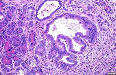
bioRxiv preprint first posted online Aug. 23, 2018; doi: http://dx.doi.org/10.1101/398347. The copyright holder for this preprint
 (which was not peer-reviewed) is the author/funder, who has granted bioRxiv a license to display the preprint in perpetuity.
                                    All rights reserved. No reuse allowed without permission.

   ABSTRACT:

   Genetically engineered mouse models (GEMMs) that recapitulate the major genetic drivers in
   pancreatic ductal adenocarcinoma (PDAC) have provided unprecedented insights into the
   pathogenesis of this lethal neoplasm. Nonetheless, generating an autochthonous model is an
   expensive, time consuming and labor intensive process, particularly when tissue specific
   expression or deletion of compound alleles are involved. In addition, many of the current PDAC
   GEMMs cause embryonic, pancreas-wide activation or loss of driver alleles, neither of which
   reflects the cognate human disease scenario. The advent of CRISPR/Cas9 based gene editing can
   potentially circumvent many of the aforementioned shortcomings of conventional breeding
   schema, but ensuring the efficiency of gene editing in vivo remains a challenge. Here we have
   developed a pipeline for generating PDAC GEMMs of complex genotypes with high efficiency
   using a single “workhorse” mouse strain expressing Cas9 in the adult pancreas under a p48
   promoter. Using adeno-associated virus (AAV) mediated delivery of multiplexed guide RNAs
   (sgRNAs) to the adult murine pancreas of p48-Cre; LSL-Cas9 mice, we confirm our ability to
                                           G12D
   express an oncogenic Kras                      allele through homology-directed repair (HDR), in conjunction
   with CRISPR-induced disruption of cooperating alleles (Trp53, Lkb1 and Arid1A). The resulting
   GEMMs demonstrate a spectrum of precursor lesions (pancreatic intraepithelial neoplasia
   [PanIN] or Intraductal papillary mucinous neoplasm [IPMN] with eventual progression to
   PDAC. Next generation sequencing of the resulting murine PDAC confirms HDR of oncogenic
   KrasG12D allele at the endogenous locus, and insertion deletion (“indel”) and frameshift
   mutations of targeted tumor suppressor alleles. By using a single “workhorse” mouse strain and
   optimal AAV serotype for in vivo gene editing with combination of driver alleles, we have
   created a facile autochthonous platform for interrogation of the PDAC genome.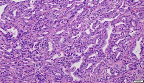
bioRxiv preprint first posted online Aug. 23, 2018; doi: http://dx.doi.org/10.1101/398347. The copyright holder for this preprint
 (which was not peer-reviewed) is the author/funder, who has granted bioRxiv a license to display the preprint in perpetuity.
                                    All rights reserved. No reuse allowed without permission.

   INTRODUCTION:

   Pancreatic ductal adenocarcinoma (PDAC) is one of the most lethal cancers known, due to the
   absence of reliable early diagnosis and the absence of effective therapeutic regimens [1]. It is
   currently the third leading cause of cancer worldwide and is projected to become second leading
   cause of cancer in the United States by 2030. The median 5-year survival rate of PDAC is only
   9% [2], and underscores the need for developing new approaches in understanding the molecular
   mechanisms underlying the disease.

   The advent of next generation sequencing (NGS) and its application as part of large publicly
   funded efforts such as The Cancer Genome Atlas (TCGA) and International Cancer Genome
   Consortium (ICGC) has identified a huge catalogue of genes that are altered in PDAC [3, 4],
   although in many instances, particularly with low frequency mutations, the functional
   consequences of the alterations remain unknown. The definitive evidence for gene perturbation
   in carcinogenesis is typically obtained via autochthonous models, which in the context of PDAC,
   is exemplified by expression of a mutant Kras allele in the pancreas alongside one or more
   cooperating mutations [5].                  First developed in 2003, PDAC GEMMs have dramatically
   accelerated our understanding of the molecular pathogenesis of this neoplasm, including how the
   genetic background of the cancer impacts the tumor microenvironment (TME) and resulting
   immune response [6-8]. In contrast to cell line or patient-derived xenograft (PDX) models,
   autochthonous models harbor the precursor lesions observed in the multistep progression of
   PDAC, as well as the stroma-dense TME observed in the corresponding human disease [9-11].

   Although autochthonous models have been instrumental in our understanding the molecular
   underpinnings of PDAC, the conventional Cre-based recombinatorial models do not faithfully
   mimic adult onset human PDAC due to their inherent limitation of inducing Ras activation and
   cooperating gene alteration(s) in every epithelial cell of the pancreas during development. In
   some respects, the conventional models are more analogous to “familial” PDAC where every cell
   in the pancreas is predisposed to neoplasia due to a germline anomaly (for example,
   CDKN2A/p16), and cancers arise due to stochastic bi-allelic gene inactivation [12, 13]. In
   addition to this conceptual limitation, conventional Cre-mediated recombinatorial models are
   also hindered by time, labor and expenses required for generating appropriate numbers of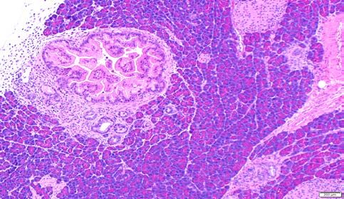
bioRxiv preprint first posted online Aug. 23, 2018; doi: http://dx.doi.org/10.1101/398347. The copyright holder for this preprint
 (which was not peer-reviewed) is the author/funder, who has granted bioRxiv a license to display the preprint in perpetuity.
                                    All rights reserved. No reuse allowed without permission.

   colonies, especially with complex genotypes where the “pups” of interest might only be a minor
   fraction of the litter. A timeframe of 12-18 months for completing the breeding of complex
   mouse genotypes is not unusual in the field. Novel platforms that bypass the pancreas-wide
   embryonic expression of mutant Kras and loss of cooperating alleles, while also greatly
   improving the efficiency of model generation would be a significant advancement in the
   preclinical arena.

   Recently, the RNA-guided endonuclease Cas9 from microbial type II CRISPR (clustered
   regularly interspaced short palindromic repeat) system has emerged as a powerful tool for
   genome engineering in mammalian cells [14-16], including somatic cell editing in a variety of
   mouse organs that includes brain, lung, liver and pancreas [17-21]. CRISPR/Cas9 can be targeted
   to the desired genomic loci by using programmable 20-bp single guide RNAs (sgRNAs) to
   generate DNA double stranded breaks which induce genome editing via one of the two DNA
   damage repair pathways: non homologous end-joining (NHEJ) resulting in insertion-deletion
   (INDELs), or homology directed repair (HDR) resulting in precise sequence substitution in the
   presence of a repair template [22-25]. The multiplexing abilities of gene editing by combining
   multiple sgRNAs to the Cas9 provides this programmable nuclease system with a unique
   advantage for conducting in vivo combinatorial gene editing, especially in the context of
   developing autochthonous cancer models [21]. There have been several approaches used for
   delivering CRISPR/Cas9 in vivo in murine pancreatic cells, including lentivirus and plasmid
   based transfection [26, 27], although many of these vectors are hindered by low efficiency of
   multiplexed gene editing and risk of on-target recombination in unintended cell types or tissues
   [28]. In recent years, adeno-associated virus (AAV) vectors, in particular, have been increasingly
   utilized for in vivo delivery approaches of sgRNAs due to their efficiency, lack of inherent
   pathogenicity and strong safety profile [29-32].

   In order to generate a “workhorse” model of genetically engineered mice that develop PDAC
   with a variety of complex genotypes, we have developed a simple injection method by
   employing AAV-based delivery of sgRNAs cargo to the pancreas of adult mice, where Cas9
   activity is restricted to the p48-expressing acinar compartment [33]. Our model overcomes
   several challenges of the current Cre-based models, including the introduction of mutant alleles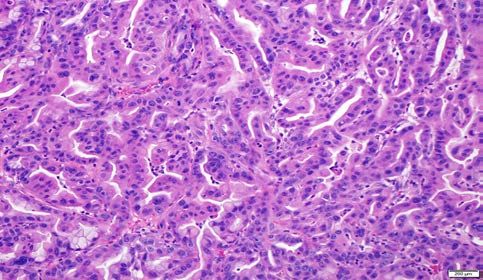
bioRxiv preprint first posted online Aug. 23, 2018; doi: http://dx.doi.org/10.1101/398347. The copyright holder for this preprint
 (which was not peer-reviewed) is the author/funder, who has granted bioRxiv a license to display the preprint in perpetuity.
                                    All rights reserved. No reuse allowed without permission.

   in a subset of adult pancreatic epithelial cells (rather than pancreas-wide embryonic expression),
   and the use of a single “workhorse” mouse genotype (p48-Cre; LSL-Cas9) from which cancers
   harboring multiple complex genotypes can be readily generated through somatic gene editing,
   thus greatly increasing efficiency. Importantly, in contrast to some of the recently described
   CRISPR/Cas9-mediated PDAC models that require an oncogenic KrasG12D allele in the pancreas
   on which cooperating mutations are then introduced by CRISPR/Cas9 [26, 27], our method
   dispenses with the need for a constitutive KrasG12D allele, by including a mutant KrasG12D
   template in the delivery cargo that recombines with, and replaces, the endogenous wild type
   allele through HDR.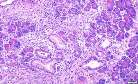
bioRxiv preprint first posted online Aug. 23, 2018; doi: http://dx.doi.org/10.1101/398347. The copyright holder for this preprint
 (which was not peer-reviewed) is the author/funder, who has granted bioRxiv a license to display the preprint in perpetuity.
                                    All rights reserved. No reuse allowed without permission.

   MATERIALS AND METHODS:

   Generation of pancreas specific Cre-dependent Cas9 mice:

   Conditional Cas9 mice (Rosa26-LSL-Cas9) was obtained from the Jackson Laboratory and was
   crossed with pancreas specific p48-Cre mice in order to generate experimental p48-Cre; LSL-
   Cas9 mice. The following primers were used to genotype Cas9 (Cas9_Common-S 5’-
   AAGGGAGCTGCAGTGGAGTA-3’; Cas9_Wild-AS 5’- CCGAAAATCTGTGGGAAGTC-3’;
   Cas9_Mut-AS 5’- CGGGCCATTTACCGTAAGTTAT-3’) and p48-Cre (ptf1a_Fw 5’-
   AACCAGGCCCAGAAGGTTAT-3';                                     ptf1a_Rv             5'-TCAAAGGGTGGTTCGTTCTC-3';
   ptf1a_Cre_Fw                    5'-ATAGGCTACCTGGCCATGCCC-3';                                          ptf1a_Cre_Rv               5'-
   CGGGCTGCAGGAATTCGTCG-3').

   CRISPR sgRNA design:

   CRISPR sgRNAs for Kras, Trp53 and Lkb1 have been previously described [19]. Genomic
   sequence for Arid1a gene was downloaded from UCSC genome browser and sgRNA cassette
   was generated using the CRISPR design tool (http://crispr.mit.edu).

   AAV vector plasmids:

   The AAV-KPL plasmid (AAV: ITR-U6-sgRNA (Kras)-U6-sgRNA (p53)-U6-sgRNA (LKB1)-
   pEFS-RLUC-2A-Cre-shortPA- KrasG12D_HDRDonor-ITR) was obtained from Addgene.
   AAV-KPLΔCre was generated by removing the Cre sequence from AAV-KPL by digesting with
   Nhe1 and HindIII followed by ligation. AAV-KPΔCre was generated by digesting AAV-
   KPLΔCre with Kpn1 and Xba1 and followed by ligation. AAV-KΔCre was made by digesting
   AAV-KPLΔCre with BamH1 and Kpn1, and followed by ligation. To construct AAV-KPAΔCre,
   first the sgRNA for mouse Arid1a exon3 was cloned into the pX459 (pSpCas9 (BB)-2A-Puro)
   vector, then the whole segment containing the U6 promoter, Arid1a sgRNA with primers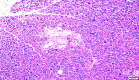
bioRxiv preprint first posted online Aug. 23, 2018; doi: http://dx.doi.org/10.1101/398347. The copyright holder for this preprint
 (which was not peer-reviewed) is the author/funder, who has granted bioRxiv a license to display the preprint in perpetuity.
                                    All rights reserved. No reuse allowed without permission.

   containing Xba1 and Kpn1 sites was PCR amplified. Finally, AAV-KPLΔCre was digested with
   Xba1 and Kpn1 to release the Lkb1 CRISPR containing unit following which the entire amplicon
   containing Arid1a CRISPR was ligated to generate AAV-KPAΔCre. AAV-KAΔCre was made
   by digesting AAV-KPAΔCre with BamH1 and Xba1 and re-ligated. All of the AAV vector
   constructs were sequence verified before virus production. All vector constructs are readily
   available from the authors upon request (the schematic of the constructs are shown in
   Supplementary Fig. 1A).

   AAV vector production:

   Human Embryonic Kidney 293T (HEK293T) cells were used for AAV production. HEK293T
   cells were maintained in Dulbecco’s Modified Eagle Medium (DMEM, Life Technologies)
   supplemented with 10% fetal bovine serum (FBS, Atlanta-Biologicals) and 1% penicillin and
   streptomycin (Life Technologies). Cells were grown as adherent cultures in 5% CO2 at 37 °C on
   15 cm cell culture plates.
   Viruses were generated by linear polyethyleneimine (PEI)-mediated triple transfection. The PEI
   was added to a DNA mixture of pAAV2/8, pAAV2/5, or pAAV2/6 rep/cap plasmid (10
   µg/plate), transgene plasmid (10 µg/plate), and pXX6-80 helper plasmid (20 µg/plate), incubated
   for 30 min at room temperature and added to poly-L-lysine-coated plates of 90% confluent
   HEK293T cells. The cells were harvested 48 h post-transfection and lysed by three cycles of
   freeze/thaw. The cell lysate was treated with 4 U/ml Benzonase (Sigma-Aldrich) for 40 min at
   37°C, and then separated through an iodixanol gradient (15/25/40/54%) in Beckman Ultra-Clear
   Quick Seal Tubes (Beckman Coulter, Brea, CA). After centrifugation at 48,000 rpm for 1.75 h in
   a 70Ti rotor, the virus was extracted from the 40% layer. Viruses were further purified through
   anion exchange chromatography (Q Sepharose, Pall) followed by concentration and buffer
   exchange through Amicon Ultra 4 (Millipore) into GB-PF68 (50 mM Tris, pH 7.6, 150 mM
   NaCl, 10 mM MgCl2, 0.001% Pluronic F68). Virus titers were determined by qPCR using
   SYBR green (Life Technologies) and primers against the Rluc portion of the transgene cassette
   (forward:                                  GCCTCGTGAAATCCCGTTAGTA,                                                               reverse:
   GCATTGGAAAAGAATCCTGGGTCC) on a BIO-RAD CFX96.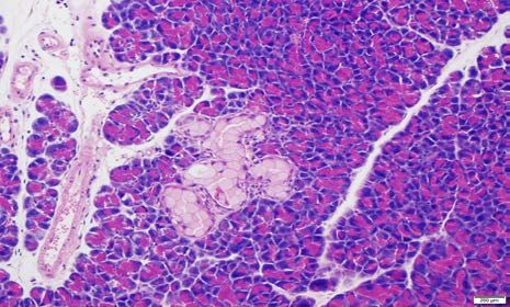
bioRxiv preprint first posted online Aug. 23, 2018; doi: http://dx.doi.org/10.1101/398347. The copyright holder for this preprint
 (which was not peer-reviewed) is the author/funder, who has granted bioRxiv a license to display the preprint in perpetuity.
                                    All rights reserved. No reuse allowed without permission.

   Surgical procedure and direct injection:

   100 μl of PBS containing AAV-CRISPR sequences was injected into the pancreatic tail using 29
   G needle (BD Biosciences) through left subcostal laparotomy under inhaled anesthesia. Leakage
   was not observed in any mice during the procedure. After the injection, the pancreas and spleen
   was carefully returned into the peritoneal cavity and the abdomen was closed using 4-0 Vicryl
   (Ethicon) and skin staplers. All the mice were monitored at least once a week by abdominal
   palpation and abdominal ultrasound to investigate the entire pancreas was performed every 4
   weeks using the Vevo 2100 (Fujifilm). Moribund mice with pancreatic tumors were euthanized
   by cervical dislocation and carbon dioxide inhalation according to the IACUC protocol. The
   assessment of primary tumor, intraperitoneal dissemination and distant metastases performed
   were based on both gross findings at necropsy and by histopathological examination. Any
   surviving mice at 100 days (~ 3 months post injection) were then euthanized and the Pancreata
   harvested for histopathological examination.

   T7 Endonuclease Gene editing assay:

   Genomic DNA was isolated from tumor samples using the Qiagen genomic DNA isolation kit.
   Subsequently ~400bp of genomic fragment encompassing the CRISPR targeted cleavage site
   was PCR amplified with high fidelity Taq polymerase. Approximately 200ng of purified PCR
   product was used for each T7 endonuclease assay. Briefly, 2ul NEBuffer 2, 200ng of purified
   PCR product and dH2O was added to a total of 19ul in a PCR tube. The hybridization reaction
   was run as follows: 5min, 95C; ramp down to 85C at -2C/s; ramp down to 25C at -0.1C/s; hold
   at 4C. We then added 1ul (10U) T7 endo I and incubated at 37C for 15min. The reaction was
   stopped by adding 2ul of 0.25M EDTA and loaded immediately on a 1.5% agarose gel.

   Immunofluorescence:

   Pancreata were harvested from the moribund mice due to tumor burden at different time point
   with maximum harvesting time around ~100 days. Pancreata were fixed in 4%
   paraformaldehyde, followed by standard paraffin embedding. Tissue was then cut into 5uM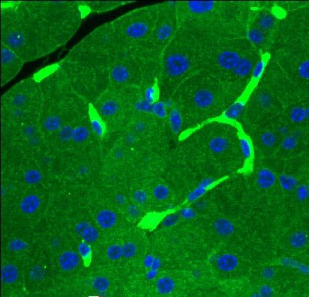
bioRxiv preprint first posted online Aug. 23, 2018; doi: http://dx.doi.org/10.1101/398347. The copyright holder for this preprint
 (which was not peer-reviewed) is the author/funder, who has granted bioRxiv a license to display the preprint in perpetuity.
                                    All rights reserved. No reuse allowed without permission.

   sections. Incubations with primary antibodies were performed overnight at 40C using standard
   techniques in PBS containing 0.2% Triton and 10% FBS using the following antibodies or
   probes: mouse anti-E-Cadherin (BD Transduction 610181, 1:50); rabbit anti-Sox9 (Millipore
   AB5535, 1:1000); hamster anti-Muc1 (Thermo Fisher HM-1630-F, 1:100); mouse anti-Arid1a
   (Santa Cruz Biotechnology sc-32761, 1:100); mouse anti-p53 (Santa Cruz Biotechnology sc126,
   1:50) and rabbit anti-LKB1 (Abcam ab185734 1:100). For immunofluorescence, secondary
   antibodies were obtained from Abcam and used at 1:300 dilution; samples were mounted with
   Fluorescence mounting media (Dako, S3023) and imaged on the Olympus Confocal Microscope.
   .

   Whole Exome Sequencing:

   Whole exome sequencing of harvested murine tumors was performed at Johns Hopkins
   University Next Generation Sequencing (NGS) Core Facility. Briefly, the Agilent SureSelect
   Mouse exome sequencing kit was used to prepare sequencing libraries and capture annotated
   exonic sequences. The resulting libraries were sequenced on a HiSeq 2500, and after quality
   control checks the fastq files were aligned to genome build mm10 with bwa mem 0.7.7. Picard
   tools 1.118 were used to remove duplicate reads and GATK 3.6 was used for base recalibration.
   To call somatic variants, a reference sequence panel of normals was first constructed from
   C57BL mouse strains, and Mutect2 was used with this panel. Annotation was done with snpEFF
   and snpSIFT.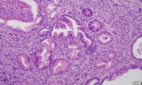
bioRxiv preprint first posted online Aug. 23, 2018; doi: http://dx.doi.org/10.1101/398347. The copyright holder for this preprint
 (which was not peer-reviewed) is the author/funder, who has granted bioRxiv a license to display the preprint in perpetuity.
                                    All rights reserved. No reuse allowed without permission.

   RESULTS:

   Strategy for virally delivered CRISPR-mediated genome editing in the adult pancreatic
   epithelium

   As a prelude for in vivo gene editing studies we first performed direct injection of AAV
   containing reporter alleles into the mouse pancreas. To test the differential efficacy of distinct
   AAV serotypes infecting pancreatic epithelial cell, we injected AAV6-Cre-GFP and AAV8-Cre-
   GFP into the adult mouse Pancreata (Supplementary Figure.1B, C) which confirmed the
   superiority of AAV8 in infecting epithelial cells as evidenced by extent of GFP expression.
   Henceforth, all of our AAV vectors were made using AAV8. Further, in order to avoid spurious
   Cre-dependent recombination in the non-epithelial pancreatic tissue including the peritoneum
   from the injected virus, we deleted the Cre sequence from the original AAV construct, AAV-
   KPL (Supplementary Figure.1A). Instead, we generated p48-Cre+/-; LSL-Cas9 mice as the
   “workhorse” mouse where Cre recombinase is only expressed in the adult pancreatic epithelium,
   specifically acinar cells.

   Multiplexed CRISPR/Cas9 mediated somatic genome-editing results in autochthonous PDAC
   harboring diverse genotypes.

   A set of six independent AAV8 vectors were generated for injection into the pancreas of p48-
   Cre+/-;LSL-Cas9 mice, containing the following sgRNAs: (i) AAV-LacZ (control) (ii)AAV-Kras
   (which included the Kras G12D HDR template and henceforth designated as “K”, (iii) AAV-Kras,
   Trp53 ( henceforth designated as “KP”), (iv)AAV-Kras; Arid1a (henceforth designated as “KA”),
   (v) AAV-Kras; Trp53; Lkb1 (henceforth designated as “KPL”) and (vi) AAV-Kras;Trp53;Arid1a
   (henceforth designated as “KPA”). The schematic of the model development and post-harvest
   correlative assays are illustrated in Figure 1A. The pancreatic development was monitored in
bioRxiv preprint first posted online Aug. 23, 2018; doi: http://dx.doi.org/10.1101/398347. The copyright holder for this preprint
(which was not peer-reviewed) is the author/funder, who has granted bioRxiv a license to display the preprint in perpetuity.
All rights reserved. No reuse allowed without permission.
real time by ultrasound as shown in Figure 1B. The survival of the mice injected with the six
vectors is depicted in Figure 1C. Excluding the control “LacZ” mice, the mice with “K” and
“KA” had the most indolent natural history with no mortality observed at ~100days (at which
point these mice were euthanized). In contrast, “KP”, “KPA” and “KPL” injected mice had
comparable median survival with progression to invasive neoplasia. In the “KPL” injected nice,
ultrasound examination demonstrated readily visible tumors as early as eight weeks after
injection and these were apparent 1-2 weeks later in “KP” and “KPA” injected mice.
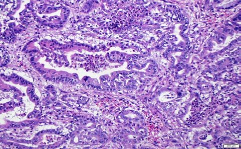
Representative histopathology photomicrographs from the mice injected with various AAV
constructs are shown in Figure 2. In mice that succumbed to invasive carcinomas, we have
captured examples of murine pancreatic intraepithelial neoplasia (mPanIN) in the surrounding
pancreas, to demonstrate that the carcinomas arose on a backdrop of what is widely accepted as
the most common precursor lesion in human PDAC. In “K” and “KA” injected mice, we only
observed focal mPanIN lesions of various grades, in the absence of invasive carcinomas (Figure
2 A-D), which is also reflected in their survival. Notabley, surrounding histologically normal
pancreatic parenchyma was readily seen in these mice even at ~ 100 days, distinct from the
pancreas-wide activation of Cre in conventional models where there is progressive replacement
of acinar tissue by precursor lesions throughout the organ. In contrast to mice injected with “K”
and “KA” viruses, the remaining cohorts developed invasive cancers, as seen with “KP” (Figure
2 F), “KPA” (Figure 2 H) and “KPL” (Figure 2 J) respectively. Of note, we also readily
observed examples of acinar ductal metaplasia (ADM) in the background parenchyma and
typically adjacent to mPanINs, as these are considered the “precursor to the precursor” in the
multistep model of murine PDAC pathogenesis (Figure 2 E, G and I). Further, the “KPL”
injected mice were distinguished by the presence of cystic precursor lesions (Intraductal
papillary mucinous neoplasms or IPMNs, illustrated in Supplementary Figure 2), a feature that
has been previously reported in conventional Cre models of Lkb1 deletion. The resulting IPMNs
expressed the apomucin MUC1 on their surface, consistent with a so-called pancreato-biliary
subtype of differentiation (Supplementary Figure 3)
Histopathological examination of the invasive carcinomas at low magnification often showed
normal pancreatic parenchyma adjacent to a region of invasive carcinoma mirroring the
“segmental” nature of disease in the human pancreas ( and distinct from the multifocal nature ofbioRxiv preprint first posted online Aug. 23, 2018; doi: http://dx.doi.org/10.1101/398347. The copyright holder for this preprint
(which was not peer-reviewed) is the author/funder, who has granted bioRxiv a license to display the preprint in perpetuity.
All rights reserved. No reuse allowed without permission.
disease seen in the conventional Cre models) (Supplementary Figure 4A). While
adenocarcinomas were seen with all three cancer forming genotypes (“KP”, “KPA” and “KPL”)
(Figure 2), the proportion of adenocarcinomas versus poorly differentiated (sarcomatoid) tumors
varied (Supplementary Figure 4B), with “KPL” and “KPA” resulting predominantly in
adenocarcinomas and “KP” in poorly differentiated lesion. These variations in the proportion of
histological subtypes for individual genotypes have been well documented with Cre- based
models [34], so was not unexpected. In addition to the primary tumor, metastatic
adenocarcinomas were observed in a subset of “KPL” and “KPA” injected mice, specifically in
the liver and lung (Supplementary Figure 5).
Correlative studies in CRISPR/Cas9 generated PDAC models
A series of correlative protein expression and sequencing studies were conducted on the
harvested tissues from the CRISPR/Cas9 generated PDAC lesions. For example, we confirmed
the loss of expression of p53, Arid1a and Lkb1 within the neoplastic cells of tumors harvested
from “KPL” and “KA” and virus injected pancreata, respectively (Figure 3). Although “KA”
injected mice did not form invasive carcinomas, partial to complete loss of expression of Arid1a
protein was demonstrable in the ADM and mPanIN lesions that resulted in the pancreata
(identified based on Sox9 expression) consistent with successful somatic editing of Arid1a loci
(Supplementary Figure 6). As previously noted “KP” mice developed multiple examples of
poorly differentiated (sarcomatoid ) carcinomas, and we confirmed the epithelial origin of these
cancers by the expression of cytokeratin 19 in the primary tumors (data not shown). Of note ,
these poorly differentiated PDAC were characterized by loss of membrane E-cadherin and co
expression of the mesenchymal marker vimentin, suggesting a phenotype of partial epithelial to
mesenchymal transformation (EMT), which has been previously described in aggressive PDAC
[35, 36].
The T7 endonuclease assay serves as a preliminary measure of the efficacy of genome targeting
based on the ability of the enzyme to cleave DNA strands that are not perfectly matched due to
non-homologous end joining. Representative examples of the T7 endonuclease assay for Kras,
Trp53 and Lkb1 from “KPL” and “KPA” tumors are shown in Figure 4A, which demonstrates
targeting of sgRNA to the corresponding locus. In contrast to the cancer samples, the controlbioRxiv preprint first posted online Aug. 23, 2018; doi: http://dx.doi.org/10.1101/398347. The copyright holder for this preprint
(which was not peer-reviewed) is the author/funder, who has granted bioRxiv a license to display the preprint in perpetuity.
All rights reserved. No reuse allowed without permission.
pancreas injected with LacZ sgRNA showed no evidence of genome editing in the T7
endonuclease assay.
In order to confirm the CRISPR/Cas9 mediated genome editing at the nucleotide level, we then
performed exome sequencing of tumor derived genomic DNA from “KPL” and “KPA” virus
injected pancreata, using reference DNA from a panel of C57/BL mice. In Figure 4B, we
demonstrate NGS reads for KRAS, showing replacement of the endogenous locus with the donor
template carrying the codon 12 non-synonymous mutation by HDR. Additional “marker”
nucleotides (in blue color on sequence read) derived from the exogenous KRAS template (and
accounting for synonymous base variations) are seen in the neoplastic DNA, confirming that the
KrasG12D mutation observed was not spontaneous but rather derived from the exogenous
template by HDR. Since “bulk” tumor DNA was sequenced, we observed a mutant fraction of
9% for the KrasG12D allele suggesting ~20 % neoplastic cellularity. Exome sequencing data also
confirmed disruption of three loci (Arid1a, Trp53 and Lkb1), with multiple insertion deletion
(“indel”) events observed for each gene (Figures 4C, D and E). This is consistent with
independent CRISPR editing events occurring during the initial injection, and the observed
PDAC “mass” at necropsy being of oligoclonal origin. The pie charts adjacent to each gene
designate an overview of the classes of indel events for each targeted locus (detailed information
for individual mouse tumors sequenced is illustrated in Supplementary Figure 7). The majority
of indels were located 1-5bp upstream or downstream of the PAM sequence. The maximum
insertion size observed was 15bp and the maximum deletion size observed was 23bp. Overall,
between 8-30% of DNA reads (based on underlying neoplastic cellularity and gene of interest)
demonstrated unequivocal indels on sequencing.
Finally, we explored whether the resulting tumors had developed secondary mutations of the 12
most commonly altered genes in PDAC, using data collated from the TCGA and ICGC datasets
[3, 4]. Besides the CRISPR targeted genes in the corresponding mouse model (Supplementary
Figure 8), we did not observe any secondary mutations in genes such as Smad4, Gnas, Rnf43,
Kdm6a and Brca2. Importantly, the notable exceptions were Kmt2c (Mll3) and Kmt2d (Mll4),
which encode for chromatin regulatory proteins [37], and demonstrated frameshift mutations or
“indels” in subsets of tumors. It is unclear whether these events represented a requirement for
tumor progression in these models, given that in the TCGA and ICGC datasets genes encoding
for chromatin regulatory proteins are one of the most commonly mutated family of genes [3, 4].bioRxiv preprint first posted online Aug. 23, 2018; doi: http://dx.doi.org/10.1101/398347. The copyright holder for this preprint
(which was not peer-reviewed) is the author/funder, who has granted bioRxiv a license to display the preprint in perpetuity.
All rights reserved. No reuse allowed without permission.
DISCUSSION:
GEMMs represent the most rigorous preclinical platform for hypothesis testing the functional
contribution of recurrent genetic alterations towards multistep pancreatic carcinogenesis, but
conventional approaches demonstrate several logistical and temporal challenges, particularly,
when complex allelotypes are desired. In this study, we have successfully demonstrated
CRISPR-induced somatic gene editing using AAV-mediated direct intra-pancreatic injection of
diverse sgRNA combinations for establishing autochthonous PDAC with high efficiency, and for
some of the genotypes, relatively short latency to cancer incidence. Specifically, while five
distinct sgRNA combinations were used as AAV cargo and all developed mPanINs, three
genotypes (“KP”, “KPA”, and “KPL”) resulted in PDAC in the arbitrarily assigned time frame
for observation (~3 months), underscoring the importance of the genetic background on the
latency of cancer formation. While it is certainly possible that the remaining two genotypes (“K”
and “KA”) might eventually develop PDAC on longer follow up, our goal in this initial
feasibility study was to keep the time frame for observation relatively short and perform
necropsy on any surviving mice at ~3 months. Importantly, all of the resulting PDAC lesions
developed on a backdrop of credentialed precursor lesions (mPanINs and IPMNs), and
progressed to metastatic disease in a subset of mice, confirming that the autochthonous models
we have developed recapitulates the multistep progression seen in conventional Cre-based
models. Loss of CRISPR-disrupted protein expression was confirmed in both pre-invasive and
invasive neoplastic lesions, and NGS on the resulting tumors validated gene targeting at the
sequence level.
The catalogue of genetic alterations targeted using CRISPR sgRNAs in the pancreas were based
on the frequency of alterations reported in human PDAC in public databases [3, 4]. For example,
KRAS, which is activated by oncogenic mutations in >90% of PDAC, and TP53, which is
inactivated by loss of function mutations of homozygous deletions in 75% of PDAC, are obviousbioRxiv preprint first posted online Aug. 23, 2018; doi: http://dx.doi.org/10.1101/398347. The copyright holder for this preprint
(which was not peer-reviewed) is the author/funder, who has granted bioRxiv a license to display the preprint in perpetuity.
All rights reserved. No reuse allowed without permission.
choices. In fact, mutations of KRAS are required not only for the cell autonomous effects in
cancer progression, but also for the paracrine effects on the tumor microenvironment that create
a tumor permissive immune milieu [38, 39]. Somatic mutations of the SWI/SNF complex
ARID1A are seen in approximately 8-10% of PDAC, and this DNA binding helicase has been
implicated in maintenance of acinar cell identity, and attenuating the progression to a precursor
ductal phenotype (acinar ductal metaplasia) on a mutant Kras background [40, 41]. In our series,
confirmed loss of Arid1a and expression of mutant Kras in “KA” mice was sufficient to induce
mPanINs, but not PDAC within the ~3 month time window. In contrast, addition of Trp53
deletion (“KPA”) resulted in PDAC with relatively short latencies, with a greater propensity
towards differentiated adenocarcinomas than the poorly differentiated carcinomas observed in
“KP” mice alone. Of note, this correlation between ARID1A mutations and histological grade of
differentiation has also been reported in other solid cancers, such as endometrial, hepatocellular,
and pancreatic carcinomas [42-44], suggesting that loss of this epigenetic regulator might
promote the acquisition of a ductal (i.e. “differentiated”) phenotype in PDAC. Finally, we also
targeted Lkb1, which encodes for a serine threonine kinase mutated in several solid cancers. In
the pancreas, LKB1 mutations or loss of Lkb1 expression has been described in cystic precursors
(IPMNs) [45, 46], and prior conventional models of conditional Lkb1 loss have demonstrated
PDAC arising on the background of cystic neoplasms [47, 48]. In our series, we did not examine
“KL” as a dual targeting construct, but “KPL” injected mice developed cystic lesions resembling
IPMNs in a subset of cases. Similar to “KPA” mice, “KPL” mice also developed predominantly
adenocarcinomas upon progression.
A few important methodological caveats emerged during the course of this study. First, we
recognized the AAV8 serotype to be significantly more efficient at viral transduction than
AAV6, which is consistent with prior reports of using AAV8 as a delivery vector to the pancreas
[49]. Second, the original CRISPR construct used in this study had a Cre expressing element
[19], which resulted in spurious Cas9 activation and extra-pancreatic mesenchymal tumors in the
abdomen due to leakage of injected virus (data not shown), therefore, we deleted the Cre
element, thus restricting recombinase expression to only the p48-expressing acinar parenchyma
in the p48-Cre;LSL-Cas9 mouse. Third, while there have been prior studies using CRISPR/Cas9
for somatic gene editing and PDAC induction in mice, there is one key distinction from ourbioRxiv preprint first posted online Aug. 23, 2018; doi: http://dx.doi.org/10.1101/398347. The copyright holder for this preprint
(which was not peer-reviewed) is the author/funder, who has granted bioRxiv a license to display the preprint in perpetuity.
All rights reserved. No reuse allowed without permission.
approach. For example, Chiou et al [26] used an intraductal lentiviral injection methodology for
delivering Cre recombinase and sgRNA against Lkb1 into the main pancreatic duct of mice
expressing conditional (“LSL”) allele of Cas9 and mutant KrasG12D. Analogously, Maresch et al
[27] used direct electroporation of plasmid based CRISPR/cas9 vectors against as many as 13
targeted loci in Ptf1-Cre; LSL-KrasG12D mice, resulting in invasive tumors with multiple
disrupted alleles. Both of these methods, however, required a conditional mutant LSL-KrasG12D
allele in the host animal (either constitutively expressed [27], or activated via virally delivered
Cre [26]), on which background, somatic gene editing was enabled. In our study, however, we
have dispensed with the requirement for a KrasG12D allele in the host animal, by incorporation of
a mutant template sequence in the AAV vector that is incorporated at the endogenous Kras locus
through HDR. This has allowed us to utilize a single uniform strain of “workhorse” mice – Ptf1-
Cre; LSL-Cas9 – for injecting various sgRNAs, thereby, greatly enhancing the efficiency of
PDAC generation when using different combinations of targeting vectors. Further, by expressing
mutant Kras only in the subset of cells where the cooperating tumor suppressor allele is also
disrupted, we ensure a bona fide “segmental” nature of disease in the pancreas, recapitulating the
cognate human pathophysiology. A potential corollary advantage of this method is the possibility
that genes of interest can be independently disrupted, including prior to the activation of mutant
Kras, by varying the timing of AAV/CRISPR delivery (for example, with sequential injections).
Of interest, NGS studies on the CRISPR-induced PDAC showed that editing events were
typically localized within a few bases of the PAM sequence, with individual “bulk” tumors
harboring multiple classes of “indels” or frameshift mutations. This is consistent with the
CRISPR-induced PDAC being an oligoclonal mix of more than one initiating clonal event, as
previously reported [26]. While we did not pursue this particular line of investigation, one can
envision that barcoding individual AAV constructs could allow for rigorous interrogation of
selectivity in tumor formation and metastatic ability of distinct genomic alterations even at one
given locus. The NGS data also revealed a somewhat robust rate of HDR for Kras in the
sequenced “bulk” tumors (9% on average across six tumors), which is substantially greater than
the 1.8% Kras HDR frequency reported in generating lung cancer models using AAV-mediated
delivery of CRISPR [19]. The precise reason for this in unclear, but might represent a higher
propensity for HDR in the pancreatic acinar component compared to the pulmonary epithelium,bioRxiv preprint first posted online Aug. 23, 2018; doi: http://dx.doi.org/10.1101/398347. The copyright holder for this preprint
(which was not peer-reviewed) is the author/funder, who has granted bioRxiv a license to display the preprint in perpetuity.
All rights reserved. No reuse allowed without permission.
or a greater ability of the AAV8 serotype used in this study to enable HDR in the mouse genome,
independent of exonuclease activity, as was recently shown for certain AAV strains [50]. Finally,
our sequencing data showed that while several recurrently mutated PDAC-associated genes,
including Smad4, Gnas, Rnf43, Kdm6a and Brca2 did not undergo secondary mutations during
PDAC development, two genes in particular - Kmt2c (Mll3) and Kmt2d (Mll4), which encode for
chromatin regulatory proteins [37] – did demonstrate recurrent alterations. The encoded proteins,
Mll3 and Mll4, are histone methyltransferase enzymes that are part of a so-called COMPASS-
like complex responsible for addition of methyl residues to lysine residues on histones, which
function as an “activation mark”. Loss of function mutations of MLL3 and other COMPASS-like
complex proteins is reported in approximately 10% of PDAC, and loss of expression has been
associated with better prognosis [37]. We are continuing to explore the relevance of these
recurrent non-targeted mutations, including their requirement for tumor formation in the context
of other targeted loci using the AAV-CRISPR system.
In summary, we have generated a “workhorse” Ptf1-Cre; LSL-Cas9 mouse that enables the
relatively facile interrogation of the functional role of mutated genes in the PDAC landscape,
including diverse combinations of targeted alleles using AAV8 as a delivery vector. Our
platform precludes the need for a germline mutant Kras allele in the host mouse, and the
resulting pancreata demonstrate the full compendium of precursor and invasive lesions observed
in human PDAC. This “workhorse” platform will allow investigators to conduct in vivo
functional genomics studies and generate reagents, such as cell lines with defined genetic
alterations, with considerable efficiency.bioRxiv preprint first posted online Aug. 23, 2018; doi: http://dx.doi.org/10.1101/398347. The copyright holder for this preprint
(which was not peer-reviewed) is the author/funder, who has granted bioRxiv a license to display the preprint in perpetuity.
All rights reserved. No reuse allowed without permission.
REFERENCES:
1. Ying, H., et al., Genetics and biology of pancreatic ductal adenocarcinoma. Genes Dev, 2016.
30(4): p. 355-85.
2. Siegel, R.L., K.D. Miller, and A. Jemal, Cancer statistics, 2018. CA Cancer J Clin, 2018. 68(1): p. 7-
30.
3. Cancer Genome Atlas Research Network. Electronic address, a.a.d.h.e. and N. Cancer Genome
Atlas Research, Integrated Genomic Characterization of Pancreatic Ductal Adenocarcinoma.
Cancer Cell, 2017. 32(2): p. 185-203 e13.
4. Bailey, P., et al., Genomic analyses identify molecular subtypes of pancreatic cancer. Nature,
2016. 531(7592): p. 47-52.
5. Tuveson, D.A. and S.R. Hingorani, Ductal pancreatic cancer in humans and mice. Cold Spring
Harb Symp Quant Biol, 2005. 70: p. 65-72.
6. DuFort, C.C., K.E. DelGiorno, and S.R. Hingorani, Mounting Pressure in the Microenvironment:
Fluids, Solids, and Cells in Pancreatic Ductal Adenocarcinoma. Gastroenterology, 2016. 150(7): p.
1545-1557 e2.
7. Singh, M., C.L. Murriel, and L. Johnson, Genetically engineered mouse models: closing the gap
between preclinical data and trial outcomes. Cancer Res, 2012. 72(11): p. 2695-700.
8. Morrison, A.H., K.T. Byrne, and R.H. Vonderheide, Immunotherapy and Prevention of Pancreatic
Cancer. Trends Cancer, 2018. 4(6): p. 418-428.
9. Colvin, E.K. and C.J. Scarlett, A historical perspective of pancreatic cancer mouse models. Semin
Cell Dev Biol, 2014. 27: p. 96-105.
10. Ponz-Sarvise, M., D.A. Tuveson, and K.H. Yu, Mouse Models of Pancreatic Ductal
Adenocarcinoma. Hematol Oncol Clin North Am, 2015. 29(4): p. 609-17.
11. Behrens, D., W. Walther, and I. Fichtner, Pancreatic cancer models for translational research.
Pharmacol Ther, 2017. 173: p. 146-158.
12. Shi, C., et al., Increased Prevalence of Precursor Lesions in Familial Pancreatic Cancer Patients.
Clin Cancer Res, 2009. 15(24): p. 7737-7743.
13. Brune, K., et al., Multifocal neoplastic precursor lesions associated with lobular atrophy of the
pancreas in patients having a strong family history of pancreatic cancer. Am J Surg Pathol, 2006.
30(9): p. 1067-76.
14. Jiang, F. and J.A. Doudna, CRISPR-Cas9 Structures and Mechanisms. Annu Rev Biophys, 2017. 46:
p. 505-529.
15. Wright, A.V., J.K. Nunez, and J.A. Doudna, Biology and Applications of CRISPR Systems:
Harnessing Nature's Toolbox for Genome Engineering. Cell, 2016. 164(1-2): p. 29-44.bioRxiv preprint first posted online Aug. 23, 2018; doi: http://dx.doi.org/10.1101/398347. The copyright holder for this preprint
(which was not peer-reviewed) is the author/funder, who has granted bioRxiv a license to display the preprint in perpetuity.
All rights reserved. No reuse allowed without permission.
16. Zhang, C., R. Quan, and J. Wang, Development and application of CRISPR/Cas9 technologies in
genomic editing. Hum Mol Genet, 2018. 27(R2): p. R79-R88.
17. Hsu, P.D., E.S. Lander, and F. Zhang, Development and applications of CRISPR-Cas9 for genome
engineering. Cell, 2014. 157(6): p. 1262-78.
18. Zhu, Z., et al., Genome Editing of Lineage Determinants in Human Pluripotent Stem Cells Reveals
Mechanisms of Pancreatic Development and Diabetes. Cell Stem Cell, 2016. 18(6): p. 755-68.
19. Platt, R.J., et al., CRISPR-Cas9 knockin mice for genome editing and cancer modeling. Cell, 2014.
159(2): p. 440-55.
20. Millette, K. and S. Georgia, Gene Editing and Human Pluripotent Stem Cells: Tools for Advancing
Diabetes Disease Modeling and Beta-Cell Development. Curr Diab Rep, 2017. 17(11): p. 116.
21. Sanchez-Rivera, F.J. and T. Jacks, Applications of the CRISPR-Cas9 system in cancer biology. Nat
Rev Cancer, 2015. 15(7): p. 387-95.
22. Pawelczak, K.S., et al., Modulating DNA Repair Pathways to Improve Precision Genome
Engineering. ACS Chem Biol, 2018. 13(2): p. 389-396.
23. Bibikova, M., et al., Enhancing gene targeting with designed zinc finger nucleases. Science, 2003.
300(5620): p. 764.
24. Jasin, M., et al., High frequency of homologous recombination in mammalian cells between
endogenous and introduced SV40 genomes. Cell, 1985. 43(3 Pt 2): p. 695-703.
25. Rudin, N., E. Sugarman, and J.E. Haber, Genetic and physical analysis of double-strand break
repair and recombination in Saccharomyces cerevisiae. Genetics, 1989. 122(3): p. 519-34.
26. Chiou, S.H., et al., Pancreatic cancer modeling using retrograde viral vector delivery and in vivo
CRISPR/Cas9-mediated somatic genome editing. Genes Dev, 2015. 29(14): p. 1576-85.
27. Maresch, R., et al., Multiplexed pancreatic genome engineering and cancer induction by
transfection-based CRISPR/Cas9 delivery in mice. Nat Commun, 2016. 7: p. 10770.
28. Liu, C., et al., Delivery strategies of the CRISPR-Cas9 gene-editing system for therapeutic
applications. J Control Release, 2017. 266: p. 17-26.
29. Moreno, A.M., et al., In Situ Gene Therapy via AAV-CRISPR-Cas9-Mediated Targeted Gene
Regulation. Mol Ther, 2018. 26(7): p. 1818-1827.
30. Kemaladewi, D.U., et al., Correction of a splicing defect in a mouse model of congenital muscular
dystrophy type 1A using a homology-directed-repair-independent mechanism. Nat Med, 2017.
23(8): p. 984-989.
31. Tabebordbar, M., et al., In vivo gene editing in dystrophic mouse muscle and muscle stem cells.
Science, 2016. 351(6271): p. 407-411.
32. Chew, W.L., et al., A multifunctional AAV-CRISPR-Cas9 and its host response. Nat Methods, 2016.
13(10): p. 868-74.
33. Kawaguchi, Y., et al., The role of the transcriptional regulator Ptf1a in converting intestinal to
pancreatic progenitors. Nat Genet, 2002. 32(1): p. 128-34.
34. Bardeesy, N., et al., Both p16(Ink4a) and the p19(Arf)-p53 pathway constrain progression of
pancreatic adenocarcinoma in the mouse. Proc Natl Acad Sci U S A, 2006. 103(15): p. 5947-52.
35. Hruban, R.H. and D.S. Klimstra, Adenocarcinoma of the pancreas. Semin Diagn Pathol, 2014.
31(6): p. 443-51.
36. Adsay, N.V., et al., Ductal neoplasia of the pancreas: nosologic, clinicopathologic, and biologic
aspects. Semin Radiat Oncol, 2005. 15(4): p. 254-64.
37. Dawkins, J.B., et al., Reduced Expression of Histone Methyltransferases KMT2C and KMT2D
Correlates with Improved Outcome in Pancreatic Ductal Adenocarcinoma. Cancer Res, 2016.
76(16): p. 4861-71.
38. Vonderheide, R.H., Tumor-promoting inflammatory networks in pancreatic neoplasia: another
reason to loathe Kras. Cancer Cell, 2014. 25(5): p. 553-4.bioRxiv preprint first posted online Aug. 23, 2018; doi: http://dx.doi.org/10.1101/398347. The copyright holder for this preprint
(which was not peer-reviewed) is the author/funder, who has granted bioRxiv a license to display the preprint in perpetuity.
All rights reserved. No reuse allowed without permission.
39. Bryant, K.L., et al., KRAS: feeding pancreatic cancer proliferation. Trends Biochem Sci, 2014.
39(2): p. 91-100.
40. Livshits, G., et al., Arid1a restrains Kras-dependent changes in acinar cell identity. Elife, 2018. 7.
41. Kimura, Y., et al., ARID1A Maintains Differentiation of Pancreatic Ductal Cells and Inhibits
Development of Pancreatic Ductal Adenocarcinoma in Mice. Gastroenterology, 2018. 155(1): p.
194-209 e2.
42. Zhang, Z.M., et al., The clinicopathologic significance of the loss of BAF250a (ARID1A) expression
in endometrial carcinoma. Int J Gynecol Cancer, 2014. 24(3): p. 534-40.
43. Abe, H., et al., Altered expression of AT-rich interactive domain 1A in hepatocellular carcinoma.
Int J Clin Exp Pathol, 2015. 8(3): p. 2763-70.
44. Zhang, L., et al., Loss of ARID1A Expression Correlates With Tumor Differentiation and Tumor
Progression Stage in Pancreatic Ductal Adenocarcinoma. Technol Cancer Res Treat, 2018. 17: p.
1533034618754475.
45. Sahin, F., et al., Loss of Stk11/Lkb1 expression in pancreatic and biliary neoplasms. Mod Pathol,
2003. 16(7): p. 686-91.
46. Sato, N., et al., STK11/LKB1 Peutz-Jeghers gene inactivation in intraductal papillary-mucinous
neoplasms of the pancreas. Am J Pathol, 2001. 159(6): p. 2017-22.
47. Hezel, A.F., et al., Pancreatic LKB1 deletion leads to acinar polarity defects and cystic neoplasms.
Mol Cell Biol, 2008. 28(7): p. 2414-25.
48. Lo, B., et al., Lkb1 regulates organogenesis and early oncogenesis along AMPK-dependent and -
independent pathways. J Cell Biol, 2012. 199(7): p. 1117-30.
49. Chen, M., et al., Efficient Gene Delivery and Expression in Pancreas and Pancreatic Tumors by
Capsid-Optimized AAV8 Vectors. Hum Gene Ther Methods, 2017. 28(1): p. 49-59.
50. Smith, L.J., et al., Stem cell-derived clade F AAVs mediate high-efficiency homologous
recombination-based genome editing. Proc Natl Acad Sci U S A, 2018. 115(31): p. E7379-E7388.bioRxiv preprint first posted online Aug. 23, 2018; doi: http://dx.doi.org/10.1101/398347. The copyright holder for this preprint
(which was not peer-reviewed) is the author/funder, who has granted bioRxiv a license to display the preprint in perpetuity.
All rights reserved. No reuse allowed without permission.
FIGURE LEGENDS:
Figure 1. Generation and natural history of CRISR/Cas9 induced genetically engineered
mouse models of pancreatic cancer.
A. Schematic of experimental procedure showing the direct injection of AAVs containing
sgRNAs into the tail of p48-Cre; LSL-Cas9 mice. Serial ultrasound imaging was performed to
follow tumor induction for ~3 months following injection, at which time point the mice were
euthanized and the pancreata were harvested for histopathological evaluation and correlative
studies,
B. Non-invasive monitoring of pancreatic tumor formation by ultrasound imaging at regular
interval following viral injection. Note the normal pancreas head (Ph) at 3 months post injection
while the pancreatic tail harbors a large tumor. Relative position of the pancreas with respect to
kidney and spleen are depicted on the ultrasound image.
C. The natural history of various tumor genotypes generated in this study – LacZ (control), “K”,
“KA”, “KP”, “KPA”, and “KPL” is show via Kaplan-Meier survival curve. Any remaining mice
alive at 100 days post-injection were euthanized and underwent necropsy. Please see text for
details of each genotype. .
Figure 2. Histopathology of CRISPR-Cas9-induced pancreatic precursor lesions and
invasive cancers in p48-Cre; LSL-Cas9 mice.
A. Low-grade murine pancreatic intraepithelial neoplasia (mPanIN) lesion on an otherwise
uninvolved pancreatic parenchyma in an AAV-Kras (“K”) mouse. No invasive cancers were
seen at ~100 days.
B. High-grade murine mPanIN lesion on an otherwise uninvolved pancreatic parenchyma in an
AAV-Kras (“K”) mouse. No invasive cancers were seen at ~100 days.bioRxiv preprint first posted online Aug. 23, 2018; doi: http://dx.doi.org/10.1101/398347. The copyright holder for this preprint
(which was not peer-reviewed) is the author/funder, who has granted bioRxiv a license to display the preprint in perpetuity.
All rights reserved. No reuse allowed without permission.
C-D. Low-grade murine mPanIN lesion on an otherwise uninvolved pancreatic parenchyma in
AAV-Kras; Arid1a (“KA”) mice. No high-grade mPanIN or invasive cancers were seen at ~100
days.
E. Entrapped high-grade murine mPanIN lesions with poorly differentiated (sarcomatoid) PDAC
in the background in an AAV-Kras, Trp53 (“KP”) mouse.
F. Invasive adenocarcinoma (PDAC) arising in an AAV-Kras, Trp53 (“KP”) mouse.
G. Low-grade murine mPanIN lesions and acinar ductal metaplasia (ADM) in a pancreas with
changes of chronic pancreatitis in an AAV-Kras, Trp53, Arid1A (“KPA”) mouse.
H. Invasive adenocarcinoma (PDAC) arising in an AAV-Kras, Trp53, Arid1A (“KPA”) mouse.
I. High-grade murine mPanIN lesions and acinar ductal metaplasia (ADM) in a pancreas with
changes of chronic pancreatitis in an AAV-Kras, Trp53, Lkb1 (“KPL”) mouse.
J. Invasive adenocarcinoma (PDAC) arising in an AAV-Kras, Trp53, Lkb1 (“KPL”) mouse.
Figure 3. Downregulation of CRISPR/Cas9 targeted protein in PDAC samples.
A-F. Immunofluorescence was performed for p53 protein expression in control pancreas (lacZ
injected) and “KPL” mouse tumors, showing absence of p53 in latter.
G-L. Immunofluorescence for Arid1a shows loss of nuclear Arid1a expression in “KPA” tumors,
with retained expression in “KP” tumors.
M-R. Immunofluorescence for Lkb1 shows loss of nuclear Lkb1 expression in “KPL” tumors,
with retained expression in “KP” tumors
In this photomicrograph panel, (A) and (D) are p53, (G) and (J) are Arid1a, and (M) and (P) are
Lkb1 stains, respectively. (B), (E), (H), (K), (N) and (Q) are DAPI stains. The scale bar = 50µ.
Figure 4. Sequencing of CRISPR/Cas9 induced tumors confirms homology directed
recombination (HDR) and gene editing at targeted loci. .
A. T7 endonuclease assay in genomic DNA isolated from “KPL” and “KPA” tumors following
injection of AAV-sgRNAs constructs demonstrates evidence of gene targeting at the
corresponding loci. Genomic DNA from AAV-LacZ injected pancreas was used as control.
B. Schematic of sgRNA position and HDR donor template design for targeting the mouse
endogenous Kras locus to incorporate a KrasG12D oncogenic mutation. Next generation
sequencing (NGS) reads confirms a G-A point mutation (box) leading to KrasG12D while bluebioRxiv preprint first posted online Aug. 23, 2018; doi: http://dx.doi.org/10.1101/398347. The copyright holder for this preprint
(which was not peer-reviewed) is the author/funder, who has granted bioRxiv a license to display the preprint in perpetuity.
All rights reserved. No reuse allowed without permission.
letters represent exogenous bases from the introduced HDR template that result in synonymous
changes. The pie chart on the right represents the percentage of HDR at the Kras locus from the
total number of NGS reads across all six of the sequenced mouse tumors.
C-E. Schematic representing the sgRNA design for targeting Arid1a, Trp53 and Lkb1,
respectively, and representative NGS reads (rn) confirming the presence of “indels” that disrupt
the target of interest (left). Notably, any given tumor demonstrated multiple indels at the targeted
locus, consistent with a oligoclonal nature of the resulting neoplasm. The distribution of indel
classes at each target site of interest is shown in the pie charts on the right.
SUPPLEMENTARY FIGURES:
Supplementary Figure 1: Generation of Adeno-associated virus (AAV)-sgRNA constructs
and evaluation of AAV vectors for intra-pancreatic injection.
A. Schematic of various AAV constructs that were used in the study. GFP, green fluorescence
protein; K, Kras; P, Trp53; A, Arid1a; L, Lkb1. KPLΔCre denotes deletion of Cre from the
parental KPL construct.
B-C. Transduction of AAV6-Cre-GFP (B) and AAV8-Cre-GFP (C) by direct injection into the
pancreas of adult B6 mouse. Diffuse GFP expression is shown in the injection site (pancreatic
tail) at one month following injection with AAV8 subtype (C).
Supplementary Figure 2: Representative examples of cystic papillary neoplasms consistent
with intraductal papillary mucinous neoplasms (IPMNs) arising in the pancreata of “KPL”
mice. The epithelial lining resembles high-grade pancreato-biliary subtype of IPMNs observed in
humans.
Supplementary Fig 3. Expression of Mucin1 in pancreatic precursor lesions of “KPL”
mice.
Low power (A-C) and high power (D-F) photomicrographs showing Mucin1 (green, A, D)
expressed on the luminal surface of papillary epithelial cells in the IPMN-like precursor lesions.
E-cadherin expression is shown in red (B, E) while DAPI in blue shown in the merged picture
(C, F). Scale bar= 50µ.You can also read