Spaceflight Effects and Molecular Responses in the Mouse Eye: Preliminary Observations After Shuttle Mission STS-133
←
→
Page content transcription
If your browser does not render page correctly, please read the page content below
Research Article

Spaceflight Effects and Molecular Responses in the Mouse Eye: Preliminary
Observations After Shuttle Mission STS-133

Susana B. Zanello1, Corey A. Theriot2, Claudia Maria Prospero Ponce3 , and Patricia Chevez-Barrios3,4

1
 Division of Space Life Sciences, Universities Space Research Association, Houston, TX; 2 Wyle Science,
Technology and Engineering, Houston, TX, Department of Preventive Medicine and Community Health, University
of Texas Medical Branch, Galveston, TX; 3 Pathology and Laboratory Medicine and Ophthalmology, Weill Medical
College of Cornell University, The Methodist Hospital, Houston, TX; 4 Department of Pathology and Genomic
Medicine, The Methodist Hospital, Houston, TX

ABSTRACT                                                  hydroxy-2'-deoxyguanosine (8-OHdG), caspase-3,
                                                          and glial fibrillary acidic protein (GFAP) and β-
    Spaceflight       exploration       presents
                                                          amyloid double-staining. 8-OHdG and caspase-3
environmental stressors including microgravity-
                                                          immunoreactivity was increased in the retina in
induced cephalad fluid shift and radiation
                                                          FLT samples at return from flight (R+1)
exposure. Ocular changes leading to visual
                                                          compared to ground controls, and decreased at
impairment in astronauts are of occupational
                                                          day 7 (R+7). β-amyloid was seen in the nerve
health relevance. The effect of this complex
                                                          fibers at the post-laminar region of the optic nerve
environment on ocular morphology and function
                                                          in the flight samples (R+7). Expression of
is poorly understood. Female 10-12 week-old
                                                          oxidative and cellular stress response genes was
BALB/cJ mice were assigned to a flight (FLT)
                                                          upregulated in the retina of FLT samples upon
group flown on shuttle mission STS-133, Animal
                                                          landing, followed by lower levels by R+7. These
Enclosure Module ground control group (AEM),
                                                          results suggest that reversible molecular damage
or vivarium-housed (VIV) ground controls. Eyes
                                                          occurs in the retina of mice exposed to spaceflight
were collected at 1, 5, and 7 days after landing
                                                          and that protective cellular pathways are induced
and were fixed for histological sectioning. The
                                                          in the retina and optic nerve in response to these
contralateral eye was used for gene expression
                                                          changes.
profiling by RT-qPCR. Sections were visualized
by hematoxylin/eosin stain and processed for 8-           INTRODUCTION
                                                              The space environment creates challenges for
Key words: Spaceflight; Retina; Cornea;                   extended human spaceflight and presents a unique
     Oxidative Stress; Visual Impairment;                 combination of stressors: microgravity, high-
     Intraocular/Intracranial Pressure; Beta-             energy-particle radiation, nutritional deficiencies,
     Amyloid; Mouse                                       hypobaric hypoxia, intermittent hyperoxia, and
                                                          psychological stress. Lack of gravity implies
Correspondence to: Susana Zanello                         reduced physical loading, fluid shift, and
     Universities Space Research Association              incompletely understood cellular responses that
     Lyndon B. Johnson Space Center                       are reflected by a number of detrimental changes,
     2101 NASA Parkway, Mail CodeSK
                                                          such as muscle atrophy and loss of bone mass,
     Houston, TX 77058
     Phone: 281.244.6779
                                                          immunosuppression, and overall gene expression
     E-Mail: susana.b.zanello@nasa.gov                    changes (Pietsch et al., 2011; Sundaresan and
                                                          Pellis, 2009). Ground models of simulated

                                                Gravitational and Space Research   Volume 1 (1) Oct 2013 -- 29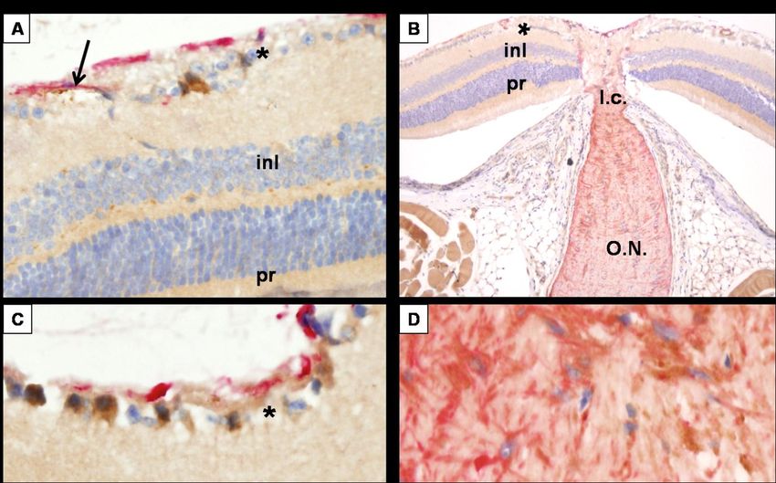
Zanello ... al. -- Spaceflight-Induced Ocular Changes in Mice

microgravity, namely hindlimb suspension (HS)            However, these studies were limited to structural
and bed rest, induce a fluid shift and concomitant       histopathologic observations of the eye. In the
vascular pressure and flow alterations (Hargens          present      work,       we      expand       the
and Watenpaugh, 1996; Wilkerson et al., 2002),           immunohistopathologic analysis to investigate the
affecting not only cardiovascular physiology but         effects of spaceflight and the elicited responses
also inducing genome-wide gene expression                observed in the eyes of mice aboard shuttle
changes in the central nervous system (Frigeri et        mission STS-133, focusing, for the first time, on
al., 2008).                                              molecular and cellular processes subjacent to the
     Ocular changes have been reported related to        histopathologic changes.
exposure to the space environment. In humans,
                                                         MATERIALS AND METHODS
the direct effect of radiation in the lens results in
cataract formation (Cucinotta et al., 2001), which       Animals
manifests with a higher incidence and earlier                 This work consisted of a tissue sharing-
onset in the astronaut population. Light flashes in      derived project that used specimens collected
the eye are an occurrence that has been observed         from a parent animal experiment aboard shuttle
by astronauts since the Apollo program (Sannita          mission STS-133. The original experiment
et al., 2006) -- a phenomenon not completely             included animals infected with respiratory
understood.                                              syncytial virus immediately after return to Earth
     Most importantly, recent medical data from          (study led by independent investigator Dr.
astronaut cohorts have reported the development          Roberto Garofalo, from the University of Texas
of optic disc edema, choroidal folds, posterior          Medical Branch in Galveston). However, the work
globe flattening, and a resulting hyperopic shift        discussed in this article only included the non-
(Kramer et al., 2012; Mader et al., 2011) in a           infected control animals. Animal procedures were
fraction of the astronaut population upon return         approved by the NASA Ames Research Center
from missions longer than 30 days (NASA, 2010).          and Kennedy Space Center institutional animal
No clear etiology has been established for these         care and use committees. The STS-133 mission
cases, but it is hypothesized that microgravity, the     occurred from February 24 to March 9, 2011, for
ensuing cephalad fluid shift, and venous                 a total duration of 12 days and 19 hours. Female
congestion may play a role. The perturbations            10 to 12 week-old BALB/cJ mice were assigned
observed in some individuals of the astronaut            to one of three experimental groups: Flight (FLT),
cohort resemble those found in papilledema               Animal Enclosure Module (AEM) ground
associated       with     idiopathic     intracranial    controls, and vivarium-housed (VIV) ground
hypertension (IIH) also known as pseudotumor             controls. The flight animals (FLT) were housed in
cerebri (Friedman, 2007; Kramer et al., 2012;            AEMs identical to the ground controls. The AEM
Mader et al., 2011). Because the etiology is still a     is a self-contained habitat that provides
matter of speculation, investigating whether             ventilation, waste management, food, water, and
exposure to microgravity represents a source of          controlled lighting (Naidu et al., 1995). It has
stress for the eye is an issue of critical               previously been used in experiments studying
occupational health importance. To this aim, this        rodent biology during spaceflight. The AEM
project examines the effects of spaceflight on the       flight unit is located in the middeck locker of the
rodent eye and the responses that occur when             shuttle and its temperature is set at 3° to 8°C
challenged with exposure to microgravity in              above the environmental middeck temperature.
combination with other stressors during                  Lighting of 14 lux is set to a 12 hour day/12 hour
spaceflight.                                             night cycle. AEM ground controls were
     Previous spaceflight studies performed on           maintained in identical conditions at the Space
rodents found evidence of retinal degeneration in        Life Sciences Laboratory, Kennedy Space Center.
neonatal rats aboard shuttle mission STS-72              Vivarium ground controls were housed in
(Tombran-Tink and Barnstable, 2006), and of cell         standard vivarium cages and conditions, on a 12-
swelling and disruption in rats aboard two               hour day/12-hour night light cycle at 200 to 215
experiments on Russian Cosmos satellites                 lux. In view of the housing and lighting conditions
(Philpott et al., 1980; Philpott et al., 1978).

30 – Gravitational and Space Research   Volume 1 (1) Oct 2013
Zanello et al. -- Spaceflight-Induced Ocular Changes in Mice

of the vivarium, the proper ground controls that        washing in phosphate buffer saline (PBS), the
allow measuring the effects attributed to               specimens were incubated with Vector ImmPress
spaceflight are the AEM-housed ground controls.         detection kit corresponding to the primary
    After sacrifice, one eye of each mouse from         antibody’s host and counterstained with
the three groups (FLT, AEM, and VIV) was                hematoxylin. For the double stain with β-amyloid
collected at 1, 5, and 7 days after landing, and was    and GFAP, antigen retrieval was performed with
fixed for histological examination. The                 Dako target retrieval solution (a modified citrate
contralateral eye was stored in RNALater and            buffer from Dako, Carpinteria, CA), steaming for
used for gene expression profiling by RT-qPCR.          25 minutes, and then treated with peroxidase
                                                        blocking buffer as above, and endogenous biotin
Materials
                                                        blocked with Vector Avidin/Biotin blocking kit
    The histological 4% paraformaldehyde-based          (Vector, Burlingame, CA). Staining for β-amyloid
fixative was obtained from Excalibur Pathology,         was done with the mouse-on-mouse peroxidase kit
Inc., Oklahoma City, OK. Goat polyclonal                according to the manufacturer’s instructions
antibody      to     8-hydroxy-2'-deoxyguanosine        (Vector Labs). Diaminobenzidine (DAB) was
(8OHdG) (ab10802) and rabbit polyclonal                 used for color labeling for β-amyloid (brown). For
antibody to activated caspase-3 (ab52181) were          GFAP immunostaining, Dako’s streptavidin
purchased from Abcam Inc., Cambridge, MA.               phosphatase kit was used with permanent red
Mouse monoclonal antibody to β-amyloid 1-16             (red) as the chromophore.
was obtained from Millipore (Temecula, CA) and
                                                        Qualitative Detection
rabbit polyclonal antibody against glial fibrillary
acidic protein (GFAP) was purchased from Dako,               Morphology and histology were interpreted
Carpinteria, CA. Paraffin embedding and                 by an ophthalmic pathologist (masked for specific
histologic sectioning were contracted from              study groups) on H&E slides. Immunostained
Excalibur Pathology. qRT-PCR reagents were              slides were evaluated for positivity of stain in a
purchased from Qiagen Inc., Valencia, CA and            graded scale from 0 to 3+, where 0 indicated
BioRad, Hercules, CA. Tissue samples were               absence of staining and 3+ indicated marked
assigned       a     different    number        for     positivity and more than 3 positive cells per layer.
immunohistochemistry evaluation and gene                Immunoreactivity was evaluated in the corneal
profiling to perform a masked analysis.                 epithelium and endothelium, iris, lens, choroid,
                                                        retinal ganglion cell (RGC) layer, inner nuclear
Histology and Immunohistochemistry
                                                        layer (INL), outer nuclear layer (ONL), and optic
    Fixed eyes were paraffin embedded, sectioned        nerve.
at 5 µm thickness, and stained with standard
                                                        Quantitative Detection
hematoxylin-eosin       (H&E)     for    histologic
examination. Four immunohistologic stains were              To quantify oxidative-related DNA damage in
performed: 8OHdG to detect oxidative-related            the retina, densitometric quantification of 8OHdG
DNA damage, activated caspase-3 to study                immunohistochemistry was performed. Briefly,
apoptosis, and double stain using β-amyloid as a        digital color images of the retina were processed
marker of neuronal and axonal injury and GFAP           using NIH ImageJ ver.1.68 (Abramoff et al.,
as an indicator of glial activation. All                2004) and converted to an 8-bit inverted gray-
immunostains had negative (omitting primary             scale image for analysis. Regions of interest were
antibody) and positive (using known tissue that         selected from each retina section, corresponding
reacts with the antibody of interest) controls. For     to the RGC, INL, and ONL as well as nearby
8OHdG and caspase-3 staining, sections were             areas without immunoreactivity for background
equilibrated in water after deparaffinization and       measurements. Five sections were analyzed for
treated sequentially in 3% hydrogen peroxide, 1%        each sample, for which the mean density per unit
acetic acid, and 2.5% serum (Vector Labs,               area (minus mean background density) was
Burlingame, CA) before incubating with the              measured.
diluted primary antibody for either 2 hours at              To quantify apoptosis in the retina, activated
room temperature or overnight at 4ºC. After             caspase-3 positive cells were identified for each

                                             Gravitational and Space Research   Volume 1 (1) Oct 2013 -- 31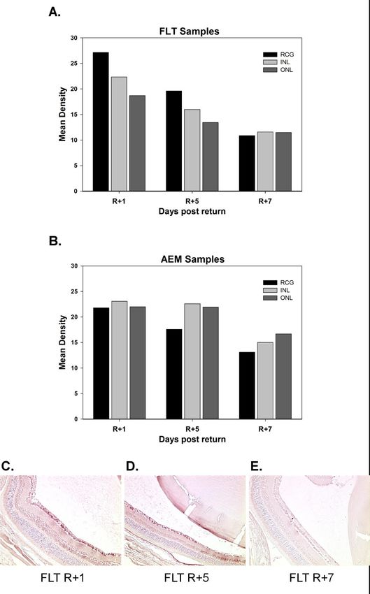
Zanello et al. -- Spaceflight-Induced Ocular Changes in Mice

retinal sample and expressed over the total              enlargement of the cell. However, irregular
number of cells in each of the following retinal         acanthosis, irregular increment of cell layers, with
layers: RGC, INL, and ONL. Cellular number was           pronounced edema was present in the VIV group
determined with the cell counting plug-in for            at R+7 (mice #41, 42). All mice had inflammatory
ImageJ ITCN (Byun et al., 2006).                         cells either in the anterior chamber or vitreous,
                                                         regardless of the group. Focal cortical cataracts,
Gene Expression Analysis
                                                         disrupted fibers, and formation of globules in the
     Mouse retina was microdissected and placed          cortex of the lens, which is located between the
in RNAlater (Life Technologies, Grand Island,            nucleus and the epithelium, were present in
NY). Total RNA was then isolated using the               several mice. As shown in Figure 1, full cortical
AllPrep DNA/RNA Micro kit (Qiagen, Valencia,             cataracts were seen only in the two mice of the
CA) and analyzed for quality using an Agilent            FLT group at R+7 group and this was associated
2100 Bioanalyzer. All samples used reported a            with caspase-3 2+ staining. The VIV group at R+7
RNA Integrity Number (RIN) >7.0.              The        had no morphologic changes of cataract but had
Quantitect Reverse Transcriptase kit (Qiagen) was        caspase-3 2+ staining as well (see below).
then used to generate cDNA templates for                 Apoptosis of neurons defined as shrinkage of the
subsequent real-time qPCR analysis. Fifty                cytoplasm with hyperchromatic nuclei and
nanograms of RNA were used in each reverse               degenerated chromatin was observed in some
transcriptase reaction in a total reaction volume        mice. These findings were quantified using
scaled to 30 μL according to manufacturer’s              immunohistochemistry and they are discussed
instructions, and the synthesis reaction was             below. Some slides showed artifacts in the
allowed to proceed for 2.5 hours.           qPCR         histology (possibly due to traumatic enucleation)
amplifications were done in a total volume of 20         that precluded complete interpretation. These
μL using 1 μL of a 1:10 dilution of the cDNA             findings are not included in the interpretation.
pool obtained in the previous step and SYBR              Only those findings that are clear and not affected
Green qPCR mastermix (BioRad, Hercules, CA)              by processing are reported.
on a Bio-Rad CFX96 real-time PCR detection
system. Samples were run in three technical              Oxidative Stress: 8OHdG
replicates each. Primers (Qiagen) were selected to       Cornea
hybridize with genes specific for various cellular
                                                             8OHdG immunoreactivity was positive in all
response pathways according to relevant findings
                                                         mice in the acanthotic areas of the cornea. In the
in the literature that reported known roles in
                                                         FLT group, positivity was evidenced in the
retinal stress, degeneration, oxidative stress,
                                                         corneal epithelium and endothelium, but we were
inflammation, and death/survival (Table 1). Three
                                                         not able to document significant differences
housekeeping genes (Hprt1, Rplp0, and Rpl13)
                                                         compared to AEM and VIV controls with the
were selected according to previously reported
                                                         present data.
expression stability (van Wijngaarden et al.,
2007). Normalization to the housekeeping genes           Retina and Optic Nerve
was performed using the geNorm algorithm                      Figure 2 summarizes 8OHdG data. The two
(Vandesompele et al., 2002) built into the CFX96         mice in the FLT group at R+1 showed frank
software, which computes a normalization factor          positivity for 8OHdG in the neuronal layer. One
for each sample from the contribution of each            of these also evidenced 8OHdG in some vessels
housekeeping gene.                                       over the ON head. Digital quantitative analysis of
RESULTS                                                  immunoreactivity in the retinal layers was more
                                                         prominent in the RGC of FLT samples at R+1
Histological Analysis of Eye Specimens                   (Figure 2B). Comparing FLT samples at the
    Results are summarized in Table 2. All groups        different tissue collection time points, 8OHdG
showed corneal acanthosis, defined as thickening         immunoreactivity decreased from R+1 to R+7
of the epithelium of more than 5 layers of cells,        (Figure 2B, C, D, and E). All mice were negative
and edema defined as clearing of cytoplasm with          at the level of the optic nerve.

32 – Gravitational and Space Research   Volume 1 (1) Oct 2013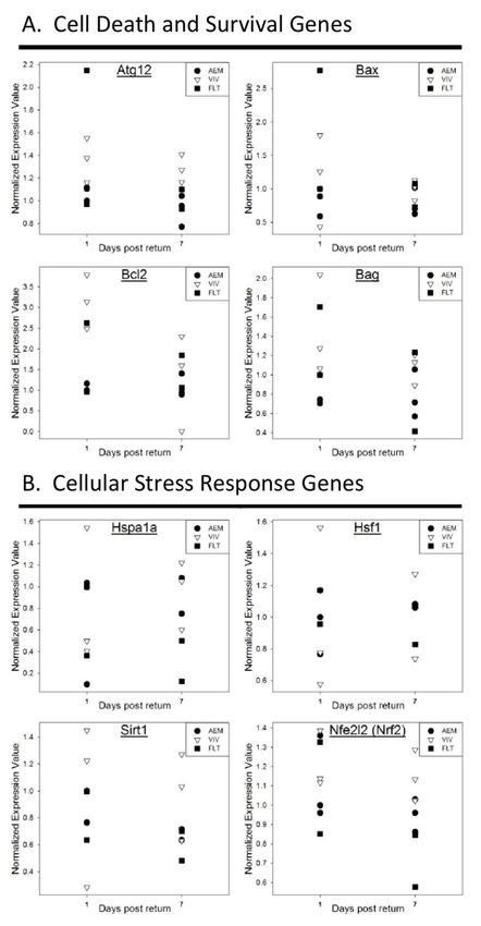
Zanello et al. -- Spaceflight-Induced Ocular Changes in Mice

Table 1. Genes of interest evaluated for expression changes in the mouse retina. Grouping was done
according to relevant cellular processes and complete gene name with gene symbol are provided, as well as
references reporting possible relevant roles in retina physiology.

  Process                        Gene Symbol              Gene name

  Cell death and survival        Bax                      Bcl2-associated X protein
  (Lohr et al., 2006)            Bcl2                     B-cell lymphoma 21
                                 Bag1                     Bcl2-associated athanogene 12
                                 Atg12                    Autophagy related 123

  Cellular Stress response       Hsf1                     Heat shock transcription factor 1
                                 Hspa1a                   Heat shock 70kDa protein 1A4
                                 Sirt1                    Sirtuin 15

                                 Nfe2l2 (Nrf2)            Nuclear factor (erythroid-derived 2)-like 26

  Oxidative stress response      Hmox1                    Heme-oxygenase 17
                                 Cat                      Catalase
                                 Sod2                     Superoxide dismutase 2, mitocondrial8
                                 Gpx4                     Glutathione peroxidase 49
                                 Prdx1                    Peroxiredoxin 1
                                 Cygb                     Cytoglobin

  Inflammation                   Nfkb1                    Nuclear factor of kappa light polypeptide gene
                                                          enhancer in B-cells 110
                                 Tgfb1                    Transforming growth factor beta 111

  Normalizing genes              Rpl13                    Ribosomal protein L13
                                 Rplp0                    Ribosomal protein, large, P0
                                 Hprt                     hypoxanthine phosphoribosyltransferase 1

  1 (Godley et al., 2002)                5 (Chen et al., 2009)                      9 (Ueta et al., 2012)
  2 (Liman et al., 2008)                 6 (Wei et al., 2011)                       10 (Wise et al., 2005)
  3 (Wang et al., 2009)                  7 (Zhu et al., 2007)                       11 (Gerhardinger et al., 2009)
  4 (Awasthi and Wagner, 2005)           8 (Justilien et al., 2007)

                                                               Retina and Optic Nerve
Apoptosis: Caspase-3
                                                                    Detection of apoptosis by activated caspase-3
Cornea
                                                               immunoreactivity was performed on retinal
    Activated caspase-3 appeared positive in the
                                                               sections and compared in the different specimens
cornea of all mice with the same intensity.
                                                               (Figures 1 and 3). All mice showed positivity in
Lens                                                           the neuronal layer regardless of day of sacrifice.
    Two mice of the FLT group at R+7 had                       Digital image quantification of caspase-3
cataract formation associated with caspase-3 2+                immunoreactivity revealed that VIV samples had
staining (Figure 1). The VIV group at R+7 had no               the highest percentage of apoptotic cells in the
morphologic changes of cataract but had caspase-               INL and RGC layer, followed by FLT samples, at
3 2+ staining as well.                                         day R+1 and R+7. Comparatively, VIV and FLT
                                                               retina samples showed more caspase-3 positive

                                                 Gravitational and Space Research        Volume 1 (1) Oct 2013 -- 33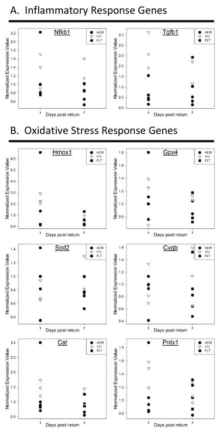
Zanello et al. -- Spaceflight-Induced Ocular Changes in Mice

Table 2. Histologic interpretation with Hematoxylin-Eosin. Data arranged according to group (FLT, AEM,
VIV) and day of sacrifice: 2 mice per group at R+1, +5, or +7, respectively.

                               Cornea                                             Lens                     Retina          ON
                  FLT                   AEM           VIV           FLT           AEM            VIV       FLT AEM VIV FLT AEM VIV
                                                                  Anterior                     Anterior
                FA and E                 FA           FA                          Nml                      Nml Nml Nml Nml Nml Nml
                                                                subcapsular C                subcapsular C
 Day 1
          Bullae*, A 1+, E 2+                                                                  Anterior
                                       A* 2+          FA            Nml           Nml                      Nml Nml Nml Nml  -  Nml
         basal layer calcification                                                           subcapsular C
                                                                                  Focal
             FA and basal E          FA*, E 1+      Central E       Nml                         Nml    Nml Nml Nml Nml Nml Nml
                                                                                cortical C
 Day 5                               Intranuclear
                                                                                  Focal      Anterior
                   FA                 inclusions,     FA            Nml                                  Nml Nml Nml Nml Nml Nml
                                                                                cortical C subcapsular C
                                      A 1+, E 2+
                                                 Irregular A 1+
                   FA                    FA                        Cortical C      Nml           Nml       Nml Nml Nml Nml Nml Nml
                                                      E 3+
 Day 7
                                                 Irregular A 1+
               A* 1+, E 2+              FA                         Cortical C      Nml           Nm        Nml Nml Nml Nml Nml Nml
                                                      E 2+
         (A)= acanthosis, (C)= cataract, (E)= edema, (FA)= focal acantosis, (Nml)=normal, anterior subcapsular C (anterior subcapsular
         cataract is disruption of the fibers with proliferation of the epithelium in the anterior subcapsular áreas of the lens)
         Comments: *Anterior chamber 1+ cell

cells than AEM samples at R+1, except for the                               at the retinal neuronal layer: 8OHdG, caspase-3,
INL in the AEM group at R+7. VIV samples also                               β-amyloid, and GFAP.
tended to increase their percentage of apoptotic                                At the level of the optic nerve, only the FLT
cells at day R+7, as seen in qualitative analysis.                          group at R+7 showed positivity for both β-
Retinal pigment epithelium (RPE) of the FLT                                 amyloid in the axons and GAFP in the astrocytes
group at R+1 and one mouse at R+5 showed                                    either at the level of the lamina cribrosa or distal
positivity with caspase-3, and one mouse AEM                                to it (Figure 4). No co-expression was seen of
R+7 showed only rare and focal RPE staining                                 GFAP and β-amyloid in same cell type.
(Figure 1). Qualitative and quantitative evaluation
                                                                            Cellular Responses              Identified   by     Gene
of ON immunoreactivity was inconclusive.
                                                                            Expression Analysis
β-amyloid and GFAP                                                               Gene expression profiling on STS-133 flight
     β-amyloid and GFAP stains were studied in                              samples and their AEM and vivarium ground
the retina and optic nerve only and                                         controls was performed targeting a set of genes
immunostained retina sections are shown in                                  focused on cellular death and survival, oxidative
Figure 4. With regard to the retina, all mice were                          stress and cellular stress response, and
positive in the neuronal layer for β-amyloid.                               inflammation. Results are shown in Figure 5 and
Overall, the vivarium mice showed a slightly                                Figure 6 and expressed as comparative normalized
higher positivity in both RGC and INL compared                              expression across the individual specimens at R+1
to the rest of the mice (VIV animals showed 2-3+                            and R+7 for all groups. Due to the limited sample
positivity at R+1 and R+5, more than any other                              size, statistical analysis was not possible and these
group; one FLT animal at R+7 showed similar 2+                              results are mainly descriptive.
reactivity). GFAP was present in astrocytes of the
                                                                            Activation of Oxidative Stress Response and
retinal neuronal layer in at least one mouse of
                                                                            Pro-Inflammatory Genes
each group, except in the FLT group at R+5,
where it was absent. No activation (positivity) of                              Figures 5 and 6 (see section below) plot gene
Muller cells was noted in any of the eyes.                                  expression data measured by real time qPCR.
     While results were not conclusive from these                           Several genes coding for key antioxidant enzymes
retinal findings, it is important to note that only                         (Hmox1, Sod2, Cat, Gpx4, Cygb, Prdx1) were
the FLT group at R+1 were positive for all stains                           elevated in retina samples obtained immediately
                                                                            after flight (Figure 5B), but this elevation returned

34 – Gravitational and Space Research                  Volume 1 (1) Oct 2013
Zanello et al. -- Spaceflight-Induced Ocular Changes in Mice
to levels closer to AEM ground control values at 7 Hmox1 showed the highest levels in those
days post-landing. A similar trend was observed samples for which a higher evidence of stress was
for inflammatory mediators Nfkb1 and Tgfb1 observed (FLT samples at R+1 and VIV ground
(Figure 5A). controls).
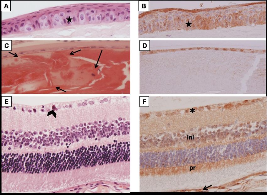
Figure 1. Histological analysis of H&E and Caspase-3 stained eye samples. Hematoxylin and Eosin stain,
original magnification 20X : Panel A. AEM R+7, Epithelium of cornea showing focal edema of cells seen as
clearing and enlargement of the cytoplasm in the basal layers (star marks the level of the basal layers) and
acanthosis (thickening of more than 5 layers of cells). Panel C. FLT R+1, anterior lens with cortical cataract
seen as disorganization of the fibers of the cortex (arrows at the level of the cortex). Notice the displaced
nucleus (nucleus of epithelial cells of the lens should only be present in the subcapsular area and not in the
cortex in the anterior portion of the lens). Panel E. FLT R+1, retina with an apoptotic neuron seen as a
shrunken cell with hyperchromatic condensed nucleus and eosinophilic cytoplasm (arrow head). Remainder
of retina appears morphologically unremarkable. Caspase 3 immunostaining: Panel B. FLT R+1 corneal
epithelium staining positively with Caspase 3 in the superficial layers and in the basal layers (star). Positive
staining of the basal cells of the corneal epithelium is seen in the focal acanthotic areas, and in the upper
differentiated layers (internal positive control). Panel D. FLT R+1 lens epithelium staining with Caspase 3;
notice that cortex is negative. Panel F. FLT R+1, retina with caspase-3 staining of cytoplasm of neurons (*)
predominantly with faint staining of the inner nuclear layer (inl) and inner segments of photoreceptors (pr).
The cytoplasm of RPE cells is also staining (arrow).
Gravitational and Space Research Volume 1 (1) Oct 2013 -- 35Zanello et al. -- Spaceflight-Induced Ocular Changes in Mice Figure 2. 8OHdG immunoreactivity in retinal neuronal layers of AEM and FLT mice. Bars indicate the mean of n=2 biological samples. Each individual neuronal cell layer was compared at R+1, R+5, and R+7 in AEM samples (panel A) and Flight samples (panel B). Representative images of 8OHdG stained histological sections of the retina in FLT samples at R+1 (panel C), R+5 (panel D), and R+7 (panel E). 36 – Gravitational and Space Research Volume 1 (1) Oct 2013
Zanello et al. -- Spaceflight-Induced Ocular Changes in Mice

Figure 3. Quantification of Caspase-3 immunoreactivity by neuronal layer. Percentage of caspase-3 positive
cells in the Inner Nuclear Layer (panel A) and the Retinal Ganglion Cell Layer (panel B) was calculated as
described in Methods for day R+1 and R+7 tissue collection time points. Representative images of
histological sections stain (red-brown) for caspase-3 of Flight (panel C), AEM (panel D), and Vivarium (panel
E) samples at day R+1. Arrows indicate caspase-3 positive stained cells identified in different layers of the
retina.

                                               Gravitational and Space Research   Volume 1 (1) Oct 2013 -- 37
Zanello et al. -- Spaceflight-Induced Ocular Changes in Mice
Figure 4. Beta amyloid (brown) and glial fibrillary acid protein (GFAP) (red) double staining
immunohistochemistry. A: FLT R+1 (mouse #13). Retina with focal positive cytoplasmic staining in neurons
of the ganglion cell layer (*) with β-amyloid (brown). Perivascular (arrow) and other astrocytes in the
ganglion cell layer stain with GFAP (red). Notice the negative staining of Muller cells with GFAP. B: FLT
R+1 optic nerve. Note the staining of the optic nerve (O.N.) in the region posterior to the lamina cribrosa
(l.c.) with GFAP and focally with β-amyloid. Non-specific staining of the orbital muscle is also seen with β-
amyloid (brown). C: FLT R+1 retina higher magnification of focal positivity with β-amyloid (brown) in
ganglion cell layer (*) and GFAP in astrocytes (red). D: FLT R+1 optic nerve higher magnification of
immediate post-laminar region. Notice the staining of oligodendrocytes and astrocytes with GFAP (red) and
the β-amyloid stain (brown) of the nerve fibers in between the glial cells.
Cell Death and Survival Genes Activation of Cellular Stress Genes
The proapoptotic gene Bax was elevated in The cellular stress response genes Hsf1and
one flight sample (#13) at day R+1 and Nrf2 (Nfe2l2) were expressed slightly higher in
moderately elevated in one flight sample (#52) at VIV samples compared to AEM controls. Among
R+7. Vivarium mice showed a higher expression the FLT mice, there was a tendency to higher
of Bax at all collection time points compared to expression at R+1 than R+7 (Figure 6B). The
AEM ground controls. FLT samples at R+1 and Hsf1 activator sirtuin 1 (Sirt1) did not show major
VIV samples exhibited higher levels of the differences across the various samples.
autophagy marker Atg12 and the survival genes Interestingly, the heat shock protein 70KDa
Bcl2 and Bag1, suggesting that cellular protection Hsp1a1 was expressed at a lower level in mouse
mechanisms may be triggered as a response to #13 that exhibited, overall, the highest signs of
cellular stress (Figure 6A). stress.
38 – Gravitational and Space Research Volume 1 (1) Oct 2013Zanello et al. -- Spaceflight-Induced Ocular Changes in Mice
Figure 5. Gene expression analysis of inflammatory and oxidative stress response genes. Inflammatory
response (panel A) and oxidative stress (panel B) gene expression levels from RNA isolated from retina
samples in Flight (FLT), AEM, and Vivarium (VIV) samples at day R+1 and R+7, measured by real time
qPCR. Y axis represents the comparative gene expression levels normalized to housekeeping genes.
Gravitational and Space Research Volume 1 (1) Oct 2013 -- 39Zanello et al. -- Spaceflight-Induced Ocular Changes in Mice Figure 6. Gene expression analysis of cell death and survival and cellular stress response genes. Cell death and survival (panel A) and cellular stress (panel B) gene expression levels from RNA isolated from retina samples in Flight (FLT), AEM, and Vivarium (VIV) samples at day R+1 and R+7, measured by real time qPCR. Y axis represents the comparative gene expression levels normalized to housekeeping genes. 40 – Gravitational and Space Research Volume 1 (1) Oct 2013
Zanello et al. -- Spaceflight-Induced Ocular Changes in Mice

DISCUSSION                                              both oxidative stress-induced DNA damage in the
                                                        neuronal layers of flight mice retinas and of an
     While the spaceflight results reported herein
                                                        oxidative stress response induced at the gene
represent pilot data due to the small sample size,
                                                        expression level in these mice. Short-term
these data offer, for the first time, direct evidence
                                                        responsiveness to DNA oxidation followed by
suggesting that oxidative stress, neuronal damage,
                                                        DNA repair has been studied longitudinally in
and mechanical injury take place in the retina,
                                                        blood of trauma patients (Oldham et al., 2002),
lens, and optic nerve of rodents flown in low-
                                                        suggesting that the attenuated DNA damage
Earth orbit for a period under two weeks. Several
                                                        observed after one week of return from flight may
previous studies have shown the occurrence of
                                                        be the result of DNA repair.
oxidative stress during spaceflight (Stein, 2002),
                                                             Of note, the ground controls kept in the
however, our work gives a first insight into the
                                                        vivarium exhibited a comparable level of retinal
impact of space-associated factors on biological
                                                        oxidative stress to the samples from flight,
processes like cell death, oxidative stress, and
                                                        especially at longer exposures (day R+7). This is
probable mechanical injury in the rodent eye.
                                                        likely due to the fact that the illumination
     Because the BALB mouse strain used in the
                                                        conditions in a standard vivarium room are
STS-133 experiment is susceptible to light-
                                                        approximately 15-fold in light flux compared to
induced retinal degeneration (LaVail et al., 1987),
                                                        the illumination of an AEM, even if both maintain
we speculate that this particular strain exhibits an
                                                        a 12 hour light-12 hour dark cycle.
enhanced sensitivity to oxidative stress and/or a
reduced stress response, making it a suitable strain        Caspase-3 is a pro-enzyme that is activated in
in which to identify alerting evidence of risks         the intrinsic apoptotic pathway in all mammals
previously unrecognized in the retinal tissue,          (D'Amelio et al., 2010). In this study, all mice
while impacting its value as a model for the study      showed positivity for caspase-3 at the level of the
of the human changes seen in-flight.                    cornea. This may be explained by the fact that
     8OHdG, a product of deoxyguanosine                 caspase-3 immunoreactivity in the stratified
oxidation, is a marker of oxidative stress-induced      epithelium of the cornea serves as an internal
DNA damage. This damage has been observed in            positive control due to the natural differentiation
mouse cornea exposed to dryness (Nakamura et            process that the basal cells suffer towards
al., 2007), ultraviolet radiation (Tanito et al.,       cornification. Apoptosis can be triggered by
2003), and in mouse retina exposed to intense           oxidative stress, brain trauma, or ischemia. In a
light (Tanito et al., 2002; Wiegand et al., 1983). In   model of brain ischemia, the area of neuronal
our study, 8OHdG was present in all acanthotic          apoptosis has been identified not in the infarct
areas of the cornea. Irregular acanthosis with          region but in the surrounding area, where the
visible edema was only seen in the VIV samples          oxygen tension is decreased, but not absent
at R+7, and it was only in this group where             (Pulsinelli et al., 1982). The presence of activated
positivity at the corneal endothelium was               caspase-3 is thus related to hypoxic environment
observed since day 1, suggesting an impaired ion        and radiation exposure. In our study, the FLT
and water transport in the cornea.                      group at R+1 showed higher positivity compared
     The retinal response to intense light in           to the rest of the groups. This may be related to
susceptible mice has been studied before and has        radiation and microgravity exposure during
been found to be related to lipid peroxidation at       spaceflight. It is important to point out that the
the ONL (Tanito et al., 2002; Wiegand et al.,           effect of high-energy-particle radiation may be
1983). Likewise, radiation-induced retinopathy is       overall increased in this susceptible mouse strain.
an ocular complication in cancer patients that              Qualitative examination revealed that VIV
receive radiation therapy (Parsons et al., 1996).       and FLT groups showed more caspase-3-positive
The processes involved in the damage by high-           cells at the retinal layers than AEM retinas. This
energy-particle radiation in these cases may share      may suggest that the damage caused by visible
commonalities (direct DNA damage and oxidative          light radiation in the albino strain in the vivarium
stress) with exposure to radiation present during       conditions may be comparable to the damage
spaceflight. The present work shows evidence of         caused by the exposure to spaceflight

                                              Gravitational and Space Research   Volume 1 (1) Oct 2013 -- 41
Zanello et al. -- Spaceflight-Induced Ocular Changes in Mice
environmental factors. We also observed positive and had the unique characteristic of being at the
microglial (astrocytes) but not Muller cell level of lamina cribrosa or immediately distal to
activation in VIV specimens, which may support it. This compares with the findings in traumatic
the notion of visible light radiation effects as the injury in children of shaken-baby syndrome where
triggering factor in inner layers of the retina only most of the axonal changes are seen in the
in these mice (Song et al., 2012). postlaminar region (Gleckman et al., 2000). This
Both mice in the FLT group at R+1 and one may be associated to the anatomy of this region
mouse at R+5 showed evidence of apoptosis in the where the nerve is anchored by the fibers of the
RPE. Apotosis in the RPE has been identified in lamina cribrosa but immediately posterior to this
ocular pathologies like age-related macular or beyond this area the nerve can move freely.
degeneration (AMD) secondary to exposure to Thus, in the event of mechanical trauma the
activated monocytes (Yang et al., 2011), or immediate fibers in the postlaminar region may be
triggered by oxidative stress with H2O2, the ones demonstrating more damage. The trauma
lipofuscin, or light irradiation (Sparrow et al., may include increased intracranial pressure that is
2000). This data also suggest oxidative stress may transmitted into the nerve, positional or whiplash
be an important component in the retinal damage (similar, although in a less intense manner to what
in these mice. Of note, in vitro experiments with happens in shaken baby syndrome), or vibration
human RPE cells cultured in simulated (as the one occurring during launch or landing).
microgravity generated by a NASA-bioreactor However, there is the need to further investigate
resulted in DNA damage and inflammatory the nature of the changes through additional
response in these cells (Roberts et al., 2006). experimental work.
Retinal pigment epithelium attenuation has been
GFAP is an intermediate filament protein
related to retinal choroidal folds previously found
known to be present in astrocytes, Muller cells,
in astronauts (Mader et al., 2011). It is yet to be
and oligodendrocytes in the post-laminar optic
determined whether or not increased RPE
nerve. GFAP is elevated when there is stress in
apoptosis may contribute to the formation of
the central nervous system and has been shown in
choroidal folds or if it increases the risk for AMD
the injured retina mostly present in the activated
in astronauts.
Muller cells (Lewis and Fisher, 2003). In this
Several advances in immunohistochemistry paper, we show that the optic nerves of several
have led to the identification of β-amyloid in mice were positive for GFAP and β-amyloid;
traumatic brain injury in humans (Iwata et al., however, it was only the FLT group at R+7 that
2002), rats, and pigs (Smith et al., 1999), by showed increased expression of GFAP at the
tracing not only the full-length protein but also postlaminar optic nerve. These findings suggest
small aminoacid peptides. β-amyloid was present that the astrocytes and oligodendrocytes were
in areas of the brain as soon as one day after brain activated in this region probable secondary to
trauma was provoked by pressure injection of mechanical trauma. The causes of this, either
saline into the cranium in a rat model (Pierce et vibration or fluid shift-related, need to be further
al., 1996). Moreover, β-amyloid deposits showed investigated.
evidence of optic nerve injury in cases of shaken- In addition, only FLT mice sacrificed at day 1
baby syndrome (Gleckman et al., 2000). Previous (FLT R+1) were immunoreactive in the neuronal
studies in animal models have shown distribution layer for all β-amyloid, GFAP, caspase-3, and
of β-amyloid in the mouse retina that suggests its 8OHdG, suggesting increased oxidative and
involvement in the pathophysiology of glaucoma possibly mechanical damage. This may be
(Kipfer-Kauer et al., 2010). We report that β- explained by the possible correlation of β-amyloid
amyloid deposition was present in the neural deposition and activation of astrocytic cells, both
retina of mice in all treatment groups and that the triggering reactive oxygen species production
VIV mice showed a slightly higher positivity in (Lamoke et al., 2012).
both RGC and INL compared to the rest of the
The gene expression profiling results with
mice. Interestingly, β-amyloid was present in the
BALB mice in flight STS-133 support the
optic nerve of both mice in the FLT group at R+7
immunohistopathologic findings and suggest that:
42 – Gravitational and Space Research Volume 1 (1) Oct 2013Zanello et al. -- Spaceflight-Induced Ocular Changes in Mice
a) Oxidative stress-induced DNA damage was These preliminary data suggest that
higher in the FLT samples compared to controls spaceflight represents a source of environmental
on R+1, and decreased on R+7. A trend toward stress that directly translates into oxidative and
higher oxidative and cellular stress response gene cellular stress in the retina, which is partially
expression was also observed on R+1 compared to reversible upon return to Earth. Moreover, the
AEM controls, and these levels decreased on R+7. optic nerve findings suggest that the lesion may be
Several genes coding for key antioxidant mechanical in nature and that does not resolve
enzymes, namely, heme-oxygenase-1, after return to Earth, at least in the animals
peroxiredoxin, and catalase, were among those studied. Further work is needed to dissect the
elevated after flight. Likewise, the inflammatory contribution of the various spaceflight factors
response genes Nfkb1and Tgfb1 were elevated (microgravity, radiation) and to evaluate the
after flight. The fact that only two mice flown on impact of the stress response on retinal and optic
STS-133 were genetically analyzed per day of nerve health. These preliminary results should
sacrifice creates a major limitation in any inform investigators on the design of future
statistical analysis. However, this does not studies utilizing a more suitable mouse strain
preclude the comparisons of samples. b) There is devoid of photic degeneration predisposition,
an apparent correlation trend in the stress male animals that better reflect the astronaut
parameters measured in the different animals and population, and statistically powered larger
there is certain variability in the stress response sample sizes.
among the individual animals. For example,
ACKNOWLEDGEMENTS
mouse # 13 in the FLT group at R+1 suffered
from overall elevated stress, demonstrated by the We would like to recognize Richard Boyle for
highest 8OHdG levels, induction of antioxidant tissue sharing and collection, Audrey Nguyen for
enzymes, induction of Nfkb1, and concomitant help with digital image analysis, and James
lower levels of the cytoprotective heat shock Fiedler for graphic work. This work was funded
protein Hsp1a1. Sirtuin 1 gene expression results by the NASA Human Research Program.
were non-conclusive, but further analysis is REFERENCES
required to determine if translocation of sirtuin 1
may occur and how this may affect the expression Abramoff, M.D., Magalhaes, P.J., and Ram, S.J.
of downstream cellular stress response genes 2004. Image processing with ImageJ.
(Jaliffa et al., 2009; Ozawa et al., 2010). Biophotonics International. 11: 36-42.
c) Spaceflight represents a source of Awasthi, N. and Wagner, B.J. 2005. Upregulation
environmental stress that translates into oxidative of heat shock protein expression by
and cellular stress in the retina, which is partially proteasome inhibition: an antiapoptotic
reversible upon return to Earth. Also, retinas mechanism in the lens. Investigative
from VIV control mice evidenced higher Ophthalmology & Visual Science. 46: 2082-
oxidative stress markers, Nfkb1 and Tgfb1, likely 2091.
due to the more intense illumination in vivarium
cages versus the AEM. Byun, J., Verardo, M.R., Sumengen, B., Lewis,
In addition, mice in FLT group at R+7 were G.P., Manjunath, B.S., and Fisher, S.K. 2006.
positive for both β-amyloid and GFAP, and it was Automated tool for the detection of cell nuclei
only in these mice that there was increase in in digital microscopic images: application to
GFAP staining adjacent to lamina cribrosa in the retinal images. Molecular Vision. 12: 949-
optic nerve. We suspect some long term damage 960.
in the optic nerve may be seen after spaceflight Chen, D., Pacal, M., Wenzel, P., Knoepfler, P.S.,
because this did not resolve after seven days on Leone, G., and Bremner, R. 2009. Division
Earth. Additional quantitative experiments are and apoptosis of E2f-deficient retinal
needed to give a better understanding on this progenitors. Nature. 462: 925-929.
finding.
Cucinotta, F.A., Manuel, F.K., Jones, J., Iszard,
G., Murrey, J., Djojonegro, B., and Wear, M.
Gravitational and Space Research Volume 1 (1) Oct 2013 -- 43Zanello et al. -- Spaceflight-Induced Ocular Changes in Mice
2001. Space radiation and cataracts in mouse retinal degeneration. Investigative
astronauts. Radiation Research. 156: 460-466. Ophthalmology & Visual Science. 50: 3562-
3572.
D'Amelio, M., Cavallucci, V., and Cecconi, F.
2010. Neuronal caspase-3 signaling: not only Justilien, V., Pang, J.J., Renganathan, K., Zhan,
cell death. Cell Death & Differentiation. 17: X., Crabb, J.W., Kim, S.R., Sparrow, J.R.,
1104-1114. Hauswirth, W.W., and Lewin, A.S. 2007.
SOD2 knockdown mouse model of early
Friedman, D.I. 2007. Idiopathic intracranial
AMD. Investigative Ophthalmology & Visual
hypertension. Current Pain and Headache
Science. 48: 4407-4420.
Reports. 11: 62-68.
Kipfer-Kauer, A., McKinnon, S.J., Frueh, B.E.,
Frigeri, A., Iacobas, D.A., Iacobas, S., Nicchia,
and Goldblum, D. 2010. Distribution of
G.P., Desaphy, J.F., Camerino, D.C., Svelto,
amyloid precursor protein and amyloid-beta in
M., and Spray, D.C. 2008. Effect of
ocular hypertensive C57BL/6 mouse eyes.
microgravity on gene expression in mouse
Current Eye Research. 35: 828-834.
brain. Experimental Brain Research. 191:
289-300. Kramer, L.A., Sargsyan, A.E., Hasan, K.M., Polk,
J.D., and Hamilton, D.R. 2012. Orbital and
Gerhardinger, C., Dagher, Z., Sebastiani, P.,
intracranial effects of microgravity: findings
Park,Y.S., and Lorenzi, M. 2009. The
at 3-T MR imaging. Radiology. 263(3): 819-
transforming growth factor-beta pathway is a
27.
common target of drugs that prevent
experimental diabetic retinopathy. Diabetes. Lamoke, F., Ripandelli, G., Webster, S.,
58: 1659-1667. Montemari, A., Maraschi, A., Martin, P.,
Marcus, D.M., Liou, G.I., and Bartoli, M.
Gleckman, A.M., Evans, R.J., Bell, M.D., and
2012. Loss of thioredoxin function in retinas
Smith, T.W. 2000. Optic nerve damage in
of mice overexpressing amyloid beta. Free
shaken baby syndrome: detection by beta-
Radical Biology and Medicine. 53: 577-588.
amyloid precursor protein immuno-
histochemistry. Archives of Pathology & LaVail, M.M., Gorrin, G.M., and Repaci, M.A.
Laboratory Medicine. 124: 251-256. 1987. Strain differences in sensitivity to light-
induced photoreceptor degeneration in albino
Godley, B.F., Jin, G.F., Guo, Y.S, and Hurst, J.S.
mice. Current Eye Research. 6: 825-834.
2002. Bcl-2 overexpression increases survival
in human retinal pigment epithelial cells Lewis, G.P. and Fisher, S.K. 2003. Up-regulation
exposed to H(2)O(2). Experimental Eye of glial fibrillary acidic protein in response to
Research. 74: 663-669. retinal injury: its potential role in glial
remodeling and a comparison to vimentin
Hargens, A.R. and Watenpaugh, D.E. 1996.
expression. International Review of Cytology.
Cardiovascular adaptation to spaceflight.
230: 263-290.
Medicine and Science in Sports and Exercise.
28: 977-982. Liman, J., Faida, L., Dohm, C.P., Reed, J.C.,
Bahr, M., and Kermer, P. 2008. Subcellular
Iwata, A., Chen, X.H., McIntosh, T.K., Browne,
distribution affects BAG1 function. Brain
K.D., and Smith, D.H. 2002. Long-term
Research. 1198: 21-26.
accumulation of amyloid-beta in axons
following brain trauma without persistent Lohr, H.R., Kuntchithapautham, K., Sharma,
upregulation of amyloid precursor protein A.K., and Rohrer, B. 2006. Multiple, parallel
genes. Journal of Neuropathology & cellular suicide mechanisms participate in
Experimental Neurology. 61: 1056-1068. photoreceptor cell death. Experimental Eye
Research. 83: 380-389.
Jaliffa, C., Ameqrane, I., Dansault, A., Leemput,
J., Vieira, V., Lacassagne, E., Provost, A., Mader, T.H., Gibson, C.R., Pass, A.F., Kramer,
Bigot, K., Masson, C., Menasche, M., and L.A., Lee, A.G., Fogarty, J., Tarver, W.J.,
Abitbol, M. 2009. Sirt1 involvement in rd10 Dervay, J.P., Hamilton, D.R., Sargsyan, A.,
44 – Gravitational and Space Research Volume 1 (1) Oct 2013Zanello et al. -- Spaceflight-Induced Ocular Changes in Mice
Phillips, J.L., Tran, D., Lipsky, W., Choi, J., experimental K-007. Aviation, Space, and
Stern, C., Kuyumjian, R., and Polk, J.D. 2011. Environmental Medicine. 49: 19-28.
Optic disc edema, globe flattening, choroidal
Pierce, J.E., Trojanowski, J.Q., Graham, D.I.,
folds, and hyperopic shifts observed in
Smith, D.H., and McIntosh, T.K. 1996.
astronauts after long-duration space flight.
Immunohistochemical characterization of
Ophthalmology. 118: 2058-2069.
alterations in the distribution of amyloid
Naidu, S., Winget, C.M., Jenner, J.W., Mele, G., precursor proteins and beta-amyloid peptide
and Holley, D.C. 1995. Effects of housing after experimental brain injury in the rat.
density on mouse physiology and behavior in Journal of Neuroscience. 16: 1083-1090.
the NASA Animal Enclosure Module
Pietsch, J., Bauer, J., Egli, M., Infanger, M., Wise,
simulators. Journal of Gravitational
P., Ulbrich, C., and Grimm, D. 2011. The
Physiology. 2: 140.
effects of weightlessness on the human
Nakamura, S., Shibuya, M., Nakashima, H., organism and mammalian cells. Current
Hisamura, R., Masuda, N., Imagawa, T., Molecular Medicine. 11: 350-364.
Uehara, M., and Tsubota, K. 2007.
Pulsinelli, W.A., Brierley, J.B., and Plum, F.
Involvement of oxidative stress on corneal
1982. Temporal profile of neuronal damage in
epithelial alterations in a blink-suppressed dry
a model of transient forebrain ischemia.
eye. Investigative Ophthalmology & Visual
Annals of Neurology. 11: 491-498.
Science. 48: 1552-1558.
Roberts, J.E., Kukielczak, B.M., Chignell, C.F.,
NASA. 2010. Longitudinal Study of Astronaut
Sik, B.H., Hu, D.N., and Principato, M.A.
Health database.
2006. Simulated microgravity induced
Oldham, K.M., Wise, S.R., Chen, L., Stacewicz- damage in human retinal pigment epithelial
Sapuntzakis, M., Burns, J., and Bowen, P.E. cells. Molecular Vision. 12: 633-638.
2002. A longitudinal evaluation of oxidative
Sannita, W.G., Narici, L., and Picozza, P. 2006.
stress in trauma patients. Journal of
Positive visual phenomena in space: A
Parenteral and Enteral Nutrition. 26: 189-
scientific case and a safety issue in space
197.
travel. Vision Research. 46: 2159-2165.
Ozawa, Y., Kubota, S., Narimatsu, T., Yuki, K.,
Smith, D.H., Chen, X.H., Nonaka, M.,
Koto, T., Sasaki, M., and Tsubota, K. 2010.
Trojanowski, J.Q., Lee, V.M., Saatman, K.E.,
Retinal aging and sirtuins. Ophthalmic
Leoni, M.J., Xu, B.N., Wolf, J.A., and
Research. 44: 199-203.
Meaney, D.F. 1999. Accumulation of amyloid
Parsons, J.T., Bova, F.J., Mendenhall, W.M., beta and tau and the formation of
Million, R.R., and Fitzgerald, C.R. 1996. neurofilament inclusions following diffuse
Response of the normal eye to high dose brain injury in the pig. Journal of
radiotherapy. Oncology. 10: 837-847; Neuropathology & Experimental Neurology.
discussion 847-838, 851-832. 58: 982-992.
Philpott, D.E., Corbett, R., Turnbill, C., Black, S., Song, D., Song, Y., Hadziahmetovic, M., Zhong,
Dayhoff, D., McGourty, J., Lee, R., Harrison, Y., and Dunaief, J.L. 2012. Systemic
G., and Savick, L. 1980. Retinal changes in administration of the iron chelator deferiprone
rats flown on Cosmos 936: A cosmic ray protects against light-induced photoreceptor
experiment. Aviation, Space, and degeneration in the mouse retina. Free
Environmental Medicine. 51: 556-562. Radical Biology and Medicine. 53: 64-71.
Philpott, D.E., Corbett, R., Turnbill, C., Harrison, Sparrow, J.R., Nakanishi, K., and Parish, C.A.
G., Leaffer, D., Black, S., Sapp, W., Klein, 2000. The lipofuscin fluorophore A2E
G., and Savik, L.F. 1978. Cosmic ray effects mediates blue light-induced damage to retinal
on the eyes of rats flown on Cosmos No. 782, pigmented epithelial cells. Investigative
Gravitational and Space Research Volume 1 (1) Oct 2013 -- 45Zanello et al. -- Spaceflight-Induced Ocular Changes in Mice
Ophthalmology & Visual Science. 41: 1981- and exosomes in the aged retinal pigment
1989. epithelium: possible relevance to drusen
formation and age-related macular
Stein, T.P. 2002. Space flight and oxidative stress.
degeneration. PLoS ONE. 4: E4160.
Nutrition. 18: 867-871.
Wei, Y., Gong, J., Yoshida, T., Eberhart, C.G.,
Sundaresan, A. and Pellis, N.R. 2009. Cellular
Xu, Z., Kombairaju, P., Sporn, M.B., Handa,
and genetic adaptation in low-gravity
J.T., and Duh, E.J. 2011. Nrf2 has a protective
environments. Annals of the New York
role against neuronal and capillary
Academy of Sciences. 1161: 135-146.
degeneration in retinal ischemia-reperfusion
Tanito, M., Nishiyama, A., Tanaka, T., Masutani, injury. Free Radical Biology and Medicine.
H., Nakamura, H., Yodoi, J., and Ohira, A. 51: 216-224.
2002. Change of redox status and modulation
Wiegand, R.D., Giusto, N.M., Rapp, L.M., and
by thiol replenishment in retinal
Anderson, R.E. 1983. Evidence for rod outer
photooxidative damage. Investigative
segment lipid peroxidation following constant
Ophthalmology & Visual Science. 43: 2392-
illumination of the rat retina. Investigative
2400.
Ophthalmology & Visual Science. 24: 1433-
Tanito, M., Takanashi, T., Kaidzu, S., Yoshida, 1435.
Y., and Ohira, A. 2003. Cytoprotective effects
Wilkerson, M.K., Colleran, P.N., and Delp, M.D.
of rebamipide and carteolol hydrochloride
2002. Acute and chronic head-down tail
against ultraviolet B-induced corneal damage
suspension diminishes cerebral perfusion in
in mice. Investigative Ophthalmology &
rats. American Journal of Physiology - Heart
Visual Science. 44: 2980-2985.
and Circulatory Physiology. 282: 328-334.
Tombran-Tink, J. and Barnstable, C.J. 2006.
Wise, K.C., Manna, S.K., Yamauchi, K., Ramesh,
Space flight environment induces
V., Wilson, B.L., Thomas, R.L., Sarkar, S.,
degeneration in the retina of rat neonates.
Kulkarni, A.D., Pellis, N.R., and Ramesh,
Advances in Experimental Medicine and
G.T. 2005. Activation of nuclear transcription
Biology. 572: 417-424.
factor-kappaB in mouse brain induced by a
Ueta, T., Inoue, T., Furukawa, T., Tamaki, Y.,
simulated microgravity environment. In Vitro
Nakagawa, Y., Imai, H., and Yanagi, Y. 2012.
Cellular & Developmental Biology - Animal.
Glutathione peroxidase 4 is required for
41: 118-123.
maturation of photoreceptor cells. Journal of
Biological Chemistry. 287: 7675-7682. Yang, D., Elner, S.G., Chen, X., Field, M.G.,
Petty, H.R., and Elner, V.M. 2011. MCP-1-
van Wijngaarden, P., Brereton, H.M., Coster, D.J.,
activated monocytes induce apoptosis in
and Williams, K.A. 2007. Stability of
human retinal pigment epithelium.
housekeeping gene expression in the rat retina
Investigative Ophthalmology & Visual
during exposure to cyclic hyperoxia.
Science. 52: 6026-6034.
Molecular Vision. 13: 1508-1515.
Zhu, Y., Zhang, Y., Ojwang, B.A., Brantley,
Vandesompele, J., De Preter, K., Pattyn, F.,
M.A., and Gidday, J.M. 2007. Long-term
Poppe, B., Van Roy, N., De Paepe, A., and
tolerance to retinal ischemia by repetitive
Speleman, F. 2002. Accurate normalization of
hypoxic preconditioning: role of HIF-1alpha
real-time quantitative RT-PCR data by
and heme oxygenase-1. Investigative
geometric averaging of multiple internal
Ophthalmology & Visual Science. 48: 1735-
control genes. Genome Biology. 3(7):
1743.
RESEARCH0034.
Wang, A.L., Lukas, T.J., Yuan, M., Du, N., Tso,
M.O., and Neufeld, A.H. 2009. Autophagy
46 – Gravitational and Space Research Volume 1 (1) Oct 2013You can also read