Mechanical Characterization and Modeling of the Porcine Cerebral Meninges - Semantic Scholar
←
→
Page content transcription
If your browser does not render page correctly, please read the page content below
ORIGINAL RESEARCH
                                                                                                                                                  published: 31 August 2020
                                                                                                                                             doi: 10.3389/fbioe.2020.00801

                                             Mechanical Characterization and
                                             Modeling of the Porcine Cerebral
                                             Meninges
                                             Baptiste Pierrat 1,2 , Louise Carroll 1 , Florence Merle 1 , David B. MacManus 1,3 , Robert Gaul 4 ,
                                             Caitríona Lally 4 , Michael D. Gilchrist 1 and Aisling Ní Annaidh 1,5*
                                             1
                                              School of Mechanical & Materials Engineering, University College Dublin, Dublin, Ireland, 2 Mines Saint-Étienne, Centre CIS,
                                             Univ Lyon, Univ Jean Monnet, INSERM, U 1059 Sainbiose, Saint-Étienne, France, 3 School of Mechanical & Manufacturing
                                             Engineering, Dublin City University, Dublin, Ireland, 4 Trinity Centre for Biomedical Engineering, School of Engineering, Trinity
                                             College Dublin, Dublin, Ireland, 5 School of Medicine and Medical Science, UCD Charles Institute of Dermatology, University
                                             College Dublin, Dublin, Ireland

                                             The cerebral meninges, made up of the dura, arachnoid, and pia mater, is a tri-layer
                                             membrane that surrounds the brain and the spinal cord and has an important function
                                             in protecting the brain from injury. Understanding its mechanical behavior is important to
                           Edited by:        ensure the accuracy of finite element (FE) head model simulations which are commonly
                          Haojie Mao,
University of Western Ontario, Canada
                                             used in the study of traumatic brain injury (TBI). Mechanical characterization of freshly
                         Reviewed by:
                                             excised porcine dura-arachnoid mater (DAM) was achieved using uniaxial tensile testing
                  Elisabetta M. Zanetti,     and bulge inflation testing, highlighting the dependency of the identified parameters on
             University of Perugia, Italy    the testing method. Experimental data was fit to the Ogden hyperelastic material model
                 Massimiliano Zingales,
             University of Palermo, Italy    with best fit material parameters of µ = 450 ± 190 kPa and α = 16.55 ± 3.16 for
                     *Correspondence:        uniaxial testing, and µ = 234 ± 193 kPa and α = 8.19 ± 3.29 for bulge inflation testing.
                       Aisling Ní Annaidh    The average ultimate tensile strength of the DAM was 6.91 ± 2.00 MPa (uniaxial), and
               aisling.niannaidh@ucd.ie
                                             the rupture stress at burst was 2.08 ± 0.41 MPa (inflation). A structural analysis using
                     Specialty section:
                                             small angle light scattering (SALS) revealed that while local regions of highly aligned
          This article was submitted to      fibers exist, globally, there is no preferred orientation of fibers and the cerebral DAM
                         Biomechanics,
                                             can be considered to be structurally isotropic. This confirms the results of the uniaxial
                 a section of the journal
        Frontiers in Bioengineering and      mechanical testing which found that there was no statistical difference between samples
                         Biotechnology       tested in the longitudinal and transversal direction (p = 0.13 for µ, p = 0.87 for α). A finite
       Received: 15 November 2019            element simulation of a craniotomy procedure following brain swelling revealed that the
           Accepted: 22 June 2020
         Published: 31 August 2020
                                             mechanical properties of the meninges are important for predicting accurate stress and
                                Citation:
                                             strain fields in the brain and meninges. Indeed, a simulation using a common linear elastic
            Pierrat B, Carroll L, Merle F,   representation of the meninges was compared to the present material properties (Ogden
       MacManus DB, Gaul R, Lally C,
                                             model) and the intracranial pressure was found to differ by a factor of 3. The current study
 Gilchrist MD and Ní Annaidh A (2020)
      Mechanical Characterization and        has provided researchers with primary experimental data on the mechanical behavior of
      Modeling of the Porcine Cerebral       the meninges which will further improve the accuracy of FE head models used in TBI.
                               Meninges.
     Front. Bioeng. Biotechnol. 8:801.       Keywords: dura mater, mechanical properties, multiaxial testing, bulge inflation, SALS, meninges, traumatic
        doi: 10.3389/fbioe.2020.00801        brain injury

Frontiers in Bioengineering and Biotechnology | www.frontiersin.org                  1                                                  August 2020 | Volume 8 | Article 801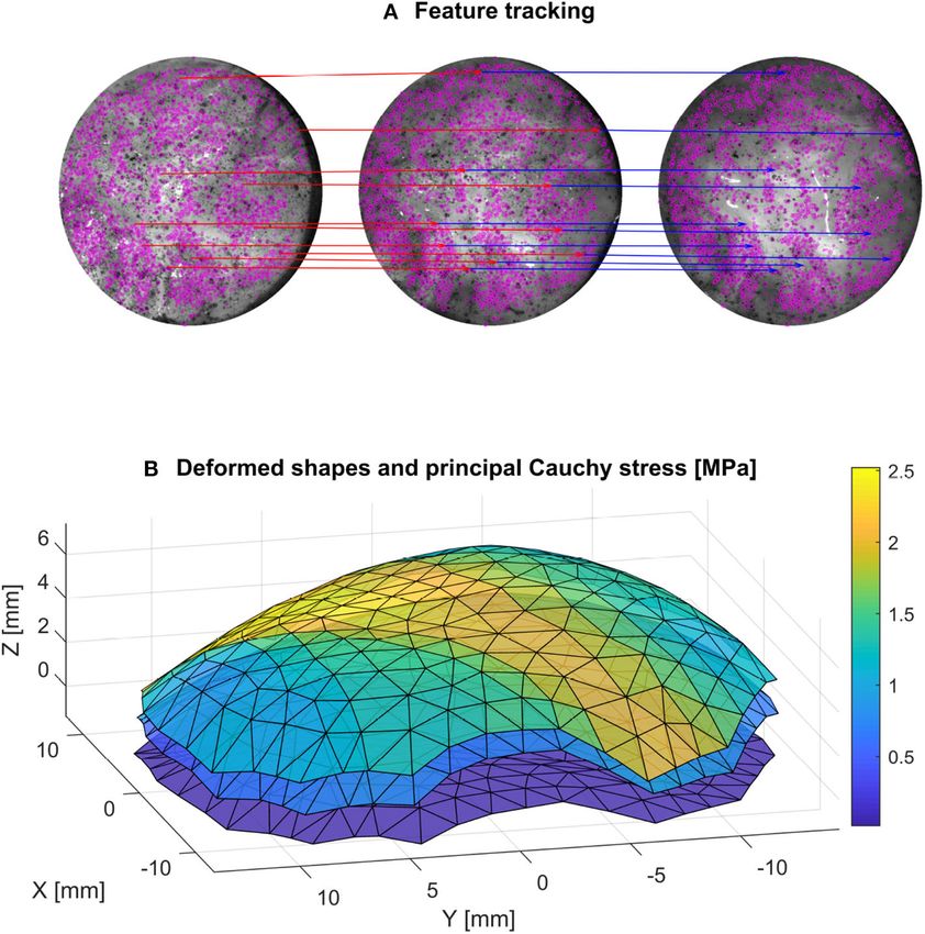
Pierrat et al.                                                                                              Mechanical Characterization of Porcine Meninges

1. INTRODUCTION                                                             are large variations in results which could be attributed to
                                                                            the nature of the testing protocol, the source of the meninges
Traumatic brain injury (TBI), caused by a fierce acceleration or            samples, storage of samples and insufficient sample sizes. In
impact to the head, is a critical public health problem throughout          addition, the main test method found in previous investigations
the world (Peeters et al., 2015; MacManus et al., 2017a). In                is either uniaxial or biaxial tensile testing. There is only one other
the U.S. alone, TBI accounts for 1.7 million injuries, 275,000              study which uses bulge inflation testing (Chauvet et al., 2010),
hospitalizations and 50,000 deaths annually (Faul et al., 2010). Its        a common experimental method used for similar membrane
primary causes are road traffic accidents and falls (Peeters et al.,        materials, however the purpose of that study was to compare the
2015), while contact sports athletes are particularly susceptible           efficacy of different surgical suturing methods. Inflation testing
to mild TBI (Gardner and Yaffe, 2015). Since the 2000s, both                offers a simple multiaxial test method for membrane materials
multibody dynamics models and finite element (FE) head models               (Avril and Evans, 2017), simply relating pressure to stresses and
have been developed by researchers to better understand the                 deflection to strains. In addition to uniaxial tension, this method
response of the human head to dynamic loads (Gilchrist et al.,              has been chosen because it more closely replicates the in vivo
2001; Horgan and Gilchrist, 2003; King et al., 2006; Kleiven,               loading condition of the meninges under intracranial pressure.
2006; Chafi et al., 2009; Li et al., 2011). These models have since         Intracranial pressure (ICP) is the pressure of the cerebrospinal
provided insights into the fundamental mechanisms of TBI in                 fluid and is an important variable in many medical conditions
road traffic accidents (Kleiven, 2007), accidental falls (Doorly and        including stroke hydrocephalus or brain tumors (Syed et al.,
Gilchrist, 2006; Raul et al., 2006; Pascoletti et al., 2019), impact        2016).
sports (Post et al., 2013), and ballistics impacts (Li et al., 2016).           In the present study, we address the dearth of primary
The accuracy of such models relies heavily on the accuracy of               experimental data on the cerebral meninges, and provide new
the constitutive data which underpins the tissue behavior. While            constitutive data for inclusion in updated FE head models. This
there is ongoing interest in the mechanical characterization                is achieved through uniaxial tension and inflation testing of
of brain tissue (Rashid et al., 2013), cranial bone (Motherway              fresh adolescent porcine DAM samples and through a structural
et al., 2013), and scalp tissue (Trotta et al., 2018a,b; Trotta and         analysis using SALS. We also seek to highlight the importance
Ni Annaidh, 2019), the cerebral meninges and its importance in              of the meninges in the mechanical response of FE head models
head impact biomechanics has been largely overlooked in recent              under complex loading, e.g., craniotomy surgery following
years. The dura mater (DM), the outermost and most substantial              elevated ICP.
of the tri-layer meninges, is one of the stiffest membranes in
the human body (Van Noort et al., 1981a) and previous works
strongly suggest that the DM helps protect the brain against                2. MATERIALS AND METHODS
damage (Van Noort et al., 1981a). A recent study by MacManus
                                                                            2.1. Sample Preparation
et al. (2017a) has shown that it can mitigate the dynamic response
                                                                            Post-mortem porcine specimens were collected from a local
of the brain by reducing the shear stress and the peak intracranial
                                                                            slaughterhouse and transported to the laboratory. The specimens
pressure, when the scalp and skull is removed. The DM lines the
                                                                            consisted of six 22 week old (tensile tests) and five 23 week
inner surface of the skull and its main functions are to collect
                                                                            old (inflation tests) mixed sex pig heads. Previous studies
venous blood and to protect and support the brain and the spinal
                                                                            have indicated that porcine tissue is a suitable model for both
cord (Hamann et al., 1998). Researchers agree that the lumbar
                                                                            human brain and human meninges tissue (Frink et al., 2011;
DM exhibits significant anisotropy (Runza et al., 1999), with the
                                                                            Mazgajczyk et al., 2012). To gain access to the cranial meninges
stiffness differing by up to 500%, however, the anisotropy of the
                                                                            and underlying brain tissue the scalp was removed from the
cerebral DM remains unclear. Van Noort et al. (1981b), using
                                                                            specimens using a scalpel, exposing the cranial bone. Following
polarized light and Hamann et al. (1998), using small angle light
                                                                            the removal of the scalp, the cranial bone was excised using an
scattering (SALS) to analyse the collagen fiber architecture of
                                                                            oscillating saw. Incisions using the oscillating saw were made in
human cerebral DM concluded that while there are regions of
                                                                            a pentagonal shape around the top, sides, and back of the skull,
highly aligned collagen fibers, this alignment is only seen over
                                                                            as depicted in Figure 1a (dashed black line). Where possible, the
short distances and the global structure of the cerebral DM can
                                                                            incisions were made outside of the cranial cavity housing the
be considered as isotropic.
                                                                            brain to ensure the meninges and brain tissue were not damaged.
    Currently, not all FE head models include a description of the
                                                                            Following removal of the skull and exposure of the meninges,
meninges: of those that do (Horgan and Gilchrist, 2003; King
                                                                            the dura mater and arachnoid mater layers (DAM)1 of the
et al., 2006; Kleiven, 2006; Chafi et al., 2009; Li et al., 2011),
                                                                            meninges were removed using surgical scissors by incising along
they often include a simplified linear response based on uniaxial
                                                                            the yellow dashed line (shown in Figure 1b). The underlying
tests performed by Galford and McElhaney (1970). There is a
                                                                            tissue, e.g., cerebral cortex and pia mater, were left within the
relative dearth of primary data on the mechanical properties of
                                                                            cranial cavity. The cerebral DAM was then tested immediately
the meninges, and with the exception of Walsh et al. (2018),
De Kegal et al. (2017) and MacManus et al. (2017a), all other               1 Following the convention used in MacManus et al. (2017a), the current samples
studies have been conducted over 30 years ago (Galford and                  are referred to as dura-arachnoid mater (DAM), accurately reflecting the samples’
McElhaney, 1970; Van Noort et al., 1981a; McGarvey et al.,                  composition. When referring to previous works, they are described as per the
1984; Bylski et al., 1986). Of these previous investigations, there         author’s description i.e., dura mater or meninges.

Frontiers in Bioengineering and Biotechnology | www.frontiersin.org     2                                               August 2020 | Volume 8 | Article 801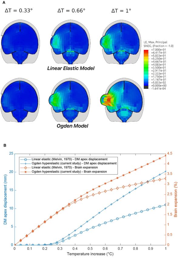
Pierrat et al.                                                                                                           Mechanical Characterization of Porcine Meninges

  FIGURE 1 | (a) Pig skull with scalp removed, skull to be removed by incising along black dashed lines. (b) Skull removed along black dashed lines, meninges to be
  removed by incising along yellow dashed lines. (c) Orientation and geometry of DM samples for uniaxial tensile tests (solid lines) and inflation tests (dotted lines)
  (MacManus et al., 2017a).

under ambient conditions (4 h after sacrifice) or was refrigerated                       The engineering (first Piola-Kirchhoff) stress in the tensile
in damp tissue for a maximum of 24 h before testing. As                                  direction, P is given as:
dehydration occurs within minutes, each sample was prepared
and tested individually, while remaining samples were kept in                                                       dwo               α
                                                                                                             P=         = µ(λα−1 − λ− 2 −1 )                              (3)
saline solution to ensure that they did not dehydrate.                                                               dλ

                                                                                         The Cauchy stress in the tensile direction, σ is derived as
2.2. Tensile Testing
Samples were cut in a dogbone shape, of overall length 63.5                                                                                   α
                                                                                                                 σ = λP = µ(λα − λ− 2 )                                   (4)
mm and width 3.2 mm. Sandpaper was glued to each end of
the sample to reduce slipping in the grips. The gauge length
                                                                                         The parameters µ and α can be related to the initial shear
and width of samples were measured optically from the camera,
                                                                                         modulus, µs , by Equation (5):
while the sample thickness was taken as an average of three
measurements along the sample using Vernier calipers. Tensile
                                                                                                                           2µs = µα                                       (5)
testing was performed on a Hounsfield universal testing machine
with a 1 kN load cell and a test speed of 50 mm/min (strain
                                                                                         Finally the Young’s modulus, E, can be related to the
rate of 0.01 s−1 ). Overall, 25 samples from six pigs were tested
                                                                                         shear modulus with the following equation for an isotropic,
until failure. The majority of samples tested (N = 17) were
                                                                                         incompressible material:
in the sagittal (longitudinal) orientation, however to investigate
the possible anisotropy of the DAM, a number of samples (N
                                                                                                                            E = 3µs                                       (6)
= 8) in the coronal (transverse) orientation were also tested.
Three DAMs were tested so that both longitudinal (N = 6)                                 Similarly, the incompressible deviatoric strain energy density
and transverse (N = 8) samples from the same DAM could be                                function for the NeoHookean model, wnh , is given by
compared (as shown in Figure 1c), thereby eliminating inter-                             Equation (7).
subject variability as a factor.
                                                                                                                wnh = C1 (λ2 + 2λ−1 − 3)                                  (7)
2.2.1. Ogden Hyperelastic Model
The incompressible deviatoric strain energy density function for                         where C1 is the NeoHookean material parameter given by:
the Ogden model, wo , is given by Equation (1).
                                                                                                                                    µs
                                                                                                                           C1 =                                           (8)
                              µ                                                                                                     2
                          wo = (λα1 + λα2 + λα3 )                              (1)
                              α                                                          Best fit Ogden and NeoHookean model parameters for each
                                                                                         experimental stress-strain curve were obtained using the non-
where µ is the first Ogden parameter and α is the second Ogden                           linear least squares function, lsqnonlin, in MATLAB.
parameter (Ogden, 1997). For an incompressible material in
uniaxial tension, λ1 =λ and λ2 =λ3 = √1 . Therefore, wo becomes:
                                               λ
                                                                                         2.3. Bulge Inflation Testing
                                                                                         A customized device was used to inflate the DAM and observe
                             µ          α                                                its deformation using stereo digital image correlation. The major
                         wo = (λα + 2λ− 2 − 3)                                 (2)       components of the device include a motorized syringe pump
                             α

Frontiers in Bioengineering and Biotechnology | www.frontiersin.org                  3                                                August 2020 | Volume 8 | Article 801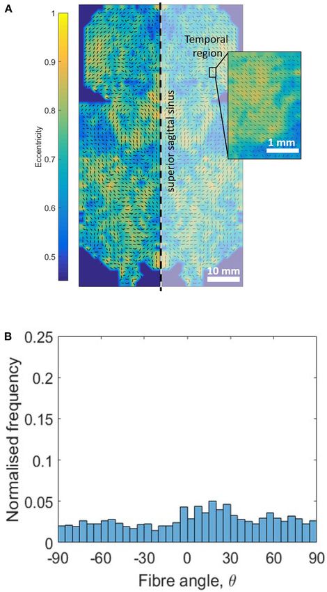
Pierrat et al.                                                                                                    Mechanical Characterization of Porcine Meninges

  FIGURE 2 | Inflation setup with stereo acquisition system. Main components
  include a motorized syringe pump to provide pressurized water to the
  specimen, which is clamped on its edges to seal the chamber; a pressure
  transducer to monitor the pressure and; two cameras (GOM, 5M LT) with a
  lighting system to acquire images for stereo vision.

                                                                                    FIGURE 3 | Image of thickness measurement device. Main components
                                                                                    include two blue laser triangulation sensors which generate two distance
containing water, a pressure transducer and a 30 mm diameter                        maps to characterize the local thickness.
circular specimen holder (Figure 2). A stereo-camera bench was
also used to capture a pair of images every second which was
synchronized with the pressure transducer.
    Five DAMs were cut into circular samples of ∼40 mm                             (∼400,000 points for the area of interest) of the surface of the
diameter. All tests were performed until rupture or irreversible                   specimen in the initial, undeformed configuration. In order to
leakage. A constant volume rate was used to control the pump so                    follow the deformation of the surface in the following states, the
that the pressure increased at a mean rate of 2.9 kPa/s.                           shape is interpolated on a coarser triangular mesh, as shown in
    As bulge inflation tests are known to be particularly sensitive                Figure 4. A quadratic LOWESS interpolation robust to outliers
to specimen thickness (Rivlin and Saunders, 1951), great care was                  was used for this purpose.
taken in its measurement. Prior to testing, the mean thickness
of each DAM specimen was measured using a custom-made                              2.3.2. Deformed Shape Measurement
device (Figure 3) based on two blue laser triangulation sensors                    A fundamental experimental challenge noticed during the
(Cavinato et al., 2019). Each laser is located on either side of the               inflation tests was the slight porosity of the DAM. This lead to
membrane where they both perform a scan of their respective                        water droplets slowly leaking out of the surface and removing the
surface to obtain two distance maps. The differential distance                     speckle pattern in some locations, as seen in Figure 5. Instead
map then characterizes the local thickness, which is then averaged                 of using a waterproof liner, which would have affected the
for the whole area of interest on the sample. Finally, a fine speckle              global mechanical response of the sample, it was decided to
pattern was sprayed on the samples with a graphite powder paint.                   base the processing of the deformed images on the remaining
During sample preparation and testing, great care was taken to                     pattern that was not altered during the test. For this purpose,
keep the specimen hydrated.                                                        instead of a dense DIC technique, a sparse feature-tracking
                                                                                   algorithm was used to track the speckle points that remained
2.3.1. Initial Shape Measurement                                                   intact during the entire test. Such techniques have worse accuracy
The intrinsic and extrinsic camera parameters were determined                      than DIC in terms of sub-pixel displacement, but are more
using a chessboard pattern with the Stereocalibration tool in                      robust to large displacements and by nature only track areas
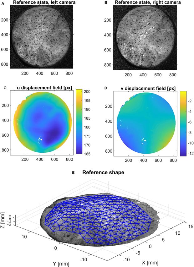
Matlab. Based on this calibration, it is possible to triangulate the               with high information. In this case, the TrackMate plugin
3D position of a point based on its pixel coordinates in the images                (Tinevez et al., 2017) in imageJ was used to detect the features
from both cameras. In order to find the point correspondences                      using a difference of gradient algorithm, and then track the
between the two images at the reference state (horizontal and                      displacement of the speckle features on the left and right cameras
vertical displacement in pixels), an open-source digital image                     (Figure 5). Only the features that were tracked for the whole
correlation software [NCorr (Blaber et al., 2015)] was used, as                    set of images were retained. Obvious outliers were removed
shown in Figure 4. This step yields a dense 3D point cloud                         based on the mean velocity of the tracked features. The nodal

Frontiers in Bioengineering and Biotechnology | www.frontiersin.org            4                                               August 2020 | Volume 8 | Article 801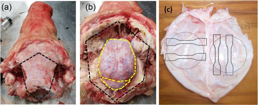
Pierrat et al.                                                                                                           Mechanical Characterization of Porcine Meninges

  FIGURE 4 | Method used to acquire the initial configuration using stereo-DIC: using the reference configuration [left (A) and right (B) images], the horizontal (C) and
  vertical (D) displacement fields between these pictures are calculated using 2D DIC. Then, triangulation provides the 3D surface of the object (E), on which an area of
  interest is meshed for further processing.

Frontiers in Bioengineering and Biotechnology | www.frontiersin.org                 5                                                 August 2020 | Volume 8 | Article 801
Pierrat et al.                                                                                                                Mechanical Characterization of Porcine Meninges

  FIGURE 5 | A feature tracking algorithm (A) is used (the tracking of 10 points for an initial, intermediate and final configuration is shown for better readability) to get the
  deformed configuration of the mesh at different stages (B). The calculated principal Cauchy stress is also represented on the mesh for these three stages.

displacement of the mesh was obtained by interpolation of the                               The strain in the third direction, i.e., the thickness variation, was
sparse displacement field of the tracked features using again a                             computed based on initial and current thickness as described in
quadratic LOWESS interpolation.                                                             the following section. These quantities were calculated for each
                                                                                            element at every frame.
2.3.3. Local Strain Measurement
The local strain is calculated based on the mesh configuration
at each frame, using the same method employed by Volino                                     2.3.4. Local Stress Measurement
et al. (2009). Briefly, the in-plane deformation gradient of each                           Under a pure membrane assumption, it is possible to calculate
triangular element is computed directly from the nodal positions.                           the local Cauchy stress at each element knowing the pressure and
Two initially perpendicular unit in-plane vectors U and V are                               the membrane shape. This procedure is described in Romo et al.
defined in the initial configuration for each element, such as U is                         (2014), Cavinato et al. (2019), and summarized below.
the projection of the medio-lateral direction on the element and                                The mesh is composed of nelements elements and nnodes nodes.
V is the complementary vector representing the antero-posterior                             Based on force equilibrium at each element (the normal force
direction. These vectors are then updated based on the nodal                                due to the pressure equilibrates the local membrane forces of the
position in the deformed configuration. The in-plane element                                three nodes), it is possible to write a set of 3 × nelements equations
deformation gradient is then:                                                               relating the three in-plane stress components expressed at each
                                                                                            node (so that the total number of unknowns is 3×nnodes ). Besides,
                                    F = [U V]                                     (9)       additional equations can be added at the edges of the membrane
                                                                                            so that the edge traction vector is normal to the edge direction (no
The true (logarithmic) in-plane strain tensor is:                                           shear) and in the plane of the membrane (no moment). In these
                                       1                                                    equations, the thickness needs to be known. The initial thickness
                                E=       log(FT F)                               (10)       h0 is given by the local thickness measurements averaged over
                                       2

Frontiers in Bioengineering and Biotechnology | www.frontiersin.org                     6                                                   August 2020 | Volume 8 | Article 801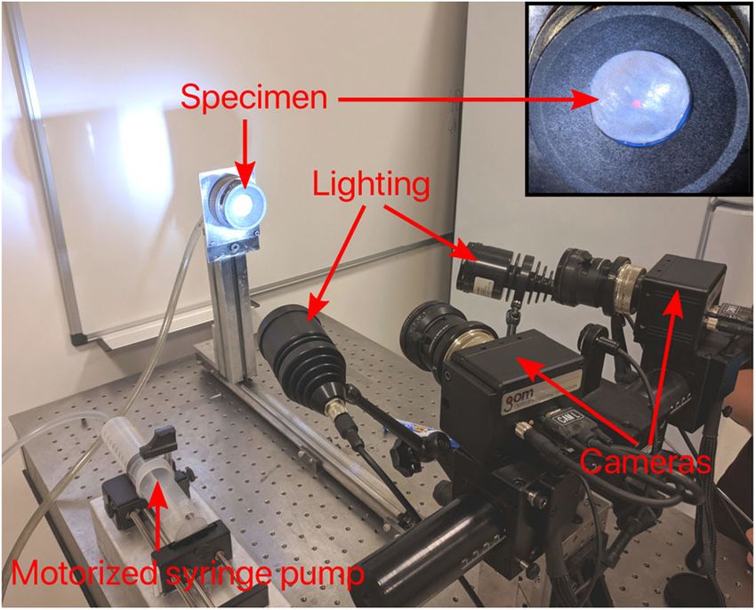
Pierrat et al.                                                                                                            Mechanical Characterization of Porcine Meninges

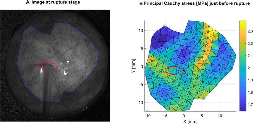
  FIGURE 6 | Local rupture stress is obtained by selecting the ruptured area (A) in red, and averaging the local principal Cauchy stress in this area (B) in the frame
  preceding rupture. Here an example is shown for sample 1R where the average value is 2.0 MPa.

the element and the current thickness h is calculated based on the                          The strain energy density w stored in the membrane can be
assumption of incompressibility:                                                         expressed as (Reddy, 2017):
                                                                                                                              Z       Eij
                                       h0                                                                               w=                  σij dEij                          (12)
                            h=                                                (11)                                                0
                               F11 F22 − F21 F12
                                                                                         The total strain energy Wexp for the membrane is then obtained
This overdetermined system of equations is solved using least-                           by integration over the whole domain:
square matrix inversion for the nodal stresses, which are then
                                                                                                                                      Z
distributed over the elements. The in-plane Cauchy stress tensor
was calculated for each element at every frame with this method,                                                       Wexp =                   w dV                          (13)
                                                                                                                                            V
as seen in Figure 5.
                                                                                         Knowing each element stress, strain and volume, this quantity
2.3.5. Local Failure Stress Measurement                                                  was calculated for each time frame based on numerical
On the first frame showing visible damage, such as water bursting                        integration and summation over the elements.
from the sample, the damaged area was manually selected                                     On the other hand, assuming an incompressible Ogden strain
(Figure 6). Then, the local principal Cauchy stress of the previous                      energy function, this quantity can also be expressed as:
frame just before failure was averaged in this area to characterize
                                                                                                                 Z
the local failure stress. Note that this process was performed only                                                   µ α
                                                                                                        Wo =           (λ + λα2 + λ−α −α
                                                                                                                                   1 λ2 − 3)dV                                (14)
for tests showing failure in an area where shape reconstruction                                                   V   α 1
was successful and away from the clamped areas.
                                                                                         Knowing the volume and the principal stretches of each
2.3.6. Global Ogden Model Identification                                                 element (derived from the deformation gradient tensor), this
Based on the local element stress and strain response throughout                         expression has only two unknowns: µ and α. Consequently,
the inflation test, it would be possible to identify the parameters                      these parameters were identified using a non-linear optimization
of a constitutive law for each element. However, due to                                  procedure, such as:
measurement uncertainties, this mapping would be noisy and                                                                  X
not necessarily an accurate representation of the local variation                                       α, µ = argmin                   i
                                                                                                                                      kWexp − Woi (µ, α)k2                    (15)
of properties. Besides, the problem of averaging the identified                                                               i
parameters to get a representative set of properties is not trivial.
Because we are more interested in a set of properties representing                       where superscript i represents a data frame.
the global response of the tissue, a global identification technique                        Numerically, this non-linear optimization was performed
based on the total strain energy is proposed.                                            using the fitnlm function in Matlab.

Frontiers in Bioengineering and Biotechnology | www.frontiersin.org                  7                                                          August 2020 | Volume 8 | Article 801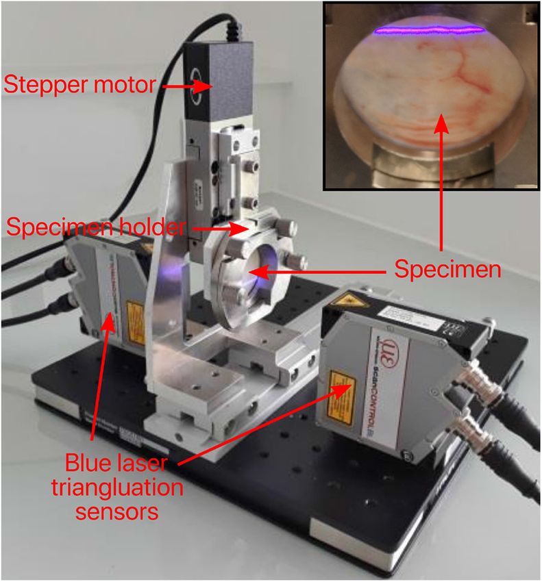
Pierrat et al.                                                                                                Mechanical Characterization of Porcine Meninges

   Similarly, an incompressible NeoHookean strain energy                            Abaqus 6.14 software, the UCDBTM (Horgan and Gilchrist,
function was also used to identify the C1 parameter:                                2003, 2004) (an existing finite element head trauma model
                         Z                                                          developed at University College Dublin) was used to simulate
                                                                                    a simplified craniotomy procedure. The mechanical properties
                 Wnh =         C1 (λ21 + λ22 + λ−2 −2
                                                1 λ2 − 3)dV             (16)
                           V                                                        of the biological tissues are given in Table 1. A section of skull
                                                                                    measuring ∼46 × 48 mm was removed from the UCDBTM
2.4. SALS                                                                           as shown in Figure 7. Here, brain swelling, as experienced
Small angle light scattering (SALS) was performed on a single                       following TBI, was simulated using a thermal expansion method,
sample to determine whether a preferred orientation of collagen                     where the coefficient of thermal expansion was 0.02◦ C−1 with
fibers exists in the DAM. Briefly, the SALS system consists of an                   an applied temperature increase of 1◦ C. In an unconstrained
unpolarized 5 mW HeNe laser (λ = 632.8 nm; JDSU, Newbury,                           environment, this will generate thermal strains ε=0.02, which
UK), focusing lens (fl = 150 mm; Edmund Optics Ltd, York, UK),                      corresponds to a volumetric brain swelling of 6%. When a
automated sample positioner, projection screen and a CMOS                           structure is not free to expand, such as the brain in the skull
USB camera mounted on a linear guide rail (Gaul et al., 2017).                      cavity, the change in temperature will cause stress. In this
The device passes a 150 µm diameter laser beam through the                          case, the true volumetric expansion will be dictated by the
tissue specimen which scatters light according to the internal                      stiffness of the enclosing structures, in this case the DAM
fiber architecture. The resulting scattered light distribution is                   in the opened area. While this approach does not represent
analyzed using a custom Matlab code (MathWorks, Cambridge,                          the physiological swelling process (Lang et al., 2014), the
UK) to determine orientation and alignment information about                        thermal expansion approach has been previously adopted by
the specimen as detailed elsewhere (Gaul et al., 2017). Due to                      (Weickenmeier et al., 2017) for the simulation of brain swelling
sample size limitations imposed by the SALS system, the sample                      in craniotomies. The response of the UCDBTM using both
was cut into 6 rectangular sections of 25 × 10 mm and each                          the original meninges material properties, as described in
section was analyzed individually. Vectors were spaced every 1                      (Horgan and Gilchrist, 2004), and the present material properties
mm with each vector corresponding to a region of 150 µm2 .                          evaluated in inflation tests (detailed in Table 2), are contrasted
                                                                                    with respect to the maximum principle strain in the brain and
2.5. Craniotomy Simulation                                                          the maximum protrusion of the brain outside the bone flap. As
Following the mechanical evaluation of the DAM and the                              high deformations were expected, geometric non-linearities were
determination of an appropriate strain energy density function                      accounted for (NLGEOM option in Abaqus), so that the only
(Ogden model), finite element simulations were undertaken                           difference between the two simulations was the material model of
to highlight the potential applications of this study. Using                        the DAM.

TABLE 1 | Mechanical properties of UCDBTM (Horgan and Gilchrist, 2003, 2004).

Tissue                                             Model                  Density              Poisson’s          Model parameters              # Elements
                                                                          [kg/m3]                ratio

Cerebellum                                         Viscoelastic            1,060                                     G0 = 10 kPa                     780
                                                                                                                     G∞ = 2 kPa
                                                                                                                Decay constant = 80−s
                                                                                                                    K = 2.19 GPa
Gray matter                                        Viscoelastic            1,060                                     G0 = 10 kPa                    5,004
                                                                                                                     G∞ = 2 kPa
                                                                                                                Decay constant = 80−s
                                                                                                                    K = 2.19 GPa
Brain stem                                         Viscoelastic            1,060                                    G0 = 22.5 kPa                    220
                                                                                                                    G∞ = 4.5 kPa
                                                                                                                Decay constant = 80−s
                                                                                                                    K = 2.19 GPa
White Mater                                        Viscoelastic            1,060                 0.499              G0 =12.5 kPa                    1,105
                                                                                                                    G∞ = 2.5 kPa
                                                                                                                Decay constant = 80−s
                                                                                                                    K = 2.19 GPa
Cortical bone                                      Linear elastic          2,000                 0.22                 E=15 GPa                      4,160
Trabecular bone                                    Linear elastic          1,300                 0.24                 E = 1 GPa                     4,096
Pia mater                                          Linear elastic          1,130                 0.45                E = 11.5 MPa                   2,786
CSF                                                Linear elastic          1,000                 0.499               E = 0.15 MPa                   2,874
Facial bone                                        Linear elastic          2,100                 0.23                E = 5.5 GPa                     406
DAM (Horgan and Gilchrist, 2003, 2004)             Linear elastic          1,130                 0.45                E = 31.5 MPa                   2,155
DAM (current)                                      Ogden (n = 1)            1130                 0.49           µ = 0.23 MPa, α = 8.2               2,155

Current mechanical properties of the DM are provided in boldface.

Frontiers in Bioengineering and Biotechnology | www.frontiersin.org             8                                         August 2020 | Volume 8 | Article 801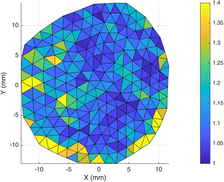
Pierrat et al.                                                                                                                 Mechanical Characterization of Porcine Meninges

3. RESULTS                                                                                 curve was fit to both the Ogden and the NeoHookean
                                                                                           material models using the lsqnonlin function in MATLAB.
3.1. Tensile Testing                                                                       It can be seen from Figure 8 that the Ogden model captures the
The measured thickness of the DAM using Venier calipers                                    non-linear response of the DAM well, with an average R2 =0.98,
was 400 ± 100 µm. For each uniaxial tensile test performed,                                while the NeoHookean model fails to capture the toe region of the
a stress-strain curve was calculated. From these curves, the                               response with an average R2 =0.82. The average Ogden material
average failure strain was found to be 22 ± 4%, and the                                    parameters for all samples tested was µ = 450 ± 190 kPa and
average ultimate tensile strength was 6.91 ± 2.00 MPa. Each                                α = 16.55 ± 3.16. As per Equations (5) and (6) this results in a
                                                                                           shear modulus, µs of 3.55 ± 1.46 MPa and a Young’s modulus,
                                                                                           E, of 10.64 ± 4.39 MPa. The average NeoHookean material
                                                                                           parameter for all samples tested was C1 = 5.87 ± 2.90 MPa,
                                                                                           which corresponds to a shear modulus, µs of 11.74 ± 5.79 MPa.

    FIGURE 7 | UCDBTM (Horgan and Gilchrist, 2003, 2004) with bone flap
    removed to simulate craniotomy with skull section removed to simulate a
    craniotomy procedure. Various colored elements represent different biological
    tissues (white = bone, gray = gray matter, orange = white matter, green =               FIGURE 8 | Average and range of stress-strain relationships of DM for uniaxial
    ventricles, yellow = cerebellum, blue = brain stem, red = membranes).                   tension tests and representative best fit Ogden model.

TABLE 2 | DAM properties identified with the inflation test for the 10 samples (two for each pig, left side and right side).

                                                                                           Ogden                                    Neo-Hookean

Pig              Side          Thickness          Burst pressure               µ              α              R2                C1              R2             Rupture stress
                                    [µm]               [kPa]                 [kPa]           [-]             [-]           [kPa]               [-]                 [MPa]

1                 L                 360                 193                  11.9           13.8           0.983               345            0.718                   *
                  R                 359                 206                   481           5.39           0.997               905            0.972                 2.00

2                 L                 377                 127                   177           8.61           0.988               598            0.942                   *
                  R                 381                 183                   497           4.50           0.987               654            0.970                 2.52

3                 L                 361                  †                     †              †              †                  †               †                     †
                  R                 390                 141                   273           5.64           0.997               490            0.973                   *

4                 L                 400                 150                   136           10.3           0.990               591            0.943                 1.72
                  R                 327                  †                     †              †              †                  †               †                     †

5                 L                 405                 150                  59.6           9.12           0.992               295            0.869                   *
                  R                 394                  †                     †              †              †                  †               †                     †

Mean                                375                 164                   234           8.19           0.991               554            0.912                 2.08
St. Dev.                            24                   30                   193           3.29           0.005               205            0.093                 0.41

*Rupture not in area of interest.
†
  Experiment failed.

Frontiers in Bioengineering and Biotechnology | www.frontiersin.org                    9                                                   August 2020 | Volume 8 | Article 801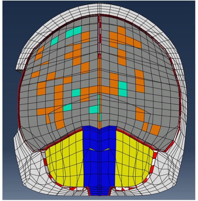
Pierrat et al.                                                                                                          Mechanical Characterization of Porcine Meninges

Figure 8 shows the average stress-strain relationship and the                            in Figure 9. This was a trend observed for all three DAMs tested
entire range of the samples tested in uniaxial tension. Also shown                       in this manner. However, a two tail T-test revealed that the
is a representative fit of the Ogden model and the NeoHookean                            Ogden model parameters for samples in the longitudinal and
model to the average stress-strain curve.                                                transverse direction were not statistically different (p = 0.13
    Analysing average stress strain curves in both the longitudinal                      for µ, p = 0.87 for α). This indicates that while there is some
(N = 17) and the transverse (N = 8) orientations, no clear                               anisotropy evident, this is not statistically significant within the
difference was evident (see Figure 9). However, when inter-                              current results and the global material behavior of the cerebral
subject variability was removed by comparing samples only                                DAM can be considered isotropic. This will be discussed further
from the same DAM, it was observed that longitudinal samples                             in section 3.3.
appeared stiffer than transverse samples also shown, for example,

                                                                                         3.2. Inflation Testing
                                                                                         The results of the inflation testing are presented in Table 2.
                                                                                         An example of the sDIC reconstruction is shown in Figure 5,
                                                                                         and an example of a successful burst is shown in Figure 6.
                                                                                         The mean measured thickness for all specimens using the laser
                                                                                         triangulation method was 375 ± 24 µm. From the 10 samples,
                                                                                         sDIC reconstruction failed for 3 due to the speckle pattern being
                                                                                         affected by water droplets. The mean constitutive parameters
                                                                                         identified for the Ogden model were µ=234 ± 193 kPa and
                                                                                         α=8.19 ± 3.29. The Ogden model fit the experimental data well
                                                                                         with a mean coefficient of determination of R2 =0.99, as can
                                                                                         be seen in Figure 10. Also, the mean confidence intervals were
                                                                                         ±20 kPa for µ and ±0.38 for α with confidence level of 95%
                                                                                         as returned by the non-linear optimization. This shows that
                                                                                         our identification procedure is sensitive to both parameters of
                                                                                         the model. The mean identified parameter for the NeoHookean
  FIGURE 9 | Directional effects of the DM. The average response of samples              model was C1 =554 ± 205 kPa. However, as this model has a more
  (shown as solid lines) in orthogonal directions is not statistically different,        linear response, it fit the data relatively poorly with a mean R2
  however, longitudinal samples appear somewhat stiffer, especially when                 of 0.91. Rupture data was successfully obtained in the area of
  inter-subject variability is eliminated, e.g., as shown by dashed lines
  representing the longitudinal and transverse response from a single DM.
                                                                                         interest for three specimens only. The mean value of rupture
                                                                                         stress was 2.08 ± 0.41 MPa.

  FIGURE 10 | Total strain energy vs. pressure for all DM samples of the inflation tests. Experimental values are shown as dots and the fit with the Ogden strain energy
  function in dashed lines.

Frontiers in Bioengineering and Biotechnology | www.frontiersin.org                 10                                               August 2020 | Volume 8 | Article 801
Pierrat et al. Mechanical Characterization of Porcine Meninges
3.3. Orientation
SALS contour plots were generated for one half of the DAM
and the results were mirrored along the superior sagittal sinus
for illustration (Figure 11A). The fiber alignment is defined by
eccentricity (values of 0–1) where areas of high eccentricity
(> 0.7) have fibers highly aligned with the vector directions
indicated and areas with low eccentricity (< 0.7) have a low
degree of fiber alignment, or highly dispersed fibers. Results
indicate that there are local regions of high alignment (as
shown in yellow and inset), however there is no global preferred
orientation, as seen in Figure 11B. The lack of a dominant angle
in Figure 11B indicates that the DAM is globally isotropic. This
result is also corroborated by the statistical analysis presented
in section 3.1, which found no statistical difference between the
Ogden model parameters for samples in the longitudinal and
transverse direction.
3.4. Craniotomy Simulation
It can be seen in Figure 12 that the linear elastic and Ogden
material models predict two significantly different responses,
with the stiffer linear elastic model predicting lower Maximum
Principal Strains and a less pronounced protrusion of the
brain following the craniotomy, caused by a 26% lower brain
expansion. Stress fields were also highly affected by the DAM
properties: brain (intracranial) pressure ranged from 13–15 to
46–50 kPa for the linear elasic and Ogden models, respectively.
This emphasizes the fact that the material properties of the DAM
have a significant impact on the overall response of the brain.
4. DISCUSSION
Comparing the main results under uniaxial testing of the current
study to previous authors (see Table 3), we find that our failure
strain of 22 ± 4% falls between the values reported by Van Noort
et al. (1981a) and McGarvey et al. (1984) of 18.72 and 31.7%,
respectively and that our Ultimate Tensile Strength of 6.91 ±
2.00 MPa also falls between the values reported by Van Noort
et al. (1981a), McGarvey et al. (1984) of 4.7 and 9.41 MPa,
respectively. The determined average material parameters were
µ = 450 ± 190 kPa and α = 16.55 ± 3.16 for the Ogden FIGURE 11 | Evaluation of DAM microstructure. (A) Global and local (inset)
model and C1 = 5.87 ± 2.90 MPa for the NeoHookean model. SALS contour plots of fiber orientation and alignment, where eccentricity
values (0–1) represent the level of fiber alignment. (B) Orientation distribution
Based on the calculated average coefficient of determination plot of global DM fiber directions. Only the left half of the DM was analyzed, the
(R2 =0.98 for Ogden model and R2 =0.82 for NeoHookean), it right half was generated through mirror symmetry.
has been determined that the Ogden model offers a better fit to
experimental data. Two previous authors, Maikos et al. (2008)
and De Kegal et al. (2017), have also suggested that the Ogden
model offers the best fit to experimental data and that it captures Inflation results, however, differ somewhat from the results
the non-linear region of the stress-strain response well. Again, of the uniaxial testing, highlighting that the identification of
the reported material parameters in the current study lie between constitutive parameters is dependent on the test method. Similar
the values reported by Maikos et al. (2008) (µ = 420 kPa) and observations have been made by Lari et al. (2012) for sclera,
De Kegal et al. (2017) (µ = 660 kPa). Given the close comparison where they compared the response of uniaxial tension and
between these results, porcine DAM can be considered a good inflation experiments suggesting that testing conditions in the
model for human DAM as previously suggested by Frink et al. uniaxial tests can induce artifacts due to sensitivity to tissue
(2011) and Mazgajczyk et al. (2012). gripping techniques, sample cutting, and Saint-Venant related
The results of the bulge inflation testing confirmed that issues. Regarding the current experiments, the identified Ogden
the Ogden model is a suitable choice of constitutive model material parameters were significantly less for bulge inflation
since the mean coefficient of determination here was R2 =0.99. testing than unaxial tensile testing (52% less for µ and 49% less
Frontiers in Bioengineering and Biotechnology | www.frontiersin.org 11 August 2020 | Volume 8 | Article 801Pierrat et al. Mechanical Characterization of Porcine Meninges FIGURE 12 | Comparison of craniotomy simulation using the Linear Elastic Model (Horgan and Gilchrist, 2004) and Ogden Model (present study) induced by brain swelling simulated by thermal expansion [1–2% swelling corresponds to typical volumetric expansion seen at high altitudes (Morocz et al., 2001)]. (A) Maximum Principal Strain contour plots. (B) DM apex displacement and brain expansion vs. temperature increase. for α). Similarly, bulge inflation testing revealed a rupture stress no other studies have previously determined the rupture strength of 2.08 ± 0.41 MPa, significantly below the Ultimate Tensile of the DAM through bulge inflation testing. Interestingly, it can Strength of 6.91 ± 2.00 MPa. While rupture data is based only be seen in Figure 6 that tissue failure is not located in the same on the analysis of three specimens, it is included here given that region as peak stress, which was observed for all three specimens. Frontiers in Bioengineering and Biotechnology | www.frontiersin.org 12 August 2020 | Volume 8 | Article 801
Pierrat et al.                                                                                                               Mechanical Characterization of Porcine Meninges

TABLE 3 | Summary of results found in previous tensile investigations.

                                                                        Summary of tensile testing

References                                  Species                    Thickness                     Model parameters                               Descriptive values

Galford and McElhaney (1970)                Human                          –                  MK: Es = 29.65 MPa, El = 3.38                                    –
                                                                                                          MPa
Van Noort et al. (1981a)                    Human                          –                                   –                                UTS = 4.7 MPa, FS = 18.4%
McGarvey et al. (1984)                      Human                          –                                   –                               UTS = 9.41MPa ± 1.54, FS =
                                                                                                                                                      31.7% ± 1.6
Bylski et al. (1986)                        Fetal human                0.57 mm                  MR: C = 1.18 MPa, α = 0.25                                     –
Maikos et al. (2008)                        Rat                         0.8 mm                OG: µ = 0.42 MPa ± 0.19, β =                     UTS = 2.49 MPa ± 2.03, FS =
                                                                                                      32.9 ± 6.65                                       39% ± 13
De Kegal et al. (2017)                      Human                      1.06 mm                OG: µ = 0.66 MPa ± 0.38, β =                                     –
                                                                        ± 0.22                       −1.09 ± 3.19
MacManus et al. (2017a)                     Porcine                        –                   NH and QLV; µ = 0.019 MPa ±                                     –
                                                                                                         0.0085
Present study                               Porcine                     0.4 mm                OG: µ = 0.45 MPa ± 0.19, α =                     UTS = 6.91 MPa ± 2.00, FS =
                                                                         ± 0.1                        16.55 ± 3.16                                      22% ± 4

MK, Maxwell-Kelvin; MR, Mooney Rivlin; OG, Ogden; NH, NeoHookean; QLV, Quasi Linear Viscoleasticity; Es , Storage Modulus; El , Loss Modulus; UTS, Ultimate Tensile Strength; FS,
Failure Strain.

Although surprising, this observation has also been reported for                            et al. (2017) found no evidence of hysteresis and therefore
arterial tissue (Cavinato et al., 2019), suggesting that rupture                            concluded that the DM could be considered purely elastic at
mechanisms may be better explained by an understanding of the                               lower strain rates. Conversely, other authors have observed
microstructure and composition heterogeneities in the tissue. As                            substantial viscoelastic behavior and modeled the meninges as
DAM is a highly fibrous medium, local rearrangement of the                                  either quasi-linear viscoelastic or linearly viscoelastic (Galford
microstructure depending on the loading conditions may also                                 and McElhaney, 1970; MacManus et al., 2017a). The present
explain the discrepancy between uniaxial and biaxial (inflation)                            study, however, did not consider the effects of viscoelasticity and
identified parameters. It should be noted that the test specimens                           therefore care should be taken when comparing these results
came from 22 week - 23 week old pigs. It is well-known that the                             to testing carried out at dynamic rates (Maikos et al., 2008;
material properties of soft tissues are affected by age (Thibault                           MacManus et al., 2017a).
and Margulies, 1998; MacManus et al., 2017b) and the results                                    Previous authors have all concluded that the behavior of the
presented here may not be generalizable across all ages.                                    DM is globally isotropic (Van Noort et al., 1981a; Hamann et al.,
   The average thickness of samples in the current study                                    1998; De Kegal et al., 2017). This statement however, is usually
measured by Vernier calipers was 0.4 ± 0.1 mm and for those                                 qualified with a suggestion that there is local anisotropy and
determined through the laser triangulation method, this was 375                             regions of aligned collagen fibers. For this reason, tensile testing
± 24 µm, indicating broad agreement between the two methods.                                and SALS was conducted with a view to determining whether the
The mean figure is lower than that of De Kegal et al. (2017)                                meninges really should be considered as an isotropic material.
and Galford and McElhaney (1970) who reported values of 1                                   SALS orientation analysis shows global isotropy in the DAM,
mm for human DM. Bylski et al. (1986) however reported an                                   highlighted by the lack of a dominant direction in the orientation
average thickness of 0.57 mm for fetal human DM and suggested                               distribution plot shown in Figure 11. There are, however, regions
that the thickness of the DM increases with age. Furthermore,                               of local fiber alignment, such as the temporal region which has
Galford and McElhaney (1970) compared the thickness of human                                been reported elsewhere for human DM (Hamann et al., 1998).
and monkey DM and found that human DM was thicker, while                                    Closer analysis of our inflation tests in Figure 13 demonstrates
Maikos et al. (2008) reported a DM thickness of 0.8 mm for                                  that the ratio of the first principal stress/the second principal
rats. Given that samples used in the current study are from                                 stress is close to one and only deviates from one at the edge.
adolescent pigs, this may explain somewhat the lower thickness                              Given that the inflation tests involve an equibiaxial tension state,
values obtained when compared to older human samples. This                                  this is further evidence of the globally isotropic response of
difference may also be attributed, in part, to the different                                the DAM. Furthermore, tensile tests revealed that there was
measurement techniques employed. O’Leary et al. (2013) noted                                no statistically significant difference between samples tested in
that optical measurement techniques, such as those employed                                 the longitudinal and transversal directions. When samples from
by De Kegal et al. (2017), can overestimate the thickness of                                the same DAM only were compared (thereby eliminating inter-
soft tissues by an average of 23%, while Vernier calipers can                               subject variability), the longitudinal samples appeared to be
underestimate the thickness by an average of 8%.                                            slightly stiffer. This difference, however, was not statistically
   Tensile tests were carried out at a speed of 50 mm/min,                                  significant within the test population. To definitively determine
corresponding to a strain rate of ∼0.01 s−1 and comparable                                  whether the cerebral meninges is isotropic, a much larger sample
to the strain rate applied in De Kegal et al. (2017). De Kegal                              size would be required. However, as previous authors have

Frontiers in Bioengineering and Biotechnology | www.frontiersin.org                    13                                                 August 2020 | Volume 8 | Article 801
Pierrat et al. Mechanical Characterization of Porcine Meninges
which is under intracranial pressure. Given that the evaluated
material parameters vary depending on the test method, it is
suggested that future finite element simulations employ material
parameters evaluated through bulge inflation testing.
The purpose of simulating the craniotomy procedure was to
emphasize the important role of the meninges in predicting the
response of the human head under various loading conditions. A
further objective was to highlight that even under physiological
strains (e.g., such as those experienced by someone following
TBI), a non-linear description of the meninges is important.
Indeed, a recent publication by Li et al. (2017) has highlighted
that non-linear models of tissues should replace linear models in
head models for more realistic simulation outcomes.
Finally, while ideally tissue should be tested in its physiological
state, or as close as is possible, the tissue here has been tested
between 4 and 24 h post-mortem at ambient conditions. There
is no global agreement on the effect of prolonged storage
of brain tissue on mechanical properties, with some authors
FIGURE 13 | Stretch ratio, calculated as first principal stretch/second principal concluding there is minimal effect up until 5 days post-mortem
stretch, for a given DAM. For an isotropic material this should be 1. It can be (Budday et al., 2015), while others conclude that as short as 6
seen that the stretch ratio is close to 1 in the centre and only deviates from 1 h of storage can affect the properties (Garo et al., 2007). As
at the edge. concluded by Budday et al. (2019) in their review paper of
brain tissue, we have considered that, provided the tissue is kept
sufficiently hydrated, the degeneration process of post-mortem
concluded, and in the absence of statistically significant data to tissues is negligible. Similarly, it has been previously reported
the contrary, the cerebral meninges can be considered isotropic. that increasing the temperature from ambient conditions to
The analysis of the inflation tests were performed under the physiological conditions (37◦ C) significantly reduces the stiffness
assumption that the DAM was isotropic and incompressible. By of brain tissue (Pogoda et al., 2014). Contrary to this, a number
identifying a single set of parameters for each sample, it was of authors have also reported the opposite; that increasing the
also assumed that the thickness and material properties were temperature from ambient to 37◦ C has negligible effect (Rashid
homogeneous throughout the membrane. Hence, homogeneous et al., 2012). As stated above, best practice is to test as close as
stresses and strains were expected. From Figure 6 it can be possible to physiological conditions, and the fact that samples
seen that the stress was not always homogeneous; this may be were tested at ambient conditions may have an effect on the
due to regions of local fiber alignment, non-uniform thickness present results.
across the sample, or heterogeneities in the tissue constituents.
The thickness measurement device allows the mapping of local
thickness, and its direct use should be considered in future works. 5. CONCLUSION
On the other hand, Chauvet et al. (2010) performed an analysis
on the displacement and strain field in uniaxial tension showing It was confirmed that the global behavior of the porcine
that the displacement field appeared homogeneous along the DAM can be considered isotropic (although it does contain
sample. It was therefore concluded that the meninges could be local regions of highly aligned collagen fibers). The mechanical
considered homogeneous. The compressibility of the meninges behavior of the DAM is well-described by the non-linear Ogden
has not been extensively discussed in the literature, but given that hyperelastic model (average parameters determined through
the meninges is primarily composed of water it is common to bulge inflation testing); µ=234 ± 193 kPa and α=8.19 ± 3.29,
assume that it is incompressible. which better captures the non-linear toe region in the stress-
Bulge inflation testing coupled with sDIC and thickness strain response compared with existing linear elastic models. A
measurement was found to offer definite advantages in terms finite element simulation of a craniotomy procedure revealed
of reliability of the results, although the experimental setup that the mechanical behavior of the meninges has a large effect
and subsequent analysis is more involved. Firstly, it provides on the overall response of the brain and that existing FE head
direct access to stresses without prior knowledge of the material models should be updated to include a non-linear description of
constitutive behavior. Secondly, direct measurement of local the meninges.
displacement on the surface of the specimen avoids the need
for arbitrary assumptions at the clamped edges (e.g., no
slippage), which is often problematic to verify. Finally, it is DATA AVAILABILITY STATEMENT
important to note that bulge inflation testing, compared with
uniaxial tensile testing, offers a more biofidelic representation The datasets generated for this study are available on request to
of the in-vivo loading conditions experienced by the DAM the corresponding author.
Frontiers in Bioengineering and Biotechnology | www.frontiersin.org 14 August 2020 | Volume 8 | Article 801Pierrat et al. Mechanical Characterization of Porcine Meninges
ETHICS STATEMENT MG and CL contributed to the design of the project and
revised the work critically. LC, FM, DM, and RG completed the
Ethical review and approval was not required for the animal study experimental aspects of the work and wrote the corresponding
because only post-mortem animal tissue obtained from a local sections of the manuscript. All authors contributed to the
slaughterhouse was used for this study. The study is therefore manuscript revision.
exempt from ethical approval.
AUTHOR CONTRIBUTIONS ACKNOWLEDGMENTS
AN and BP contributed to the conception, design and oversight The authors would like to acknowledge Dawn Pork & Bacon,
of the project, and wrote the first draft of the manuscript. Waterford, for supplying the porcine specimens.
REFERENCES Gaul, R. T., Nolan, D. R., and Lally, C. (2017). Collagen fibre characterisation
in arterial tissue under load using SALS. J. Mech. Behav. Biomed. Mater. 75,
Avril, S., and Evans, S. (2017). Material Parameter Identification and Inverse 359–368. doi: 10.1016/j.jmbbm.2017.07.036
Problems in Soft Tissue Biomechanics. New York, NY: CISM International Gilchrist, M. D., O’Donoghue, D., and Horgan, T. J. (2001). A two dimensional
Centre for Mechanical Sciences. analysis of the biomechanics of frontal and occipital head impact injuries. Int.
Blaber, J., Adair, B., and Antoniou, A. (2015). Ncorr: open source 2D J. Crashworth. 6, 253–262. doi: 10.1533/cras.2001.0176
digital image correlation matlab software. Exp. Mech. 55, 1105–1122. Hamann, J., C, M., Sacks, M. S., and Malinin, T. I. (1998). Quantification of the
doi: 10.1007/s11340-015-0009-1 collagen fibre architecture of human cranial dura mater. J. Anat. 192, 99–106.
Budday, S., Nay, R., de Rooij, R., Steinmann, P., Wyrobek, T., Ovaert, doi: 10.1046/j.1469-7580.1998.19210099.x
T. C., et al. (2015). Mechanical properties of gray and white matter Horgan, T. J., and Gilchrist, M. D. (2003). The creation of three-dimensional finite
brain tissue by indentation. J. Mech. Behav. Biomed. Mater. 46, 318–330. element models for simulating head impact biomechanics. Int. J. Crashworth.
doi: 10.1016/j.jmbbm.2015.02.024 8, 353–366. doi: 10.1533/ijcr.2003.0243
Budday, S., Ovaert, T., Holzapfel, G. A., Steinmann, P., and Kuhl, E. (2019). Fifty Horgan, T. J., and Gilchrist, M. D. (2004). Influence of FE model variability in
shades of brain: a review on the mechanical testing and modeling of brain tissue. predicting brain motion and intracranial pressure changes in head impact
Archiv. Comput. Methods Eng. doi: 10.1007/s11831-019-09352-w. [Epub ahead simulations. Int. J. Crashworth. 9, 401–418. doi: 10.1533/ijcr.2004.0299
of print]. King, H., Nicholas, A., and Albert, I. (2006). Development of numerical models for
Bylski, D., Kriewall, T., Akkas, N., and Melvin, J. (1986). Mechanical-behavior of injury biomechanics research: a review of 50 years of publications in the stapp
fetal dura-mater under large deformation biaxial tension. J. Biomech. 19, 19–26. car crash conference. Stapp Car Crash J. 50, 429–490.
doi: 10.1016/0021-9290(86)90105-3 Kleiven, S. (2006). Evaluation of head injury criteria using a finite element
Cavinato, C., Molimard, J., Curt, N., Campisi, S., Orgéas, L., and Badel, P. model validated against experiments on localized brain motion, intracerebral
(2019). Does the knowledge of the local thickness of human ascending thoracic acceleration, and intracranial pressure. Int. J. Crashworth. 11, 65–79.
aneurysm walls improve their mechanical analysis? Front. Bioeng. Biotechnol. doi: 10.1533/ijcr.2005.0384
7:169. doi: 10.3389/fbioe.2019.00169 Kleiven, S. (2007). Predictors for Traumatic Brain Injuries Evaluated Through
Chafi, M., Dirisala, V., Karami, G., and Ziejewski, M. (2009). A finite element Accident Reconstructions. Reading: SAE International, 22.
method parametric study of the dynamic response of the human brain with Lang, G. E., Stewart, P. S., Vella, D., Waters, S. L., and Goriely, A. (2014). Is the
different cerebrospinal fluid constitutive properties. Proc. Inst. Mech. Eng. H J. donnan effect sufficient to explain swelling in brain tissue slices? J. R. Soc.
Eng. Med. 223, 1003–1019. doi: 10.1243/09544119JEIM631 Interface 11:20140123. doi: 10.1098/rsif.2014.0123
Chauvet, D., Carpentier, A., Allain, J.-M., Polivka, M., Crepin, J., and George, Lari, D. R., Schultz, D. S., Wang, A. S., Lee, O.-T., and Stewart, J. M. (2012). Scleral
B. (2010). Histological and biomechanical study of dura mater applied to mechanics: comparing whole globe inflation and uniaxial testing. Exp. Eye Res.
the technique of dura splitting decompression in Chiari type I malformation. 94, 128–135. doi: 10.1016/j.exer.2011.11.017
Neurosurg. Rev. 33, 287–294. doi: 10.1007/s10143-010-0261-x Li, X., Gao, X. L., and Kleiven, S. (2016). Behind helmet blunt trauma induced
De Kegal, D., amd Vastmansa, J., Fehervarya, H., Depreitere, B., Vander Sloten, by ballistic impact: a computational model. Int. J. Impact Eng. 91, 56–67.
J., and Famaey, N. (2017). Biomechanical characterization of human dura doi: 10.1016/j.ijimpeng.2015.12.010
mater. J. Mech. Behav. Biomed. Mater. 79, 122–134. doi: 10.1016/j.jmbbm.2017. Li, X., Sandler, H., and Kleiven, S. (2017). The importance of nonlinear tissue
12.023 modelling in finite element simulations of infant head impacts. Biomech. Model.
Doorly, M. C., and Gilchrist, M. D. (2006). The use of accident reconstruction Mechanobiol. 16, 823–840. doi: 10.1007/s10237-016-0855-5
for the analysis of traumatic brain injury due to head impacts arising Li, Z., Hu, J., Reed, M. P., Rupp, J. D., Hoff, C. N., Zhang, J., et al. (2011).
from falls. Comput. Methods Biomech. Biomed. Eng. 9, 371–377. Development, validation, and application of a parametric pediatric head finite
doi: 10.1080/10255840601003551 element model for impact simulations. Ann. Biomed. Eng. 12, 2984–2997.
Faul, M., Xu, L., Wald, M. M., and Coronado, V. G. (2010). Traumatic Brain Injury doi: 10.1007/s10439-011-0409-z
in the United States: Emergency Department Visits, Hospitilizations, and Deaths. MacManus, D. B., Pierrat, B., Murphy, J. G., and Gilchrist, M. D. (2017a).
Washington, DC: U.S. Department of Health and Human Services. 2002–2006. Protection of cortex by overlying meninges tissue during dynamic
Frink, M., Andruszkow, H., Zeckey, C., Krettek, C., and Hildebrand, F. (2011). indentation of the adolescent brain. Acta Biomater. 57, 384–394.
Experimental trauma models: an update. J. Biomed. Biotechnol. 2011:797383. doi: 10.1016/j.actbio.2017.05.022
doi: 10.1155/2011/797383 MacManus, D. B., Pierrat, B., Murphy, J. G., and Gilchrist, M. D. (2017b).
Galford, J. E., and McElhaney, J. H. (1970). Viscoelastic study of scalp, brain, and Region and species dependent mechanical properties of adolescent and
dura. J. Biomech. 3, 211–221. doi: 10.1016/0021-9290(70)90007-2 young adult brain tissue. Sci. Rep. 7:13729. doi: 10.1038/s41598-017-
Gardner, R. C., and Yaffe, K. (2015). Epidemiology of mild traumatic brain 13727-z
injury and neurodegenerative disease. Mol. Cell. Neurosci. 66, 75–80. Maikos, J. T., Elias, R. A., and Shreiber, D. I. (2008). Mechanical properties of
doi: 10.1016/j.mcn.2015.03.001 dura mater from the rat brain and spinal cord. J. Neurotrauma 25, 35–81.
Garo, A., Hrapko, M., Van Dommelen, J., and Peters, G. (2007). Towards a reliable doi: 10.1089/neu.2007.0348
characterisation of the mechanical behaviour of brain tissue: the efects of Mazgajczyk, E., Ścigała, K., Czyż, M., Jarmundowicz, W., and Bȩdziński, R. (2012).
post-mortem time and sample preparation. Biorheology 44, 51–58. Mechanical properties of cervical dura mater. Acta Bioeng. Biomech. 14, 51–58.
Frontiers in Bioengineering and Biotechnology | www.frontiersin.org 15 August 2020 | Volume 8 | Article 801You can also read


