Small Vessel Disease, a Marker of Brain Health: What the Radiologist Needs to Know - American ...
←
→
Page content transcription
If your browser does not render page correctly, please read the page content below
Published October 7, 2021 as 10.3174/ajnr.A7302

                                                                                                                                        REVIEW ARTICLE

             Small Vessel Disease, a Marker of Brain Health: What the
                                          Radiologist Needs to Know
              A. Mahammedi,             L.L. Wang,         B.J. Williamson,      P. Khatri,   B. Kissela,   R.P. Sawyer,    R. Shatz,     V. Khandwala, and
                                                                                                                                                   A. Vagal

      ABSTRACT
      SUMMARY: Small vessel disease, a disorder of cerebral microvessels, is an expanding epidemic and a common cause of stroke and de-
      mentia. Despite being almost ubiquitous in brain imaging, the clinicoradiologic association of small vessel disease is weak, and the under-
      lying pathogenesis is poorly understood. The STandards for ReportIng Vascular changes on nEuroimaging (STRIVE) criteria have
      standardized the nomenclature. These include white matter hyperintensities of presumed vascular origin, recent small subcortical infarcts,
      lacunes of presumed vascular origin, prominent perivascular spaces, cerebral microbleeds, superficial siderosis, cortical microinfarcts, and
      brain atrophy. Recently, the rigid categories among cognitive impairment, vascular dementia, stroke, and small vessel disease have
      become outdated, with a greater emphasis on brain health. Conventional and advanced small vessel disease imaging markers allow a
      comprehensive assessment of global brain heath. In this review, we discuss the pathophysiology of small vessel disease neuroimaging no-
      menclature by means of the STRIVE criteria, clinical implications, the role of advanced imaging, and future directions.

      ABBREVIATIONS: BOLD ¼ blood oxygen level–dependent; CAA ¼ cerebral amyloid angiopathy (SVD type 2); CMB ¼ cerebral microbleeds; cSS ¼ cortical
      superficial siderosis; HA ¼ hypertensive arteriolosclerosis (SVD type 1); ICH ¼ intracerebral hemorrhage; PVS ¼ prominent perivascular spaces; STRIVE ¼
      STandards for ReportIng Vascular changes on nEuroimaging; SVD ¼ small vessel disease; WMH ¼ white matter hyperintensities

S    mall vessel disease (SVD) is an umbrella term of disease proc-
     esses affecting the small arteries, arterioles, venules, and capil-
laries of the brain. The small vessel injuries cause lesions that can
                                                                                        prominent perivascular spaces, microinfarcts, and brain atro-
                                                                                        phy.11,12 There have been competing systems in the past regard-
                                                                                        ing the neuroimaging standards for classification of SVD.
be detected on pathologic examination and that are now increas-                         However, due to the substantial variation in how studies have
ingly being recognized on brain imaging.1,2 Although SVD can                            defined SVD on conventional MR imaging,13,14 a most recent
be asymptomatic, it commonly coexists with neurodegenerative                            international effort to establish clinical and research standards to
diseases and can exacerbate cognitive impairment, increase the                          facilitate a more consistent approach in describing SVD neuroi-
risk of stroke, and worsen outcome after stroke.3-6 In fact, SVD is                     maging was proposed by the STandards for ReportIng Vascular
an enormous health burden, causing about 25% of ischemic                                changes on nEuroimaging (STRIVE).11
strokes and most hemorrhagic strokes, is the most common cause                              There are different types and etiopathogenic classifications of
of vascular dementia, and contributes to about half of dementias
                                                                                        SVD, with a proposed classification (Table 1).1 Type 1, hyperten-
worldwide.3,5-8 As the rigid boundaries among vascular demen-
                                                                                        sive arteriolosclerosis (HA), and type 2, cerebral amyloid angiopa-
tia, stroke, and SVD fade, the common theme now is brain
                                                                                        thy (CAA) are, by far, the most common sporadic types of SVD in
health.9,10 The spectrum of SVD includes white matter hyperin-
                                                                                        older adults and the most radiologically well-established entities.15
tensities (WMH) of presumed vascular origin, recent small sub-
                                                                                        Although most SVD is sporadic, likely related to hypertension or
cortical infarcts, lacunes, microbleeds, superficial siderosis,
                                                                                        other vascular risk factors, a few forms are related to rare genetic
                                                                                        diseases (for example, CADASIL; cerebral autosomal recessive
Received April 18, 2021; accepted after revision July 5.
                                                                                        arteriopathy with subcortical infarcts and leukoencephalopathy;
From the Departments of Neuroradiology (A.M., L.L.W., B.J.W., V.K., A.V.) and
Neurology (P.K., B.K., R.P.S., R.S.), University of Cincinnati Medical Center,          Fabry disease; Hereditary Endotheliopathy with Retinopathy,
Cincinnati, Ohio.
                                                                                        Nephropathy, and Stroke syndrome; and COL4AI), of which
Please address correspondence to Abdelkader Mahammedi, MD, Department of
Neuroradiology, University of Cincinnati Medical Center, 234 Goodman St,                CADASIL is the most common (Table 1).5
Cincinnati, OH; e-mail: abdelkm2@gmail.com                                                  In this review, we discuss the underlying mechanisms and
     Indicates open access to non-subscribers at www.ajnr.org                           pathophysiology of SVD neuroimaging nomenclature using the
     Indicates article with online supplemental data.                                   STRIVE criteria, clinical implications, the role of advanced imag-
http://dx.doi.org/10.3174/ajnr.A7302                                                    ing, and future directions. Because SVD is almost ubiquitous in
                                                                                                     AJNR Am J Neuroradiol :    2022    www.ajnr.org     1

                                        Copyright 2021 by American Society of Neuroradiology.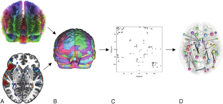
Table 1: Etiopathogenic classification of cerebral small vessel diseases
                                                                   Classification
 Type 1: HA
 Fibrinoid necrosis, lipohyalinosis, microatheroma, microaneurysms
 Type 2: Sporadic and hereditary CAA
 Type 3: Inherited or genetic SVD (distinct from CAA)
 For example, CADASIL, CARASIL, MELAS, Fabry disease, COL4A1 mutations, and so forth
 Type 4: Inflammatory- and immunologically-mediated SVD
 Examples: Wegener granulomatosis, Churg-Strauss syndrome, microscopic polyangiitis, Henoch-Schönlein purpura, cryoglobulinemic
   vasculitis, cutaneous leukocytoclastic angiitis, primary angiitis of the CNS, Sjögren syndrome, rheumatoid vasculitis, scleroderma,
   dermatomyositis, and so forth
 Type 5: Venous collagenosis
 Type 6: Other SVD
 Examples: postradiation angiopathy and nonamyloid microvessel degeneration in Alzheimer disease
Note:—MELAS indicates Mitochondrial Encephalopathy, Lactic Acidosis, and Stroke-like episodes; CARASIL, cerebral autosomal recessive arteriopathy with subcortical
infarcts and leukoencephalopathy.

                                                                                                       lacunar infarcts. There are four possible
                                                                                                       mechanisms for lacunar ischemic stroke
                                                                                                       that have been proposed: atheroma of
                                                                                                       parent arteries (usually MCA) or perfo-
                                                                                                       rating arterioles, embolism from the
                                                                                                       heart or carotid arteries, and intrinsic
                                                                                                       SVD (lipohyalinosis or fibrinoid
                                                                                                       necrosis).19,20
                                                                                                           Although hypertension is a key risk
                                                                                                       factor, many patients with SVD are not
                                                                                                       hypertensive.2,21-24 Recent studies sug-
                                                                                                       gest more complex mechanisms other
                                                                                                       than arteriolosclerosis that can cause
                                                                                                       capillary wall dysfunction. The deposi-
                                                                                                       tion of lipids in arteriolar walls (microa-
                                                                                                       theroma) can damage the endothelium
                                                                                                       (Fig 1),2,5 leading to leakage of plasma
                                                                                                       proteins and inflammatory cells into the
                                                                                                       perivascular tissues.5,25 The BBB dys-
FIG 1. Possible entry points in the pathophysiology of SVD. The order of these events has not          function involving the neurogliovascu-
been determined.                                                                                       lar unit can result in vessel stiffening,
                                                                                                       inflammation, myelin damage, and sec-
                                                                                                       ondary neurodegeneration.2,5,26-28 The
adult brain imaging, it is important for radiologists to be familiar     neurogliovascular unit has multiple possible entry points for disease
with the imaging spectrum, nomenclature, and clinical relevance.         mechanisms (ie, endothelial cells, pericytes, astrocytes, oligodendro-
                                                                         cytes, neurons, and the extracellular matrix). At a cellular level, the
                                                                         disruption of normal astrocyte function decreases neuronal energy
Pathophysiology and Epidemiology of SVD                                  supply, resulting in BBB leakage.2,25,29 Although the exact order of
The exact pathogenesis of SVD is incompletely understood, but the        these events is not well-established, these cellular changes of endo-
most common abnormalities are diffuse arteriolosclerosis, lipohyali-     thelial failure and inflammation are more dynamic and widespread
nosis, and fibrinoid necrosis of small arterioles. 2,16-18
                                                           Additionally, than previously thought.2,5 It was also hypothesized that in rodents,
embolism or atheroma and other vascular risk factors (diabetes mel-      prominent perivascular spaces can be associated with a blockage of
litus, hypertension, and hypercholesterolemia) are also believed to      the drainage of interstitial fluid from increased vessel stiffness and
play a role.2 Fisher16 suggested that symptomatic lacunar infarcts       increased arteriolar pulsatility such as with arterial hypertension,
were caused by atherosclerosis or embolism in the larger arterioles,     which results in fluid stagnation in the perivascular spaces and thus
whereas the silent infarcts were likely attributable to lipohyalinosis   decreased interstitial fluid flushing.5,30 This result may impede the
                                                2
or fibrinoid necrosis in the smaller arterioles. Lacunar infarcts were   clearance of metabolites (including b -amyloid and other proteins)
traditionally considered to be mainly caused by lipohyalinosis of        from tissues. Alzheimer disease, cerebral amyloid angiopathy, and
small perforating arteries. However, atherosclerosis in the parental     several monogenic small vessel diseases are thus being considered as
artery can produce a similar single lacunar infarct by blocking the      protein elimination failure angiopathies.5,30
                              19
orifice of branching arteries. A systematic review of Asian studies          Although there is significant variation in the epidemiology of
reported that parent artery atherosclerosis resulted in 20% of single    SVD markers, the prevalence of these markers is associated with
2     Mahammedi        2022    www.ajnr.org
Table 2: Visual grading scales for SVD markers and total SVD score
Total SVD Scorea
MR Imaging Sequence and Visual Rating Type 1: Hypertensive42 Type 2: CAA33
Classic markers
Lacune T2WI/FLAIR Any lacunes: 1 point
WMH FLAIR/Fazekas scale (0–3): Deep WMH (Fazekas 2 or 3) Deep WMH (Fazekas 2 or 3) or
Periventricular WMH: or periventricular WMH periventricular WMH
0 ¼ absent; (Fazekas 3): 1 point (Fazekas 3): 1 point
1 ¼ caps or pencil-thin;
2 ¼ smooth halo;
3 ¼ irregular extending to deep WM
Deep WMH:
0 ¼ absent;
1 ¼ punctate foci;
2 ¼ beginning confluence;
3 ¼ large confluent areas
PVS T2WI 4-point visual rating scale: .20 PVS in the basal .20 PVS in CSO: 1 point
0: no PVS; 1 (mild): #10 PVS; 2 (moderate): 11–20 ganglia: 1 point
PVS; 3 (moderate to severe): 21–40 PVS; and
4 (severe): .40 PVS
Microbleeds T2* GRE/SWI; several visual scores are Any microbleed: 1 point 2–4 Cortical CMB: 1 point;
available including MARS and BOMBS $5 cortical CMB: 2 points
New markers
cSS T2* GRE/SWI, categorized as focal or Focal cSS: 1 point; disseminated
disseminated cSS: 2 points
Cortical High-field MR imaging
microinfarct (.3T)
3D T1WI
3D DIR
Note:—GRE indicates gradient recalled-echo; MARS, Microbleed Anatomical Rating Scale; BOMBS, Brain Observer MicroBleed Scales; CSO, centrum semiovale; cSS, corti-
cal superficial siderosis; BG, basal ganglia; DIR, double inversion recovery.
a
Type 1: Hypertensive; type 2: CAA.
greater age. The association between white matter hyperintensities, infarct occurring in the territory of a perforating arteriole with
lacunes, prominent perivascular spaces (PVS), and cerebral micro- symptoms or imaging features that suggest a timeline of a previous
bleeds (CMB) and aging has been well-documented.31 Other demo- few weeks. However, in most patients, these lesions can appear
graphic factors such as sex and ethnicity/race can also explain the without apparent clinical symptoms (silent infarcts) (Online
variability in SVD, though the exact mechanism is poorly under- Supplemental Data).11 The term “recent” is preferred instead of
stood.31 It was reported that although Blacks and Asians have a “acute” because it includes both the hyperacute stage and the first
high burden of SVD compared with Whites, Blacks had a lower few weeks of the lesion.11 These lesions typically measure #20 mm
prevalence of WMH but a higher prevalence of severe WMH.32 in their maximum diameter in the axial plane, though in the coro-
Several population-based studies have characterized SVD in the nal plane, they can appear slightly larger and elongated, reflecting
“healthy population” and explored the risk factors for SVD.32 the territory of the occluded perforating arteriole (Fig 2A). They are
best identified with hyperintense signal on DWI, hypointense signal
Neuroimaging of SVD on an ADC map, and hyperintense signal on T2-weighted and
Nomenclature and Definitions. Radiologists are well-versed with FLAIR sequences. The most common fate of these lesions on long-
the variations in the radiology reports describing SVD, for exam- term follow-up is cavitation to become a lacune (in 28%–94%,
ple, WMH, cerebral white matter disease, leukoaraiosis, and age- depending on the duration of follow-up), turning into a nonspecific
related white matter disease. The STRIVE guidelines and standards focus of T2/FLAIR hyperintensity, or eventually completely disap-
were developed to address this variability (Online Supplemental pearing.2,8 Furthermore, STRIVE proposes that lesions in the basal
Data).11 The recommendations were initially applied to research ganglia and internal capsule of .20 mm are a different subtype
studies with the potential goal of widespread clinical use to stand- and, therefore, should be classified as striatocapsular infarcts instead
ardize image interpretation, acquisition, and reporting. The classic of small subcortical infarcts. Similarly, infarcts of the anterior cho-
MR imaging markers of SVD include recent small subcortical roidal artery are etiologically and anatomically distinct.
infarctions, lacunes of presumed vascular origin, WMH of pre-
sumed vascular origin, PVS, and CMB. Two additional MR imag- Lacunes of Presumed Vascular Origin. The STRIVE guidelines
ing markers of SVD that occur with high prevalence in CAA are define lacunes of presumed vascular origin as round or ovoid, sub-
cortical superficial siderosis (cSS) and cortical microinfarcts cortical, fluid-filled cavities (with signal similar to that of CSF in all
(Table 2).33 sequences), measuring between 3 and 15 mm in diameter, consist-
ent with a previous, small subcortical infarct or hemorrhage located
Recent Small Subcortical Infarcts. According to the STRIVE cri- in deep gray and white matter and in a territory of perforating arte-
teria, the term “recent small subcortical infarcts” refers to a recent riole (Fig 2B and Online Supplemental Data). These lesions
AJNR Am J Neuroradiol : 2022 www.ajnr.org 3brainstem. Although the Fazekas visual
rating scale (Fig 3) is the most com-
monly used method to assess the bur-
den of WMH, there is a lack of a
rigorous method to accurately quantify
the WMH burden.34 Furthermore, a
fronto-occipital gradient was used in
few studies to describe the difference in
the severity of WMH between the fron-
tal and occipital lobes.33,35 It was sug-
gested that patients with obvious
occipital-dominant WMH were more
likely to develop complications from
SVD such as lobar intracranial hemor-
rhage than healthy controls.35 The se-
verity of WMH is strongly associated
with cerebrovascular disease, vascular
risk factors, gait disturbance, cognitive
symptoms, and poststroke functional
outcomes.3,11
Perivascular Spaces, Also Called
Virchow-Robin Spaces. Perivascular
spaces are extensions of the extracere-
bral fluid space that are covered by the
pia mater, which surrounds cerebral
vessels from the brain surface into and
through the brain parenchyma. These
are commonly microscopic and not
FIG 2. A, Recent subcortical infarct in the right lentiform nucleus in a 59-year-old woman with a visible on conventional neuroimaging,
history of hypertension. From left to right, axial DWI, ADC map, FLAIR, and coronal T2WI show
restricted diffusion (solid white arrow, A). On coronal T2WI, the elongated morphology of the
but when they become prominent and
acute infarct in the craniocaudal axis (dashed white arrow, A) is related to the territory of a per- numerous, they are considered a
forating artery. B, A lacune of presumed vascular origin in a 67-year-old man with a history of de- marker of SVD. PVS have been shown
mentia. From left to right, axial DWI, T2WI, FLAIR, and T1WI show a remote lacunar infarct in the to be associated with other SVD
left frontal corona radiata, which demonstrates T2 hyperintensity with a peripheral rim of gliosis,
markers such as WMH and lacunes,
best seen on FLAIR image (solid white arrow, B). C, Prominent perivascular spaces in a 63-year-old
man with a history of dementia. A prominent perivascular space is noted in the left insular region but not atrophy.11 PVS have a signal
with a centrally traversing vessel (solid white arrow, C), without peripheral gliosis on the FLAIR intensity similar to that of CSF in all
image (dashed arrow, C). Additionally, there are .20 dilated perivascular spaces in the bilateral sequences and appear round or ovoid
basal ganglia on axial T2WI (circles, C). D, Cerebral microbleeds with cSS in an 87-year-old man with a diameter of ,3 mm but can
with a history of CAA who presented with a worsening mental status. Axial SWI demonstrate
appear more elongated when imaged
multiple foci of susceptibility artifacts predominantly involving the basal ganglia and thalami, con-
sistent with microbleeds. Additional areas of scattered cortical subarachnoid hemorrhagic stain- parallel to the course of the penetrat-
ing (arrows, D) indicate cortical superficial siderosis. ing vessel. They are typically located in
the inferior basal ganglia, centrum
semiovale, and midbrain and do not
have a surrounding gliotic rim (in
typically demonstrate a surrounding gliotic rim of T2 FLAIR hyper- contrast with lacunes) (Fig 2C and Online Supplemental Data).
intensity (Figs 3 and 4), which can be a useful feature when present. Although lacunes and PVS can have similar MR imaging features,
A peripheral gliotic rim can also surround perivascular spaces when pathologic studies have demonstrated that lesions that
they pass through an area of white matter hyperintensity. Although are ,3 mm in diameter are more likely to be perivascular spaces
lacunes are commonly seen in older patients without symptoms, than lacunes.11 A traversing vessel can sometimes be seen in the
they are associated with an increased risk of stroke, dementia, and center of a perivascular space when imaged at high resolution,
gait impairment.5,11 another differentiator of PVS versus lacunes (Fig 2C). The sever-
ity of PVS is typically assessed on axial T2-weighted images using
WMH of Presumed Vascular Origin. WMH of presumed vascular a validated 4-point visual rating scale based on the total number
origin are characterized by hyperintense lesions on T2 FLAIR of PVS (0 indicating no PVS; 1 [mild], #10 PVS; 2 [moderate],
and decreased attenuation on CT in the periventricular/deep cere- 11–20 PVS; 3 [moderate to severe], 21–40 PVS; and 4 [severe],
bral white matter, subcortical gray matter, basal ganglia, and .40 PVS) in the basal ganglia and centrum semiovale (Table
4 Mahammedi 2022 www.ajnr.org2).33 The topography of PVS can be a
helpful characteristic of the underlying
SVD type: PVS are likely associated
with arteriolosclerosis when located in
the basal ganglia, and they are more
likely to be related to CAA when
located in the centrum semiovale (Fig
4).10 However, the clinical significance
of PVS remains unclear; a few studies
have reported that PVS can be associ-
ated with an increased risk of demen-
tia if located in the basal ganglia and
white matter.5,10,36
CMB. CMB are small round or ovoid
lesions (#10 mm in diameter) of
marked hypointensity with associated
blooming on T2 gradient-echo (T2*) or
other sequences that are sensitive to
susceptibility effects, especially SWI
(Fig 2D and Online Supplemental
Data). CMB correspond to hemosid-
erin-laden macrophages in perivascular
tissue, consistent with vascular leakage
of blood cells.5 Several visual scores are
available, including the Microbleed
Anatomical Rating Scale and the Brain
FIG 3. Fazekas scale of WMH grading. Observer MicroBleed Scales, which
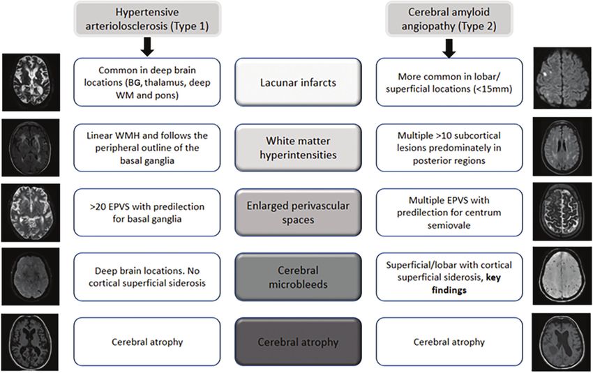
FIG 4. SVD MR imaging markers of the two most common etiopathogenic types of cerebral SVD in older adults, HA and CAA. BG indicates ba-
sal ganglia; EPVS: enlarged prominent perivascular spaces.
AJNR Am J Neuroradiol : 2022 www.ajnr.org 5markers and using a more comprehen-
sive approach to assess total SVD bur-
den.33,40 Total SVD score is a simple
and pragmatic way of assessing overall
brain health and has been shown to be
a prognostic indicator of cognitive
decline and recurrent stroke (Fig 5).
The total burden of lesions in specific
anatomic locations can also be used for
lesion-symptom associations and has
been shown to be associated with cog-
nitive impairment and gait and mood
disorders.5,40,41 For instance, a total
SVD score in CAA was proposed by
Charidimou et al33 and can potentially
provide a more practical framework to
better evaluate the effect of CAA-
related brain damage on clinical out-
comes. A similar total MR imaging
SVD burden approach was proposed in
HA by Klarenbeek et al.42 The imaging
rating points of total SVD burden are
outlined in Table 2.
Imaging in HA versus CAA
HA (type 1) and CAA (type 2) are the
most common sporadic SVD types in
older adults (Fig 4 and Table 2) with
intrinsically different pathophysiology,
clinical significance, and prognosis.15
HA predominantly affects small perfo-
rating end arteries of the deep gray
nuclei and deep white matter, whereas
FIG 5. High SVD burden in a 55-year-old man with a history of arterial hypertension. Note multi-
CAA results from b -amyloid deposi-
ple microbleeds in the left thalamus and occipital lobe (dotted arrows) on SWI (A), remote lacu-
nar infarcts in the right centrum semiovale (white arrows, B), and WMH Fazekas 2 on FLAIR (B). tion within the cortical and leptomenin-
More than 20 dilated perivascular spaces are seen on axial T2WI (circle, C) and remote lacunar geal arteries. Both types are common
infarct in right lentiform nucleus (white arrow, C). The total SVD score is 4. The patient developed causes of ischemic manifestation, intra-
an acute lacunar infarct (black arrow) on DWI (D) 10 months later. cranial hemorrhage, and cognitive
impairment. Although both types can
be associated with similar imaging
have reasonable intrarater and interrater reliability for the presence markers, including CMB, WMH, lacunar infarcts, and PVS, the
of definite microbleeds.37,38 In fact, a few recent publications have anatomic distribution of these markers can be helpful to differenti-
proposed a novel automatic method to detect CMB from MR ate these 2 entities radiologically. Lobar/superficial and cortical dis-
images by exploiting the 3D convolutional neural network.39 CMB tribution is consistently associated with CAA, whereas the
are associated with an increased risk of cognitive decline and ische- involvement of deep brain regions and the basal ganglia is most of-
mic and hemorrhagic stroke, though the risk of dementia appears ten associated with HA (Fig 4 and Table 2).15,33,36,42-44
higher in patients with CMB restricted to a superficial/lobar loca- Furthermore, deep CMB are typically associated with arterioloscle-
tion compared with those with deep involvement.5,10 Two other rosis, whereas a strictly superficial/lobar location is characteristic of
hemorrhagic manifestations of SVD include cSS and spontaneous CAA.10 CAA is characteristically associated with lobar CMV, cSS,
intracerebral hemorrhage (in contrast to secondary hemorrhage centrum semiovale perivascular spaces, and multiple punctate
due to other causes such as trauma and vascular malformations). FLAIR WMH (typically .10) with a predilection for the posterior
regions.15,33,44 A peri-basal ganglia pattern of WMH is strongly
Total SVD Score linked to arteriolosclerosis (Fig 4).15
Although the neuroimaging markers have typical characteristics,
in practice, it is difficult to distinguish lacunes, PVS, and WMH Clinical Implications
of presumed vascular origin because they are often closely related. Clinical manifestations of SVD and poor brain health can be var-
A few studies have suggested combining these MR imaging ied. The presenting clinical symptoms can be abrupt, such as a
6 Mahammedi 2022 www.ajnr.orgFIG 6. Sample pipeline for studying brain connectivity for assessing global brain health. A, Data are processed to generate structural (tractogra-
phy) or functional (fMRI) connectivity measures. B, A parcellation scheme is applied to obtain connectivity between different regions. C, The
connectivity data are represented as an adjacency matrix. D, Network properties are calculated on the adjacency matrix. In the sample image,
regions in the network are sized by importance (ie, centrality) and edges are sized by the strength of the connection.
resulting intracerebral hemorrhage (ICH) or, more insidious, SVD and Cognitive Decline. SVD coexists with Alzheimer disease
manifesting as progressive cognitive decline, mood disorders, and Alzheimer disease–related dementia, can worsen cognitive
extrapyramidal symptoms, and depression. This variability can outcome, and is the most common cause of vascular dementia,
be due to multiple factors including characteristics of vascular contributing to about 50% of dementias.5-7,10 Recent data suggest
injury (type, location, and extent), the degree of comorbidities, that WMH burden is associated with an increased risk of devel-
and resilience factors such as brain reserve, which refers to the oping dementia, including the Alzheimer disease type,3 and that
capacity to cope with brain pathology.5 Many clinical symp- arteriolosclerosis increases the odds of Alzheimer disease demen-
toms relate to secondary neurodegeneration due to the global tia, independent of the effect of Alzheimer disease pathology.10
effects of SVD such as brain atrophy.5,45-50 Several studies have
shown that subcortical infarcts and WMH can induce loss of a SVD and Parkinsonism. SVD at baseline, particularly WMH bur-
connected cortex distal to the infarct through degeneration of den and lacunes, was associated with an increased risk of parkin-
white matter tracts, which results in secondary cortical thinning, sonism.52 Furthermore, microbleeds and gray matter atrophy
and neurodegeneration.45,47,48 The main clinical manifestations of were found to increase the risk of vascular parkinsonism.47
SVD follow.
Advanced Imaging for SVD
While SVD markers are extremely common findings on conven-
SVD and Ischemic Stroke. SVD causes approximately 25% of all
tional MR imaging, there are still weak associations of the imaging
acute ischemic strokes, mainly in the form of lacunar infarction.10
markers and the heterogeneous clinical expression of SVD.8,53 One
Although stroke recurrence is similar to that in other stroke sub-
potential reason is that conventional MR imaging measures tend to
types, the proportion of patients with lacunar infarct who are de-
be focal, whereas SVD is increasingly thought of as a global, whole-
pendent at 3 years is 42%.10 Approximately 30% of patients with
brain phenomenon. Furthermore, the severity and nature of clinical
lacunar stroke will develop cognitive impairment, mostly involving
symptoms can be vastly different in patients with radiologically
executive functions, attention, and psychomotor speed.10
identical SVD lesions.47 A network-based and/or global approach
to observe subclinical SVD manifestations may explain this discrep-
SVD and ICH. ICH presumed due to SVD can result in devastat- ancy. Advanced imaging techniques such as high-field MR imaging,
ing complications with high morbidity and mortality and a signif- DTI, blood oxygen level–dependent (BOLD) MR imaging, and per-
icant risk of recurrence.51 CMB are present in 52% of patients fusion imaging show considerable promise.2
with first-ever ICH and 83% of those with recurrent ICH.51 The
location of recurrent ICH can relate to the distribution and sever- High-Field MR Imaging. A recent MR imaging–identifiable marker
ity of underlying SVD, particularly CMB and superficial siderosis, of SVD is cortical microinfarcts. Microinfarcts, especially those in
which could help in identifying patients at high risk of ICH recur- the deep gray matter, are best detected with an MR imaging field
rence.51 Furthermore, it is reported that the presence of $5 CMB strength of $3T. High-field (7T) MR imaging shows a greater abil-
can be associated with recurrent lobar ICH.33 ity to detect remote microinfarcts.54 Most interesting, postmortem
AJNR Am J Neuroradiol : 2022 www.ajnr.org 7studies have shown that the penumbra of microinfarcts can extend have shown a positive correlation of SVD severity (ie, worse
up to 12 times larger than the microinfarct core, which is the only SVD = more compromised BBB) and decreased BBB integrity in
component visible with conventional MR imaging.47 Characterizing patients with lacunar stroke and worse clinical outcome.66,67 The
the penumbra and distribution of microinfarcts may be clinically second method, which does not involve a contrast agent, is arterial
important because they are known to lead to a reduced capability of spin-labeling.68 While arterial spin-labeling has not been used to
adjacent axons to conduct action potentials.55 detect early biomarkers of SVD, it has been used to differentiate
subgroups of patients with SVD. For example, a recent study
DTI. In addition to measures related to white matter integrity, showed that CBF, measured by 3D arterial spin-labeling, was sig-
such as fractional anisotropy, one can also characterize the struc- nificantly decreased globally and locally in the temporal lobe, fron-
tural connectivity with tractography, an important marker for tal lobe, hippocampus, thalamus, and insula in patients with
global brain health and cognition (Fig 6). In connectivity space, increasing subcortical vascular cognitive impairment.69 There is a
regions are often called “nodes” and the connections between rich research opportunity to fully characterize the properties of the
them are called “edges.” The collection of all nodes and edges for microvasculature in patients with SVD using arterial spin-labeling.
a network can be represented as a connectivity matrix, where The multitude of advanced imaging techniques being used
each row and column represent a node and entries represent con- now and yet to be developed will undoubtedly lead to a redefini-
nections.56 Network properties can be derived from these matri- tion of how we understand and think about SVD.
ces that describe network integration and which nodes are the
most important for successful functioning. Studies using these Vessel Wall Imaging
network measures have shown that patients with SVD have MRA and CTA are important to detect abnormalities in the lumen
decreased local and global network integration in the prefrontal of vessels but cannot characterize the vessel wall.70 Vessel wall
and interhemispheric tracts, compared with healthy controls.57 imaging has recently been developed to address this limitation, en-
Additionally, studies have suggested that SVD may lead to loss of abling better detection of nonstenotic lesions and better characteri-
energy-dependent long-range white matter connections, which zation of stenotic lesions.71,72 Briefly, vessel wall imaging is
disrupt the structure of the network. This can result in lowered achieved using a sequence with high spatial resolution with opti-
efficiency and connectivity of the most important regions in the mal contrast-to-noise, attained by suppressing luminal blood.71
brain for normal functioning (sometimes called the “rich-club”), Several parameters affect the quality of the resulting images, such
leading to impaired cognition.58 The connectivity and whole as field strength, 2D-versus-3D imaging, and a blood-nulling tech-
brain–based studies are gaining popularity and may lead to the
nique, but there has not yet been a thorough analysis of the trade-
discovery of novel biomarkers and explanations for the heteroge-
offs among these permutations.71 Intracranial atherosclerosis pro-
neity of symptoms seen in SVD.
vides a key example of how vessel wall imaging can advance our
knowledge of SVD. Intracranial atherosclerosis has been down-
BOLD MRI. BOLD MR imaging is a technique that measures cere-
bral blood flow by detecting relative regional differences in oxy- played as a contributor to SVD, likely because traditional vascular
genated and deoxygenated blood supply.59 Cerebrovascular sequences (ie, MRA) can only assess luminal stenosis in large
reactivity can be derived using this technique in tandem with arteries.73 Vessel wall imaging has revealed that intracranial athero-
induced hypercapnia (called a CO2 challenge). A recent study in sclerosis burden, which is dependent on features of both the lumen
patients with SVD showed that lower cerebrovascular reactivity, and the vessel wall, is related to cortical and subcortical infarcts,
increased venous pulsatility, and decreased foramen magnum microinfarcts, and WMH.73 Several other pathologies can be cap-
CSF volume were associated with a greater burden of WMH and tured by vessel wall imaging and may elucidate the etiology of
a more severe basal ganglia rating of PVS.60 Other studies have SVD, such as atherosclerotic plaque, large-vessel vasculitis, and dis-
indicated the role of aortic arch stiffening as a potential contribu- crimination of reversible cerebral vasoconstriction syndrome.70
tor in SVD through excessive pulsatile energy transmitted to the
cerebral vasculature as part of brain-heart coupling.61 Artificial Intelligence Advances in SVD
Another use of BOLD MR imaging is fMRI. Like tractogra- While identifying the presence of at least 1 lacune or cerebral
phy, fMRI can be analyzed in terms of brain connectivity. Using microbleed is straightforward in most situations, visual grading
connectivity-based techniques with resting-state fMRI data, of WMH and PVS poses a unique challenge because the rating
researchers have shown that patients with SVD have reduced scales are inherently subjective, with poor agreement among neu-
connectivity in the prefrontal, parietal, and cingulate cortices, roradiologists.74 MR imaging, specifically T2 FLAIR imaging, is
with corresponding increases in cerebellar connectivity and the primary way that WMH are assessed due to even higher inter-
impaired deactivation of the default mode network.62,63 reader variability using CT scans. An automated algorithm using
CT scans was able to perform as well as experts at delineating
Perfusion Imaging. Recently, compromised BBB integrity has WM lesions, but it varied widely with .90% around the mean
gained traction as an early biomarker for SVD and may play an estimate.75 Previous studies have shown some success in predict-
important role in understanding SVD pathogenesis.64 There are 2 ing the evolution of WMH and cognitive outcomes in patients
primary methods for measuring BBB integrity and microvascula- with WMH.76,77 However, more research is needed on the quan-
ture with MR imaging. The first, and most reliable method, is tification of WMH because the studies with this focus have been
dynamic contrast-enhanced MR imaging with the use of an appro- largely in patients with multiple sclerosis.78,79 WMH in patients
priate pharmacokinetic model.65 Using this technique, researchers with additional findings, such as lacunar or cortical infarcts,
8 Mahammedi 2022 www.ajnr.orgpresent a difficult challenge for current algorithms. Furthermore, 6. Kapasi A, DeCarli C, Schneider JA. Impact of multiple pathologies
fully automated quantification of WMH, either by the Fazekas on the threshold for clinically overt dementia. Acta Neuropathol
scale or volumetric measurement using a standard of care medi- 2017;134:171–86 CrossRef Medline
7. Bos D, Wolters FJ, Darweesh SKL, et al. Cerebral small vessel disease
cal imaging (rather than research sequences) is difficult.
and the risk of dementia: a systematic review and meta-analysis of
Attempting to grade PVS with automated artificial intelligence population-based evidence. Alzheimers Dement 2018;14:1482–12
tools is even more of a challenge, with only 1 study that showed CrossRef Medline
good performance in the centrum semiovale but poor perform- 8. Shibuya M, da Costa Leite C, Lucato LT. Neuroimaging in cerebral
ance in the basal ganglia.80 There are great opportunities for small vessel disease: update and new concepts. Dement Neuropsychol
future research to explore these rating scales for greater intersub- 2017;11:336–42 CrossRef Medline
9. Gorelick PB, Furie KL, Iadecola C, et al. Defining optimal brain
ject agreement, particularly in routine imaging, and come up
health in adults. Stroke 2017;48:e284–303 CrossRef Medline
with efficient and reproducible artificial intelligence brain health 10. Pasi M, Cordonnier C. Clinical relevance of cerebral small vessel
solutions. diseases. Stroke 2020;51:47–53 CrossRef Medline
11. Wardlaw JM, Smith EE, Biessels GJ, et al; STandards for ReportIng
Vascular changes on nEuroimaging (STRIVE v1). Neuroimaging
CONCLUSIONS AND FUTURE DIRECTIONS standards for research into small vessel disease and its contribu-
Small vessel disease is a rising epidemic associated with detri- tion to ageing and neurodegeneration. Lancet Neurol 2013;12:822–
mental brain health. Neuroimaging plays a fundamental role in 38 CrossRef Medline
identifying SVD. The recognition of endothelial and neuroglio- 12. van Veluw SJ, Shih AY, Smith EE, et al. Detection, risk factors, and
vascular unit dysfunction as the main underlying mechanisms functional consequences of cerebral microinfarcts. Lancet Neurol
of SVD is fundamental to develop the role of advanced neuroi- 2017;16:730–40 CrossRef Medline
13. Potter GM, Marlborough FJ, Wardlaw JM. Wide variation in defini-
maging techniques. A more comprehensive approach to gauge
tion, detection, and description of lacunar lesions on imaging.
total SVD burden, rather than individualizing and classifying Stroke 2011;42:359–66 CrossRef Medline
each of these markers separately, is needed in future studies. 14. Zhu YC, Dufouil C, Tzourio C, et al. Silent brain infarcts. Stroke
This may provide a more complete estimate of the full impact of 2011;42:1140– 45 CrossRef Medline
SVD on the brain to better assess total brain health and the 15. Charidimou A, Boulouis G, Haley K, et al. White matter hyperinten-
effect of SVD-related brain damage. Finally, as radiologists, it is sity patterns in cerebral amyloid angiopathy and hypertensive arte-
riopathy. Neurology 2016;86:505–11 CrossRef Medline
critical that we use standardized terminology to describe SVD
16. Fisher CM. Lacunar infarcts: a review. Cerebravascular Diseases
in both clinical and research settings. 1991;1:311–20 CrossRef
17. Fisher CM. Lacunar strokes and infarcts. Neurology 1982;32:871–76
Disclosures: Pooja Khatri—RELATED: Grant: National Institutes of Health.* CrossRef CrossRef Medline
UNRELATED: Consultancy: Lumosa Therapeutics, Diamedica Therapeutics*; Grants/ 18. Bailey EL, Smith C, Sudlow CL, et al. Pathology of lacunar ischemic
Grants Pending: Cerenovus*; Royalties: UpToDate; Other: Bayer, Basking stroke in humans: a systematic review. Brain Pathol 2012;22:583–91
Biosciences, Comments: Bayer, National Trial Leader; Basking Biosciences, Scientific
CrossRef Medline
Advisory Board. Brett Kissela—RELATED: Grant: National Institutes of Health/
National Institute of Neurological Disorders and Stroke.* Rhonna Shatz—RELATED: 19. Kim JS, Yoon Y. Single subcortical infarction associated with pa-
Grant: National Institutes of Health.* Achala Vagal—RELATED: Grant: National rental arterial disease: important yet neglected sub-type of athero-
Institutes of Health, Cerenovus,* Comments: R01, National Institutes of Health/ thrombotic stroke. Int J Stroke 2013;8:197–203 CrossRef Medline
National Institute of Neurological Disorders and Stroke,* NS103824; RF1, National 20. Shi Y, Wardlaw JM. Update on cerebral small vessel disease: a
Institute of Neurological Disorders and Stroke/National Institute on Aging, dynamic whole-brain disease. Stroke Vasc Neurol 2016;1:83–92
NS117643; R01, National Institutes of Health/National Institute of Neurological CrossRef Medline
Disorders and Stroke, NS100417; National Institutes of Health/National Institute of
21. Weber R, Weimar C, Blatchford J, et al; PRoFESS Imaging Substudy
Neurological Disorders and Stroke, 1U01NS100699; National Institutes of Health/
National Institute of Neurological Disorders and Stroke, U01NS110772; Principal
Group. Telmisartan on top of antihypertensive treatment does
Investigator, Imaging Core Lab, ENDOLOW Trial, Cerenovus. *Money paid to the not prevent progression of cerebral white matter lesions in the
institution. Prevention Regimen for Effectively Avoiding Second Strokes
(PRoFESS) MRI substudy. Stroke 2012;43:2336–42 CrossRef
Medline
REFERENCES 22. Jackson CA, Hutchinson A, Dennis MS, et al. Differing risk factor
1. Pantoni L. Cerebral small vessel disease: from pathogenesis and profiles of ischemic stroke subtypes: evidence for a distinct lacunar
clinical characteristics to therapeutic challenges. Lancet Neurol arteriopathy? Stroke 2010;41:624–29 CrossRef Medline
2010;9:689–701 CrossRef Medline 23. Lammie GA, Brannan F, Slattery J, et al. Nonhypertensive cerebral
2. Wardlaw JM, Smith C, Dichgans M. Mechanisms of sporadic cerebral small-vessel disease. Stroke 1997;28:2222–29 CrossRef Medline
small vessel disease: insights from neuroimaging. Lancet Neurol 24. Godin O, Tzourio C, Maillard P, et al. Antihypertensive treatment
2013;12:483–97 CrossRef Medline and change in blood pressure are associated with the progression
3. Debette S, Schilling S, Duperron MG, et al. Clinical significance of of white matter lesion volumes. Circulation 2011;123:266–73
magnetic resonance imaging markers of vascular brain injury: a CrossRef Medline
systematic review and meta-analysis. JAMA Neurol 2019;76:81–94 25. Bosetti F, Galis ZS, Bynoe MS, et al; “Small Blood Vessels: Big Health
CrossRef Medline Problems” Workshop Participants. Small blood vessels: big health
4. Georgakis MK, Duering M, Wardlaw JM, et al. WMH and long-term problems?”: scientific recommendations of the National Institutes
outcomes in ischemic stroke: a systematic review and meta-analy- of Health Workshop. J Am Heart Assoc 2016;5:e0043898 CrossRef
sis. Neurology 2019;92:e1298–1308 CrossRef Medline Medline
5. Wardlaw JM, Smith C, Dichgans M. Small vessel disease: mechanisms 26. Petersen MA, Ryu JK, Akassoglou K. Fibrinogen in neurological dis-
and clinical implications. Lancet Neurol 2019;18:684–96 CrossRef eases: mechanisms, imaging and therapeutics. Nat Rev Neurosci
Medline 2018;19:283–301 CrossRef Medline
AJNR Am J Neuroradiol : 2022 www.ajnr.org 927. Wuerfel J, Haertle M, Waiczies H, et al. Perivascular spaces: MRI 49. Chabriat H, Hervé D, Duering M, et al. Predictors of clinical wor-
marker of inflammatory activity in the brain? Brain 2008;131:2332– sening in cerebral autosomal dominant arteriopathy with subcorti-
40 CrossRef Medline cal infarcts and leukoencephalopathy. Stroke 2016;47:4–11 CrossRef
28. Hawkins BT, Davis TP. The blood-brain barrier/neurovascular unit in Medline
health and disease. Pharmacol Rev 2005;57:173–85 CrossRef Medline 50. Rizvi B, Narkhede A, Last BS, et al. The effect of white matter hyper-
29. Bechmann I, Galea I, Perry VH. What is the blood–brain barrier intensities on cognition is mediated by cortical atrophy. Neurobiol
(not)? Trends Immunol 2007;28:5–11 CrossRef Medline Aging 2018;64:25–32 CrossRef Medline
30. Iliff JJ, Wang M, Zeppenfeld DM, et al. Cerebral arterial pulsation 51. Charidimou A, Imaizumi T, Moulin S, et al. Brain hemorrhage re-
drives paravascular CSF–interstitial fluid exchange in the murine currence, small vessel disease type, and cerebral microbleeds: a
brain. J Neurosci 2013;33:18190–99 CrossRef Medline meta-analysis. Neurology 2017;89:820–29 CrossRef Medline
31. Caunca MR, De Leon-Benedetti A, Latour L, et al. Neuroimaging of 52. van der Holst HM, De Leeuw FE. Author response. Neurology
cerebral small vessel disease and age-related cognitive changes. 2016;86:1268–69 CrossRef Medline
Front Aging Neurosci 2019;11:145 CrossRef Medline 53. Gouw AA, Seewann A, van der Flier WM, et al. Heterogeneity of
32. Das AS, Regenhardt RW, Vernooij MW, et al. Asymptomatic cere- small vessel disease: a systematic review of MRI and histopathol-
bral small vessel disease: insights from population-based studies. J ogy correlations. J Neurol Neurosurg Psychiatry 2011;82:126–35
Stroke 2019;21:121–38 CrossRef Medline CrossRef Medline
33. Charidimou A, Martinez-Ramirez S, Reijmer YD, et al. Total mag- 54. van Rooden S, Goos JD, van Opstal AM, et al. Increased number of
netic resonance imaging burden of small vessel disease in cerebral microinfarcts in Alzheimer disease at 7-T MR imaging. Radiology
amyloid angiopathy: an imaging-pathologic study of concept vali- 2014;270:205–11 CrossRef
dation. JAMA Neurol 2016;73:994–1001 CrossRef Medline 55. Coban H, Tung S, Yoo B, et al. Molecular disorganization of axons
34. Fazekas F, Chawluk J, Alavi A, et al. MR signal abnormalities at 1.5 adjacent to human cortical microinfarcts. Front Neurol 2017;8:405
T in Alzheimer’s dementia and normal aging. AJR Am J Roentgenol CrossRef Medline
1987;149:351–56 CrossRef Medline 56. Rubinov M, Sporns O. Complex network measures of brain connec-
35. Zhu YC, Chabriat H, Godin O, et al. Distribution of white matter tivity: uses and interpretations. Neuroimage 2010;52:1059–69 CrossRef
hyperintensity in cerebral hemorrhage and healthy aging. J Neurol Medline
2012;259:530–36 CrossRef Medline 57. Lawrence AJ, Chung AW, Morris RG, et al. Structural network effi-
36. Passiak BS, Liu D, Kresge HA, et al. Perivascular spaces contribute ciency is associated with cognitive impairment in small-vessel dis-
to cognition beyond other small vessel disease markers. Neurology ease. Neurology 2014;83:304–11 CrossRef Medline
2019;92:e1309–21 CrossRef Medline 58. Marebwa BK, Adams RJ, Magwood GS, et al. Cardiovascular risk
37. Gregoire SM, Chaudhary UJ, Brown MM, et al. The Microbleed factors and brain health: impact on long-range cortical connec-
Anatomical Rating Scale (MARS). Neurology 2009;73:1759–66 tions and cognitive performance. J Am Heart Assoc 2018;7:e010054
CrossRef Medline CrossRef Medline
38. Cordonnier C, Potter GM, Jackson CA, et al. Improving interrater 59. Ogawa S, Tank DW, Menon R, et al. Intrinsic signal changes accom-
agreement about brain microbleeds. Stroke 2009;40:94–99 CrossRef panying sensory stimulation: functional brain mapping with mag-
Medline netic resonance imaging. Proc Natl Acad Sci U S A 1992;89:5951–55
39. Dou Q, Chen H, Yu L, et al. Automatic detection of cerebral micro- CrossRef Medline
bleeds from MR images via 3D convolutional neural networks. 60. Blair GW, Thrippleton MJ, Shi Y, et al. Intracranial hemodynamic
IEEE Trans Med Imaging 2016;35:1182–95 CrossRef Medline relationships in patients with cerebral small vessel disease. Neurology
40. Staals J, Makin SD, Doubal FN, et al. Stroke subtype, vascular risk 2020;94:e2258–69 CrossRef Medline
factors, and total MRI brain small-vessel disease burden. Neurology 61. Aghilinejad A, Amlani F, King KS, et al. Dynamic effects of aortic
2014;83:1228–34 CrossRef Medline arch stiffening on pulsatile energy transmission to cerebral vascula-
41. Banerjee G, Jang H, Kim HJ, et al. Total MRI small vessel disease ture as a determinant of brain-heart coupling. Sci Rep 2020;10:8784
burden correlates with cognitive performance, cortical atrophy, CrossRef Medline
and network measures in a memory clinic population. J Alzheimers 62. Papma JM, den Heijer T, de Koning I, et al. The influence of cerebral
Dis 2018;63:1485–97 CrossRef Medline small vessel disease on default mode network deactivation in mild
42. Klarenbeek P, van Oostenbrugge RJ, Rouhl RPW, et al. Ambulatory cognitive impairment. Neuroimage Clin 2012;2:33–42 CrossRef
blood pressure in patients with lacunar stroke. Stroke Medline
2013;44:2995–99 CrossRef Medline 63. Schaefer A, Quinque EM, Kipping JA, et al. Early small vessel disease
43. Pasi M, Boulouis G, Fotiadis P, et al. Distribution of lacunes in cere- affects frontoparietal and cerebellar hubs in close correlation with
bral amyloid angiopathy and hypertensive small vessel disease. clinical symptoms: a resting-state fMRI study. J Cereb Blood Flow
Neurology 2017;88:2162–68 CrossRef Medline Metab 2014;34:1091–95 CrossRef Medline
44. Shams S, Martola J, Charidimou A, et al. Topography and determi- 64. Li Y, Li M, Zuo L, et al. Compromised blood-brain barrier integ-
nants of magnetic resonance imaging (MRI)-visible perivascular rity is associated with total magnetic resonance imaging burden
spaces in a large memory clinic cohort. J Am Heart Assoc 2017;6: of cerebral small vessel disease. Front Neurol 2018;9:221 CrossRef
e006229 CrossRef Medline Medline
45. Duering M, Righart R, Wollenweber FA, et al. Acute infarcts cause 65. Heye AK, Thrippleton MJ, Armitage PA, et al. Tracer kinetic model-
focal thinning in remote cortex via degeneration of connecting ling for DCE-MRI quantification of subtle blood-brain barrier
fiber tracts. Neurology 2015;84:1685–92 CrossRef permeability. Neuroimage 2016;125:446–55 CrossRef Medline
46. Dickie DA, Karama S, Ritchie SJ, et al. Progression of white matter 66. Topakian R, Barrick TR, Howe FA, et al. Blood-brain barrier perme-
disease and cortical thinning are not related in older community- ability is increased in normal-appearing white matter in patients
dwelling subjects. Stroke 2016;47:410–16 CrossRef Medline with lacunar stroke and leukoaraiosis. J Neurol Neurosurg
47. Ter Telgte A, van Leijsen EM, Wiegertjes K, et al. Cerebral small ves- Psychiatry 2010;81:192–97 CrossRef Medline
sel disease: from a focal to a global perspective. Nat Rev Neurol 67. Wardlaw JM, Doubal FN, Valdes-Hernandez M, et al. Blood–brain bar-
2018;14:387–98 CrossRef Medline rier permeability and long-term clinical and imaging outcomes in cer-
48. Peres R, De Guio F, Chabriat H, et al. Alterations of the cerebral cor- ebral small vessel disease. Stroke 2013;44:525–27 CrossRef Medline
tex in sporadic small vessel disease: a systematic review of in vivo 68. Hendrikse J, Petersen ET, Golay X. Vascular disorders: insights
MRI data. J Cereb Blood Flow Metab 2016;36:681–95 CrossRef from arterial spin-labeling. Neuroimaging Clin N Am 2012;22:259–
Medline 69 CrossRef Medline
10 Mahammedi 2022 www.ajnr.org69. Sun Y, Cao W, Ding W, et al. Cerebral blood flow alterations as 75. Chen L, Carlton Jones AL, Mair G, et al; IST-3 Collaborative Group. Rapid
assessed by 3D ASL in cognitive impairment in patients with sub- automated quantification of cerebral leukoaraiosis on CT images: a
cortical vascular cognitive impairment: a marker for disease sever- multicenter validation study. Radiology 2018;288:573–81 CrossRef
ity. Front Aging Neurosci 2016;8:211 CrossRef Medline Medline
70. Mandell DM, Mossa-Basha M, Qiao Y, et al; Vessel Wall Imaging Study 76. Rachmadi MF, Valdés-Hernández M del C, Makin S, et al.
Group of the American Society of Neuroradiology. Intracranial vessel Automatic spatial estimation of white matter hyperintensities evo-
wall MRI: Principles and Expert Consensus Recommendations of lution in brain MRI using disease evolution predictor deep neural
the American Society of Neuroradiology. AJNR Am J Neuroradiol networks. April 21, 2020. bioRxiv. https://www.biorxiv.org/content/
2017;38:218–29 CrossRef Medline 10.1101/738641v3. Accessed April 21, 2020
71. Lindenholz A, van der Kolk AG, Zwanenburg JJM, et al. The use and 77. Schirmer MD, Dalca AV, Sridharan R, et al. White matter hyperin-
pitfalls of intracranial vessel wall imaging: how we do it. Radiology tensity quantification in large-scale clinical acute ischemic stroke
2018;286:12–28 CrossRef Medline cohorts: the MRI-GENIE study. Neuroimage Clin 2019;23:101884
72. Farag S, El-Dien HZ, Abdelazeem Y, et al. Value of vessel wall mag- CrossRef Medline
netic resonance imaging in the diagnosis of cerebrovascular dis- 78. Jiang J, Liu T, Zhu W, et al. UBO Detector: a cluster-based, fully
eases. Egypt J Neurol Psychiatry Neurosurg 2020;56:114 CrossRef automated pipeline for extracting white matter hyperintensities.
73. Zwartbol MH, van der Kolk AG, Kuijf HJ, et al; UCC-SMART Study Neuroimage 2018;174:539–49 CrossRef Medline
Group. Intracranial vessel wall lesions on 7T MRI and MRI fea- 79. Schmidt P, Pongratz V, Küster P, et al. Automated segmentation of
tures of cerebral small vessel disease: the SMART-MR study. J changes in FLAIR-hyperintense white matter lesions in multiple
Cereb Blood Flow Metab 2021;41:1219–28 CrossRef Medline sclerosis on serial magnetic resonance imaging. Neuroimage Clin
74. Mårtensson G, Ferreira D, Cavallin L, et al; Alzheimer’s Disease 2019;23:101849 CrossRef Medline
Neuroimaging Initiative. AVRA: Automatic visual ratings of atro- 80. Dubost F, Yilmaz P, Adams H, et al. Enlarged perivascular spaces in
phy from MRI images using recurrent convolutional neural net- brain MRI: Automated quantification in four regions. Neuroimage
works. Neuroimage Clin 2019;23:101872 CrossRef Medline 2019;185:534–44 CrossRef Medline
AJNR Am J Neuroradiol : 2022 www.ajnr.org 11You can also read