PIRT the TRP Channel Regulating Protein Binds Calmodulin and Cholesterol-Like Ligands - MDPI
←
→
Page content transcription
If your browser does not render page correctly, please read the page content below
biomolecules

Article
PIRT the TRP Channel Regulating Protein Binds
Calmodulin and Cholesterol-Like Ligands
Nicholas J. Sisco 1,2,† , Dustin D. Luu 1,2,† , Minjoo Kim 1,2 and Wade D. Van Horn 1,2, *
 1   The School of Molecular Sciences, Arizona State University, Tempe, AZ 85287, USA; njsisco@asu.edu (N.J.S.);
     ddluu@asu.edu (D.D.L.); mkim89@asu.edu (M.K.)
 2   The Virginia G. Piper Biodesign Center for Personalized Diagnostics, Biodesign Institute, Arizona State
     University, Tempe, AZ 85281, USA
 *   Correspondence: wade.van.horn@asu.edu; Tel.: +1-(480)-965-8322; Fax: +1-(480)-965-2747
 †   These authors contributed equally to this work.

 Received: 1 February 2020; Accepted: 17 March 2020; Published: 21 March 2020

 Abstract: Transient receptor potential (TRP) ion channels are polymodal receptors that have been
 implicated in a variety of pathophysiologies, including pain, obesity, and cancer. The capsaicin and
 heat sensor TRPV1, and the menthol and cold sensor TRPM8, have been shown to be modulated
 by the membrane protein PIRT (Phosphoinositide-interacting regulator of TRP). The emerging
 mechanism of PIRT-dependent TRPM8 regulation involves a competitive interaction between PIRT
 and TRPM8 for the activating phosphatidylinositol 4,5-bisphosphate (PIP2 ) lipid. As many PIP2
 modulated ion channels also interact with calmodulin, we investigated the possible interaction
 between PIRT and calmodulin. Using microscale thermophoresis (MST), we show that calmodulin
 binds to the PIRT C-terminal α-helix, which we corroborate with a pull-down experiment, nuclear
 magnetic resonance-detected binding study, and Rosetta-based computational studies. Furthermore,
 we identify a cholesterol-recognition amino acid consensus (CRAC) domain in the outer leaflet of the
 first transmembrane helix of PIRT, and with MST, show that PIRT specifically binds to a number of
 cholesterol-derivatives. Additional studies identified that PIRT binds to cholecalciferol and oxytocin,
 which has mechanistic implications for the role of PIRT regulation of additional ion channels. This is
 the first study to show that PIRT specifically binds to a variety of ligands beyond TRP channels and
 PIP2 .

 Keywords: PIRT; TRP channels; PIP2 ; calmodulin; β-estradiol; nuclear magnetic resonance;
 microscale thermophoresis

1. Introduction
      Transient receptor potential (TRP) ion channels respond to a variety of stimuli with
pathophysiological implications in pain, obesity, and cancer [1–6]. TRPV1 is activated by heat,
protons, and capsaicin, which is a pungent vanilloid compound [7,8]. TRPV1 function is further
modulated by calcium [9], calmodulin (a calcium-binding protein) [10,11], PIP2 [11], and a small
membrane protein named PIRT (phosphoinositide interacting regulator of TRPs) [12–14]. Functionally
opposite to TRPV1, TRPM8 is activated by cold, basic pH, and menthol. Like TRPV1, TRPM8 function
is also modulated by calcium [15], calmodulin [11,16–21], PIP2 [22], and PIRT [23,24]. PIRT modulation
of these channels is the least well-characterized.
      PIRT, Phosphoinositide-interacting regulator of TRP, was originally identified as a membrane
protein that modulates TRP channel function and which binds phosphoinositides [9]. Additionally,
it was shown to be expressed in the dorsal root ganglia and trigeminal ganglia of the peripheral
nervous system, where, in mice, it enhances TRPV1-dependent currents [12]. More recently, PIRT was

Biomolecules 2020, 10, 478; doi:10.3390/biom10030478                         www.mdpi.com/journal/biomolecules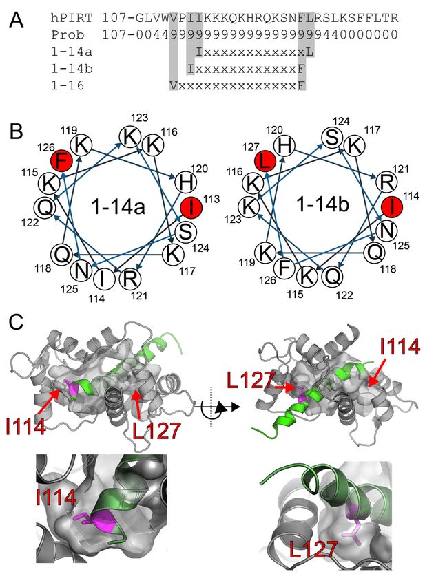
Biomolecules 2020, 10, 478                                                                         2 of 17

shown to regulate TRPV1 with neuropathic pain implications [25], and TRPV1-dependent uterine
contraction pain [14]. PIRT is also a regulatory subunit for TRPM8, where it displays species-dependent
effects enhancing and attenuating TRPM8-dependent conductance in the rodent and human proteins,
respectively [23]. PIRT regulation of TRPM8 appears to arise through competitively binding and
modulating access of the lipid PIP2 [22,23].
      Despite a preponderance of data showing that PIRT regulates TRP channels, there is a growing body
of evidence showing that PIRT has regulatory functions that may not be directly related to TRP channels.
It has been reported to interact with P2 × 3 channels and that it could impact overactive bladder [26],
and also co-localizes with P2X2 within the enteric nervous system [27]. Given that PIRT interacts with
PIP2 , and that a number of PIP2 -dependent channels are also calmodulin-regulated [11,16,28–30], we
sought to investigate if PIRT interacts with calmodulin and other common modalities that impact ion
channel function.
      The highly conserved calcium sensor, calmodulin (CaM), is a known ion channel regulator [17,
19,31–33] that downregulates Ca2+ -permeable cation channels, including TRP channels, through a
negative feedback mechanism that prevents excessive calcium influx [1]. In TRPV1, CaM competes for
a binding site with PIP2 and another calcium-binding protein, S100A1 [11]. There is direct evidence
that CaM binds to isolated TRPV1 peptides [10]. Complementing this evidence are cryo-EM structures
of a related channel, TRPV6, which identifies the location and stoichiometry of the interaction [18]. The
structure reveals that CaM binds to TRPV6 in a region that is structurally homologous to the bacterial
potassium channel KCNQ1 [21]. It is plausible, based on a conserved CaM binding region in TRPV6,
that TRPV1 and TRPM8 bind to CaM in an analogous manner.
      TRPM8 is functionally modulated by CaM, where it desensitizes channel conductance in a PIP2
dependent manner [16] with calcium influx leading to CaM and PIP2 -dependent desensitization of
TRPM8. It is now understood that TRPM8 binds to and is regulated by both PIRT and PIP2 [24,34];
however, it is not currently clear how the CaM-dependent modulatory process functions in the context
of PIRT. Nonetheless, with the evidence that PIRT, TRPV1, and TRPM8 bind PIP2 [11,12,22–24,35–38],
and given the common interdependence of PIP2 –CaM regulation of ion channels, we hypothesize that
PIRT may have a yet to be a determined role in CaM-dependent TRP channel regulation.
      Herein, we predict a CaM binding site in the human PIRT using established bioinformatic
techniques [39] and then use this information to guide binding assays to identify the amino acids
that bind CaM. Our results show that PIRT and CaM bind to each other, which likely leads to the
modulation of ion channel access to PIP2 and calcium, both of which regulate TRPM8, TRPV1, and
other channels with which PIRT may yet interact. Encouraged by the CaM binding data, we used
additional bioinformatics tools and surveyed the literature to identify additional potential PIRT
binding molecules. Using microscale thermophoresis (MST), our ligand screen focused on a number of
cholesterol-like molecules, including cholesteryl-hemisuccinate, cortisol, and β-estradiol, and other TRP
channel modulator ligands, including oxytocin and cholecalciferol, we show that the cholesterol-like
compounds bind PIRT at a previously unrecognized cholesterol-binding domain (CRAC) [40] and that
PIRT shows specificity for β-estradiol but not testosterone and for oxytocin over vasopressin. The
ability of PIRT to bind these ligands suggests that it functions as a multimodal TRP channel modulator
with implications in other ion channel functions.

2. Materials and Methods

2.1. Rosetta Flexible Peptide and High-Resolution Docking
      We used Rosetta ab initio flexible peptide docking protocols [41] to fold and dock the PIRT
C-terminal α-helix (residues 112-137) into the NMR structure of CaM (PDB: 2K0F). We generated
3, 5, and 9-mer fragments for PIRT according to established chemical shift quota filtered fragment
generation protocols that are commonly used for Chemical-Shift-Rosetta (CS-Rosetta) [42]. We used
these fragments and ensemble state number one from 2K0F to calculate 80,000 docking decoys. The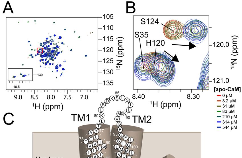
Biomolecules 2020, 10, 478                                                                           3 of 17

lowest scoring decoy was used to rescore the docked decoys, where the corresponding energy funnel
signifies convergence. As per standard docking and Rosetta analysis protocols, we selected the decoy
with the lowest interface score decoy with the lowest RMSD as the converged or working model to do
a final standard fast relax [43].

2.2. Protein Expression and Purification
      The expression and purification of PIRT were carried out following the established protocols in
our previous work on PIRT [23,24].
      Human CaM was expressed at 37 ◦ C with a hexahistidine tag comprising MGHHHHHHG- inserted
into a pET29 vector with kanamycin resistance and overexpressed in BL21 (DE3) cells. The cells were
grown in 14 N-M9 minimal media (42 mM disodium phosphate (Sigma-Aldrich), 17 mM dipotassium
phosphate (Sigma-Aldrich), 8 mM sodium chloride (Sigma-Aldrich), 17 mM ammonium chloride
(Sigma-Aldrich), 1× working solution of MEM vitamin solution (Corning), 1 mM magnesium sulfate
(Sigma-Aldrich), 100 µM calcium chloride (Sigma-Aldrich), and 22 mM D-glucose (Sigma-Aldrich)).
For NMR studies on 15 N-calmodulin, 15 N-ammonium chloride was the sole nitrogen source in the M9
minimal media. The cells were induced at OD600 = 0.6 with 0.5 mM IPTG (Sigma-Aldrich). The cells
were pelleted after 5 hr of induction at 6000×g at 4 ◦ C for 20 min, which resulted in 3 g of cellular mass
per 500 mL of M9 culture.
      The cell pellet was resuspended and homogenized by tumbling for 1 hr in lysis buffer consisting
of lysozyme (0.2 mg/mL), RNase (0.02 mg/mL), DNase (0.02 mg/mL), 1 mM phenylmethanesulfonylfluoride
(PMSF, Sigma-Aldrich), 5 mM magnesium acetate (Sigma-Aldrich), 50 mM HEPES (Sigma-Aldrich)
at pH 7.7, and 300 mM NaCl. The suspended cells were lysed using a sonicator (QSonica Q500) at
50% duty cycle and 50% amplitude for 7.5 min total on time. Cellular debris was removed with
centrifugation of cell lysate at 38,000× g at 4 ◦ C for 20 min. The supernatant was used for the following
steps after discarding the pellet. The supernatant was tumbled for 1 hr and then loaded onto 2 mL of
pre-equilibrated Ni-NTA (QIAGEN: 2 mL of resin per gram of cell pellet) within a gravity column. The
Ni-NTA was pre-equilibrated with lysis buffer (50 mM HEPES (Sigma-Aldrich) pH 7.7 and 300 mM
NaCl). Purification was then carried out with a flow-through buffer (50 mM HEPES pH 7.5), low
imidazole wash (50 mM HEPES pH 7.5, 10 mM imidazole), and finally an elution buffer (50 mM HEPES
pH 7.5, 300 mM imidazole). Following Ni-NTA chromatography, the eluent was concentrated to a
volume of 500 µL and loaded directly onto a 60 mL column volume Superdex 200 (GE Healthcare
Life Sciences) pre-equilibrated with 50 mM HEPES at pH 7.5 and separated by size. CaM purity was
assessed with SDS-PAGE and the identity confirmed by western blot analysis with a penta-histidine
primary antibody and anti-mouse alkaline phosphatase detection (Figure S1). We then used NMR to
show that our CaM construct retains functional properties by heteronuclear single-quantum coherence
(HSQC) NMR in the presence and absence of calcium. The resulting spectra show characteristic
chemical shift perturbation for CaM (Figure S1).
      For the pull-down experiment, the His-tagged CaM was buffer exchanged using a 10 kDa cutoff
Amicon Ultra centrifugal filter (Millipore) into a thrombin cleavage buffer (25 mM Na2 HPO4 , 150 mM
NaCl, pH 7.8) following Ni-NTA purification. The sample was then tumbled with 3 units of thrombin
(Novagen) for 24 h at room temperature before flowing over Ni-NTA. The collected flow-through
contained His-tag cleaved CaM, which was further purified by gel filtration chromatography.

2.3. Microscale Thermophoresis
     Human PIRT was fluorescently labeled according to previous protocols [24]. For the MST
measurements, the concentration of PIRT was kept consistent at 200 nM for all ligands in MST buffer
(0.1% DPC (w/v), 50 mM HEPES, pH 7.0). Apo-CaM was purified as mentioned above, with 0.5 mM
EDTA added to the size exclusion buffer and kept consistent throughout the MST measurements.
     Cortisol, β-estradiol, testosterone, cholesteryl-hemisuccinate, and cholecalciferol were all
solubilized as stock solutions in neat chloroform. From the ligand stock solutions, the desired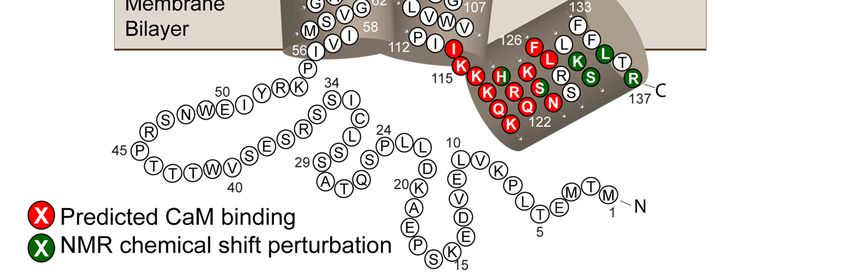
Biomolecules 2020, 10, 478                                                                         4 of 17

amount of the ligand for MST experiments was aliquoted into 200 µL PCR tubes, and the chloroform
was evaporated off under streaming N2 at room temperature. The compounds were then resuspended
in MST buffer with 200 nM PIRT. The disposal of testosterone was carried out according to U.S. Drug
Enforcement Administration (DEA) standards under Title 21 Code of Federal Regulations. Oxytocin,
vasopressin, calcium chloride, and nicotinamide stock solutions were prepared directly in MST buffer
and aliquoted to desired concentrations for MST analysis.
     The MST labeled PIRT was added to the solutions and incubated for 1 hr at room temperature.
After incubation, a standard MST glass capillary tube (NanoTemper) was drawn into the tube by
capillary action (~5 µL). MST experiments were carried out in triplicate at room temperature with 50%
infrared laser power and green channel using 10% excitation power.
     All of the data from the MST ligand screen that showed ligand-dependent thermophoresis were
normalized to free PIRT and bound PIRT following established protocols [44,45], from which the
dissociation constant was calculated. Ligand-independent (i.e., non-binding) thermophoresis is evident
in the controls (Figures S2A and S3). Ligand titration concentrations were optimized to show saturation
and minima within the bounds of solubility; i.e., the cholesterol-like ligands tended to become insoluble
above the concentrations used. The data were fit with in-house Python scripts where the errors are
reported as the root-mean-square error (RMSE) of the fit. Typically, membrane protein binding studies
of hydrophobic compounds use units of mole percent [24,44,46]. However, given that we screened both
hydrophobic and hydrophilic compounds, we used molar-dependent units with a constant PIRT and
membrane mimic concentration in order to compare the binding studies across chemical environments.

2.4. PIRT and Calmodulin Pull-Down Assay
     The pull-down experiment was conducted by mixing 10 µg His-tagged PIRT to 20 µg CaM (sans
His-tag) and tumbled at room temperature to allow the mixture to come to equilibrium. The sample
was then exposed to 200 µL of Ni2+ -bound nitrilotriacetic acid (Ni-NTA) resin and washed with
50 column volumes of 50 mM HEPES, 0.1% DDM, pH 7.5 to elute any unbound CaM. The bound
sample was then eluted with 50 mM HEPES, 0.1% DDM, 500 mM imidazole, pH 7.5, and analyzed by
SDS-PAGE gel.

2.5. Nuclear Magnetic Resonance-Detected Binding Assay
      15 N-human  PIRT (180 µL, 3 mm NMR tube) and 15 N-human CaM (550 µL, 5 mm NMR tube) were
measured in NMR buffer (4% D2 O (v/v, Sigma Aldrich), 20 mM sodium phosphate (Fisher Scientific),
500 µM DSS (Sodium trimethylsilylpropanesulfonate, Sigma Aldrich), and 0.5 mM EDTA (Sigma
Aldrich) at pH 6.5) on a Bruker 850 MHz 1 H magnet with Avance III console. Two thousand forty-eight
direct points and 128 indirect points were collected with 128 transients, processed in NMRpipe [47], and
analyzed in CCPNMR [48]. Optimization of NMR conditions for PIRT was carried out previously [24],
resulting in dodecylphosphocholine (DPC) as the most suitable detergent for investigations with PIRT
and at a temperature of 40 ◦ C. An HSQC of 15 N-human CaM was measured with and without CaCl2 to
show that it was properly folded in the conditions we tested.
     To validate the cholesterol-like and β-estradiol binding site, a saturating concentration of
β-estradiol (3.82 mole %) was dissolved in DMSO before adding to a 15 N-human PIRT sample.
An HSQC of human PIRT was measured before and after adding β-estradiol, and the resonances with
significant chemical shift perturbation identify the amino acids that comprise the binding site.

3. Results

3.1. Bioinformatic Analysis of PIRT Predicts Calmodulin-Binding Motifs
     The CaM 1-14 motif has a consensus sequence of (FLIVW)-X12 -(FLIVW), where the X is any amino
acid flanked by hydrophobic Phe, Leu, Ile, Val, or Trp. With the CaM target database web server [49],
PIRT was identified to contain a highly conserved 1-14 calmodulin-binding motif in the C-terminal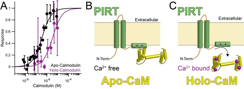
Biomolecules 2020, 10, 478                                                                                      5 of 17
Biomolecules 2020, 10, x; doi:                                                                                  5 of 17

C-terminal
-helix, and -helix,  andtwo
              there are    there are two
                              possible     possible
                                         motif       motiflocated
                                               positions   positions   located atIle113
                                                                    at residues    residues    Ile113toorPhe126
                                                                                          or Ile114        Ile114 to
                                                                                                                   or
Phe126   or  Leu127    (Figures  1 and   S4). Residue   number    127  is not  100%   conserved
 Leu127 (Figure 1 and Figure S4). Residue number 127 is not 100% conserved for a leucine; however,   for a  leucine;
however,    sequence shows
 sequence homology      homology    shows that phenylalanine
                               that phenylalanine                   can occupy
                                                     can occupy position           position
                                                                            127, which        127,
                                                                                         is still    which iswith
                                                                                                  consistent     stilla
consistent  with  a 1–14   motif. Worth   noting  is that PIRT  may   have   an additional    CaM
1–14 motif. Worth noting is that PIRT may have an additional CaM motif of 1–16 from position Val111  motif   of 1–16
from  position
 to Phe126      Val111
            (Figure  1A).toHowever,
                            Phe126 (Figure    1A). However,
                                     if this motif            if this
                                                   location truly     motif
                                                                  binds      location
                                                                          PIRT, we aretruly
                                                                                        unablebinds   PIRT, we
                                                                                                  to resolve      are
                                                                                                              it with
unable  to resolve
 our available  NMR it with
                        data.our available NMR data.

       Figure 1. PIRT has a highly conserved calmodulin-binding motif found in the C-terminal α-helix.
      Figure 1. PIRT has a highly conserved calmodulin-binding motif found in the C-terminal α-helix. (A)
       (A) We performed bioinformatic analysis on the PIRT sequence and show here that it has putative 1–14
      We performed bioinformatic analysis on the PIRT sequence and show here that it has putative 1–14
       and 1–16 motifs. We exclude the 1–16 motif as Val111 is likely found well within the membrane bilayer,
      and 1–16 motifs. We exclude the 1–16 motif as Val111 is likely found well within the membrane
       preventing calmodulin-binding. The helical projections in (B) show that both 1–14 motifs are consistent
      bilayer, preventing calmodulin-binding. The helical projections in (B) show that both 1–14 motifs are
       with an α-helix with hydrophobic residues that would occupy the calmodulin-binding pockets in the
      consistent with an α-helix with hydrophobic residues that would occupy the calmodulin-binding
       N- and C-terminal lobes. (C) Using Rosetta ab initio flexible peptide docking, we folded the C-terminal
      pockets in the N- and C-terminal lobes. (C) Using Rosetta ab initio flexible peptide docking, we folded
       α-helix into the calmodulin binding pocket and highlight that the peptide that fits best is the 1–14 motif
      the C-terminal α-helix into the calmodulin binding pocket and highlight that the peptide that fits best
       with Ile114 and Leu127.
      is the 1–14 motif with Ile114 and Leu127.
      Using the GPS 2.0 tool, we identified two previously unknown calmodulin-dependent kinase
      Using the
 II (CaMKII)     GPS with
              motifs  2.0 tool,
                            the we  identified
                                sequence        two previously
                                           R-X-X-S/T            unknown
                                                       in PIRT, both       calmodulin-dependent
                                                                      of which                      kinase II
                                                                               have very high conservation
(CaMKII)   motifs
 (Figure S4) [50]. with  the sequence
                    The amino           R-X-X-S/T
                                 acids Arg36         in PIRT,
                                                to Ser39       bothconserved
                                                         are 100%    of which have  very
                                                                               across thehigh   conservation
                                                                                           phosphoinositide
(Figure  S4) [50]. The  amino   acids  Arg36   to Ser39  are 100%   conserved  across the  phosphoinositide
 interaction proteins and are consistent with the minimal recognition site for CaMKII [50]. There is less
interaction
 conservationproteins
               for the and  areCaMKII
                       second   consistent
                                         sitewith  the minimal
                                             at amino            recognition
                                                        acid locations Arg121site for CaMKII
                                                                               to Ser124,      [50]. There is
                                                                                          but conservation  is
less conservation for the second CaMKII site at amino acid locations Arg121 to Ser124, but
Biomolecules 2020, 10, 478 6 of 17
Biomolecules 2020, 10, x; doi: 6 of 17
conservation is still high with Arg121 to being overall highly conserved. We did not attempt to
still highthe
validate with Arg121 to
predicted being overall
CaMKII but 185
motifs, highly conserved. We did
mention them notfor
here attempt to validate the predicted
completeness.
CaMKII motifs, but 185 mention them here for completeness.
3.2. Flex Pep Dock
3.2. Flex Pep Dock
Using Rosetta flexible peptide ab initio docking protocols, we were able to model how the
Usingcharged
positively RosettaC-terminal
flexible peptide
α-helixabofinitio
PIRT docking protocols,
might bind with thewe were able
negatively to model
charged how the
calmodulin.
positively charged C-terminal α-helix of PIRT might bind with the negatively
We previously assigned the amino acid backbone resonances for the C-terminal amino acids from charged calmodulin.
We previously assigned
Lys117–Arg137 and usedthe amino
these acid backbone
assignments to makeresonances for the constrained
experimentally C-terminal amino acidswhich
fragments, from
Lys117–Arg137 and used these assignments to make experimentally constrained
take into account the φ/ψ angles derived from NMR as well as evolutionarily conserved φ/ψ angles fragments, which
takestretches
for into account the ϕ/ψ
of similar aminoangles derived
acids. from NMR
The Rosetta as well show
calculations as evolutionarily conserved
clear convergence angles
ϕ/ψS5)
(Figure and
for stretches of similar amino acids. The Rosetta calculations show clear convergence
localize the C-terminal α-helix to our predicted 1–14 CaM-binding motif (Figure 1A). The helical (Figure S5) and
localize the C-terminal α-helix to our predicted 1–14 CaM-binding motif (Figure
wheel representation (Figure 1B), made with using the European Molecular Biology Open Software 1A). The helical wheel
representation
Suite [51], shows(Figure
that 1B),
it ismade with possible
certainly using thefor European
there toMolecular Biology Open
be two different Software
1–14 site Suite [51],
that would be
shows that it is certainly possible for there to be two different 1–14 site
amenable for binding within the CaM hydrophobic pockets; however, our models predominately that would be amenable for
bindingthe
found within the CaM
position Ile114hydrophobic
to Leu127 topockets; however, our models
be the lowest-scoring; predominately
i.e., lowest found
energy, and the position
therefore, best
Ile114 to Leu127
docking (Figure S5). to be the lowest-scoring; i.e., lowest energy, and therefore, best docking (Figure S5).
3.3. PIRT Specifically Interacts with Calmodulin
3.3. PIRT Specifically Interacts with Calmodulin
PIRT has a high affinity for calcium-free CaM (apo-CaM) and a reduced affinity for calcium
PIRT has a high affinity for calcium-free CaM (apo-CaM) and a reduced affinity for calcium
bound calmodulin. The MST binding screen shows that PIRT binds tightly in the mid to low nM
bound calmodulin. The MST binding screen shows that PIRT binds tightly in the mid to low nM
range, K = 350 ± 40 nM for apo-CaM (Table 1, Figure 2A). Calcium bound CaM (holo-CaM) has a
range, Kdd = 350 ± 40 nM for apo-CaM (Table 1, Figure 2A). Calcium bound CaM (holo-CaM) has a
decreased affinity for PIRT(i.e., weaker binding) and right-shifts the binding curve by about 200-fold to
decreased affinity for PIRT(i.e., weaker binding) and right-shifts the binding curve by about 200-fold
an apparent K = 60 ± 30 µM (Table 1, Figure 2A). PIRT does not bind to calcium chloride (Figures S2A
to an apparentd Kd = 60 ± 30 μM (Table 1, Figure 2A). PIRT does not bind to calcium chloride (Figures
and S3A).
S2A and S3A).
Table 1. Ligands bound to PIRT using MST.
Table 1. Ligands bound to PIRT using MST.
Ligand Kd (M) RMSE (M) Ligand Type
Ligand Kd (M) RMSE (M) Ligand Type
Calmodulin (apo, free) (apo, free) 350 ×
Calmodulin 10×−910−9
350 40 × 1040−9× 10⁻⁹ CaM, Intracellular
CaM, Intracellular Protein Protein
Calmodulin (holo, bound) −6 30−6× 10⁻⁶ Calcium Bound Calcium
60 ×
Calmodulin (holo, bound) 60 × 10−610 30 × 10 CaMBound CaM
CholesterylCholesteryl
HS × −6 −6 SteroidPrecursor
Hormone Precursor
HS 103 10
103 × 10−6 6 × 10⁶−6× 10 Steroid Hormone
Cortisol Cortisol 790 × 10 −6 50 × 10−6 Cortical Steroid Hormone
790 × 10−6 50 × 10 −6 Cortical Steroid Hormone
β-Estradiolβ-Estradiol 800 × 10×−610−6
800 100
100 × 10 −6× 10 −6
Sex Steroid Hormone Hormone
Sex Steroid
Cholecalciferol
Cholecalciferol 10×−310−3
2.1 ×2.1 0.4
0.4 × 10 −3 × 10 −3 Secosteroid Hormone
Secosteroid Hormone
Oxytocin 7 × 10⁻⁶ −6 1−6× 10⁻⁶ Peptide Hormone
Oxytocin 7 × 10 1 × 10 Peptide Hormone
Figure 2. PIRT
PIRT (Phosphoinositide-interacting
(Phosphoinositide-interacting regulator
regulator of
of TRP)
TRP) binds
binds to calcium-free calmodulin
(apo-CaM) andandcalcium
calciumbound
bound calmodulin
calmodulin (holo-CaM). (A) (A)
(holo-CaM). PIRTPIRT
bindsbinds
calcium-free calmodulin
calcium-free (apo-
calmodulin
CaM, black)
(apo-CaM, withwith
black) approximately
approximately200-fold higher
200-fold affinity
higher affinitythan
thanCaCa2+2+
-bound
-boundcalmodulin
calmodulin(holo-CaM,
(holo-CaM,
purple). (B)
(B) The
The differences
differences in
in affinity
affinity suggest
suggest the possibility
possibility that
that PIRT is bound to apo-CaM until
intracellular calcium levels rise high enough for holo-CaM to bind calcium and release release PIRT
PIRT (C).
(C).Biomolecules 2020, 10, 478 7 of 17
To verify the CaM–PIRT interaction, a pull-down assay was conducted by mixing purified
Biomolecules 2020, 10, x; doi: 7 of 17
His-tagged PIRT with His-cleaved CaM at room temperature and then tumbled with Ni-NTA resin.
To verify the CaM–PIRT interaction, a pull-down assay was conducted by mixing purified His-
The sample was washed extensively with 50 column volumes to remove any unbound protein. CaM
tagged PIRT with His-cleaved CaM at room temperature and then tumbled with Ni-NTA resin. The
co-elutes
samplewith
wasthe imidazole
washed elution
extensively of PIRT.
with The Coomassie
50 column stain gel
volumes to remove anyshows that protein.
unbound PIRT and CaM
CaM co- were
co-eluted confirming direct binding between PIRT and CaM (Figure S6).
elutes with the imidazole elution of PIRT. The Coomassie stain gel shows that PIRT and CaM were
co-eluted confirming direct binding between PIRT and CaM (Figure S6).
3.4. NMR Titration Shows Residues with Ligand-Dependent Perturbation Including the Predicted Calmodulin
Binding
3.4.Site
NMR Titration Shows Residues with Ligand-Dependent Perturbation Including the Predicted
Calmodulin Binding15Site
NMR-detected N labeled PIRT titrations with CaM show chemical shift perturbations for
calcium-free CaM (Figure
NMR-detected 15N 3A) within
labeled thetitrations
PIRT C-terminal withα-helix
CaM show(Figure 3B). Interestingly,
chemical Ser124for
shift perturbations shows
calcium-free CaM (Figure 3A) within the C-terminal α-helix (Figure 3B). Interestingly,
perturbations suggestive of slow-exchange on the NMR timescale with the disappearance of the Ser124 shows
perturbations
initial suggestive
peak, presumably theofligand-free
slow-exchange
state,onandthereappearance
NMR timescaleofwith the disappearance
the apparent of the
bound peak across
initial peak,
the titration presumably the
concentrations; ligand-freeof
a hallmark state, and reappearance
slow-exchange and of the binding.
tight apparent bound peak across
In addition to Ser124,
the titration concentrations; a hallmark of slow-exchange and tight binding. In addition to Ser124,
chemical shift perturbation is also seen for His120 in the predicted 1–14 calmodulin-binding motif,
chemical shift perturbation is also seen for His120 in the predicted 1–14 calmodulin-binding motif,
while residues Lys131, Ser132, Leu135, and Arg137 also show chemical shift perturbation and are
while residues Lys131, Ser132, Leu135, and Arg137 also show chemical shift perturbation and are
found in close
found proximity
in close to to
proximity thethe
binding
bindingmotif
motif (Figure 1C).
(Figure 1C).
FigureFigure 3. PIRT
3. PIRT hashas residuesthat
residues thatbind
bindto
to calcium-free
calcium-free calmodulin
calmodulin in in
thethe
C-terminal α-helix
C-terminal that is
α-helix that is
located
located in orinnear
or near the 1‒14
the 1-14 motif.
motif. (A)(A)
TheThe
HSQCHSQC follows
follows thetitration
the titrationofofNMR
NMRinvisible 14N-calcium-
invisible 14 N-calcium-free
free calmodulin with NMR-detected 15N-PIRT with concentrations of 0, 3.2, 31, 83, 210, 314, 544 μM
calmodulin with NMR-detected 15 N-PIRT with concentrations of 0, 3.2, 31, 83, 210, 314, 544 µM
calcium-free calmodulin. (B) Highlighted from the red inset on the HSQC are Ser35 showing no
calcium-free calmodulin. (B) Highlighted from the red inset on the HSQC are Ser35 showing no
perturbation, His120 showing chemical shift perturbation show with the arrow, and Ser124 showing
perturbation, His120 showing chemical shift perturbation show with the arrow, and Ser124 showing
an apparent slow exchanging resonance with the arrow highlighting the movement. In (C), the
an apparent slow exchanging resonance with the arrow highlighting the movement. In (C), the
calmodulin-binding motif is highlighted in red from Ile114 to Leu127 that was shown with Rosetta
calmodulin-binding
flexible docking tomotif
be theis highlighted
best inHighlighted
fitting motif. red from Ile114
in darkto Leu127
green that was
are residues shown
that with Rosetta
show chemical
flexible docking
shift to be the
perturbations withbest
the fitting motif. Highlighted in dark green are residues that show chemical
NMR titration.
shift perturbations with the NMR titration.Biomolecules 2020, 10, 478 8 of 17
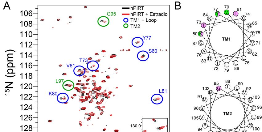
3.5. Two Cholesterol-Binding Motifs Found on PIRT with Bioinformatic Analysis
PIRT has two putative cholesterol binding motifs in the first transmembrane α-helix. Cholesterol-
binding proteins share a consensus sequence motif named cholesterol-recognition amino acid consensus
called CRAC, and CARC, where CARC is the mirror image of CRAC. The CRAC amino acid sequence
is (L/V)-X1-5 -(Y)-X1-5 -(K/R), with N to C terminal directionality, and X stands for any amino acids [40].
Using the known molecular motifs for cholesterol-binding proteins, we report two cholesterol-like
binding sites in the first transmembrane α-helix of human PIRT with CRAC in the outer leaflet of the
membrane bilayers and CARC in the inner leaflet (Figure 4 and Figure S4).
Biomolecules 2020, 10, x; doi: 9 of 17
Figure Figure
4. PIRT4. has
PIRT has a conserved
a conserved cholesterol-binding
cholesterol-binding CRACand
CRAC andCARC
CARCmotif.
motif. (A)
(A) We
We analyzed
analyzedthe the PIRT
PIRT sequence and identify that it contains a CRAC motif in the outer leaflet of the first
sequence and identify that it contains a CRAC motif in the outer leaflet of the first transmembrane
transmembrane α-helix with amino acids highlighted in green and a CARC domain in the inner leaflet
withamino
α-helixwith amino acids highlighted in green and a CARC domain in the inner leaflet with amino
acids highlighted in cyan. Shown in the helical wheel projection, the CRAC domain is
acids highlighted in
supported with the cyan. Shown
evidence thatin the helical
within wheel
a standard projection,
α-helix, the CRAC domain
the Val70-Tyr77-Lys80 are on theissame
supported
with the evidence that within a standard α-helix, the Val70-Tyr77-Lys80 are on the
interface and are confirmed to bind to cholesterol with the NMR data below. (B) The amino acids that same interface
and aremayconfirmed to bind to
bind to cholesterol cholesterolβ-estradiol,
hemisuccinate, with the NMR dataand
and cortisol below. (B) The
are shown hereamino acids that
as an example of may
bind tohow PIRT could
cholesterol bind these ligands.
hemisuccinate, To bind the
β-estradiol, andβ-estradiol
cortisolorandcortisol, we show
are shown hereas
here that
anPIRT could of how
example
incorporate
PIRT could bind Thr73, which is onTothe
these ligands. samethe
bind interface as the CRAC
β-estradiol domainwe
or cortisol, residues.
show In Figure
here thatS4,PIRT
our could
alignment highlights a second cholesterol-binding domain, CARC, it resides outside of the membrane
incorporate Thr73, which is on the same interface as the CRAC domain residues. In Figure S4, our
bilayer, does not have a helical interface consistent with a standard α-helix, and is not highlighted
alignment highlights a second cholesterol-binding domain, CARC, it resides outside of the membrane
here. (C) PIRT ligand-dependent thermophoresis of cholesteryl-hemisuccinate, β-estradiol, and
bilayer,cortisol.
does not Thehave a helical
Kd values interface
are listed consistent
in Table 1. with a standard α-helix, and is not highlighted here.
(C) PIRT ligand-dependent thermophoresis of cholesteryl-hemisuccinate, β-estradiol, and cortisol. The
Kd values are listed in Table 1.Biomolecules 2020, 10, 478 9 of 17
3.6. PIRT and Cholesterol Ligands Screening with Microscale Thermophoresis
We used microscale thermophoresis to investigate PIRT–cholesterol interactions. Given the
solubility limitations of cholesterol, we tested the predicted cholesterol-binding with the more soluble
cholesteryl-hemisuccinate, which shows an affinity of 103 ± 6 µM (Figure 4). Cholesterol derivatives
also bind PIRT with cortisol and β-estradiol Kd values of 790 ± 60 µM and 800 ± 100 µM, respectively
(Figure 4). Surprisingly, testosterone does not bind to PIRT (Figure S3B) despite its structural similarity
to cholesterol, cortisol, and β-estradiol.
3.7. β-Estradiol Induced PIRT Chemical Shift Perturbation
To validate the predicted CRAC cholesterol-binding site in PIRT, a saturating concentration of
β-estradiol was added to 15 N labeled PIRT. The resulting TROSY-HSQC spectra show chemical shift
perturbations primarily in the upper transmembrane α-helix 1 (TM1) with a few that breach to the
upper transmembrane α-helix 2 (TM2) (Figure 5). More specifically, chemical shift perturbations were
seen at residues Ser60, Val61, Thr73, Tyr77, Lys80, and Leu81 in TM1 while Gly95 and Leu97 are seen
in the TM2. The NMR data confirm the CRAC binding site location (Figures 4 and 5). The NMR data
also show limited perturbation away from the CRAC site upon β-estradiol binding, with chemical
shift perturbation of Ser60 and Val61 in TM1 and Gly95 and Leu97 in TM2, which are outside of the
predicted CRAC domain, this could indicate that β-estradiol induces a change in PIRT conformation
or dynamics.
Biomolecules 2020, 10, x; doi: 10 of 17
Figure
Figure 5.5. Identification
Identificationof of the
the PIRT-β-estradiol
PIRT-β-estradiolbinding
bindingsitesitebybyNMR.
NMR.(A) (A) The
The NMR
NMR HSQCHSQC ofof PIRT
PIRT
(black) againstPIRT
(black) overlaid against PIRTwith
witha a3.82-mole
3.82-mole percent
percent β-estradiol
β-estradiol (red).
(red). Chemical
Chemical shift
shift perturbations
perturbations are
are
seenseen
in theinfirst
thetransmembrane
first transmembrane α-helix
α-helix (S60, V61,(S60, V61, K80,
T73, Y77, T73,andY77,
L81) K80,
and and
secondL81) and second
transmembrane
α-helix (G95 andα-helix
transmembrane L97). (B)(G95
Helical
andwheel
L97).projection
(B) Helicalof the first and
wheel second of
projection transmembrane
the first andα-helices
second
(TM1 and TM2, respectively)
transmembrane α-helices (TM1 withand
the TM2,
predicted CRAC domain
respectively) with (green) compared
the predicted to thedomain
CRAC NMR chemical
(green)
shift perturbation
compared to the NMR (pink). The chemical
chemical shift perturbation
shift perturbation seenchemical
(pink). The in the upper sections of the
shift perturbation α-helices
seen in the
supports
upper the CRAC
sections of thedomain
α-helicesprediction.
supportsResidues
the CRAC S60domain
and V61 in TM1 and
prediction. G95 andS60
Residues L97and
in TM2 suggest
V61 in TM1
there
and G95might
andbe L97 a conformational change
in TM2 suggest there when
might beβ-estradiol binds with
a conformational PIRT.
change when β-estradiol binds with
PIRT.
To show the significance of the MST binding data, we used nicotinamide (Figure S3D) as a
control because it has well-known metabolism and should not interact with PIRT. In previous MST
PIRT binding studies using similar conditions, DMSO was previously shown to not bind to PIRT [24].
4. DiscussionBiomolecules 2020, 10, 478 10 of 17
3.8. Microscale Thermophoresis Shows PIRT Binds to Other Trp Channel Modulators
Cholecalciferol, the pro-hormone form of vitamin-D synthesized in the skin, has low structural
similarity to cholesterol-like molecules but is known to affect TRPV6 [52]. The MST data show that PIRT
specifically interacts, albeit with low affinity, to this secosteroid with an apparent Kd = 2.1 ± 0.4 mM
(Figure S7). Similarly, PIRT affects oxytocin-induced uterine contraction pain in a TRPV1-dependent
manner [14], which we used to test the hypothesis that PIRT may bind to oxytocin. Remarkably, PIRT
binds to oxytocin with a Kd = 7 ± 1 µM (Figure S7), and yet it does not bind to the structurally similar
peptide hormone R8-arginine vasopressin (Figure S3C).
To show the significance of the MST binding data, we used nicotinamide (Figure S3D) as a control
because it has well-known metabolism and should not interact with PIRT. In previous MST PIRT
binding studies using similar conditions, DMSO was previously shown to not bind to PIRT [24].
4. Discussion
Regulation of TRP channels by PIP2 , Ca2+ , and CaM is an active area of research. Several reviews
highlight the important features of these TRP channel modulatory mechanisms [53–57]. For some
TRPV channels, it is clear that CaM directly binds these channels and regulates function [58,59].
Structural studies of TRPV5 indicate that a single CaM can bridge two distinct TRPV5 binding sites [58].
Similarly, TRPV1 has at least two CaM binding sites, one in the N-terminal ankyrin repeat domain
(ARD) and another in the distal C-terminus (CT) [59]. The ARD CaM-binding site functions in
desensitization, while the CT CaM interaction has a higher affinity (lower Kd ), it has a less defined
functional consequence [59]. Titration of holo-CaM to the isolated TRPV1 distal C-terminus (CT) as
monitored by tryptophan fluorescence emission identifies an affinity of Kd = 5.4 × 10⁻⁸ M [59] which is
on par with the affinities identified between PIRT and CaM in this study. However, one significant
distinction is that PIRT has a higher affinity for apo-CaM; whereas, it is generally thought that holo-CaM,
the Ca2+ -bound form, is the regulatory form that plays a role in TRP channel desensitization [55].
While CaM modulation can be direct (i.e., direct binding to TRP channels), modulation can also
happen indirectly by impacting other TRP regulatory proteins such as CaM-dependent regulation
of phosphatases or kinases [16]. In TRPM8, CaM has been clearly shown to downregulate function
indirectly [16]. There is also some evidence that CaM directly binds to TRPM8 [60,61], which would
provide an additional mechanism of CaM-mediated functional downregulation. Beyond Ca2+ –CaM
modulation, at least for TRPM8 and other TRPM channels, it is clear that Ca2+ can directly bind and
desensitize these channels in the absence of CaM [62]. Our PIRT studies identify a CaM-binding site
that adds an additional layer of complexity to TRP channel function and modulation, where PIRT
could impact both direct and indirect CaM regulatory functions.
Using purified full-length human PIRT, we show it binds to CaM at the predicted C-terminal
α-helix, which suggests a possible role in regulating TRP channel function in concert with calcium and
PIP2 and suggests a broader modulatory role in ion channel regulation. With MST, calcium bound CaM
showed an approximately 200× decrease in affinity for PIRT than for the apo (calcium unbound) form,
which likely arises from known CaM conformational changes upon calcium loading and suggests that
calcium-binding of CaM may induce the dissociation of the PIRT–CaM interaction (Figure 2). The
PIRT–CaM interaction is confirmed with a pull-down experiment showing that CaM co-eluted with
PIRT elution.
We further tested PIRT binding to CaM with NMR titrations to provide amino acids specific details
and validate our theoretical CaM binding site on PIRT. Using our previously published PIRT amino acid
resonance assignments [24], we show that calcium-free CaM binds to PIRT in the predicted C-terminal
α-helix and that the changes in chemical shift indeed correspond to the predicted CaM-binding motif
(Figure 1C). The CaM-dependent chemical shift perturbations show several amino acids that display
binding, highlighting a possible allosteric effect arising from the interaction. To model our biochemical
and biophysical measurements, we used the chemical shift assignments to make Rosetta fragments
allowing us to computationally model CaM bound to the PIRT C-terminal α-helix. Our dockedBiomolecules 2020, 10, 478 11 of 17
model shows that the C-terminal α-helix fits well within the conserved CaM-binding motif with the
hydrophobic Ile113 fitting in the C-terminal lobe and Leu127 in the N-terminal lobe of CaM from PDB
entry 2K0F (Figure 1).
Apo-CaM binding to PIRT can be used to expand on known CaM downregulation of the TRPM8
function [16]. Cryo-EM structures of TRPM8 show that Ca2+ binds to the human TRPM8 S1-S4
membrane domain with Ca2+ chelated by Glu782, Gln785, Tyr793, Asn799, and Asp802 to prime
the channel for activation [34,62]. These amino acids, as well as Glu1068, are conserved for all
Ca2+ dependent TRPM channels (TRPM2, M4, M5, and M8) [15,63–65]. This conservation for Ca2+
dependent TRP channels, and their structural homology, suggest that TRPM8 potentially binds to Ca2+
in the same location and potentially has a similar effect on the channel. In TRPM8 channels, especially,
elevated levels of intracellular Ca2+ causes CaM downregulation of TRPM8 with a mechanism that
depends on PIP2 [16]. We previously showed that PIRT reduces the human TRPM8-dependent currents
by binding directly to the human TRPM8 S1-S4 domain [23] and shuttles PIP2 to TRPM8 [24], and this
mechanism is plausible for PIP2 specific inactivation of TRPM8. The data presented here support a
more complex mechanism that contextualizes CaM, PIP2 , Ca2+ , and PIRT downregulation of TRPM8
dependent currents.
Figure 6 contextualizes the known interactions that have been identified and relate CaM, Ca2+ ,
PIRT, and PIP2 to TRPM8 modulation. TRPM8 integrates a variety of stimuli, including, but not
limited to, temperature, menthol, and pH. These inputs are then modulated by an additional layer
of interactions between PIRT, CaM, and PIP2 , where PIRT can bind to apo-CaM and PIP2 , thereby
modulating TRPM8 access to these modulators. Currently, the regulatory cross-talk between CaM and
PIP2 is unknown, but given the spatial locations and apparent overlap of the respective PIRT binding
sites, it likely impacts the regulatory network. TRPM8 gating by its canonical stimuli (i.e., cold) is
thereby further tuned by a variety of modulators, including PIRT, that will ultimately regulate calcium
influx and thereby initiating signal transduction. Increases in intracellular calcium concentrations
result in direct Ca2+ -dependent TRPM8 desensitization [62]. Increased available calcium will also
impact CaM leading to higher concentrations of holo-CaM. As a result, because of the decreased affinity
of the PIRT—holo-CaM complex, we speculate that the complex would dissociate. Once the calcium
levels return to equilibrium, then the PIRT—CaM interaction cycle would continue with apo-CaM
rebinding PIRT. The apo-CaM and PIRT complex could potentially allow the channel to become active
again by allowing PIP2 to be shuttled to TRPM8 when the complex is formed. More studies that test
the intricacies of these putative interactions are still needed, but our data support a more detailed
negative regulation from Ca2+ for TRPM8-dependent currents.
PIRT appears to regulate function via diverse means. In this vein, we identified a conserved
cholesterol-binding domain called CRAC [40] in the first transmembrane α-helix of PIRT, and we show
that it does indeed bind to several cholesterol-like molecules with potential implication in ion channel
regulation [66]. We chose these ligands based on bioinformatics predictions on the PIRT sequence
matching cholesterol-binding motifs as well as ligands implicated in uterine contraction pain (oxytocin
and β-estradiol) and a pro-hormone form of vitamin D with ties to TRPV6 (cholecalciferol) [67].
Our data is the first to show that PIRT binds to cholesterol-like molecules with specificity for
cholesteryl-hemisuccinate, cortisol, and β-estradiol; however, it does not specifically bind testosterone.
Its interaction with cortisol suggests that it may play a more intimate role in TRP channel-specific
stress-induced inflammatory processes.
Binding to cholesteryl-hemisuccinate supports cellular electrophysiology measurements where
PIRT was shown to interact with the TRPM8 pore domain [23], and it is known that TRPM8 interacts
with cholesterol to partition it into cholesterol-rich membrane domains [68]. Additionally, TRPA1
also has a CRAC domain, which affects both membrane partitioning and function [69]. Furthermore,
cholesteryl-hemisuccinate was bound in cryo-EM structures of a handful of TRP channels, including
TRPC4, TRPM4, TRPV3, and TRPM8 [62,63,70,71]. With this physiological and structural data, andBiomolecules 2020, 10, 478 12 of 17
Biomolecules 2020, 10, x; doi: 12 of 17
our cholesterol-binding data and bioinformatics, we have uncovered another layer of PIRT regulation
is formed. More studies that test the intricacies of these putative interactions are still needed, but our
that may implicate it in is much more diverse physiology than previously thought.
data support a more detailed negative regulation from Ca2+ for TRPM8-dependent currents.
Figure 6.6. A
Figure A diagram
diagram relaying
relaying the
the interactions
interactions known
known so so far
far between
between CaM,
CaM, PIRT,
PIRT,PIP
PIP22,, TRPM8,
TRPM8, andand
Ca2+
Ca 2+. Inputs are taken and are modulated by the interactions between
. Inputs are taken and are modulated PIRT and
CaM, with PIRT and PIRT
PIRT with
with
PIP22. These interactions are then integrated to TRPM8, where
PIP where thethe channel
channel responds
responds by
by either
either opening
opening
or closing.
or closing. Once
Once the
the channels
channels open,
open, calcium
calcium influx
influx occurs,
occurs, causing
causing signal
signal transduction,
transduction, and
and the
the influx
influx
of calcium
of calciumcauses
causes aa feedback
feedback loop
loop where
where calcium
calcium will
will bind
bind CaM
CaM while
while also
also sending
sending negative
negative feedback
feedback
to
to TRPM8
TRPM8and anddesensitize
desensitizeit.
it.
Interestingly,
PIRT appearswhile β-estradiol
to regulate (female
function viasex hormone)
diverse means. bindsIn tightly
this vein, to PIRT, testosterone
we identified (male sex
a conserved
hormone) shows no evidence of binding (Figure S3B). This suggests
cholesterol-binding domain called CRAC [40] in the first transmembrane α-helix of PIRT, and we that there may be a sex-specific
regulatory
show that it role in indeed
does which PIRT bind plays a part
to several and may partially
cholesterol-like explain
molecules with thepotential
role PIRTimplication
plays in uterine
in ion
contraction pain [14]. In addition to uterine contraction pain, TRP
channel regulation [66]. We chose these ligands based on bioinformatics predictions on the PIRT channels have been implicated
to play a role
sequence in cancer
matching cells where expression
cholesterol-binding motifs ofasthese
well channels
as ligandscan affect cancer
implicated cell proliferation,
in uterine contraction
metastasis,
pain (oxytocin and even
and cell death [72].
β-estradiol) andIn abreast cancer cells,
pro-hormone formtheof TRPV vitaminfamily D and
withTRPM8
ties tochannels
TRPV6
expression
(cholecalciferol) [67]. Our data is the first to show that PIRT binds to cholesterol-like molecules data
are regulated by β-estradiol and estrogen receptors [73,74]. Our MST and NMR with
show that PIRT
specificity binds tightly to β-estradiol
for cholesteryl-hemisuccinate, and induce
cortisol, some evidence
and β-estradiol; of conformational
however, change in
it does not specifically
the
bind HSQC, which suggests
testosterone. PIRT might
Its interaction play a role
with cortisol between
suggests that TRP
it may channels
play a morewith β-estradiol
intimate role in in
breast
TRP
cancer cells. Supporting the role of PIRT
channel-specific stress-induced inflammatory processes. in sex-dependent function, a recent study of PIRT knock-out
(-/- ) mice indicate
Binding that there are subtle PIRT-dependent
to cholesteryl-hemisuccinate supports cellularimpacts in metabolismmeasurements
electrophysiology and obesity that are
where
female-specific [75].
PIRT was shown to interact with the TRPM8 pore domain [23], and it is known that TRPM8 interacts
withOur ligand screen
cholesterol showsitthat
to partition intoPIRT binding is not
cholesterol-rich limited todomains
membrane cholesterol-like hormones and
[68]. Additionally, that
TRPA1
cholecalciferol
also has a CRAC (Vitamin
domain, D3) specifically
which affects interacts with PIRT.
both membrane Cholecalciferol
partitioning is a hormone
and function synthesized
[69]. Furthermore,
in
cholesteryl-hemisuccinate was bound in cryo-EM structures of a handful of TRP channels, the
the skin from a cholesterol derivative but is structurally distinct from cholesterol with sterol
including
backbone
TRPC4, TRPM4, disrupted from UV
TRPV3, andradiation
TRPM8 provided by the
[62,63,70,71]. sun.this
With Vitamin D3 is welland
physiological known to be intimately
structural data, and
involved in bone formation,
our cholesterol-binding data andwithbioinformatics,
recent researchwe onhave
dietary calciumanother
uncovered and vitamin
layer ofD3 being
PIRT shown
regulation
to regulate the epithelial ion channels TRPV5 and TRPV6 [67].
that may implicate it in is much more diverse physiology than previously thought. It is unknown what role PIRT would
directly have on TRPV6
Interestingly, whileand β-estradiol
TRPV5; however,
(female sex based on emerging
hormone) evidence
binds tightly to of PIRTtestosterone
PIRT, as a general(male
TRP
sex hormone) shows no evidence of binding (Figure S3B). This suggests that there may be a sex-
specific regulatory role in which PIRT plays a part and may partially explain the role PIRT plays inBiomolecules 2020, 10, 478 13 of 17
channel regulator, it is plausible that PIRT regulates TRPV5 and TRPV6 in concert with the calcium
sensor CaM.
Lastly, our studies indicate that PIRT interacts specifically with peptide hormones. We show
that PIRT specifically binds oxytocin. It is plausible that PIRT is involved with oxytocin regulation
given that the oxytocin receptor is a Gqα type G protein-coupled receptor (GPCR), Gqα GPCRs directly
influence PIP2 levels, GPCRs are implicated with TRP channels, and PIRT is a PIP2 binding protein that
functionally modulates TRP channels [12–14,23,25,76–78]. Similarly, PIRT was shown to be important
for uterine contraction pain under oxytocin insult during birth in mice [14]. Interestingly, while PIRT
specifically interacts with oxytocin, it does not bind R8-arginine vasopressin, despite only two amino
acid differences in the structurally similar cyclic nonapeptides. It is notable that these hormones have
drastically distinct roles in endocrinology, controlling birth induction and blood pressure regulation,
respectively. In fact, oxytocin-induced childbirth can result in off-target effects complicating childbirth
that are thought to be caused by the similarity of these hormone structures [79,80]. These effects are
not fully understood, but with the identification of PIRT, specifically interacting with these hormones,
potentially new avenues to understand these side effects can be investigated.
5. Conclusions
The data presented here show several binding partners for PIRT with specific endocrinology roles
as well as implicate an enhanced model for TRPM8 regulation. These are the first data to suggest
PIRT interactions beyond those with TRP channels and PIP2 , which opens up multiple avenues of
PIRT-related research.
Supplementary Materials: The following are available online at http://www.mdpi.com/2218-273X/10/3/478/s1,
Figure S1: Characterization of Purified Calmodulin, Figure S2: Representative microscale thermophoresis trace data
with PIRT, Figure S3: PIRT Ligand-independent thermophoresis, Figure S4: PIRT conservation across mammalian
species, Figure S5: Unguided Rosetta ab initio flexible peptide docking, Figure S6: PIRT and calmodulin pull-down
Coomassie-stained gel, Figure S7: PIRT ligand-dependent thermophoresis. A representative coordinate file from
the Rosetta modeling of the calmodulin–PIRT interaction.
Author Contributions: Conceptualization, W.D.V.H and N.J.S.; investigation, N.J.S., D.D.L., M.K., and W.D.V.H.;
writing—original draft preparation, N.J.S., D.D.L., and W.D.V.H.; writing—review and editing, N.J.S., D.D.L.,
M.K., and W.D.V.H.; visualization, N.J.S., D.D.L., and W.D.V.H.; funding acquisition, W.D.V.H. All authors have
read and agreed to the published version of the manuscript.
Funding: This research was funded by the National Institutes of General Medical Sciences, grant number
R01GM112077 (W.D.V.H.).
Acknowledgments: We acknowledge the generosity of Professors Walter Chazin (Vanderbilt University School of
Medicine) and Jeremy Mills (Arizona State University) for their material and intellectual support in expression
and purification of human calmodulin.
Conflicts of Interest: The authors declare no conflict of interest.
References
1. Veldhuis, N.A.; Poole, D.P.; Grace, M.; McIntyre, P.; Bunnett, N.W. The G protein-coupled receptor-transient
receptor potential channel axis: Molecular insights for targeting disorders of sensation and inflammation.
Pharmacol. Rev. 2015, 67, 36–73. [CrossRef] [PubMed]
2. Proudfoot, C.J.; Garry, E.M.; Cottrell, D.F.; Rosie, R.; Anderson, H.; Robertson, D.C.; Fleetwood-Walker, S.M.;
Mitchell, R. Analgesia mediated by the TRPM8 cold receptor in chronic neuropathic pain. Curr. Biol. 2006,
16, 1591–1605. [CrossRef] [PubMed]
3. Tsavaler, L.; Shapero, M.H.; Morkowski, S.; Laus, R. Trp-p8, a novel prostate-specific gene, is up-regulated in
prostate cancer and other malignancies and shares high homology with transient receptor potential calcium
channel proteins. Cancer Res. 2001, 61, 3760–3769. [PubMed]
4. Zhang, L.; Barritt, G.J. Evidence that TRPM8 is an androgen-dependent Ca2+ channel required for the
survival of prostate cancer cells. Cancer Res. 2004, 64, 8365–8373. [CrossRef] [PubMed]Biomolecules 2020, 10, 478 14 of 17
5. Ma, S.; Yu, H.; Zhao, Z.; Luo, Z.; Chen, J.; Ni, Y.; Jin, R.; Ma, L.; Wang, P.; Zhu, Z.; et al. Activation of the
cold-sensing TRPM8 channel triggers UCP1-dependent thermogenesis and prevents obesity. J. Mol. Cell Biol.
2012, 4, 88–96. [CrossRef] [PubMed]
6. Rossi, H.L.; Jenkins, A.C.; Kaufman, J.; Bhattacharyya, I.; Caudle, R.M.; Neubert, J.K. Characterization of
bilateral trigeminal constriction injury using an operant facial pain assay. Neuroscience 2012, 224, 294–306.
[CrossRef]
7. Tominaga, M.; Caterina, M.J.; Malmberg, A.B.; Rosen, T.A.; Gilbert, H.; Skinner, K.; Raumann, B.E.;
Basbaum, A.I.; Julius, D. The cloned capsaicin receptor integrates multiple pain-producing stimuli. Neuron
1998, 21, 531–543. [CrossRef]
8. Garami, A.; Shimansky, Y.P.; Rumbus, Z.; Vizin, R.C.L.; Farkas, N.; Hegyi, J.; Szakacs, Z.; Solymar, M.;
Csenkey, A.; Chiche, D.A.; et al. Hyperthermia induced by transient receptor potential vanilloid-1 (TRPV1)
antagonists in human clinical trials: Insights from mathematical modeling and meta-analysis. Pharmacol.
Tarmacol. 2020, 1, 107474. [CrossRef]
9. Koplas, P.A.; Rosenberg, R.L.; Oxford, G.S. The role of calcium in the desensitization of capsaicin responses
in rat dorsal root ganglion neurons. J. Neurosci. Res. 1997, 17, 3525–3537. [CrossRef]
10. Hetenyi, A.; Nemeth, L.; Weber, E.; Szakonyi, G.; Winter, Z.; Josvay, K.; Bartus, E.; Olah, Z.; Martinek, T.A.
Competitive inhibition of TRPV1-calmodulin interaction by vanilloids. FEBS Lett. 2016, 590, 2768–2775.
[CrossRef]
11. Grycova, L.; Holendova, B.; Lansky, Z.; Bumba, L.; Jirku, M.; Bousova, K.; Teisinger, J. Ca(2+) binding protein
S100A1 competes with calmodulin and PIP2 for binding site on the C-terminus of the TPRV1 receptor. ACS
Chem. Neurosci. 2015, 6, 386–392. [CrossRef]
12. Kim, A.Y.; Tang, Z.; Liu, Q.; Patel, K.N.; Maag, D.; Geng, Y.; Dong, X. Pirt, a phosphoinositide-binding
protein, functions as a regulatory subunit of TRPV1. Cell 2008, 133, 475–485. [CrossRef]
13. Patel, K.N.; Liu, Q.; Meeker, S.; Undem, B.J.; Dong, X. Pirt, a TRPV1 modulator, is required for
histamine-dependent and -independent itch. PLoS ONE 2011, 6, e20559. [CrossRef]
14. Wang, C.; Wang, Z.; Yang, Y.; Zhu, C.; Wu, G.; Yu, G.; Jian, T.; Yang, N.; Shi, H.; Tang, M.; et al. Pirt contributes
to uterine contraction-induced pain in mice. Mol. Pain 2015, 11, 57. [CrossRef]
15. McKemy, D.D.; Neuhausser, W.M.; Julius, D. Identification of a cold receptor reveals a general role for TRP
channels in thermosensation. Nature 2002, 416, 52–58. [CrossRef]
16. Sarria, I.; Ling, J.; Zhu, M.X.; Gu, J.G. TRPM8 acute desensitization is mediated by calmodulin and requires
PIP2 : Distinction from tachyphylaxis. J. Neurophysiol. 2011, 106, 3056–3066. [CrossRef]
17. Halling, D.B.; Liebeskind, B.J.; Hall, A.W.; Aldrich, R.W. Conserved properties of individual Ca2+ -binding
sites in calmodulin. Proc. Natl. Acad. Sci. USA 2016, 113, E1216–E1225. [CrossRef]
18. Singh, A.K.; McGoldrick, L.L.; Twomey, E.C.; Sobolevsky, A.I. Mechanism of calmodulin inactivation of the
calcium-selective TRP channel TRPV6. Sci. Adv. 2018, 4, eaau6088. [CrossRef]
19. Kovalevskaya, N.V.; van de Waterbeemd, M.; Bokhovchuk, F.M.; Bate, N.; Bindels, R.J.; Hoenderop, J.G.;
Vuister, G.W. Structural analysis of calmodulin binding to ion channels demonstrates the role of its plasticity
in regulation. Pflugers Arch. 2013, 465, 1507–1519. [CrossRef]
20. Han, B.; He, K.; Cai, C.; Tang, Y.; Yang, L.; Heinemann, S.H.; Hoshi, T.; Hou, S. Human EAG channels are
directly modulated by PIP2 as revealed by electrophysiological and optical interference investigations. Sci.
Rep. 2016, 6, 23417. [CrossRef]
21. Sun, J.; MacKinnon, R. Cryo-EM structure of a KCNQ1/CaM complex reveals insights into congenital long
QT syndrome. Cell 2017, 169, 1042–1050. [CrossRef] [PubMed]
22. Rohacs, T.; Lopes, C.M.; Michailidis, I.; Logothetis, D.E. PI(4,5)P2 regulates the activation and desensitization
of TRPM8 channels through the TRP domain. Nat. Neurosci. 2005, 8, 626–634. [CrossRef] [PubMed]
23. Hilton, J.K.; Salehpour, T.; Sisco, N.J.; Rath, P.; Van Horn, W.D. Phosphoinositide-interacting regulator of TRP
(PIRT) has opposing effects on human and mouse TRPM8 ion channels. J. Biol. Chem. 2018, 293, 9423–9434.
[CrossRef] [PubMed]
24. Sisco, N.J.; Helsell, C.V.M.; Van Horn, W.D. Competitive interactions between PIRT, the cold sensing ion
channel TRPM8, and PIP2 suggest a mechanism for regulation. Sci. Rep. 2019, 9, 14128. [CrossRef] [PubMed]
25. Wang, C.; Gu, L.; Ruan, Y.; Gegen, T.; Yu, L.; Zhu, C.; Yang, Y.; Zhou, Y.; Yu, G.; Tang, Z. Pirt together with
TRPV1 is involved in the regulation of neuropathic pain. Neural Plast. 2018, 2018, 4861491. [CrossRef]
[PubMed]You can also read