CAN NANOTECHNOLOGY AND MATERIALS SCIENCE HELP THE FIGHT AGAINST SARS-COV-2? - MDPI
←
→
Page content transcription
If your browser does not render page correctly, please read the page content below
nanomaterials

Communication
Can Nanotechnology and Materials Science Help the
Fight against SARS-CoV-2?
Maria Chiara Sportelli 1,2,3 , Margherita Izzi 1 , Ekaterina A. Kukushkina 1 ,
Syed Imdadul Hossain 1 , Rosaria Anna Picca 1,3 , Nicoletta Ditaranto 1,3 and
Nicola Cioffi 1,3, *
 1   Chemistry Department, University of Bari “Aldo Moro”, via E. Orabona 4, 70126 Bari, Italy;
     maria.sportelli@uniba.it (M.C.S.); margherita.izzi@uniba.it (M.I.); ekaterina.kukushkina@uniba.it (E.A.K.);
     syedimdadul.hossain@uniba.it (S.I.H.); rosaria.picca@uniba.it (R.A.P.); nicoletta.ditaranto@uniba.it (N.D.)
 2   IFN-CNR, Physics Department “M. Merlin”, Bari, Italy, via Amendola 173, 70126 Bari, Italy
 3   CSGI (Center for Colloid and Surface Science) c/o Dept. Chemistry, via Orabona 4, 70125 Bari, Italy
 *   Correspondence: nicola.cioffi@uniba.it; Tel.: +39-080-544-2020; Fax: +39-080-544-2026

 Received: 11 April 2020; Accepted: 16 April 2020; Published: 21 April 2020

 Abstract: Since 2004, we have been developing nanomaterials with antimicrobial properties, the
 so-called nanoantimicrobials. When the coronavirus disease 2019 (COVID-19) emerged, we started
 investigating new and challenging routes to nanoantivirals. The two fields have some important
 points of contact. We would like to share with the readership our vision of the role a (nano)materials
 scientist can play in the fight against the COVID-19 pandemic. As researchers specifically working
 on surfaces and nanomaterials, in this letter we underline the importance of nanomaterial-based
 technological solutions in several aspects of the fight against the virus. While great resources are
 understandably being dedicated to treatment and diagnosis, more efforts could be dedicated to limit
 the virus spread. Increasing the efficacy of personal protection equipment, developing synergistic
 antiviral coatings, are only two of the cases discussed. This is not the first nor the last pandemic: our
 nanomaterials community may offer several technological solutions to challenge the ongoing and
 future global health emergencies. Readers’ feedback and suggestions are warmly encouraged.

 Keywords: COVID-19; SARS-CoV-2; nanoantiviral; PPE; copper; silver; nanomedicine; contagion;
 mask; air conditioning

Dear Editor,
     “Are we ready for pandemic influenza?”
     With an as-titled article, published in Science in 2003 and heavily cited later, Webby and Webster
proposed some interrogatives about the threat of emerging infectious diseases [1]. After the global
spread of severe acute respiratory syndrome caused by a novel coronavirus (SARS-CoV) in 2002 and
the H5N1 (avian) influenza virus in the 2003, the scientific community tried to set some medical
key points to better respond to potential future pandemic diseases. The demand for a sufficiently
large supply of antiviral drugs and vaccines was clear. Other infectious diseases arose over the years,
including the pandemic H1N1 influenza (swine flu), the Middle East respiratory syndrome coronavirus
(MERS-CoV), and the current pandemic, named coronavirus disease 19 (COVID-19).
     In late 2019, a novel pneumonia caused by an unknown pathogen emerged in the city of Wuhan,
central China [2]. After few months, and specifically on 19 April 2020, the pandemic counts more
than 2,200,000 contagions around the world, according to the World health Organization (WHO) daily
reports [3].

Nanomaterials 2020, 10, 802; doi:10.3390/nano10040802                         www.mdpi.com/journal/nanomaterials
Nanomaterials 2020, 10, 802 2 of 12
Currently, many different antiviral agents, including re-purposed drugs, are under testing in
clinical trials to assess their efficacy against the new virus, but the quest for an effective treatment
against COVID-19 is still open [4–7].
Different strategies were pursued, to treat coronavirus related to MERS and SARS infections,
including the use of inhibitors of viral and host proteases, interferons (IFNs), and host-directed
therapies. One of the first antiviral drugs tested, the ribavirin, a nucleoside analog that acts as
RNA polymerase inhibitor [8], was used on patients with SARS and MERS [9]. However, ribavirin
monotherapy had limited activity against SARS-CoV and led to significant hemolysis collateral
effect [10]. Ribavirin is more commonly used together with lopinavir, ritonavir and HIV protease
inhibitors [11]. These combinations were demonstrated to improve the outcome of patients with
SARS [9].
Based on antiviral treatment against previous coronavirus SARS and MERS, lopinavir/ritonavir
combination was tested [12] too. There are also signs of the benefit of both interferon-α (IFN-α) and
interferon-β (IFN-β) treatments [13]. In addition, some studies in vitro and in non-human primates’
trials demonstrated that the combination treatment with ribavirin and INFs improves clinical outcomes
in MERS-CoV infection [14]. Translating the findings from these studies into clinical trials remains of
particular importance, especially taking into account drugs availability, pharmacokinetic properties,
and possible side effects, including long term or permanent ones.
The vital role of the spike protein (S protein) of coronaviruses makes this glycoprotein an important
therapeutic target, because it guides coronavirus entry into host cells, by providing the binding and
the fusion of virus on the host cell membrane. S protein is composed of two subunits: S1 recognizes
and binds to host receptors, and S2 facilitates fusion between the viral envelope and the host cell
membrane [15]. Numerous studies have explored how to target this first stage of the virus lifecycle.
These methods mainly involve peptidic fusion inhibitors, anti-CoV neutralizing monoclonal antibodies,
and entry receptor antagonists. However, none of these curative agents is approved for commercial
use in humans [16]. Regarding the SARS-CoV-2 infection, in the absence of a clinically proved effective
antiviral therapy against COVID-19, a combination of different drugs has been often supplemented
(Table 1).
Table 1. Non-exhaustive list of the most common antiviral agents used to fight coronavirus disease 19
(COVID-19).
Antiviral Drug Virus Infection Action Mechanism References
Protease inhibitors, they are usually used
Ritonavir/Lopinavir HIV, SARS, MERS [12]
in combination with other drugs.
Remdesivir Ebola, SARS, MERS Pre-mature termination of RNA. [6,12]
Arbidol A and B influenza, hepatitis C, SARS Blocking viral fusion. [17]
Chloroquine and
Malaria Blocking virus infection. [17–19]
Hydroxychloroquine
Remdesivir, an adenosine analogue, is a broad-spectrum antiviral with potent in vitro efficacy
against multiple genetically unrelated RNA viruses [20]. It is currently under clinical development
for the treatment of Ebola virus infection and it also demonstrated a good antiviral result in vitro
against SARS and MERS coronaviruses [6]. Remdesivir also revealed as an effective antiviral agent
in vitro against COVID-19 infection [21]. Among small-sized molecular agents approved for viral
human diseases, an immune modulator, chloroquine, shows inhibitory effects against SARS-CoV-2 [19].
It is known as a potential broad spectrum antiviral drug, widely used as anti-malarial drug [22].
Chloroquine (CQ) blocks virus infection by increasing the endosomal pH required for the fusion
between virus and cell. It also interferes with the glycosylation of cellular receptors of SARS-CoV [5,22].
A recent in vitro study demonstrated that CQ functions at both entry- and at post-entry-stages of the
SARS-CoV-2 infection [19,23]. Besides its antiviral activity, CQ also presents immune-modulatingNanomaterials 2020, 10, 802 3 of 12
activity, which can enhance its antiviral effect in vivo. Notably, CQ could be replaced by the less toxic
hydroxychloroquine
Nanomaterials 2020, 10, (HCQ) [23].
x FOR PEER REVIEW 3 of 12
In this scenario of non-univocal medical treatments and vaccine unavailability, it is evident
that, besidesIn thisthe
scenario of non-univocal
population medical treatments
above 75-year-old and peopleand with
vaccine unavailability, it is evident
immunodeficiency, that,and
or chronic
besides diseases,
oncological the population above that
the category 75-year-old
is most and people
exposed with immunodeficiency,
to COVID-19 or chronic
is that of hospital personneland[24].
oncological diseases, the category that is most exposed to COVID-19 is that of hospital personnel [24].
It is imperative to ensure the protection of health-care workers. This is not only crucial to guarantee
It is imperative to ensure the protection of health-care workers. This is not only crucial to guarantee
continuous patient care (in all nosocomial environments), but also to prevent virus transmission
continuous patient care (in all nosocomial environments), but also to prevent virus transmission by
by unaware SARS-CoV-2 carriers [25]. Besides, contagion protection for the whole population is
unaware SARS-CoV-2 carriers [25]. Besides, contagion protection for the whole population is
mandatory
mandatory in allinthose casescases
all those when social
when confinement
social confinementis not possible
is not duedue
possible to force majeure
to force majeure(work, health
(work,
problems,
health problems, caregiving, etc.). Finally, in the first stages of the removal of confinement it is
caregiving, etc.). Finally, in the first stages of the removal of confinement measurements,
envisaged that we will
measurements, it is all need safer
envisaged thatandwe more effective
will all personal
need safer protective
and more equipment
effective personal (PPE).
protective
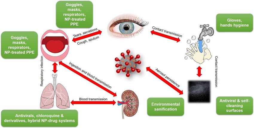
Contagion(PPE).
equipment rates of COVID-19 are much higher than those reported for the well-known SARS [26].
This brought to the rates
Contagion fast diffusion of the
of COVID-19 arevirus
much inhigher
all continents [27,28].
than those COVID-19
reported can easily propagate
for the well-known SARS
[26]. This
via cough brought to droplets,
or respiratory the fast diffusion
contact withof thebodily
virus fluids,
in all continents [27,28]. COVID-19
or from contaminated can (Figure
surfaces easily 1).
propagate
Secondary via cough
infection routesorinvolve
respiratory droplets,
touching contact with bodily
dirty/contaminated fluids,
surfaces, or frombycontaminated
followed self-inoculation
surfaces (Figure 1). Secondary infection routes involve touching
of mucous membranes. A study carried out on 29 patients [29] estimated that secondary dirty/contaminated surfaces,
routes are
followed by self-inoculation of mucous membranes. A study carried out on 29 patients
less probable than direct contagion (including eye-, nose-, oral-, etc.). Nevertheless, this risk is not [29] estimated
that secondary routes are less probable than direct contagion (including eye-, nose-, oral-, etc.).
considered negligible by different other sources, including WHO [30]. Many commonly touched
Nevertheless, this risk is not considered negligible by different other sources, including WHO [30].
surfaces, along with nosocomial environments and PPE have been tested against this risk. Based on a
Many commonly touched surfaces, along with nosocomial environments and PPE have been tested
previously published paper [31], which revealed the persistence of human coronavirus 229E on the
against this risk. Based on a previously published paper [31], which revealed the persistence of
surface
humanof common
coronavirus materials,
229E onG. Kampf
the surface recently
of commonreviewed the topic
materials, for SARS,
G. Kampf MERS
recently and SARS-CoV-2
reviewed the topic
viruses [32,33].
for SARS, MERS and SARS-CoV-2 viruses [32,33].
Figure
Figure 1. Transmission
1. Transmission pathways
pathways ofofSARS-CoV-2
SARS-CoV-2and andpossible
possible containment
containmentmeans.
means.NP
NP= nanoparticle;
= nanoparticle;
PPE = personal protective equipment. This figure was created using images
PPE = personal protective equipment. This figure was created using images under under thethe
Creative
Creative
Commons licenses. This figure is a derivative of: “Human anatomy” by Wikimedia Commons, used
Commons licenses. This figure is a derivative of: “Human anatomy” by Wikimedia Commons, used
under CC; “SugarandSkullDesigns” by Pixabay, used under CC; “Blubberfisch“ by Pixabay, used
under CC; “SugarandSkullDesigns” by Pixabay, used under CC; “Blubberfisch“ by Pixabay, used under
under CC; “Shutterstock” by FreeSVG, used under CC-BY; “OpenClipart” by FreeSVG, used under
CC; “Shutterstock” by FreeSVG, used under CC-BY; “OpenClipart” by FreeSVG, used under CC-BY;
CC-BY; “James Gathany” CDC Public Health Image library used under CC-BY.
“James Gathany” CDC Public Health Image library used under CC-BY.
Surface contamination has recently been found to be more significant than originally thought in
Surface contamination has recently been found to be more significant than originally thought in the
the spread of this viral disease. In fact, despite the use of proper PPE [34], health-care workers
spread of this viral
continued disease.
to contract In fact, despite
SARS-CoV-2, even the
afteruse of proper
barrier PPE [34],
precautions were health-care workers continued
widely implemented. The
to contract
reason behind this was found in the air, environmental, and PPE contamination inThe
SARS-CoV-2, even after barrier precautions were widely implemented. reason[35].
hospitals behind
this Besides
was found in thetouching
common air, environmental, andbeds,
surfaces (tables, PPEdoorcontamination inswitches,
handles, light hospitalstoilet
[35].sites,
Besides
etc.),common
even
touching surfaces
anteroom floors(tables,
and air beds, door handles,
fans outlets were found light switches,
to be toilet [36].
contaminated sites,The
etc.), even
latter factanteroom
is explainedfloors
by the persistence of SARS-CoV-2 in aerosols, up to several hours [37].Nanomaterials 2020, 10, 802 4 of 12
and air fans outlets were found to be contaminated [36]. The latter fact is explained by the persistence
of SARS-CoV-2 in aerosols, up to several hours [37].
Many resources and biotechnological capabilities are currently directed towards the development
of vaccines and treatments against COVID-19. Anyway, given the significance of the surface and air
contamination in the spread of the virus, attention should be paid also to the development of antiviral
and antibacterial surfaces, along with decontamination equipment and technologies.
We hypothesize that using proper air filtering may reduce the viral load in the environment,
sufficiently to decrease the probability of health-care worker infection through flaws in PPE, or in
confined public transportation [37]. According to a study of the Purdue University (West Lafayette,
IN, USA) [38], air conditioning (AC) systems are not designed to filter out particles as small as
the coronavirus. Consequently, the disease could rapidly circulate to other individuals in closed
communities as it presumably happened, as an example, in cruise ships [39].
Inverse computational fluid dynamics (CFD) models are available to identify the spread of air
particles in passenger vehicles [40]. CFD models, and the airborne nature of SARS-CoV, could be
considered to understand the spread of coronavirus in airplane passengers [41].
High-Efficiency Particulate Air (HEPA) filters can limit the spread of airborne fungi, viruses, and
bacteria [42,43]. Their use could further benefit from the implementation of a new generation of safe
and effective multifunctional antibacterial and antiviral agents.
The contamination of latex/nitrile gloves, N95 respirators, hospital scrubs, overshoes, and floors
in a nosocomial environment is considered a serious issue [35,44,45], because it helps the uncontrolled
spreading of the disease: health-care workers are anxious about passing the infection to other patients
and to their families [46].
Besides nosocomial environments, the contamination of surfaces can be regarded as responsible
for many other contagion episodes. As an example, Australian Health Authorities have recently
confirmed several cases of COVID-19 among baggage handlers. The circumstances gave rise to
concerns about cleaning the luggage and about the virus survival time on hard, smooth materials, such
as plastic and metal. The detailed explanation of contamination sources and the current number of
contagions among baggage handlers worldwide are still unknown. However, extensive cleaning and
disinfecting procedures were implemented in international airports, to reduce the surface contamination
of luggage [47].
More generally, many disinfection protocols have been developed so far, which mainly
involve the use of sodium hypochlorite [35], 70–85% ethanol [33,45,48], iodine-based and
quaternary-ammonium-salt-based disinfectants [32,48–51]. Van Doremalen et al. [36] tested the
viability of SARS-CoV-2 in different environmental conditions (aerosols, plastic, stainless steel, copper,
and cardboard). The longest viability was on stainless steel and plastic surfaces; the estimated median
virus half-life being approximately 5.6 h on stainless steel and 6.8 h on plastic. Copper was found to
be effective in inactivating the virus in a shorter time. These findings are in agreement with what
already reported on CoV-229E in 2015 [31]: brasses containing at least 70% copper were very effective
at inactivating CoV-229E, and the rate of inactivation was directly proportional to copper percentage.
Copper ion release and the generation of reactive oxygen species (ROS) were demonstrated to be
responsible for the inactivation of coronaviruses on copper and copper alloy surfaces [36]. Several
already-approved biocides based on silver or zinc oxide are just waiting to be tested against SARS-CoV-2
as well.
Based on these findings, we make a heartfelt appeal to the (Nano)Materials Science community:
we can exploit the well-known antimicrobial properties of formulations and nanostructures containing
copper, silver, and zinc species [52,53] to fight COVID-19 and, more specifically, to prevent and limit
both contamination and contagion.
Specifically, the use of copper salt nanoparticles and/or solutions (chloride, iodide, sulfide, etc.),
which are known for having an antiviral effect [54–58], could be helpful in the development of PPE
with improved shielding properties. As an example, the treatment of non-woven overshoes, surgicalNanomaterials 2020, 10, 802 5 of 12
gowns, hair cups, respirators, etc. with copper ions could help in preventing the unwanted nosocomial
virus spreading by medical personnel. In principle, the presence of metal ions could reduce (or set to
zero) the viability of CoV on these substrates, which can be considered as simple carrier interfaces for
the infection spread. Analogously, the treatment of common touching surfaces with Cu, or the use of
copper brasses for all those surfaces which need to be kept sterile, could be extremely helpful.
Nanotechnology can offer a great support in the design of contamination-safe equipment in this
era of pandemic diseases. Metal-loaded nanocomposites are known to be extremely effective in all
those cases in which a controlled and long-lasting ionic release is required [59,60]. The controlled
release of ionic copper is the key to tune the antimicrobial and antiviral properties of surfaces [61,62].
Metal nanoparticles can act as ion reservoirs for the controlled release of bioactive ions, thus tuning
the production of ROS species too. The embedding of metal nanoparticles (copper ones, in principle)
in polymer matrices could help in the tuning of metal release properties, and at the same time in
minimizing the risk of nanoparticle release into the environment [53]. Moreover, very simple and
reliable routes to synergistic nanoparticles combining a copper core and a quaternary ammonium shell
(both of which are capable, in principle, of expressing a strong antiviral action) are available in recent
literature [63].
To the best of our knowledge, there are few publications about the treatment of nosocomial
PPE with copper nanoparticles or copper oxides and salts. It is evident that inadequate PPE and
inappropriate PPE guidelines can be responsible for the death of many health-care workers and for viral
nosocomial spreading. Bhattacharjee and co-workers reviewed the topic in 2019, before the spreading
of SARS-CoV-2, taking into account other viral pandemic diseases like Ebola, SARS, and MERS [64].
They reported that metal-grafted graphene oxide (GO), for the modification of non-woven tissues,
showed to have very effective antimicrobial properties. Graphene derivatives have been reported to
be used as antimicrobial composites with different metals (Ag, Fe, Cu, Zn, etc.), and photocatalysts
(TiO2 , CdS, MnS2 , etc.) [65]. GO grafted with metal nanoparticles has been investigated as a potential
treatment for PPE [66]. Specifically, it is known that silver and copper nano-based systems loaded on
GO are very effective against both enveloped and non-enveloped viruses [65,67]. Anti-influenza copper
oxide-loaded polypropylene respirators were proposed in 2010; Borkow et al. demonstrated that copper
oxide-impregnated masks safely reduced the risk of influenza virus environmental contamination,
without altering the filtration capabilities of the masks [68]. However, no reports about the impregnation
of masks and other PPE with copper salt solutions were found in literature [68,69]. Their use is actually
part of our future research plans, which will involve both nanophases and reference compounds and
salts. In order to prevent the potential nano-toxicity of metal nanoparticles on masks and respirators
through inhalation, we think that, besides embedding nanoparticles in polymer matrices (vide infra),
the best strategy could be the use of copper salts for the impregnation of PPE pieces. In fact, when
used at low concentration levels, Cu(II) ions have low cytotoxicity on eukaryotic cells [70].
Recently, polyurethane/CuO nanocomposites were developed, acting as an effective antimicrobial
filter for air purification. It is worth noting that microparticles of CuO are a more suitable additive
for the modification of polyurethane filters than nanoparticles [71], thus reducing nanotoxicity risks.
A recent example of an antiviral air filtering system for transportation was based on the use of SiO2 -Ag
NPs as active material against MS2 bacteriophage [72].
The importance of Nanotechnology in fighting viruses is not enough explored. One of the possible
directions of further investigation is related to the use of nanomaterials to fight virus resistance to
conventional therapies; this resistance can be due to the accelerated virus adaptation in peripheral
protein sequence, thus resulting in the development of a new viral strain [73].
The antiviral action of metal nanoparticles, notably of silver nanoparticles (AgNPs), is well
known. They act as viral reproduction inhibitors, and their viricidal activity depends on the target
virus. For example, the AgNP ability to inhibit the viral entry in host cells, in the case of the HIV-1
virus, was reported, demonstrating that AgNPs are able to interact with cell receptors [73]. Regarding
double strain RNA (dsRNA) viruses, AgNP interaction with a viral genome was found, inhibiting viralNanomaterials 2020, 10, 802 6 of 12
replication [73]. Likewise, gold nanoparticles (AuNPs) stabilized by biocompatible polymers showed
antiviral activity against HIV-1 and some subtypes of influenza virus (e.g., H1N1, H3N2, H5N1) [73].
It was also demonstrated that gold nanoparticles coated with sulfated ligands, silver nanoparticles,
and hybrid silver-copper nanoparticles are able to bind the HIV envelope glycoprotein gp120 and to
inhibit in vitro HIV-1 infection in cellular models [74,75]. In addition, a size-dependent interaction with
HIV-1 (size in the range of 1–10 nm) was reported [75]. It was also demonstrated that functionalized
AgNPs (e.g., with tannic acid and mercaptoethane-sulfonate) may have the ability to prevent HSV
infection by the direct inhibition of virus attachment, penetration and post-infection spread [76,77].
Single-walled carbon nanotubes (SWCNTs) were also proposed as antiviral carriers. Specifically,
isoprinosine and ribavirin were chemically linked on the SWCNT surface to carry drugs across
biological membranes; then, the increment of the antiviral activity of hybrid materials, compared to
that of antiviral drugs, was tested [78,79].
It is known that very small metal NPs (Nanomaterials 2020, 10, 802 7 of 12
Reverting to the prevention stage, we strongly believe that contagion-safe PPE (from respirators
to surgical gowns, overshoes, hair cups, etc.) and nanotechnology-enabled highly effective antiviral
disinfectants, can be considered the most effective way to prevent viruses from spreading.
The readers working in the nanotechnology area are warmly encouraged to contact us with their
comments or proposals. Sharing our ideas and setting common actions against this pandemic will be
the key to success.
The present pandemic is not the first, nor will it be the last one. As researchers working on
nanomaterials for the life sciences, we need to ensure that we have tools in place to deal with the
COVID-19, as well as with any future pandemics.
Nanomaterial-based antiviral and antibacterial textiles, non-woven disposable products,
packaging solutions, antiviral coatings, synergistic/multifunctional surfaces, air-conditioning filters,
PPE, are just few of the possible examples requiring our prompt technological answer. While many
good research papers have been published on metal nanoparticles to be used as antibacterial or
antiviral agents, the commercialization of novel functional nanomaterials still appears to be limited by
nanotoxicology concerns or by several other practical aspects (off-target and/or any other unpredictable
effects, costs, production yield, durability, environmental impact, etc.). This is a good time to ask why
and how we can facilitate the turning of these research projects into safe and viable products.
In the field of nanoantimicrobials, we have always pursued the use of bioactive nanoparticles as
water-insoluble, polymer-confined nano-reservoirs, providing a source of ionic release, without being
released as entire nanophases in the contact matrices (e.g., physiological solutions, food, sweat, humid
air filtered through air-conditioners, etc.) [52,62].
Blocking the nanoantivirals in an adequately stable embedding matrix could be, again, the right
way to ensure safety, preventing a priori nano-toxicological risks. Other technological solutions need
to be envisaged in order to promote nanosafety and support technological knowledge transfer and
commercialization. This is becoming more and more a part of our academic mission [89,90].
We encourage our nanomaterials community to actively exploit its impressive nanotechnological
background to challenge the ongoing global health emergency.
Author Contributions: Conceptualization: M.C.S., R.A.P., N.D. and N.C.; investigation and literature overview:
M.C.S., M.I., E.A.K. and S.I.H.; first draft preparation: M.C.S. and M.I.; review and final version: M.C.S., R.A.P.,
N.D. and N.C.; supervision and funding acquisition: N.C. All authors have read and agreed to the published
version of the manuscript.
Funding: The work is part of a project that has received funding from the European Union’s Horizon 2020 research
and innovation program under the Marie Sklodowska-Curie Grant Agreement No. 813439. Partial financial
support is also acknowledged from the Italian MIUR project “E-Design” ARS01_01158.
Acknowledgments: We thank the Academic Editor and the Reviewers for their constructive comments and
valuable suggestions.
Conflicts of Interest: The authors declare no conflict of interest. The funders had no role in the design of the
study; in the collection, analyses, or interpretation of data; in the writing of the manuscript, or in the decision to
publish the results.
References
1. Webby, R.J.; Webster, R.G. Are We Ready for Pandemic Influenza? Science 2003, 302, 1519–1522. [CrossRef]
[PubMed]
2. Li, Q.; Guan, X.; Wu, P.; Wang, X.; Zhou, L.; Tong, Y.; Ren, R.; Leung, K.S.M.; Lau, E.H.Y.; Wong, J.Y.; et al.
Early Transmission Dynamics in Wuhan, China, of Novel Coronavirus–Infected Pneumonia. N. Engl. J. Med.
2020, 382, 1199–1207. [CrossRef]
3. WHO—World Health Organization. Available online: http://www.who.int/en/index.html (accessed on
14 April 2020).Nanomaterials 2020, 10, 802 8 of 12
4. Gao, J.; Tian, Z.; Yang, X. Breakthrough: Chloroquine phosphate has shown apparent efficacy in treatment of
COVID-19 associated pneumonia in clinical studies. Biosci. Trends 2020, 14, 72–73. [CrossRef] [PubMed]
5. Colson, P.; Rolain, J.-M.; Lagier, J.-C.; Brouqui, P.; Raoult, D. Chloroquine and hydroxychloroquine as
available weapons to fight COVID-19. Int. J. Antimicrob. Agents 2020, 105932. [CrossRef]
6. Wang, M.; Cao, R.; Zhang, L.; Yang, X.; Liu, J.; Xu, M.; Shi, Z.; Hu, Z.; Zhong, W.; Xiao, G. Remdesivir and
chloroquine effectively inhibit the recently emerged novel coronavirus (2019-nCoV) in vitro. Cell Res. 2020,
30, 269–271. [CrossRef] [PubMed]
7. Chang, Y.-C.; Tung, Y.-A.; Lee, K.-H.; Chen, T.-F.; Hsiao, Y.-C.; Chang, H.-C.; Hsieh, T.-T.; Su, C.-H.; Wang, S.-S.;
Yu, J.-Y.; et al. Potential Therapeutic Agents for COVID-19 Based on the Analysis of Protease and RNA
Polymerase Docking. Preprints 2020, 2020020242. [CrossRef]
8. Mercorelli, B.; Palù, G.; Loregian, A. Drug Repurposing for Viral Infectious Diseases: How Far Are We?
Trends Microbiol. 2018, 26, 865–876. [CrossRef]
9. De Wit, E.; van Doremalen, N.; Falzarano, D.; Munster, V.J. SARS and MERS: Recent insights into emerging
coronaviruses. Nat. Rev. Microbiol. 2016, 14, 523–534. [CrossRef]
10. Hui, D.S.C.; Zumla, A. Severe Acute Respiratory Syndrome: Historical, Epidemiologic, and Clinical Features.
Infect. Dis. Clin. 2019, 33, 869–889. [CrossRef]
11. Agbowuro, A.A.; Huston, W.M.; Gamble, A.B.; Tyndall, J.D.A. Proteases and protease inhibitors in infectious
diseases. Med. Res. Rev. 2018, 38, 1295–1331. [CrossRef]
12. Lai, C.-C.; Shih, T.-P.; Ko, W.-C.; Tang, H.-J.; Hsueh, P.-R. Severe acute respiratory syndrome coronavirus 2
(SARS-CoV-2) and coronavirus disease-2019 (COVID-19): The epidemic and the challenges. Int. J. Antimicrob.
Agents 2020, 55, 105924. [CrossRef] [PubMed]
13. Momattin, H.; Al-Ali, A.Y.; Al-Tawfiq, J.A. A Systematic Review of therapeutic agents for the treatment of
the Middle East Respiratory Syndrome Coronavirus (MERS-CoV). Travel Med. Infect. Dis. 2019, 30, 9–18.
[CrossRef] [PubMed]
14. Chafekar, A.; Fielding, B.C. MERS-CoV: Understanding the Latest Human Coronavirus Threat. Viruses 2018,
10, 93. [CrossRef] [PubMed]
15. Du, L.; He, Y.; Zhou, Y.; Liu, S.; Zheng, B.-J.; Jiang, S. The spike protein of SARS-CoV—A target for vaccine
and therapeutic development. Nat. Rev. Microbiol. 2009, 7, 226–236. [CrossRef] [PubMed]
16. Song, Z.; Xu, Y.; Bao, L.; Zhang, L.; Yu, P.; Qu, Y.; Zhu, H.; Zhao, W.; Han, Y.; Qin, C. From SARS to MERS,
Thrusting Coronaviruses into the Spotlight. Viruses 2019, 11, 59. [CrossRef] [PubMed]
17. Wang, Z.; Chen, X.; Lu, Y.; Chen, F.; Zhang, W. Clinical characteristics and therapeutic procedure for four
cases with 2019 novel coronavirus pneumonia receiving combined Chinese and Western medicine treatment.
Biosci. Trends 2020, 14, 64–68. [CrossRef] [PubMed]
18. Li, G.; Clercq, E.D. Therapeutic options for the 2019 novel coronavirus (2019-nCoV). Nat. Rev. Drug Discov.
2020, 19, 149–150. [CrossRef]
19. Yao, X.; Ye, F.; Zhang, M.; Cui, C.; Huang, B.; Niu, P.; Liu, X.; Zhao, L.; Dong, E.; Song, C.; et al. In Vitro
Antiviral Activity and Projection of Optimized Dosing Design of Hydroxychloroquine for the Treatment of
Severe Acute Respiratory Syndrome Coronavirus 2 (SARS-CoV-2). Clin. Infect. Dis. 2020, ciaa237. (in press).
[CrossRef]
20. Sheahan, T.P.; Sims, A.C.; Leist, S.R.; Schäfer, A.; Won, J.; Brown, A.J.; Montgomery, S.A.; Hogg, A.; Babusis, D.;
Clarke, M.O.; et al. Comparative therapeutic efficacy of remdesivir and combination lopinavir, ritonavir, and
interferon beta against MERS-CoV. Nat. Commun. 2020, 11, 1–14. [CrossRef]
21. Savarino, A.; Trani, L.D.; Donatelli, I.; Cauda, R.; Cassone, A. New insights into the antiviral effects of
chloroquine. Lancet Infect. Dis. 2006, 6, 67–69. [CrossRef]
22. Savarino, A.; Boelaert, J.R.; Cassone, A.; Majori, G.; Cauda, R. Effects of chloroquine on viral infections:
An old drug against today’s diseases. Lancet Infect. Dis. 2003, 3, 722–727. [CrossRef]
23. Touret, F.; de Lamballerie, X. Of chloroquine and COVID-19. Antivir. Res. 2020, 177, 104762. [CrossRef]
[PubMed]
24. Chang, D.; Xu, H.; Rebaza, A.; Sharma, L.; Cruz, C.S.D. Protecting health-care workers from subclinical
coronavirus infection. Lancet Respir. Med. 2020, 8, e13. [CrossRef]Nanomaterials 2020, 10, 802 9 of 12
25. Cheung, J.C.-H.; Ho, L.T.; Cheng, J.V.; Cham, E.Y.K.; Lam, K.N. Staff safety during emergency airway
management for COVID-19 in Hong Kong. Lancet Respir. Med. 2020, 8, e49. [CrossRef]
26. Liu, Y.; Gayle, A.A.; Wilder-Smith, A.; Rocklöv, J. The reproductive number of COVID-19 is higher compared
to SARS coronavirus. J. Travel Med. 2020, 27, taaa021. [CrossRef]
27. Luo, W.; Majumder, M.S.; Liu, D.; Poirier, C.; Mandl, K.D.; Lipsitch, M.; Santillana, M. The role of absolute
humidity on transmission rates of the COVID-19 outbreak. medRxiv 2020. [CrossRef]
28. Wu, J.T.; Leung, K.; Bushman, M.; Kishore, N.; Niehus, R.; de Salazar, P.M.; Cowling, B.J.; Lipsitch, M.;
Leung, G.M. Estimating clinical severity of COVID-19 from the transmission dynamics in Wuhan, China.
Nat. Med. 2020, 26, 506–510. [CrossRef]
29. Li, H.; Wang, Y.; Shao, L.; Ji, M.; Zhao, Q.; Zhou, Y.; Pei, F.; Wang, J.; Wang, M.; Hong, Y.; et al. Transmission
Routes of SARS-CoV-2: Based on the Epidemiological and Clinical Characteristics of 29 Cases in Jinan, China.
Lancet 2020. [CrossRef]
30. WHO—World Health Organization Infection Prevention and Control during Health Care When Novel
Coronavirus (nCoV) Infection is Suspected—Interim Guidance. Available online: https://extranet.who.int/
(accessed on 24 March 2020).
31. Warnes, S.L.; Little, Z.R.; Keevil, C.W. Human Coronavirus 229E Remains Infectious on Common Touch
Surface Materials. mBio 2015, 6, e01697-15. [CrossRef]
32. Kampf, G. Potential role of inanimate surfaces for the spread of coronaviruses and their inactivation with
disinfectant agents. Infect. Prev. Pract. 2020, 2, 100044. [CrossRef]
33. Kampf, G.; Todt, D.; Pfaender, S.; Steinmann, E. Persistence of coronaviruses on inanimate surfaces and their
inactivation with biocidal agents. J. Hosp. Infect. 2020, 104, 246–251. [CrossRef] [PubMed]
34. Holland, M.; Zaloga, D.J.; Friderici, C.S. COVID-19 Personal Protective Equipment (PPE) for the emergency
physician. Vis. J. Emerg. Med. 2020, 19, 100740. [CrossRef] [PubMed]
35. Ong, S.W.X.; Tan, Y.K.; Chia, P.Y.; Lee, T.H.; Ng, O.T.; Wong, M.S.Y.; Marimuthu, K. Air, Surface Environmental,
and Personal Protective Equipment Contamination by Severe Acute Respiratory Syndrome Coronavirus 2
(SARS-CoV-2) From a Symptomatic Patient. JAMA 2020, (in press). [CrossRef] [PubMed]
36. Van Doremalen, N.; Bushmaker, T.; Morris, D.H.; Holbrook, M.G.; Gamble, A.; Williamson, B.N.; Tamin, A.;
Harcourt, J.L.; Thornburg, N.J.; Gerber, S.I.; et al. Aerosol and Surface Stability of SARS-CoV-2 as Compared
with SARS-CoV-1. N. Engl. J. Med. 2020, (in press). [CrossRef]
37. Blake, E.; Yaneer, B.-Y. Could Air Filtration Reduce COVID-19 Severity and Spread? Available online:
https://necsi.edu/could-air-filtration-reduce-covid19-severity-and-spread (accessed on 14 April 2020).
38. Purdue News Service Cruise Ship AC Systems Could Promote Rapid Coronavirus Spread, Prof Says. Available
online: https://www.purdue.edu/newsroom/releases/2020/Q1/cruise-ship-ac-systems-could-promote-rapid-
coronavirus-spread,-prof-says.html (accessed on 14 April 2020).
39. Fang, Z.; Huang, Z.; Li, X.; Zhang, J.; Lv, W.; Zhuang, L.; Xu, X.; Huang, N. How many infections of COVID-19
there will be in the “Diamond Princess”-Predicted by a virus transmission model based on the simulation of
crowd flow. arXiv 2020, arXiv:2002.10616.
40. Zhang, T.F.; Chen, Q. Identification of contaminant sources in enclosed environments by inverse CFD
modeling. Indoor Air 2007, 17, 167–177. [CrossRef]
41. Mazumdar, S.; Poussou, S.B.; Lin, C.-H.; Isukapalli, S.S.; Plesniak, M.W.; Chen, Q. Impact of scaling and body
movement on contaminant transport in airliner cabins. Atmos. Environ. 2011, 45, 6019–6028. [CrossRef]
42. Kte’pi, B. High-Efficiency Particulate Air System|Air Filtration System; Encyclopedia Britannica: Chicago, IL,
USA, 2019.
43. European Standards Agency Standards EN 1822 and EN ISO 29463—EPA, HEPA and ULPA Filters. Available
online: https://www.en-standard.eu/set-en-1822-and-en-iso-29463-standards-for-heigh-efficiency-air-filters-
epa-hepa-and-ulpa/ (accessed on 14 April 2020).
44. Otter, J.A.; Donskey, C.; Yezli, S.; Douthwaite, S.; Goldenberg, S.D.; Weber, D.J. Transmission of SARS and
MERS coronaviruses and influenza virus in healthcare settings: The possible role of dry surface contamination.
J. Hosp. Infect. 2016, 92, 235–250. [CrossRef]
45. Dowell, S.F.; Simmerman, J.M.; Erdman, D.D.; Wu, J.-S.J.; Chaovavanich, A.; Javadi, M.; Yang, J.-Y.;
Anderson, L.J.; Tong, S.; Ho, M.S. Severe Acute Respiratory Syndrome Coronavirus on Hospital Surfaces.
Clin. Infect. Dis. 2004, 39, 652–657. [CrossRef]Nanomaterials 2020, 10, 802 10 of 12
46. The Lancet. COVID-19: Protecting health-care workers. Lancet 2020, 395, 922. [CrossRef]
47. Davies, A. More Qantas Flights Revealed to Have Been Crewed by Staff with Covid-19. Available
online: https://www.theguardian.com/business/2020/apr/09/more-qantas-flights-revealed-crewed-staff-
covid-19-coronavirus (accessed on 14 April 2020).
48. Rabenau, H.F.; Kampf, G.; Cinatl, J.; Doerr, H.W. Efficacy of various disinfectants against SARS coronavirus.
J. Hosp. Infect. 2005, 61, 107–111. [CrossRef]
49. Wood, A.; Payne, D. The action of three antiseptics/disinfectants against enveloped and non-enveloped
viruses. J. Hosp. Infect. 1998, 38, 283–295. [CrossRef]
50. Kariwa, H.; Fujii, N.; Takashima, I. Inactivation of SARS Coronavirus by Means of Povidone-Iodine, Physical
Conditions and Chemical Reagents. Dermatology 2006, 212 (Suppl. 1), 119–123. [CrossRef]
51. Eggers, M.; Eickmann, M.; Zorn, J. Rapid and Effective Virucidal Activity of Povidone-Iodine Products
Against Middle East Respiratory Syndrome Coronavirus (MERS-CoV) and Modified Vaccinia Virus Ankara
(MVA). Infect. Dis. Ther. 2015, 4, 491–501. [CrossRef] [PubMed]
52. Sportelli, M.C.; Picca, R.A.; Cioffi, N. Recent advances in the synthesis and characterization of
nano-antimicrobials. Tractrends Anal. Chem. 2016, 84, 131–138. [CrossRef]
53. Cioffi, N.; Rai, M. Nano-Antimicrobials: Progress and Prospects, 1st ed.; Springer: Berlin/Heidelberg, Germany,
2012; ISBN 978-3-642-24427-8.
54. Khodashenas, B.; Ghorbani, H.R. Synthesis of copper nanoparticles: An overview of the various methods.
Korean J. Chem. Eng. 2014, 31, 1105–1109. [CrossRef]
55. Fujimori, Y.; Sato, T.; Hayata, T.; Nagao, T.; Nakayama, M.; Nakayama, T.; Sugamata, R.; Suzuki, K. Novel
Antiviral Characteristics of Nanosized Copper(I) Iodide Particles Showing Inactivation Activity against 2009
Pandemic H1N1 Influenza Virus. Appl. Environ. Microbiol. 2012, 78, 951. [CrossRef] [PubMed]
56. Krzyzowska, M.; Tomaszewska, E.; Ranoszek-Soliwoda, K.; Bien, K.; Orlowski, P.; Celichowski, G.; Grobelny, J.
Chapter 12—Tannic acid modification of metal nanoparticles: Possibility for new antiviral applications.
In Nanostructures for Oral Medicine; Andronescu, E., Grumezescu, A.M., Eds.; Elsevier: Amsterdam,
The Netherlands, 2017; pp. 335–363. ISBN 978-0-323-47720-8.
57. Broglie, J.J.; Alston, B.; Yang, C.; Ma, L.; Adcock, A.F.; Chen, W.; Yang, L. Antiviral Activity of Gold/Copper
Sulfide Core/Shell Nanoparticles against Human Norovirus Virus-Like Particles. PloS ONE 2015, 10, e0141050.
[CrossRef] [PubMed]
58. Sucipto, T.H.; Churrotin, S.; Setyawati, H.; Kotaki, T.; Martak, F.; Soegijanto, S. Antiviral activity of
copper(II)chloride dihydrate against dengue virus type-2 in vero cell. Indones. J. Trop. Infect. Dis. 2017, 6,
84–87. [CrossRef]
59. Palza, H.; Nuñez, M.; Bastías, R.; Delgado, K. In situ antimicrobial behavior of materials with copper-based
additives in a hospital environment. Int. J. Antimicrob. Agents 2018, 51, 912–917. [CrossRef]
60. Cioffi, N.; Torsi, L.; Ditaranto, N.; Tantillo, G.; Ghibelli, L.; Sabbatini, L.; Bleve-Zacheo, T.; D’Alessio, M.;
Zambonin, P.G.; Traversa, E. Copper Nanoparticle/Polymer Composites with Antifungal and Bacteriostatic
Properties. Chem. Mater. 2005, 17, 5255–5262. [CrossRef]
61. Cioffi, N.; Ditaranto, N.; Sabbatini, L.; Tantillo, G.; Torsi, L.; Zambonin, P.G. Bioactive Metal Nanomaterials
Stabilized by Bioactive Agents and Preparation Process. European Patent Application EP 2157211 B1,
2 September 2015.
62. Cioffi, N.; Ditaranto, N.; Sabbatini, L.; Torsi, L.; Zambonin, P.G. Nanomaterials for Controlled Metal Release
and Process for Their Production. European Patent Application EP 2123797 B1, 25 November 2009.
63. Sportelli, M.C.; Longano, D.; Bonerba, E.; Tantillo, G.; Torsi, L.; Sabbatini, L.; Cioffi, N.; Ditaranto, N.
Electrochemical Preparation of Synergistic Nanoantimicrobials. Molecules 2020, 25, 49. [CrossRef]
64. Bhattacharjee, S.; Joshi, R.; Chughtai, A.A.; Macintyre, C.R. Graphene Modified Multifunctional Personal
Protective Clothing. Adv. Mater. Interfaces 2019, 6, 1900622. [CrossRef]
65. Chen, Y.-N.; Hsueh, Y.-H.; Hsieh, C.-T.; Tzou, D.-Y.; Chang, P.-L. Antiviral Activity of Graphene–Silver
Nanocomposites against Non-Enveloped and Enveloped Viruses. Int. J. Environ. Res. Public Health 2016, 13,
430. [CrossRef] [PubMed]
66. Perreault, F.; de Faria, A.F.; Nejati, S.; Elimelech, M. Antimicrobial Properties of Graphene Oxide Nanosheets:
Why Size Matters. ACS Nano 2015, 9, 7226–7236. [CrossRef] [PubMed]
67. Hang, X.; Peng, H.; Song, H.; Qi, Z.; Miao, X.; Xu, W. Antiviral activity of cuprous oxide nanoparticles against
Hepatitis C Virus in vitro. J. Virol. Methods 2015, 222, 150–157. [CrossRef]Nanomaterials 2020, 10, 802 11 of 12
68. Iyigundogdu, Z.U.; Demir, O.; Asutay, A.B.; Sahin, F. Developing Novel Antimicrobial and Antiviral Textile
Products. Appl. Biochem. Biotechnol. 2017, 181, 1155–1166. [CrossRef] [PubMed]
69. Sunada, K.; Minoshima, M.; Hashimoto, K. Highly efficient antiviral and antibacterial activities of solid-state
cuprous compounds. J. Hazard. Mater. 2012, 235–236, 265–270. [CrossRef] [PubMed]
70. Shaligram, S.; Campbell, A. Toxicity of copper salts is dependent on solubility profile and cell type tested.
Toxicol. Vitr. 2013, 27, 844–851. [CrossRef]
71. Ungur, G.; Hrůza, J. Modified polyurethane nanofibers as antibacterial filters for air and water purification.
RSC Adv. 2017, 7, 49177–49187. [CrossRef]
72. Krähling, V.; Stein, D.A.; Spiegel, M.; Weber, F.; Mühlberger, E. Severe Acute Respiratory Syndrome
Coronavirus Triggers Apoptosis via Protein Kinase R but Is Resistant to Its Antiviral Activity. J. Virol. 2009,
83, 2298–2309. [CrossRef] [PubMed]
73. Kerry, R.G.; Malik, S.; Redda, Y.T.; Sahoo, S.; Patra, J.K.; Majhi, S. Nano-based approach to combat emerging
viral (NIPAH virus) infection. Nanomed. Nanotechnol. Biol. Med. 2019, 18, 196–220. [CrossRef] [PubMed]
74. Di Gianvincenzo, P.; Marradi, M.; Martínez-Ávila, O.M.; Bedoya, L.M.; Alcamí, J.; Penadés, S. Gold
nanoparticles capped with sulfate-ended ligands as anti-HIV agents. Bioorganic Med. Chem. Lett. 2010, 20,
2718–2721. [CrossRef]
75. Elechiguerra, J.L.; Burt, J.L.; Morones, J.R.; Camacho-Bragado, A.; Gao, X.; Lara, H.H.; Yacaman, M.J.
Interaction of silver nanoparticles with HIV-1. J. Nanobiotechnology 2005, 3, 6. [CrossRef] [PubMed]
76. Orlowski, P.; Tomaszewska, E.; Gniadek, M.; Baska, P.; Nowakowska, J.; Sokolowska, J.; Nowak, Z.;
Donten, M.; Celichowski, G.; Grobelny, J.; et al. Tannic Acid Modified Silver Nanoparticles Show Antiviral
Activity in Herpes Simplex Virus Type 2 Infection. PloS ONE 2014, 9, e104113. [CrossRef] [PubMed]
77. Baram-Pinto, D.; Shukla, S.; Perkas, N.; Gedanken, A.; Sarid, R. Inhibition of Herpes Simplex Virus Type
1 Infection by Silver Nanoparticles Capped with Mercaptoethane Sulfonate. Bioconjugate Chem. 2009, 20,
1497–1502. [CrossRef]
78. Zhu, S.; Li, J.; Huang, A.-G.; Huang, J.-Q.; Huang, Y.-Q.; Wang, G.-X. Anti-betanodavirus activity of
isoprinosine and improved efficacy using carbon nanotubes based drug delivery system. Aquaculture 2019,
512, 734377. [CrossRef]
79. Zhu, B.; Liu, G.-L.; Ling, F.; Wang, G.-X. Carbon nanotube-based nanocarrier loaded with ribavirin against
grass carp reovirus. Antivir. Res. 2015, 118, 29–38. [CrossRef]
80. Rai, M.; Deshmukh, S.D.; Ingle, A.P.; Gupta, I.R.; Galdiero, M.; Galdiero, S. Metal nanoparticles: The
protective nanoshield against virus infection. Crit. Rev. Microbiol. 2016, 42, 46–56. [CrossRef]
81. Nanotech Surface Coronavirus: Nanotech Surface Sanitizes Milan with Nanomaterials Remaining
Self-Sterilized for Years|STATNANO. Available online: https://statnano.com//news/67531/Coronavirus-
Nanotech-Surface-Sanitizes-Milan-with-Nanomaterials-Remaining-Self-sterilized-for-Years (accessed on 6
April 2020).
82. Leung, N.H.L.; Chu, D.K.W.; Shiu, E.Y.C.; Chan, K.-H.; McDevitt, J.J.; Hau, B.J.P.; Yen, H.-L.; Li, Y.; Ip, D.K.M.;
Peiris, J.S.M.; et al. Respiratory virus shedding in exhaled breath and efficacy of face masks. Nat. Med. 2020,
1–5. [CrossRef]
83. Fanning, J.C.; Taylor, L.T. Some transition metal complexes of 8-aminoquinoline. J. Inorg. Nucl. Chem. 1965,
27, 2217–2223. [CrossRef]
84. Phopin, K.; Sinthupoom, N.; Treeratanapiboon, L.; Kunwittaya, S.; Prachayasittikul, S.; Ruchirawat, S.;
Prachayasittikul, V. Antimalarial and antimicrobial activities of 8-Aminoquinoline-Uracils metal complexes.
EXCLI J. 2016, 15, 144–152. [CrossRef] [PubMed]
85. Sona Nanotech. Sona Develops Rapid Screening Test for Coronavirus; Sona Nanotech: Halifax, Canada, 2020.
86. WHO—World Health Organization WHO|International Clinical Trials Registry Platform (ICTRP). Available
online: http://www.who.int/ictrp/en/ (accessed on 27 March 2020).
87. Mansour, H.M.; Rhee, Y.S.; Wu, X. Nanomedicine in pulmonary delivery. IJN 2009, 4, 299–319. [CrossRef]
[PubMed]
88. Pontes, J.F.; Grenha, A. Multifunctional Nanocarriers for Lung Drug Delivery. Nanomaterials 2020, 10, 183.
[CrossRef] [PubMed]Nanomaterials 2020, 10, 802 12 of 12
89. Caulfield, T.; Ogbogu, U. The commercialization of university-based research: Balancing risks and benefits.
BMC Med. Ethics 2015, 16, 70. [CrossRef] [PubMed]
90. Rasmussen, E.; Moen, Ø.; Gulbrandsen, M. Initiatives to promote commercialization of university knowledge.
Technovation 2006, 26, 518–533. [CrossRef]
© 2020 by the authors. Licensee MDPI, Basel, Switzerland. This article is an open access
article distributed under the terms and conditions of the Creative Commons Attribution
(CC BY) license (http://creativecommons.org/licenses/by/4.0/).You can also read