Association of Polymorphisms of MASP1/3, COLEC10, and COLEC11 Genes with 3MC Syndrome - MDPI
←
→
Page content transcription
If your browser does not render page correctly, please read the page content below
International Journal of
            Molecular Sciences

Review
Association of Polymorphisms of MASP1/3, COLEC10,
and COLEC11 Genes with 3MC Syndrome
Gabriela Gajek * , Anna S. Świerzko and Maciej Cedzyński
 Laboratory of Immunobiology of Infections, Institute of Medical Biology, Polish Academy of Sciences,
 93-232 Lodz, Poland; aswierzko@cbm.pan.pl (A.S.Ś.); mcedzynski@cbm.pan.pl (M.C.)
 * Correspondence: ggajek@cbm.pan.pl

 Received: 30 June 2020; Accepted: 30 July 2020; Published: 31 July 2020

 Abstract: The Malpuech, Michels, Mingarelli, Carnevale (3MC) syndrome is a rare, autosomal
 recessive genetic- disorder associated with mutations in the MASP1/3, COLEC1,1 or COLEC10 genes.
 The number of 3MC patients with known mutations in these three genes reported so far remains
 very small. To date, 16 mutations in MASP-1/3, 12 mutations in COLEC11 and three in COLEC10
 associated with 3MC syndrome have been identified. Their products play an essential role as factors
 involved in the activation of complement via the lectin or alternative (MASP-3) pathways. Recent
 data indicate that mannose-binding lectin-associated serine protease-1 (MASP-1), MASP-3, collectin
 kidney-1 (collectin-11) (CL-K1), and collectin liver-1 (collectin-10) (CL-L1) also participate in the
 correct migration of neural crest cells (NCC) during embryogenesis. This is supported by relationships
 between MASP1/3, COLEC10, and COLEC11 gene mutations and the incidence of 3MC syndrome,
 associated with craniofacial abnormalities such as radioulnar synostosis high-arched eyebrows, cleft
 lip/palate, hearing loss, and ptosis.

 Keywords: 3MC syndrome; MASP1/3; COLEC11; COLEC10; MASP-1; MASP-3; CL-K1; CL-L1

1. Introduction
      The Malpuech, Michels, Mingarelli and Carnevale syndrome [1–4] is commonly called 3MC
syndrome. In 1989, Carnevale et al. reported a phenotype consisting of downslanting palpebral
fissures, ptosis of the eyelids, periumbilical depression, hypertelorism, radioulnar synostosis, and
developmental delay in two Italian siblings (MIM 265050). In 1996, two sisters with similar ocular, facial,
skeletal, and abdominal defects, but with normal intelligence, were reported by Mingarelli et al. (also
MIM 265050). Since the clinical picture of patients suffering from Carnevale and Mingarelli syndromes
overlapped with Michels (MIM 257920) and Malpuech syndromes (MIM 248340), it was suggested that
all four disorders should be reclassified into one “3MC syndrome”. It is a rare, autosomal recessive
genetic disorder, characterized by a wide spectrum of developmental abnormalities that could include
high-arched eyebrows, cleft lip/palate, hearing loss, short stature, umbilical hernias/omphalocele, and
urogenital abnormalities. The prevalence of 3MC syndrome is unknown. The largest number of
affected persons is located in the Middle East. 3MC syndrome disorders are caused by mutations in
the mannose-binding lectin-associated serine protease (MASP)1/3 [5], COLEC11 [6], or COLEC10 [7]
genes. The number of 3MC patients carrying known mutations in these three genes reported so far still
remains rather small but the disease is more likely to occur in families with consanguineous parents.
In total, forty-six 3MC patients from 34 families with mutations in the above-mentioned genes have
been diagnosed. Among them, 26 persons from 20 families had mutations in the MASP1/3 (Table 1),
17 individuals from 12 families had mutations in the COLEC11 (Table 2), and three patients from two
families had mutations in the COLEC10 gene (Table 3). Those mutations abort or impair function
of their corresponding proteins, resulting in defective control of cell migration at an early stage of

Int. J. Mol. Sci. 2020, 21, 5483; doi:10.3390/ijms21155483                          www.mdpi.com/journal/ijms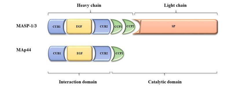
Int.Int.
J. Mol. Sci.Sci.
J. Mol. 2020, 21,21,
2020, 5483
x FOR PEER REVIEW 2 of2 14
of 14
migration interferes with ontogenesis of tissues and organs and leads to the various abnormalities
embryonal development [6]. Impaired cell migration interferes with ontogenesis of tissues and organs
manifested. The proteins expressed are also factors involved in the activation of complement via the
and leads to the various abnormalities manifested. The proteins expressed are also factors involved
lectin pathway, an important branch of the innate immune system [6,8]. It was suggested that
in the activation of complement via the lectin pathway, an important branch of the innate immune
dysfunction of the lectin pathway can be compensated by other defense mechanisms, which seems
system [6,8]. It was suggested that dysfunction of the lectin pathway can be compensated by other
to explain why immune system dysfunctions are not part of the 3MC syndrome.
defense mechanisms, which seems to explain why immune system dysfunctions are not part of the
3MC syndrome.
2. The MASP1/3 Gene and Its Products—MASP-1, MASP-3, and MAp44
2. The MASP1/3 Gene and Its Products—MASP-1, MASP-3, and MAp44
2.1. Gene and Protein Structure
2.1. Gene Theand Protein Structure
MASP1/3 gene is located on chromosome 3 (3q27-28). It contains 76 kbp and encompasses
18 The
exons. This gene
MASP1/3 gene is encodes
located for two enzymes,
on chromosome MASP-1 It
3 (3q27-28). and MASP-3
contains (mannose-binding
76 kbp and encompasses lectin-
18
associated serine proteases) as well as the non-enzymatic MAp44
exons. This gene encodes for two enzymes, MASP-1 and MASP-3 (mannose-binding lectin-associated (mannose-binding lectin-associated
protein,
serine 44 kDa)
proteases) as[9].
wellAllasthree MASP1/3 alternative
the non-enzymatic MAp44splicing products share
(mannose-binding four N-terminal
lectin-associated domains,
protein, 44
encoded
kDa) [9]. Allbythree
exons 2–8: CUB1-EGF-CUB2-CCP1
MASP1/3 alternative splicing products (explainedsharebelow), while exon
four N-terminal 1 encodes
domains, the 5′-
encoded
byuntranslated region [8]. Exons 10 and(explained
exons 2–8: CUB1-EGF-CUB2-CCP1 11 encode below),
the CCP2 domain,
while exoncommon
1 encodestothe MASP-1 and MASP-
50 -untranslated
region [8]. Exons 10 and 11 encode the CCP2 domain, common to MASP-1 and MASP-3.exons
3. Single exon 12 is responsible for the serine protease (SP) domain of MASP-3, whereas 13—
Single
18 are
exon 12 isresponsible
responsible forfor
thethecorresponding
serine protease MASP-1 fragment.
(SP) domain of The ninth whereas
MASP-3, exon encodes
exonsthe unique
13—18 areC-
terminal 17 AA sequence of MAp44. According to the NCBI database
responsible for the corresponding MASP-1 fragment. The ninth exon encodes the unique C-terminal a fourth splice variant has been
recorded, however the resulting RNA does not constitute mRNA,
17 AA sequence of MAp44. According to the NCBI database a fourth splice variant has been recorded, and presumably is degraded
throughthe
however theresulting
nonsense-mediated
RNA does not mRNA decay pathway
constitute mRNA, and [10].presumably is degraded through the
Therefore, MASP-1 and
nonsense-mediated mRNA decay pathway [10]. MASP-3 are proteases composed of six well-characterized domains, of
which five constitute
Therefore, MASP-1the and heavy
MASP-3chainare
while the sixth
proteases (SP) constitutes
composed the light chain. Upon
of six well-characterized activation
domains, of
which five constitute the heavy chain while the sixth (SP) constitutes the light chain. Upon activation a
of proenzymes, the peptide bond between them is cleaved and both chains remain connected via
of disulphide
proenzymes, bond [11,12]. The
the peptide bondCUB (C1r/C1s-Uegf
between them is cleaved (uchrin andepidermal
both chains growth
remainfactor)-BMP
connected (bonevia
morphogenic protein)) and EGF (epidermal growth factor)
a disulphide bond [11,12]. The CUB (C1r/C1s-Uegf (uchrin epidermal growth factor)-BMP (bonedomains are responsible for forming
MASP/MAp44
morphogenic complexes
protein)) and with
EGF such pattern-recognition
(epidermal growth factor) molecules
domains(PRM) as collectins
are responsible forand ficolins
forming
MASP/MAp44 complexes with such pattern-recognition molecules (PRM) as collectins and ficolins [13].in
[13]. It should be stressed that most of the proteins possessing the CUB domain are involved
developmental
It should be stressed processes,
that mostsuch asproteins
of the bone morphogenetic
possessing theprotein 1, the dorso-ventral
CUB domain are involved inpatterning
developmentalprotein
tolloid, a family of spermadhesins, and the neuronal recognition molecule
processes, such as bone morphogenetic protein 1, the dorso-ventral patterning protein tolloid, a family A5 [8,14]. The CCP
of (complement
spermadhesins, control
and theprotein)
neuronaldomains are common
recognition molecule to A5a variety of complement
[8,14]. The CCP (complementfactors control
[15]. The
serine protease (SP) domain with catalytic activity is characteristic for
protein) domains are common to a variety of complement factors [15]. The serine protease (SP) domain chymotrypsin-like proteases
[8].catalytic
with There areactivity
severalissingle nucleotide
characteristic forpolymorphisms
chymotrypsin-like of theproteases
MASP1/3[8]. gene strongly
There associated
are several with
single
protein serum
nucleotide levels. Among
polymorphisms of thethem,
MASP1/3 homozygosity
gene strongly in associated
intron 8 (rs3774275,
with protein G/G) waslevels.
serum associated
Among with
decreased MASP-3 but increased MASP-1 and MAp44. Variant alleles
them, homozygosity in intron 8 (rs3774275, G/G) was associated with decreased MASP-3 but increased for rs698090 and rs67143992
are associated
MASP-1 and MAp44. with Variant
an increase in for
alleles MASP-1
rs698090 andand MAp44 and a are
rs67143992 decrease in MASP-3,
associated with anwhile
increasevariant
in
MASP-1 and MAp44 and a decrease in MASP-3, while variant alleles of rs72549154 and rs35089177 of
alleles of rs72549154 and rs35089177 result in decreases of MASP-1 and MAp44 and an increase
MASP-3
result [9], Figure
in decreases 1.
of MASP-1 and MAp44 and an increase of MASP-3 [9], Figure 1.
Figure 1. Scheme of mannose-binding lectin-associated serine protease-1 (MASP-1), MASP-3, and
Figure 1. Scheme
mannose-binding of mannose-binding
lectin-associated lectin-associated
protein, 44 serine structures
kDa (MAp44) protein protease-1(acc.
(MASP-1), MASP-3,
to Gaboriaud and
et al.,
mannose-binding
2013 [16]. lectin-associated protein, 44 kDa (MAp44) protein structures (acc. to Gaboriaud et
al., 2013 [16].Int. J. Mol. Sci. 2020, 21, 5483 3 of 14
2.2. Tissue Expression of the MASP-1, MASP-3, and MAp44
MASP-1 is mainly expressed in the liver although specific mRNA was also found in the brain
and cervix. Similarly, the major site of MASP-3 synthesis is the liver but its expression was also noted
in a variety of other sites, including the colon, bladder, and uterus. MAp44 is synthesized in the
heart while weaker expression takes place in the liver, brain, and cervix [10,17,18]. The expression of
MASP-3 has been considered ubiquitous compared with other isoforms [10]. These results emphasize
the importance of alternative splicing mechanisms in the regulation of expression in different tissues.
The widespread expression of the MASP1/3 gene may indicate local functions of its products MASP-1,
MASP-3, and MAp44. The median serum levels of MASP-1, MASP-3, and MAp44 in healthy adults
were found to equal 10.8, 6.7 and 2.2 µg/mL, respectively. Concentrations of MASP-1 and MASP-3
showed gender differences—the first mentioned was higher in women while the second was higher in
men [19]. Moreover, MASP-3 and MAp44 also correlate with age [19].
2.3. Biological Activities of the MASP-1, MASP-3, and MAp44
Mannose-binding lectin-associated serine protease-1 (MASP-1) functions as a factor involved
in the activation of complement, coagulation, and kallikrein–kinin systems. As mentioned, it exists
in the form of a zymogen occurring in complexes with some collectins (MBL, collectin liver-1 (also
known as collectin 10, CL-10), collectin kidney-1 (or collectin 11, CL-11)), and ficolins (ficolin-1,
-2, -3 or M-, L-, H-ficolin, respectively). MASP-1 is auto-activated when the complex binds to the
target, recognized by PRM (collectins and ficolins, as lectins recognize mainly polysaccharides or
glycoconjugates expressed on the surface of pathogens or abnormal self-cells). MASP-1 then cleaves
MASP-2 zymogen (another protease of the MASP-2 family, encoded by the distinct MASP-2 gene),
which next activates complement C4 factor. Both MASP-1 and MASP-2 further activate C2 forming
C3 convertase (C4bC2a) (identical to that of the classical pathway) [20]. Moreover, MASP-1 is able to
cleave fibrinogen, factor XIII, prothrombin, and thrombin-activatable fibrinolysis inhibitor (TAFI) thus
promoting coagulation [21–24]. The clots can gather around microbes and hinder their spreading [25].
Furthermore, the high-molecular-weight kininogen was reported to be another MASP-1 substrate.
Its digestion enables release of bradykinin, a pro-inflammatory mediator of the contact system [26].
Moreover, MASP-1 can activate endothelial cells [27], enhance endothelial E-selectin expression [28],
and increase the production of interleukins IL-6 and IL-8 [29].
MASP-3 is believed to contribute to the activation of complement via the alternative pathway
through pro-factor D cleavage [20]. Another substrate is insulin-like growth factor-binding protein 5
(IGFBP-5) that modulates the effects of IGF on cell survival, differentiation, and proliferation [30].
MAp44 functions as an inhibitor of the complement pathway by competing with MASP for
PRM-binding sites [31]. It moreover contributes to regulation of cardiac development [32].
Some clinical associations of MASP-1, MASP-3, and MAp44 other than 3MC have been reported.
For example, Weinschutz Mendes et al. (2020) found that polymorphisms influencing serum
concentrations of MASP-3/MAp44 modulate susceptibility to leprosy [33]. Larsen et al. (2019)
showed that low MASP-1 concentrations were associated with clotting disorders in patients with
septic shock [34]. Michalski et al. (2019) demonstrated that children undergoing surgical correction of
congenital heart disease with low pre-operative MAp44 concentration were more likely to suffer from
post-operative complications such as systemic inflammatory response syndrome (SIRS), renal failure,
multiorgan dysfunction (MODS), or low cardiac output syndrome (LCOS), while high MASP-1 and
low MASP-3 were often associated with fatal outcome [35].
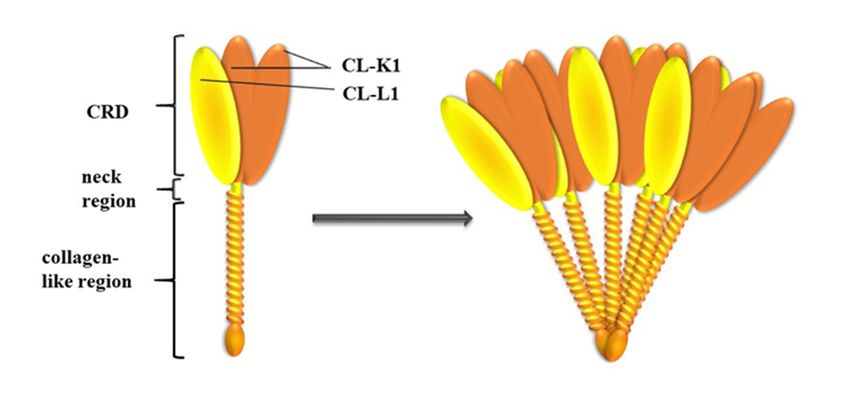
3. COLEC10 and COLEC11 Genes and Their Products, CL-L1 and CL-K1
3.1. Genes and Protein Structures
Collectins constitute a group of 400–800 kDa oligomeric, calcium-dependent lectins consisting of
basic subunits that are triplets of 30–35 kDa polypeptide chains. Collectin liver-1 (collectin-10) (CL-L1)Int. J. Mol. Sci. 2020, 21, 5483 4 of 14
Int. J. Mol. Sci. 2020, 21, x FOR PEER REVIEW 4 of 14
and
[36].collectin kidney-1
The collectin (collectin-11)
polypeptide chain (CL-K1)
consists are
ofclosely related, both
an N-terminal region structurally
containingand functionally
cysteine, [36].
a collagen-
The collectin polypeptide chain consists of an N-terminal region containing
like domain with Gly-Xaa-Yaa repeats (where Xaa and Yaa represent any amino acid residues), a cysteine, a collagen-like
domain with Gly-Xaa-Yaa
neck region, and a C-terminal repeats (where Xaa and Yaa represent
carbohydrate-recognition domainany (CRD).
amino acid Theresidues), a neck region,
CRD is responsible for
and a C-terminal carbohydrate-recognition domain (CRD). The CRD
interactions with sugar residues on microbial surfaces or aberrant host cells, whereas the collagen-is responsible for interactions
with sugar residues
like domain on microbial
forms complexes withsurfaces
proteins or aberrant
of the MASP host cells,
familywhereas
as welltheascollagen-like
interacting with domain cell
forms complexes with proteins of the MASP family as well as interacting
receptors. CL-L1 and CL-K1 form heterocomplexes called CL-LK, existing in blood. Each molecule is with cell receptors. CL-L1
and CL-K1by
stabilized form heterocomplexes
a disulphide called CL-LK,
bridge (created by Cysexisting
residuesinpresent
blood. in Each moleculedomains)
N-terminal is stabilized withby twoa
disulphide
CL-K1 andbridge one CL-L1 (created by Cys residues
polypeptide chains [37].present
CL-K1 in N-terminal
is present indomains) with two
species ranging fromCL-K1 and one
the zebrafish
CL-L1 polypeptide chains [37]. CL-K1 is present in species ranging from
to humans (with protein identities of full length protein: 72–98%). The protein identity between the zebrafish to humans (with
the
protein identities of full length protein: 72–98%). The protein
CRDs of CL-K1 and CL-L1 reaches 54%. This compares with only 25–32% for the corresponding identity between the CRDs of CL-K1
and CL-L1
domains ofreaches 54%. This
other collectins. compares with only 25–32% for the corresponding domains of other
[38].
collectins. [38].
The human COLEC10 gene is localized to chromosome 8 (8q23–q24.1) and includes six exons.
The The
first human
one encodesCOLEC10 gene
for the is localized
untranslated to chromosome
region, the N-terminal 8 (8q23–q24.1)
domain, and and theincludes six exons.
first Gly-Xaa-Yaa
The first one
sequence encodes
of the for the untranslated
collagen-like region. The region,
rest of thethe collagen-like
N-terminal domain,domainand the firstby
is encoded Gly-Xaa-Yaa
exons 2–5.
sequence of the collagen-like region. The rest of the collagen-like
Exon 5 is also responsible for the sequence of alpha-helical coiled-coil neck region while domain is encoded by exonsexon2–5.6
Exon
encodes5 is for
alsothe
responsible
CRD domain for the sequence
[39]. Twentyofpolymorphic
alpha-helical sitescoiled-coil neck region
were identified inwhile exon 6 encodes
the promoter, exons,
for the CRD domain [39]. Twenty polymorphic sites were identified
and flanking regions of the COLEC10 gene [40]. None of promoter polymorphisms were found in the promoter, exons, and to
flanking regions of the COLEC10 gene [40]. None of promoter polymorphisms
influence CL-L1 serum level. The +3654 C>T polymorphism (rs149331285, exon 5) affects the protein were found to influence
CL-L1
structureserumduelevel.
to anThe +3654
amino C>T
acid polymorphism
exchange (rs149331285,
(p.Arg125Trp). exon 5) affects
Heterozygosity wasthe protein structure
demonstrated to be
due to an amino acid exchange (p.Arg125Trp). Heterozygosity was demonstrated
associated with significantly-higher CL-L1 serum concentration, compared with C/C homozygosity. to be associated with
significantly-higher CL-L1 serum concentration,
Moreover, the -161/-157AAAATdel (rs148350292)compared may disturb withtheC/C homozygosity.
binding of severalMoreover,
transcription the
-161/-157AAAATdel (rs148350292) may disturb the binding
factors essential for liver development or immune response modulation [40]. of several transcription factors essential for
liver development or immune response modulation [40].
The COLEC11 gene is localized to chromosome 2 (2p25.3). It includes eleven exons, of which
seven The COLEC11
encode for the gene is localized
dominant isoform to chromosome
CL-11-1. With2 the (2p25.3).
exceptionIt includes
of the firstelevenone exons, of which
that encodes the
seven encode for the dominant isoform CL-11-1. With the exception
untranslated region, the other six exons are arranged and encode similar regions to that of the of the first one that encodes
the untranslated
COLEC10 region,
gene [39]. the other
Several six exons
variations in theare promoter,
arranged and exons encode similar regions
and introns have been to that of the
identified.
COLEC10
Among them, gene promoter
[39]. Several variations in the
polymorphism -9570 promoter, exons andwas
C>T (rs3820897) introns
shown havetobeen identified.
influence CL-K1Among serum
them, promoter polymorphism -9570 C>T (rs3820897) was shown
level. The non-synonymous SNP (p.His219Arg) in exon 7 (+39618 C>G, rs7567833) has no impact to influence CL-K1 serum level.
on
The non-synonymous
protein concentrationSNP (p.His219Arg)
in serum, howeverin it exon 7 (+39618
is suspected to C>G, rs7567833)
influence ligand has no impact
binding on protein
[40], Figure 2.
concentration in serum, however it is suspected to influence ligand binding [40], Figure 2.
Figure 2. Subunit and oligomeric structure of CL-LK. A total of three polypeptide chains of (two of
CL-K1
Figure and one of and
2. Subunit CL-L1) join to form
oligomeric a heteromeric
structure of CL-LK. subunit,
A total ofwhich
three may further oligomerize
polypeptide into
chains of (two of
structures ranging from dimer to hexamer (acc. to Hansen et al., 2018 [41]).
CL-K1 and one of CL-L1) join to form a heteromeric subunit, which may further oligomerize into
structures
3.2. Tissue rangingoffrom
Expression dimer to hexamer
the Collectin-Liver (acc.
1 and to Hansen et al.,12018 [41]).
Collectin-Kidney
The tissue
3.2. Tissue expression
Expression of both CL-K11 and
of the Collectin-Liver CL-L1 is ubiquitous.
and Collectin-Kidney 1 The highest expression of the
first mentioned was found in the liver, kidney, adrenal gland, ovary and gallbladder (and to a lower
The tissue expression of both CL-K1 and CL-L1 is ubiquitous. The highest expression of the first
extent, in the lung, ovary, testis, and retina) whereas expression of CL-L1 was found mainly in the liver
mentioned was found in the liver, kidney, adrenal gland, ovary and gallbladder (and to a lower
extent, in the lung, ovary, testis, and retina) whereas expression of CL-L1 was found mainly in the
liver and adrenal gland [38,41,42]. In the adrenal gland, CL-K1 was reported to be expressed in cellsInt. J. Mol. Sci. 2020, 21, 5483 5 of 14
and adrenal gland [38,41,42]. In the adrenal gland, CL-K1 was reported to be expressed in cells of all
three layers—spinal, banded, and reticular, whereas in the kidney, it is expressed in the distal canals,
as well as the glomeruli and proximal tubules [38]. In the ovary, it is associated with granulosa and
theca lutein cells while in the testis, the main expression sites are seminiferous tubules [38]. In the
liver, the expression of both CL-K1 and CL-L1 is associated with hepatocytes [38,43]. Furthermore,
high expression of both collectins was detected in placenta [42]. Average human CL-L1 serum level
was estimated to be 0.5 µg/mL, while CL-K1 was estimated to be approximately 0.4 µg/mL. Their
concentrations are strongly correlated [19]. No significant differences with sex, age (18–70 years), or
diurnal variations were found [19,40,44,45].
3.3. Biological Activity of Collectin-Liver 1 and Collectin-Kidney 1
CL-K1 was reported to recognize certain Gram-negative bacteria (E. coli O126 and O60, Pseudomonas
aeruginosa, and Klebsiella pneumoniae), mycobacteria (Mycobacterium tuberculosis), fungi (Candida albicans
and Saccharomyces cerevisiae), and viruses (influenza A virus) [38,42,46]. Surface structures identified
amongst those recognized by CL-K1 include lipopolysaccharides (LPS core region of E. coli mutants of Ra
and Rd, but not Re chemotypes), S. cerevisiae mannan, mycobacterial mannosylated lipoarabinomannan
(ManLAM) [42,46], and HIV gp120 [47]. Venkatraman Girija et al., (2015) demonstrated the affinity of
CL-K1 to mannose-rich glycans containing the disaccharide Man(α1-2)Man as terminal motif [47].
The microbial structures recognized by CL-L1 are not yet established, but formation of
heterodimers with CL-K1 could possibly lead to an increased range of interactions with microorganisms,
as well as to their higher affinity. Thus, CL-LK is characterized by high affinity to d-mannose
(d-Man), n-acetyl-d-glucosamine (d-GlcNAc), d-galactose (d-Gal), l- and d-fucose (l-Fuc, d-Fuc), and
n-acetyl-d-mannosamine (d-ManNAc) [41]. CL-LK, due to cooperation with MASP, may initiate
activation of complement via the lectin pathway (and cross-talk with the coagulation cascade). They
may also aggregate and opsonize microorganisms contributing to enhanced phagocytosis [48].
Through the collagen-like domain, but also via its CRD, CL-K1 binds to mammalian and bacterial
DNA in a calcium-independent manner [49]. It also binds to DNA exposed on apoptotic/necrotic cells
and may facilitate their clearance, preventing formation of anti-DNA antibodies. The CL-K1–DNA
interaction may lead to complement activation. However, it is much weaker compared to other
collectins, probably due to different degrees of protein oligomerization [50].
Only a few reports concerning CL-LK disease associations and the clinical significance of
CL-L1/CL-K1 have been published to date. Increased blood CL-L1 levels were observed at early stages
of acute liver failure and cirrhosis. Smedbråten et al. (2017) reported that high plasma concentrations
of CL-L1 and CL-K1 at the time of transplantation are correlated with increased mortality in kidney
transplant recipients [51]. Troldborg et al. (2018) found lower CL-L1 and CL-K1 levels in systemic
lupus erythematosus (SLE patients), compared with healthy controls, however no association with
SLE disease activity index (SLEDAI) score was found [52]. Storm et al. (2014) reported that CL-L1
may significantly distinguish between patients with colorectal cancer (CRC), patients with adenomas,
and individuals without neoplastic bowel lesions [53]. Higher levels of CL-L1 were associated with
lower odds of CRC [53]. Świerzko et al. (2018), reported higher serum levels of CL-LK complex in
multiple myeloma patients compared with healthy individuals [45]. Farrar et al., (2016) showed that
deficiency of CL-K1 is protective in the case of ischemia, preventing tubular damage and loss of renal
function [54]. Similar conclusions were also drawn by Wu et al. (2018), who demonstrated that CL-K1
plays a harmful role in the development of tubulointerstitial fibrosis by leukocyte chemotaxis and
the impact on the proliferation of renal fibroblasts [55]. Koipallil Gopalakrishnan Nair et al. (2015)
observed an approximately 3-fold increase in CL-K1 and CL-L1 transcript expression in human ectopic
endometriotic mesenchymal stem cells (MSCs) in comparison with eutopic MSCs [56].Int. J. Mol. Sci. 2020, 21, 5483 6 of 14
4. Associations of COLEC10, COLEC1,1 and MASP1/3 Gene Mutations with 3MC Syndrome
Association of MASP1/3 gene polymorphisms with 3MC were first reported by Sirmaci et al.
(2010), in three individuals from two related Turkish families. They found a missense c.2059 G>A
(p.Gly687Arg) mutation specific to the MASP-3 isoform and nonsense c.870 G>A (p.Trp290*) in exon 6,
which is shared by all gene products. In silico results suggested that the p.Gly687Arg mutation may be
damaging for the MASP-3 catalytic domain [5]. Other abnormalities of the same gene were identified
by Rooryck et al. (2011). They described three homozygous missense mutations, all located within
exon 12, encoding the MASP-3 protease domain, in the 3MC-syndrome-affected patients (c.1489C>T
p.His497Tyr, c.1888T>C p.Cys630Arg, and c.1997G>A p.Gly666Glu). All of them are predicted to affect
protein activity [6]. Atik et al. (2015) reported two splice site mutations, c.1012 - 2 A>G in one proband
and c.891 + 1 G >T in two propositi, as well as three missense mutations, c.1451 G>A (p. Gly484Glu),
c.1657 G >A (p. Asp553Asn), and c.1987 G >T (p. Asp663Tyr) in three other propositi. Splice site
mutations affect both MASP-1 and MASP-3, while missense mutations affect MASP-3 only. A total lack
of lectin pathway activity and a 2.5-fold lower alternative pathway activity in a proband homozygous
for c.891 + 1 G > T were found [57]. Urquhart et al. (2016) identified three other mutations within the
MASP1/3 gene (c.9 G>A p.Trp3*, c.547 G>T p.Val183Leu, and c.760 A>T p.Leu254*) [58]. The missense
alteration, p.Asp663Tyr (reported also, as mentioned, by Atik et al. (2015)) probably changes the
enzymatic activity of the catalytic domain [58]. Pihl et al. (2017) described the proposita from a German
family with a homozygous missense mutation c.1993G.A (p.G665S) in exon 12 of the MASP1/3 gene,
confirming the clinical diagnosis of 3MC syndrome. The serum concentration of MASP-3 in this patient
was lower than the median for a healthy population [59]. Later, Graul-Neumann et al. (2018) reported
a Turkish individual carrying a deletion of 2306 bp (c.1895_*1602+411del). It affects the terminal part
of exon 12 and accordingly the C-terminal serine protease domain specific to MASP-3 [60]. A separate
study of two individuals from a Turkish family was conducted by Basdemirci et al. (2019). They
identified a new missense homozygous mutation c.2111T>G (p. Val704Gly) (NM_139125.3) in exon
11 of the relevant gene [61]. Çakmaklı et al. (2019) found the afore-mentioned missense c.1987G>T;
(p.Asp663Tyr) MASP1/3 mutation (in a homozygous state) in a patient from Syria [62]. In summary, so
far 16 3MC-syndrome-associated mutations in the MASP-1/3 gene in 26 individuals have been reported
(Table 1). The majority of them (11) affect the serine protease domain of MASP-3.
Rooryck et al. (2011) demonstrated an association of homozygosity for three non-synonymous
mutations and one in-frame deletion in the COLEC11 gene: c.496 T>C (rs387907075, p.Ser169Pro, exon 8),
c.45delC (RCV000023960, p.Phe16Serfs X85, exon 2), c.610 G>A (rs387907076, p.Gly204Ser, exon 8), and
c.648-650delCTC (RCV000023962, p.Ser217del, exon 8). Moreover, they identified a 27-kb homozygous
deletion encompassing exons 1–3 of COLEC11, predicting partial loss of the collagen-like domain,
and complete loss of the N-terminal (cysteine-rich) domain. Furthermore, a homozygous single-base
deletion (c.300delT/G101VfsX113 in exon 6) was suggested to lead to premature termination of the
COLEC11 gene product [6]. Gardner et al. (2017) identified a homozygous C-terminal deletion resulting
in the complete loss of exon 8 and the partial loss of the carbohydrate-recognition domain [63] while
Urquhart et al. (2016) reported a frame-shift mutation (deletion of two nucleotides), c.627_628delGC
and p. (Ala213Leufs 5) [58]. Carriers of the above-mentioned mutations are suspected to be
CL-K1-functionally-deficient. Two novel non-synonymous homozygous COLEC11 mutations in 3MC
patients were later described by Munye et al. (2017). One of them, c.309delT (p.Gly104Valfs29 in exon 4)
again seems to predict premature appearance of a stop codon, while another one, c.G496A (p.Ala166Thr
in exon 6), causes a change in the structure, affecting protein activity. Additionally, they found deletion
of 10 nucleotides (89_98del ATGACGCCTG in exon 2) which predicted a frameshift change and the
introduction of a premature stop codon (p.Asp30Alafs68) [7]. To summarize, 11 mutations have so far
been described in the COLEC11 gene in 17 3MC patients (Table 2). Six of them affect the CRD region of
the collectin.Int. J. Mol. Sci. 2020, 21, 5483 7 of 14
Table 1. Families affected by the Malpuech, Michels, Mingarelli, Carnevale (3MC) syndrome, associated
with mutations in MASP1/3 gene.
Family (Number of Amino acid Change/Protein
Origin Nucleotide Change References
Carriers) Change
c.2059G>A p.Gly687Arg
F1 (2) Turkey
exon 12 MASP-3 SP
Sirmaci et al., 2010 [5]
p.Trp290*
c.870G>A
F2 (1) Turkey MASP-1/3
exon 6
CUB1-EGF-CUB2-CCP1
c.1489 C>T p.His497Tyr
F3 (1) Greece
exon 12 MASP-3 SP
c.1888T>C p.Cys630Arg
F4 (2) Italy
exon 12 MASP-3 SP
Rooryck et al., 2011 [6]
c.1997G>A p.Gly666Glu
F5 (1) Brazil
exon 12 MASP-3 SP
c.1997G>A p.Gly666Glu
F6 (1) Brazil
exon 12 MASP-3 SP
c.1012-2A>G
F7 (1) Turkey Splice site mutation
intron 7
c.891 +1G>T
F8 (1) Turkey Splice site mutation
intron 6
c.891 +1G>T
F9 (1) Turkey Splice site mutation
intron 6
Atik et al., 2015 [57]
c.1451G>A p. Gly484Glu
F10 (1) Pakistan
exon 12 MASP-3 SP
c.1657G>A p. Asp553Asn
F11 (1) Turkey
exon 12 MASP-3 SP
c.1987G>T p. Asp663Tyr
F12 (1) Syria
exon 12 MASP-3 SP
c.1987G>T p.Asp663Tyr
F13 (1) Israel
exon 12 MASP-3 SP
p.Trp3*
c.9G>A
F14 (1) Sri Lanka MASP-1/3
exon 2
CUB1-EGF-CUB2-CCP1
Urquhart et al., 2016 [58]
p.Leu 254*
c.760A>T
F14 (2) Sri Lanka MASP-1/3
exon 6
CUB1-EGF-CUB2-CCP1
p.Val 183Leu
c.547G>T
F15 (1) India MASP-1/3
exon 4
CUB1-EGF-CUB2-CCP1
c.1993G>A p.G665S
F16 (1) Germany Pihl et al., 2017 [59]
exon 12 MASP-3 SP
p. Trp3*
c.9G>A
F17 (1) Pakistan MASP-1/3 Munye et al., 2017 [7]
exon 2
CUB1-EGF-CUB2-CCP1
c.1895_*1602+411del p.Arg637Cysfs*1 Graul-Neumann et al., 2018
F18 (1) Turkey
exon 12 MASP-3 SP [60]
c.2111T>G p. Val704Gly
F19 (3) Turkey Basdemirci et al., 2019 [61]
exon 12 MASP-1/MASP-3 CCP2
c.1987G>T p.Asp663Tyr
F20 (1) Syria Çakmaklı et al., 2019 [62]
exon 12 MASP-3 SP
Three mutations of the COLEC10 gene were found associated with 3MC syndrome: c.25 C>T
(exon 1), c.226delA (exon 3), and c.528 C>G (exon 6). The last-mentioned results in severe impairment
of protein secretion, whereas the two others lead to the nonsense-mediated decay of transcripts [7].
All mutations in the COLEC10 gene occurring in patients with 3MC syndrome are shown in Table 3.Int. J. Mol. Sci. 2020, 21, 5483 8 of 14
Table 2. Families affected by 3MC syndrome, associated with mutations in the COLEC11 gene.
Family (Number of Amino Acid Change/Protein
Origin Nucleotide Change References
Carriers) Change
c.496T>C p.Ser169Pro
F1 (2) Tunisia
exon 8 CRD
c.45delC p.Phe16SerfsX 85
F2 (2) Bangaldesh
exon 2 N-terminal Collagen-like region
c.610G>A p.Gly204Ser
F3 (2) Afganistan
exon 8 CRD
c.648_650delCTC p.Ser217del
F4 (1) Saudi Arabia
exon 8 CRD
Rooryck et al., 2011 [6]
c.610G>A p.Gly204Ser
F5 (1) Pakistan
exon 8 CRD
c.300delT p.Gly101ValfsX 113
F6 (1) Italy
exon 6 Neck domain
Predicted: complete loss of
ex 1-3 deletion N-terminus and partial loss of the
F7 (1) Italy
exons 1,2,3 collagen-like domains
N-terminal collagen-like region
c.627_628delGC p. Ala213Leufs 5
F8 (2) Israel Urquhart et al., 2016 [58]
Exon 8 CRD
Predicted: complete loss of C
ex 8 deletion terminus and at least partial loss of
F9 (2) Pakistan Gardner et al., 2017 [63]
exon 8 the carbohydrate-recognition domain
(CRD)
c.309delT p.Gly104Valfs29
F10 (1) Pakistan
exon 4 Collagen-like domain
c.G496A p.Ala166Thr
F11 (1) Somalia
exon 6 Neck domain Munye et al., 2017 [7]
89_98del
United Arab p.Asp30Alafs68
F12 (1) ATGACGCCTG
Emirates N-terminal domain
exon 2
Table 3. Families affected by 3MC syndrome, associated with mutations in COLEC10 gene.
Amino Acid
Family (Number of Nucleotide
Origin Change/Protein References
Ccarriers) Change
Change
c.25C>T p. Arg9Ter
exon 1 Signal peptide
F1 (2) Pakistan
c.226delA p.Gly77Glufs*66
exon 3 Collagen-like region Munye et al.,
c.25C>T p. Arg9Ter 2017 [7]
exon 1 Signal peptide
F2 (1) Pakistan
c.528C>G p.Cys176Trp
exon 6 Neck domain
5. Concluding Remarks
The craniofacial disruptions observed in patients suffering from 3MC syndrome are similar to
neural crest migration disorders. The proper migration of neural crest cells (NCC) is essential for the
formation of bones, cartilage, ganglia, and muscles in the head [64]. Control and regulation of NCC
migration is complex and involves many genetic pathways. Thus, there is a possibility that proteins
with collagen-like regions linked to a CRD domain play dual roles, contributing to immunity and
development. Certain complement components have previously been shown to play an essential role
in cell migration. C3a and its C3aR receptor co-attracted crest cells, to coordinate migration in the
first stages of NCC regulation [65]. Furthermore, surfactant protein D [66,67] and surfactant protein
A [68] being collectins (although lacking complement activation property), have been also described as
chemoattractants. Studies employing zebrafish [6] demonstrated that loss of CL-K1 and MASP-1/-3 isInt. J. Mol. Sci. 2020, 21, 5483 9 of 14
associated with craniofacial abnormalities. Both proteins were proposed to act as guidance cues for
neural crest cell migration [47]. Moreover, CL-L1 was shown to regulate development of craniofacial
structures acting as a migratory chemoattractant [7]. Three CL-K1 gene mutations associated with
3MC syndrome, resulting in Ser169Pro and Gly204Ser substitutions and Ser217 deletion, prevent
normal secretion from mammalian cells due to structural changes caused by the failure to bind Ca2+
during biosynthesis. The destabilization of CRD probably leads to elimination of protein via the
endoplasmic-reticulum-associated protein degradation pathway [47]. Gorelik et al. (2017) also reported
that MASP-1 plays an important role in radial neuronal migration in the development of the cerebral
cortex. Deficiency of MASP-1 and other components of the lectin pathway (C3 and MASP-2) leads to
impairments in radial migration resulting in improper positioning of neurons and disorganized cortical
layers [69]. Because CL-LK heterocomplexes were found to bind MASP-1 or MASP-3 homodimers
via their collagen-like regions [47,70] it is possible that correct migration of neural crest cells requires
cooperation between CL-LK and MASP-1/-3. This may be supported by the relationship between
MASP1/3, COLEC10, and COLEC11 mutations and the incidence of 3MC syndrome, associated with
craniofacial abnormalities. During embryogenesis, these three genes are strongly expressed in the
craniofacial cartilage, palatal structures, bronchi, heart, and kidneys and the corresponding proteins
act as chemoattractants for the cranial crest nerve cells (crucial for the formation of the head skeleton),
recognizing certain endogenous carbohydrate epitopes [6]. It should be however mentioned, that mice
with knockout in MASP1/3 or COLEC11 genes developed normally and no defects similar to 3MC
syndrome were noticed [71–73]. It moreover still remains to be clarified which defense pathways may
compensate for MASP-1/3, CL-L1, and CL-K1 dysfunctions and why this rare disease is generally not
accompanied by impaired immune response.
As mentioned, so far 11 mutations in the COLEC11 gene, three in the COLEC10 gene, and 16 in
the MASP1/3 gene associated with 3MC syndrome have been described. Further studies, however, are
necessary to better understand the mechanisms by which dysfunction of MASP1/3, COLEC10, and
COLEC11 genes may lead to the 3MC syndrome.
Author Contributions: Conceptualization, G.G. and A.S.Ś.; writing—original draft preparation, G.G.;
writing—review and editing: G.G., A.S.Ś. and M.C.; supervision, M.C. All authors have read and agreed
to the published version of the manuscript.
Funding: This research received no external funding
Acknowledgments: The authors are very grateful to David C. Kilpatrick for critical reading of the manuscript
and helpful discussion.
Conflicts of Interest: The authors declare no conflict of interest.
Abbreviations
3MC Malpuech, Michels, Mingarelli, Carnevale syndrome
MASP Mannose-binding lectin-associated serine proteases
MAp44 Mannose-binding lectin-associated protein, 44kDa
SP Serine protease
BMP Bone morphogenic protein
SNP Single nucleotide polymorphism
MBL Mannose-binding lectin
PRM Pattern recognition molecule
TAFI Thrombin-activatable fibrinolysis inhibitor
IL Interleukin
MODS Multiorgan dysfunction
SIRS Systemic inflammatory response syndrome
LCOS Low cardiac output syndrome
CRD Carbohydrate-recognition domainInt. J. Mol. Sci. 2020, 21, 5483 10 of 14
CL-L1 Collectin liver-1 (collectin-10)
CL-K1 Collectin kidney-1 (collectin-11)
SP-A Surfactant protein A
SP-D Surfactant protein D
LPS Lipopolysaccharide
HIV Human immunodeficiency virus
D-Man D-MANNOSE
D-GlcNAc N-ACETYL-D-GLUCOSAMINE
D-Gal D-GALACTOSE
L-Fuc L-FUCOSE
D-Fuc D-FUCOSE
D-ManNAc N-ACETYL-D-MANNOSAMINE
CRC Colorectal cancer
MSCs Mesenchymal stem cells
References
1. Mingarelli, R.; Scanderbeg, A.C.; Dallapiccola, B. Two sisters with a syndrome of ocular, skeletal, and
abdominal abnormalities (OSA syndrome). J. Med. Genet. 1996, 33, 884–886. [CrossRef] [PubMed]
2. Malpuech, G.; Deméocq, F.; Palcoux, J.B.; Vanlieferinghen, P.; Opitz, J.M. A previously undescribed
autosomal recessive multiple congenital anomalies/mental retardation (MCA/MR) syndrome with growth
failure, lip/palate cleft(s), and urogenital anomalies. Am. J. Med. Genet. 1983, 16, 475–480. [CrossRef]
3. Michels, V.V.; Hittner, H.M.; Beaudet, A.L. A clefting syndrome with ocular anterior chamber defect and lid
anomalies. J. Pediatr. 1978, 93, 444–446. [CrossRef]
4. Carnevale, F.; Krajewska, G.; Fischetto, R.; Greco, M.G.; Bonvino, A. Ptosis of eyelids, strabismus, diastasis
recti, hip defect, cryptorchidism, and developmental delay in two sibs. Am. J. Med. Genet. 1989, 33, 186–189.
[CrossRef] [PubMed]
5. Sirmaci, A.; Walsh, T.; Akay, H.; Spiliopoulos, M.; Şakalar, Y.B.; Hasanefendioğlu-Bayrak, A.; Duman, D.;
Farooq, A.; King, M.C.; Tekin, D. MASP1 Mutations in Patients with Facial, Umbilical, Coccygeal, and
Auditory Findings of Carnevale, Malpuech, OSA, and Michels Syndromes. Am. J. Hum. Genet. 2010, 87,
679–686. [CrossRef]
6. Rooryck, C.; Díaz-Font, A.; Osborn, D.P.; Chabchoub, E.; Hernandez-Hernandez, V.; Shamseldin, H.; Kenny, J.;
Waters, A.; Jenkins, D.; Al Kaissi, A.; et al. Mutations in lectin complement pathway genes COLEC11 and
MASP1 cause 3MC syndrome. Nat. Genet. 2011, 43, 197–203. [CrossRef] [PubMed]
7. Munye, M.M.; Diaz-Font, A.; Ocaka, L.; Henriksen, M.L.; Lees, M.; Brady, A.; Jenkins, D.; Morton, J.;
Hansen, S.W.K.; Bacchelli, C.; et al. COLEC10 is mutated in 3MC patients and regulates early craniofacial
development. PLoS Genet. 2017, 13, e1006679. [CrossRef]
8. Degn, S.E.; Jensenius, J.C.; Thiel, S. Disease-Causing Mutations in Genes of the Complement System. Am. J.
Hum. Genet. 2011, 88, 689–705. [CrossRef]
9. Ammitzbøll, C.G.; Steffensen, R.; Nielsen, H.J.; Thiel, S.; Stengaard-Pedersen, K.; Bøgsted, M.; Jensenius, J.C.
Polymorphisms in the MASP1 Gene Are Associated with Serum Levels of MASP-1, MASP-3, and MAp44.
PLoS ONE 2013, 8, e73317. [CrossRef]
10. Degn, S.E.; Hansen, A.G.; Steffensen, R.; Jacobsen, C.; Jensenius, J.C.; Thiel, S. MAp44, a Human Protein
Associated with Pattern Recognition Molecules of the Complement System and Regulating the Lectin
Pathway of Complement Activation. J. Immunol. 2009, 183, 7371–7378. [CrossRef]
11. Fujita, T. Evolution of the lectin—Complement pathway and its role in innate immunity. Nat. Rev. Immunol.
2002, 2, 346–353. [CrossRef] [PubMed]
12. Matsushita, M.; Endo, Y.; Fujita, T. Structural and Functional Overview of the Lectin Complement Pathway:
Its Molecular Basis and Physiological Implication. Arch. Immunol. Ther. Exp. 2013, 61, 273–283. [CrossRef]
[PubMed]
13. Gregory, L.A.; Thielens, N.M.; Matsushita, M.; Sorensen, R.; Arlaud, G.J.; Fontecilla-Camps, J.C.; Gaboriaud, C.
The X-ray Structure of Human Mannan-binding Lectin-associated Protein 19 (MAp19) and Its Interaction
Site with Mannan-binding Lectin and L-ficolin. J. Biol. Chem. 2004, 279, 29391–29397. [CrossRef] [PubMed]Int. J. Mol. Sci. 2020, 21, 5483 11 of 14
14. Bork, P.; Beckmann, G. The CUB Domain. A Widespread Module in Developmentally Regulated Proteins. J.
Mol. Biol. 1993, 23, 539–545. [CrossRef] [PubMed]
15. Bally, I.; Rossi, V.; Thielens, N.M.; Gaboriaud, C.; Arlaud, G.J. Functional Role of the Linker between the
Complement Control Protein Modules of Complement Protease C1s. J. Immunol. 2005, 175, 4536–4542.
[CrossRef] [PubMed]
16. Gaboriaud, C.; Gupta, R.K.; Martin, L.; Lacroix, M.; Serre, L.; Teillet, F.; Arlaud, G.J.; Rossi, V.; Thielens, N.M.
The Serine Protease Domain of MASP-3: Enzymatic Properties and Crystal Structure in Complex with Ecotin.
PLoS ONE 2013, 8, e67962. [CrossRef] [PubMed]
17. Lynch, N.J.; Stover, C.M.; Sandrini, S.M.; Marston, D.; Presanis, J.S.; Schwaeble, W.J. Composition of the
Lectin Pathway of Complement in Gallus gallus: Absence of Mannan-Binding Lectin-Associated Serine
Protease-1 in Birds. J. Immunol. 2005, 174, 4998–5006. [CrossRef]
18. Skjoedt, M.-O.; Hummelshoj, T.; Palarasah, Y.; Hein, E.; Munthe-Fog, L.; Koch, C.; Skjodt, K.; Garred, P.
Serum concentration and interaction properties of MBL/ficolin associated protein-1. Immunobiology 2011, 216,
625–632. [CrossRef]
19. Troldborg, A.; Hansen, A.; Hansen, S.W.K.; Jensenius, J.C.; Stengaard-Pedersen, K.; Thiel, S. Lectin
complement pathway proteins in healthy individuals. Clin. Exp. Immunol. 2017, 188, 138–147. [CrossRef]
20. Dobó, J.; Pál, G.; Cervenák, L.; Gál, P. The emerging roles of mannose-binding lectin-associated serine
proteases (MASPs) in the lectin pathway of complement and beyond. Immunol. Rev. 2016, 274, 98–111.
[CrossRef]
21. Krarup, A.; Gulla, K.C.; Gál, P.; Hajela, K.; Sim, R. The action of MBL-associated serine protease 1 (MASP1)
on factor XIII and fibrinogen. Biochim. Biophys. Acta BBA Proteins Proteom. 2008, 1784, 1294–1300. [CrossRef]
[PubMed]
22. Jenny, L.; Dobó, J.; Gal, P.; Schroeder, V. MASP-1 of the complement system promotes clotting via prothrombin
activation. Mol. Immunol. 2015, 65, 398–405. [CrossRef]
23. Krarup, A.; Wallis, R.; Presanis, J.S.; Gal, P.; Sim, R. Simultaneous Activation of Complement and Coagulation
by MBL-Associated Serine Protease 2. PLoS ONE 2007, 2, e623. [CrossRef] [PubMed]
24. Gulla, K.C.; Gupta, K.; Krarup, A.; Gal, P.; Schwaeble, W.J.; Sim, R.; O’Connor, C.D.; Hajela, K. Activation of
mannan-binding lectin-associated serine proteases leads to generation of a fibrin clot. Immunology 2009, 129,
482–495. [CrossRef]
25. Hess, K.; Ajjan, R.; Phoenix, F.; Dobó, J.; Gál, P.; Schroeder, V. Effects of MASP-1 of the complement system on
activation of coagulation factors and plasma clot formation. PLoS ONE 2012, 7, e35690. [CrossRef] [PubMed]
26. Dobó, J.; Major, B.; Kékesi, K.A.; Szabo, I.; Megyeri, M.; Hajela, K.; Juhász, G.; Závodszky, P.; Gál, P. Cleavage
of Kininogen and Subsequent Bradykinin Release by the Complement Component: Mannose-Binding
Lectin-Associated Serine Protease (MASP)-1. PLoS ONE 2011, 6, e20036. [CrossRef] [PubMed]
27. Megyeri, M.; Jani, P.K.; Kajdácsi, E.; Dobó, J.; Schwaner, E.; Major, B.; Rigó, J.; Závodszky, P.; Thiel, S.;
Cervenak, L.; et al. Serum MASP-1 in complex with MBL activates endothelial cells. Mol. Immunol. 2014, 59,
39–45. [CrossRef] [PubMed]
28. Jani, P.K.; Schwaner, E.; Kajdácsi, E.; Debreczeni, M.; Ungai-Salánki, R.; Dobó, J.; Doleschall, Z.; Rigó, J.;
Geiszt, M.; Szabó, B.; et al. Complement MASP-1 enhances adhesion between endothelial cells and
neutrophils by up-regulating E-selectin expression. Mol. Immunol. 2016, 75, 38–47. [CrossRef]
29. Jani, P.K.; Kajdácsi, E.; Megyeri, M.; Dobó, J.; Doleschall, Z.; Futosi, K.; Timár, C.I.; Mocsai, A.; Makó, V.;
Gal, P.; et al. MASP-1 Induces a Unique Cytokine Pattern in Endothelial Cells: A Novel Link between
Complement System and Neutrophil Granulocytes. PLoS ONE 2014, 9, e87104. [CrossRef]
30. Cortesio, C.L.; Jiang, W. Mannan-binding lectin-associated serine protease 3 cleaves synthetic peptides and
insulin-like growth factor-binding protein 5. Arch. Biochem. Biophys. 2006, 449, 164–170. [CrossRef]
31. Skjoedt, M.-O.; Roversi, P.; Hummelshøj, T.; Palarasah, Y.; Rosbjerg, A.; Johnson, S.; Lea, S.M.; Garred, P.
Crystal Structure and Functional Characterization of the Complement Regulator Mannose-binding Lectin
(MBL)/Ficolin-associated Protein-1 (MAP-1). J. Biol. Chem. 2012, 287, 32913–32921. [CrossRef] [PubMed]
32. Mortensen, S.A.; Skov, L.L.; Kjaer-Sorensen, K.; Hansen, A.G.; Hansen, S.W.K.; Dagnæs-Hansen, F.;
Jensenius, J.C.; Oxvig, C.; Thiel, S.; Degn, S.E. Endogenous Natural Complement Inhibitor Regulates
Cardiac Development. J. Immunol. 2017, 198, 3118–3126. [CrossRef] [PubMed]Int. J. Mol. Sci. 2020, 21, 5483 12 of 14
33. Mendes, H.W.; Boldt, A.B.W.; Stahlke, E.V.R.S.; Jensenius, J.C.; Thiel, S.; Messias-Reason, I.J.T. Adding
MASP1 to the lectin pathway—Leprosy association puzzle: Hints from gene polymorphisms and protein
levels. PLoS Negl. Trop. Dis. 2020, 14, e0007534. [CrossRef]
34. Larsen, J.B.; Laursen, M.A.; Hvas, C.L.; Larsen, K.M.; Thiel, S.; Hvas, A.-M. Reduced Mannose-Binding
Lectin-Associated Serine Protease (MASP)-1 is Associated with Disturbed Coagulation in Septic Shock.
Thromb. Haemost. 2019, 119, 952–961. [CrossRef] [PubMed]
35. Michalski, M.; Pagowska-Klimek,
˛ I.; Thiel, S.; Świerzko, A.S.; Hansen, A.G.; Jensenius, J.C.; Cedzynski, M.
Factors involved in initiation and regulation of complement lectin pathway influence postoperative outcome
after pediatric cardiac surgery involving cardiopulmonary bypass. Sci. Rep. 2019, 9, 1–9. [CrossRef]
36. Fanelli, G.; Cordero, A.G.; Gardner, P.J.; Peng, Q.; Fernando, M.; Kloc, M.; Farrar, C.A.; Naeem, A.; Garred, P.;
Ali, R.R.; et al. Human stem cell-derived retinal epithelial cells activate complement via collectin 11 in
response to stress. Sci. Rep. 2017, 7, 1–13. [CrossRef]
37. Henriksen, M.L.; Brandt, J.; Andrieu, J.-P.; Nielsen, C.; Jensen, P.H.; Holmskov, U.; Jørgensen, T.; Palarasah, Y.;
Thielens, N.M.; Hansen, S.W.K. Heteromeric Complexes of Native Collectin Kidney 1 and Collectin Liver
1 Are Found in the Circulation with MASPs and Activate the Complement System. J. Immunol. 2013, 191,
6117–6127. [CrossRef]
38. Hansen, S.; Selman, L.; Palaniyar, N.; Ziegler, K.; Brandt, J.; Kliem, A.; Jonasson, M.; Skjoedt, M.-O.;
Nielsen, O.; Hartshorn, K.; et al. Collectin 11 (CL-11, CL-K1) Is a MASP-1/3–Associated Plasma Collectin
with Microbial-Binding Activity. J. Immunol. 2010, 185, 6096–6104. [CrossRef]
39. Selman, L.; Henriksen, M.; Brandt, J.; Palarasah, Y.; Waters, A.; Beales, P.; Holmskov, U.; Jørgensen, T.J.D.;
Nielsen, C.; Skjodt, K.; et al. An enzyme-linked immunosorbent assay (ELISA) for quantification of human
collectin 11 (CL-11, CL-K1). J. Immunol. Methods 2012, 375, 182–188. [CrossRef]
40. Bayarri-Olmos, R.; Hansen, S.W.K.; Henriksen, M.L.; Storm, L.; Thiel, S.; Garred, P.; Munthe-Fog, L. Genetic
Variation of COLEC10 and COLEC11 and Association with Serum Levels of Collectin Liver 1 (CL-L1) and
Collectin Kidney 1 (CL-K1). PLoS ONE 2015, 10, e0114883. [CrossRef]
41. Hansen, S.W.K.; Aagaard, J.B.; Bjerrum, K.B.; Hejbøl, E.K.; Nielsen, O.; Schrøder, H.D.; Skjoedt, K.;
Sørensen, A.L.; Graversen, J.H.; Henriksen, M.L. CL-L1 and CL-K1 Exhibit Widespread Tissue Distribution
With High and Co-Localized Expression in Secretory Epithelia and Mucosa. Front. Immunol. 2018, 9, 1757.
[CrossRef] [PubMed]
42. Keshi, H.; Sakamoto, T.; Kawai, T.; Ohtani, K.; Katoh, T.; Jang, S.; Motomura, W.; Yoshizaki, T.; Fukuda, M.;
Koyama, S.; et al. Identification and characterization of a novel human collectin CL-K1. Microbiol. Immunol.
2006, 50, 1001–1013. [CrossRef] [PubMed]
43. Ohtani, K.; Suzuki, Y.; Eda, S.; Kawai, T.; Kase, T.; Yamazaki, H.; Shimada, T.; Keshi, H.; Sakai, Y.; Fukuoh, A.;
et al. Molecular Cloning of a Novel Human Collectin from Liver (CL-L1). J. Biol. Chem. 1999, 274,
13681–13689. [CrossRef] [PubMed]
44. Axelgaard, E.; Jensen, L.; Dyrlund, T.F.; Nielsen, H.J.; Enghild, J.J.; Thie, L.S.; Jensenius, J.C. Investigations on
collectin-liver 1 (CL-L1 or CL-10). J. Biol. Chem. 2013, 288, 23407–23420.
45. Świerzko, A.S.; Michalski, M.; Sokołowska, A.; Nowicki, M.; Eppa, Ł.; Szala-Poździej, A.; Mitrus, I.;
Szmigielska-Kapłon, A.; Sobczyk-Kruszelnicka, M.; Michalak, K.; et al. The Role of Complement Activating
Collectins and Associated Serine Proteases in Patients With Hematological Malignancies, Receiving High-Dose
Chemotherapy, and Autologous Hematopoietic Stem Cell Transplantations (Auto-HSCT). Front. Immunol.
2018, 9, 2153. [CrossRef]
46. Troegeler, A.; Lugo, G.; Hansen, S.W.K.; Rasolofo, V.; Henriksen, M.L.; Mori, K.; Ohtani, K.; Duval, C.;
Mercier, I.; Bénard, A.; et al. Collectin CL-LK Is a Novel Soluble Pattern Recognition Receptor for
Mycobacterium tuberculosis. PLoS ONE 2015, 10, e0132692. [CrossRef]
47. Girija, U.V.; Furze, C.M.; Gingras, A.R.; Yoshizaki, T.; Ohtani, K.; Marshall, J.E.; Wallis, A.K.; Schwaeble, W.J.;
El-Mezgueldi, M.; Mitchell, D.A.; et al. Molecular basis of sugar recognition by collectin-K1 and the effects of
mutations associated with 3MC syndrome. BMC Biol. 2015, 13, 1–14. [CrossRef]
48. Hansen, S.W.K. Lung Surfactant Protein D (SP-D) and the Molecular Diverted Descendants: Conglutinin,
CL-43 and CL-46. Immunobiology 2002, 205, 498–517. [CrossRef]
49. Henriksen, M.L.; Brandt, J.; Iyer, S.S.; Thielens, N.M.; Hansen, S. Characterization of the interaction between
collectin 11 (CL-11, CL-K1) and nucleic acids. Mol. Immunol. 2013, 56, 757–767. [CrossRef]Int. J. Mol. Sci. 2020, 21, 5483 13 of 14
50. Selman, L.; Hansen, S. Structure and function of collectin liver 1 (CL-L1) and collectin 11 (CL-11, CL-K1).
Immunobiology 2012, 217, 851–863. [CrossRef]
51. Smedbråten, J.; Sagedal, S.; Åsberg, A.; Hartmann, A.; Rollag, H.; Mjøen, G.; Fagerland, M.W.; Hansen, S.;
Mollnes, T.E.; Thiel, S. Collectin Liver 1 and Collectin Kidney 1 of the Lectin Complement Pathway Are
Associated With Mortality After Kidney Transplantation. Am. J. Transplant. 2016, 17, 265–271. [CrossRef]
[PubMed]
52. Troldborg, A.; Thiel, S.; Trendelenburg, M.; Friebus-Kardash, J.; Nehring, J.; Steffensen, R.; Hansen, S.;
Laska, M.J.; Deleuran, B.; Jensenius, J.C.; et al. The Lectin Pathway of Complement Activation in Patients
with Systemic Lupus Erythematosus. J. Rheumatol. 2018, 45, 1136–1144. [CrossRef] [PubMed]
53. Storm, L.; The Danish Study Group on Early Detection of Colorectal Cancer; Christensen, I.J.; Jensenius, J.C.;
Nielsen, H.J.; Thiel, S. Evaluation of complement proteins as screening markers for colorectal cancer. Cancer
Immunol. Immunother. 2014, 64, 41–50. [CrossRef]
54. Farrar, C.A.; Tran, D.; Li, K.; Wu, W.; Peng, Q.; Schwaeble, W.; Zhou, W.; Sacks, S.H. Collectin-11 detects
stress-induced L-fucose pattern to trigger renal epithelial injury. J. Clin. Investig. 2016, 126, 1911–1925.
[CrossRef] [PubMed]
55. Wu, W.; Liu, C.; Farrar, C.A.; Ma, L.; Dong, X.; Sacks, S.H.; Li, K.; Zhou, W. Collectin-11 Promotes the
Development of Renal Tubulointerstitial Fibrosis. J. Am. Soc. Nephrol. 2017, 29, 168–181. [CrossRef]
[PubMed]
56. Nair, A.R.K.G.; Pandit, H.; Warty, N.; Madan, T. Endometriotic mesenchymal stem cells exhibit a distinct
immune phenotype. Int. Immunol. 2014, 27, 195–204. [CrossRef]
57. Atik, T.; Koparir, A.; Bademci, G.; Foster, J.; Altunoglu, U.; Mutlu, G.Y.; Bowdin, S.; Elcioglu, N.; Tayfun, G.A.;
Atik, S.S.; et al. Novel MASP1 mutations are associated with an expanded phenotype in 3MC1 syndrome.
Orphanet J. Rare Dis. 2015, 10, 128. [CrossRef]
58. Urquhart, J.E.; Roberts, R.; De Silva, D.; Shalev, S.; Chervinsky, E.; Nampoothiri, S.; Sznajer, Y.; Revencu, N.;
Gunasekera, R.; Suri, M.; et al. Exploring the genetic basis of 3MC syndrome: Findings in 12 further families.
Am. J. Med Genet. Part A 2016, 170, 1216–1224. [CrossRef]
59. Pihl, R.; Jensen, L.; Hansen, A.G.; Thøgersen, I.B.; Andres, S.; Dagnæs-Hansen, F.; Oexle, K.; Enghild, J.J.;
Thiel, S. Analysis of Factor D Isoforms in Malpuech-Michels-Mingarelli-Carnevale Patients Highlights the
Role of MASP-3 as a Maturase in the Alternative Pathway of Complement. J. Immunol. 2017, 199, 2158–2170.
[CrossRef]
60. Graul-Neumann, L.M.; Mensah, M.A.; Klopocki, E.; Uebe, S.; Ekici, A.B.; Thiel, C.T.; Reis, A.; Zweier, C.
Biallelic intragenic deletion in MASP1 in an adult female with 3MC syndrome. Eur. J. Med. Genet. 2018, 61,
363–368. [CrossRef]
61. Basdemirci, M.; Sen, A.; Ceylaner, S. Novel mutation in MASP1 gene in a new family with 3MC syndrome.
Clin. Dysmorphol. 2019, 28, 91–93. [CrossRef] [PubMed]
62. Çakmaklı, S.; Kandur, Y. 3MC syndrome: A case report. Arch. Clin. Exp. Med. 2019, 4, 107–109. [CrossRef]
63. Gardner, O.K.; Haynes, K.; Schweitzer, D.; Magee, W.P.; Urata, M.M.; Sanchez-Lara, P.A.; Johns, A. Familial
Recurrence of 3MC Syndrome in Consanguineous Families: A Clinical and Molecular Diagnostic Approach
with review of the Literature. Cleft Palate Craniofacial J. 2017, 54, 739–748. [CrossRef] [PubMed]
64. Minoux, M.; Rijli, F.M. Molecular mechanisms of cranial neural crest cell migration and patterning in
craniofacial development. Development 2010, 137, 2605–2621. [CrossRef]
65. Carmona-Fontaine, C.; Theveneau, E.; Tzekou, A.; Tada, M.; Woods, M.; Page, K.M.; Parsons, M.; Lambris, J.D.;
Mayor, R. Complement Fragment C3a Controls Mutual Cell Attraction during Collective Cell Migration.
Dev. Cell 2011, 21, 1026–1037. [CrossRef]
66. Cai, G.-Z.; Griffin, G.L.; Senior, R.M.; Longmore, W.J.; Moxley, M.A. Recombinant SP-D carbohydrate
recognition domain is a chemoattractant for human neutrophils. Am. J. Physiol. Content 1999, 276, L131–L136.
[CrossRef]
67. Crouch, E.C.; Persson, A.; Griffin, G.L.; Chang, D.; Senior, R.M. Interactions of pulmonary surfactant protein
D (SP-D) with human blood leukocytes. Am. J. Respir. Cell Mol. Biol. 1995, 12, 410–415. [CrossRef]
68. Schagat, T.L.; Wofford, J.A.; Greene, K.E.; Wright, J.R. Surfactant protein A differentially regulates peripheral
and inflammatory neutrophil chemotaxis. Am. J. Physiol. Cell. Mol. Physiol. 2003, 284, L140–L147. [CrossRef]
69. Gorelik, A.; Sapir, T.; Haffner-Krausz, R.; Olender, T.; Woodruff, T.M.; Reiner, O. Developmental activities of
the complement pathway in migrating neurons. Nat. Commun. 2017, 8, 1–12. [CrossRef]You can also read