ER targeting of non-imported mitochondrial carrier proteins is dependent on the GET pathway
←
→
Page content transcription
If your browser does not render page correctly, please read the page content below
Published Online: 21 January, 2021 | Supp Info: http://doi.org/10.26508/lsa.202000918
Downloaded from life-science-alliance.org on 2 August, 2021

Research Article

ER targeting of non-imported mitochondrial carrier
proteins is dependent on the GET pathway
Tianyao Xiao , Viplendra PS Shakya , Adam L Hughes

Deficiencies in mitochondrial import cause the toxic accumula-                        IMM potential (Wiedemann & Pfanner, 2017). Thus, in response to
tion of non-imported mitochondrial precursor proteins. Numer-                        mitochondrial dysfunction, mitochondrial protein import is im-
ous fates for non-imported mitochondrial precursors have been                        paired and non-imported proteins accumulate outside of mito-
identified in budding yeast, including proteasomal destruction,                       chondria (Hughes & Gottschling, 2012; Wang & Chen, 2015; Wrobel
deposition into protein aggregates, and mistargeting to other                        et al, 2015; Boos et al, 2020).
organelles. Amongst organelles, the ER has emerged as a key                              Previous studies found that non-imported mitochondrial pro-
destination for a subset of non-imported mitochondrial proteins.                     teins trigger proteotoxicity, initially termed mitochondrial precur-
However, how ER targeting of various types of mitochondrial                          sor overaccumulation stress (Wang & Chen, 2015; Wrobel et al, 2015).
proteins is achieved remains incompletely understood. Here, we                       To date, several studies have shown that mitochondrial protein-
show that the ER delivery of endogenous mitochondrial trans-                         induced stress triggers a cascade of cellular responses that help to
membrane proteins, especially those belonging to the SLC25A                          promote cellular survival, including translational suppression and
mitochondrial carrier family, is dependent on the guided entry of                    proteasomal destruction in the cytoplasm, nucleus and at the
tail-anchored proteins (GET) complex. Without a functional GET                       mitochondrial surface (Wang & Chen, 2015; Wrobel et al, 2015;
pathway, non-imported mitochondrial proteins destined for the                        Itakura et al, 2016; Hansen et al, 2018; Mårtensson et al, 2019; Boos
ER are alternatively sequestered into Hsp42-dependent protein                        et al, 2020; Shakya et al, 2020 Preprint). In a recent screen to
foci. Loss of the GET pathway is detrimental to yeast cells ex-                      elucidate fates of non-imported mitochondrial proteins, we
periencing mitochondrial import failure and prevents re-import                       identified the ER as an organelle to which many non-imported
of mitochondrial proteins from the ER via the ER-SURF pathway.                       mitochondrial membrane proteins were targeted (Shakya et al,
Overall, this study outlines an important role for the GET complex                   2020 Preprint). This observation is consistent with other studies
in ER targeting of non-imported mitochondrial carrier proteins.                      that have also identified alternative targeting of mitochondrial
                                                                                     proteins to the ER under a variety of conditions (Friedman et al,
DOI 10.26508/lsa.202000918 | Received 25 September 2020 | Revised 6
                                                                                     2018; Hansen et al, 2018; Vitali et al, 2018; McKenna et al, 2020; Qin et
January 2021 | Accepted 6 January 2021 | Published online 21 January 2021
                                                                                     al, 2020). Although these studies support the role of the ER as a
                                                                                     major destination for non-imported mitochondrial proteins, our
                                                                                     understanding of the mechanisms underlying alternative ER de-
Introduction                                                                         livery of mitochondrial proteins remains incompletely understood.
                                                                                     It was recently shown that the guided entry of tail-anchored
Mitochondria play crucial roles in ATP production, metabolite                        proteins (GET) pathway, a known posttranslational ER insertion
synthesis, cell immunity, and apoptosis (Friedman & Nunnari, 2014).                  pathway for C-terminal tail-anchored (TA) proteins (Schuldiner et
Abnormal mitochondrial function disrupts cellular homeostasis                        al, 2008), increases the risk of mistargeting of mitochondrial outer
and is tightly linked to aging and many metabolic diseases (Wallace,                 membrane proteins to the ER (Vitali et al, 2018). Interestingly, our
2005). A major consequence of mitochondrial dysfunction is the                       prior screen identified a variety of different types of mitochondrial
impairment of mitochondrial protein import. The vast majority of                     proteins that localized to the ER upon mitochondrial import failure
the mitochondrial proteome, which contains more than 1,000                           outside of those identified as GET-dependent, including single-
proteins, is encoded in the nucleus and synthesized in the cyto-                     pass OMM proteins and single- and multi-pass IMM proteins
plasm (Pagliarini etal, 2008). Mitochondrial precursor proteins are                  (Shakya et al, 2020 Preprint). Thus, it remains an open question as to
imported into mitochondria by translocase complexes located in                       how ER targeting of all these various types of non-imported mi-
the outer and inner mitochondrial membranes (OMM and IMM)                            tochondrial proteins is achieved.
(Wiedemann & Pfanner, 2017). The translocation of mitochondrial proteins                 Here, we sought to identify factors required for ER targeting of
containing mitochondrial targeting sequences is dependent on                         non-imported mitochondrial proteins during conditions of mitochondrial

Department of Biochemistry, University of Utah School of Medicine, Salt Lake City, UT, USA

Correspondence: hughes@biochem.utah.edu

© 2021 Xiao et al.                                                                   https://doi.org/10.26508/lsa.202000918   vol 4 | no 3 | e202000918   1 of 11
impairment. In synergy with previous observations (Vitali et al, 2018), we localization of these mitochondrial proteins was not due to their
found that the GET complex is indispensable for targeting endogenous C-terminal GFP fusion. In addition to FCCP, we also used genetic
non-imported mitochondrial carrier proteins to the ER. Specifically, we tools to specifically block mitochondrial import via deletion of
find that Get3, the cytosolic ATPase of the GET pathway (Schuldiner et al, TOM70 and TOM71. Tom70 and Tom71 reside on the OMM and fa-
2008), colocalizes with non-imported mitochondrial carrier proteins. In cilitate the import of both Alo1 and Oac1 (Wiedemann & Pfanner,
the absence of a functional GET pathway, ER-destined non-imported 2017). In tom70/tom71Δ mutants but not in wild-type cells, Alo1-GFP
mitochondrial proteins instead localize to Hsp42-dependent cytosolic and Oac1-GFP colocalized with Sec61-mCherry (Fig 1F–H). Although
foci that associate with both mitochondria and the ER. We further show it remains unclear at this point whether these proteins are inserted
that in cells lacking core components of the GET pathway, pharmaceutical to the ER or peripherally associated with the ER membrane, our
or genetic inhibition of mitochondrial protein import causes dramatically data do confirm that several mitochondrial proteins are alterna-
reduced cellular survival. In addition, GET-dependent ER-localized non- tively localized to the ER in response to either acute or constitutive
imported mitochondrial proteins are potential substrates for the ER-SURF mitochondrial import blockade.
pathway (Hansen et al, 2018) that promotes re-import of these proteins to
mitochondria. Thus, it appears that the GET pathway plays a significant The GET complex is required for localization of non-imported
role in quality control of non-imported mitochondrial carrier proteins. mitochondrial carrier proteins to the ER
To investigate the cellular machinery required for targeting these
non-imported mitochondrial proteins to the ER, we surveyed non-
Results imported mitochondrial protein localization by microscopy in a set
of strains with deficiencies in known ER-import pathways, including
A subset of non-imported mitochondrial proteins are targeted to the Sec61 translocon that imports ER proteins through either the
the ER signal recognition particle (SRP)-dependent or SRP-independent
pathways, the ER membrane protein complex (EMC), the SRP-
We previously conducted a microscopy-based screen using the independent targeting (SND) complex, and the GET complex
budding yeast GFP clone collection to study the localization and (Schuldiner et al, 2008; Ast & Schuldiner, 2013; Aviram et al, 2016;
abundance of more than 400 mitochondrial proteins under con- Aviram & Schuldiner, 2017; Chitwood et al, 2018; Guna et al, 2018;
ditions of mitochondrial membrane depolarization induced by the Shurtleff et al, 2018). Alo1 or Oac1 were endogenously tagged with
ionophore trifluoromethoxy carbonyl cyanide phenylhydrazone GFP in mutants with deletion of either SRP-independent Sec61
(FCCP) (Shakya et al, 2020 Preprint). Through the screen, the ER was translocon component SEC72, EMC component EMC2, SND complex
identified as a destination for ~3% of the non-imported mito- components SND2, or GET pathway insertases GET1/2 (Schuldiner
chondrial proteome, in agreement with prior observations of mi- et al, 2008; Wang et al, 2014a; Aviram et al, 2016; Shurtleff et al, 2018).
tochondrial proteins aberrantly localizing to the ER (Hansen et al, In response to FCCP, the ER localization of Alo1 was unaffected in
2018; Vitali et al, 2018; McKenna et al, 2020; Qin et al, 2020). We any of these mutants (Fig S3A and B). Likewise, the ER localization of
verified the localization of eight ER-localized candidates using Oac1 in sec72Δ, emc2Δ, and snd2Δ upon FCCP treatment was similar
newly generated yeast strains in which mitochondrial proteins of to wild type (Fig S3C). In contrast, an obvious reduction in FCCP-
interest were endogenously tagged with GFP at their C-termini, and induced ER targeting of Oac1 was observed upon disruption of the
the OMM protein Tom70 was fused to mCherry to mark mito- GET pathway (Fig 2A and B), which normally facilitates post-
chondria (Hughes & Gottschling, 2012; Hughes et al, 2016). Using translational insertion of TA proteins to the ER (Schuldiner et al,
super-resolution microscopy, we found that in untreated cells, all 2008). Interestingly, in get1/2Δ mutant cells, Oac1 was sequestered
eight proteins localized to mitochondria as expected (Figs 1A and B in bright protein foci that were distinct from Tom70-labeled mi-
and S1A–F). Upon FCCP treatment, the eight proteins of interest now tochondria fragments (Fig 2A and C), consistent with previous
localized to structures characteristic of yeast ER, in addition to observations that TA proteins localize to protein foci in GET mutants
residual localization to collapsed mitochondria fragments that (Schuldiner et al, 2008; Powis et al, 2013). We examined additional
we confirmed were not protein aggregates (Figs 1A and B and ER-targeted non-imported mitochondrial proteins in cells lacking
S1A–H) (Lee et al, 2018). Most of these proteins were mitochondrial Get1/2 and found that the FCCP-induced ER targeting of Mir1 and Dic1,
membrane proteins, including both OMM proteins, for example, both members of the multi-pass mitochondrial carrier protein family
Alo1 (Fig 1A), and IMM proteins, for example, Oac1 (Fig 1B). ER lo- like Oac1 (Palmieri et al, 2006), was also dependent on the GET
calization of these mitochondrial proteins was confirmed by their pathway (Fig S3D and E). Om45, an OMM protein, localized to the
colocalization with mCherry-tagged Sec61, a component of the ER- vacuole instead of the ER in get1/2Δ mutants upon FCCP treatment
localized translocon (Young et al, 2012; Aviram & Schuldiner, 2017) (Fig S3F). In contrast, other ER-destined non-imported mitochondrial
(Figs 1C–E and S1A–F). In the presence of cycloheximide, which proteins still localized to the ER in GET-deficient cells when treated
inhibits protein synthesis, ER localization of Alo1 and Oac1 was with FCCP (Fig S3G–I), suggesting that like Alo1, their targeting is
undetectable upon FCCP treatment (Fig S2A and B), indicating only independent of the GET machinery and that multiple mechanisms
newly synthesized Alo1 and Oac1 were targeted to the ER. C-ter- exist to target different non-imported proteins to the ER.
minal FLAG-tagged Alo1 and Oac1 were also targeted to the ER upon To further investigate the involvement of the GET complex in ER
FCCP treatment as determined by indirect immunofluorescence, targeting of non-imported mitochondrial proteins, we tested the
similar with their GFP-tagged counterparts (Fig S2C and D). Thus, ER requirement of upstream GET components in delivery of Oac1 to the
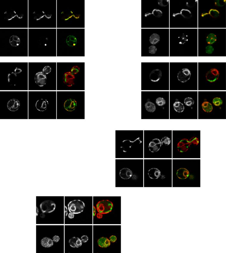
Mitoprotein ER delivery is GET-dependent Xiao et al. https://doi.org/10.26508/lsa.202000918 vol 4 | no 3 | e202000918 2 of 11Figure 1. Non-imported mitochondrial proteins are targeted to the ER. (A, B) Super-resolution images and line scan analysis of yeast expressing Alo1-GFP (A) or Oac1-GFP (B) and Tom70-mCherry −/+ FCCP. (C, D) Super-resolution images and line scan analysis of yeast expressing Alo1-GFP (C) or Oac1-GFP (D) and Sec61-mCherry −/+ FCCP. (E) Quantification of cells with ER localization of Alo1- or Oac1-GFP −/+ FCCP. N > 100 cells per replicate, error bars = SEM of three replicates. (F, G) Super-resolution images and line scan analysis of wild type or tom70/71Δ expressing Alo1-GFP (F) or Oac1-GFP (G) and Sec61-mCherry. (H) Quantification of cells with ER localization of Alo1- or Oac1- GFP in wild-type cells or tom70/71Δ mutants. N > 100 cells per replicate, error bars = SEM of three replicates. For (A, B, C, D, F, G), white arrow marks perinuclear ER. White line marks fluorescence intensity profile position. Left and right y-axis (line scan graph) corresponds to GFP and mCherry fluorescence intensity, respectively. Black arrow (line scan graph) marks colocalization. Images show single focal plane. Scale bar = 2 μm. See also Figs S1 and S2. ER, including the cytosolic ATPase Get3, which binds and recruits treatment (Fig S4B) and appearance of non-mitochondrial Oac1 foci substrates to the ER insertases Get1/2, and components of the pre- (Fig S4A and C). Knockout of GET4, GET5, or SGT2, however, had no targeting complex Get4, Get5, and Sgt2, which bind and stabilize effect on Oac1 localization (Fig S4A–C). This latter result is in line substrates to promote downstream ER targeting by Get1/2/3 (Wang with previous studies showing that deletion of upstream factors of et al, 2010, 2014a). Like Get1/2, loss of Get3 also impacted targeting the GET complex, including Get4, Get5 or Sgt2, does not completely of Oac1 to the ER (Fig S4A), with reduced ER localization upon FCCP prevent functionality of the GET pathway (Kohl et al, 2011). Thus, Mitoprotein ER delivery is GET-dependent Xiao et al. https://doi.org/10.26508/lsa.202000918 vol 4 | no 3 | e202000918 3 of 11
Figure 2. The GET complex is required for ER targeting of non-imported mitochondrial carrier proteins.
(A) Super-resolution images of wild-type or get1/2Δ mutant cells expressing Oac1-GFP and Tom70-mCherry −/+ FCCP. White arrow marks perinuclear ER. White arrowhead
marks protein foci containing Oac1-GFP. Images show single focal plane. (B, C) Quantification of (A) showing the percentage of cells with Oac1-GFP localized to the ER (B) or
protein foci (C). N > 100 cells per replicate, error bars = SEM of three replicates. (D) Super-resolution images of wild-type or get1/2Δ cells expressing Oac1-GFP and Get3-mCherry
−/+ FCCP. White arrow marks perinuclear ER. White arrowhead marks protein foci containing Oac1-GFP. Yellow arrowheads mark protein foci containing Get3-mCherry.
Images show single focal plane. (E) Quantification of (D) showing the number of foci only containing Oac1-GFP (green), Get3-mCherry (magenta), or colocalized/associated
Oac1-GFP and Get3-mCherry (yellow) per 100 cells −/+ FCCP. N > 100 cells per replicate of three replicates, values are normalized to number of foci per 100 cells. (F) Western blot
probing for GFP and FLAG in input or elution products of immunoprecipitated Get3-GFP in the indicated yeast strains. Scale bar = 2 μm. See also Figs S3–S5.

core GET components, including Get1/2 and partially Get3, are                               Sed5 and Ysy6 (Fig S5A and B), consistent with the idea that these puncta
required for targeting mitochondrial carrier proteins to the ER, but                        are the same as reported previously in get1/2Δ mutants (Schuldiner
other components of the GET pathway are dispensable.                                        et al, 2008). Furthermore, co-immunoprecipitation analysis indicated
   We also analyzed whether non-imported Oac1 colocalize with                               that a small fraction of GFP-tagged Get3 constitutively co-purified with
components of the GET machinery in cells. To do this, we created a                          FLAG-tagged Oac1 (Fig 2F), which persisted regardless of the nature of
strain expressing an mCherry-tagged version of Get3, the cytosolic                          the epitope tags on the protein, or which of the proteins was used as the
ATPase that normally resides in the cytoplasm and recruits cyto-                            bait (Fig S5C). Together, these data indicate that Oac1 associates with
solic GET substrates to ER-localized Get1/2 (Schuldiner et al, 2008;                        Get3 in cells, further supporting an interplay between the GET pathway
Wang et al, 2010). In get1/2Δ mutants, it has been shown that Get3                          and non-imported mitochondrial carrier proteins.
localizes to cytosolic foci containing GET substrates (Schuldiner
et al, 2008; Powis et al, 2013). Like canonical TA substrates of the GET                    Oac1 localizes to mitochondria- and ER-associated Hsp42-
pathway (Powis et al, 2013), we found that in get1/2Δ mutant cells,                         dependent foci in the absence of a functional GET pathway
half of the Oac1-GFP foci were colocalized or closely associated with
Get3-mCherry foci, even in times where foci were observed in the                            In cells with a non-functional GET pathway, mitochondrial carrier
absence of FCCP (Fig 2D and E). These foci also contained the TA proteins                   proteins were sequestered into protein foci (Figs 2A and C, S3D and

Mitoprotein ER delivery is GET-dependent Xiao et al.                                        https://doi.org/10.26508/lsa.202000918       vol 4 | no 3 | e202000918         4 of 11
E, and S4A). We sought to further characterize the nature of these       proteins from the ER membrane to mitochondria, promoting additional
foci. To do this, we analyzed their localization in cells with fluo-      attempts of mitochondrial import (Hansen et al, 2018). A question sur-
rescently tagged organelle markers using super-resolution mi-            rounding this pathway, termed ER-SURF, is the nature of the cellular
croscopy. In get1/2Δ mutant cells, 97% of protein foci containing        machinery that promotes initial targeting of mitochondrial proteins to the
Oac1 were associated with mitochondria marked by Tom70 (Fig 3A)          ER (Fig 5A). With our discovery that ER targeting of non-imported mi-
or the ER marked by Sec61 (Fig 3B), which is similar with previously     tochondrial membrane proteins is perturbed in GET mutants, we won-
characterized cytosolic protein aggregates (Zhou et al, 2014). To        dered whether the loss of the GET machinery limits accessibility of the ER-
verify whether these foci corresponded to protein aggregates, we         SURF pathway to non-imported mitochondrial precursor substrates. To
labeled Hsp42 and Hsp104, chaperones that commonly localize to           test whether GET-dependent delivery of non-imported mitochondrial
cytosolic protein aggregates in yeast (Zhou et al, 2014; Miller et al,   proteins to the ER is an upstream step for mitochondrial re-import
2015; Lee et al, 2018), with mCherry and examined localization with      mediated by Djp1, we tagged Oac1, which requires the GET complex to be
Oac1-GFP foci. We found that nearly all Oac1-GFP foci contained          localized to the ER, with GFP, in djp1Δ mutant cells. Interestingly, ER-
Hsp42 and Hsp104, even in untreated cells (Fig 3C–F). Deletion of        localized Oac1 was observed in 55% of djp1Δ cells without FCCP treatment
HSP42, but not HSP104, diminished the formation of Oac1 foci in          (Fig 5B and C) and more than 60% with FCCP treatment (Fig 5C). Both rates
get1/2Δ mutants (Fig 3G and H), leading to predominantly diffuse         are higher than observed in wild-type cells (Fig 5C). This result, combined
cytoplasmic localization (Fig 3G and I). Interestingly, the formation    with the fact that Oac1 colocalizes with Get3 (Figs 2D–F and S5C) and
of Get3-foci in get1/2Δ was unaffected by HSP42 deletion (Fig 3J),       localizes to Hsp42-dependent protein foci in get1/2Δ cells even without
indicating that Hsp42 is required for Oac1 deposition into Get3-         FCCP treatment (Fig 3), suggests that a portion of Oac1 is constitutively
containing protein foci. Consistent with an important role for Hsp42     targeted to the ER and shuttled to the mitochondria through the ER-SURF
in the handling of non-imported Oac1, co-immunoprecipitation             pathway. Consistent with this hypothesis, the ER localization of Oac1 in
analysis identified an interaction between that FLAG-tagged Oac1          djp1Δ cells was dramatically reduced in the absence of GET1/2 (Fig 5C),
and GFP-tagged Hsp42 (Fig 3K). Thus, Hsp42 mediates sequestration        and protein foci containing Oac1 were present in djp1Δget1/2Δ triple
of non-imported Oac1 into protein foci in the absence of a func-         mutants (Fig 5D). These data support a model in which non-imported
tional GET pathway.                                                      mitochondrial proteins are delivered to the ER in a GET-dependent
                                                                         manner for mitochondrial re-import via the ER-SURF pathway.
Dual loss of the GET pathway and mitochondrial import is
detrimental to cells

Because non-imported mitochondrial proteins are harmful to cells         Discussion
(Wang & Chen, 2015; Wrobel et al, 2015; Boos et al, 2020), we in-
vestigated whether loss of ER targeting of non-imported mito-            We previously identified the ER as a destination for a subset of non-
chondrial proteins during times of mitochondrial deficiency led to        imported mitochondrial membrane proteins during times of mi-
reduced cellular fitness. To do this, we tested the growth of GET         tochondrial dysfunction (Shakya et al, 2020 Preprint). Our current
mutants under stress of mitochondrial import failure. In compar-         work now shows that the GET complex is required for ER targeting of a
ison to wild-type cells, get1/2Δ cells exhibited more severely di-       specific group of proteins, the mitochondrial carrier proteins. In cells
minished growth in the presence of FCCP (Fig 4A). Likewise, cells        with a dysfunctional GET pathway, the ER delivery of mitochondrial
lacking the mitochondrial import receptors Tom70/71 showed               carrier proteins is impaired, and these proteins are instead se-
stronger fitness defects when combined with deletion of GET1 or           questered into Hsp42-dependent mitochondrion- and ER-associated
GET2 (Fig 4B and C). Interestingly, the growth deficiencies of get1/2Δ    cytosolic protein foci (Fig 5E). Overall, our data support a requirement
mutant cells in the presence of FCCP were largely suppressed by          for the GET pathway in targeting of non-imported mitochondrial
deletion of GET3 (Fig 4D), suggesting the presence of Get3 in the        carrier proteins to the ER.
cytoplasm without its ER receptors is problematic under these               This work synergizes well with a recent report that showed that
conditions. Overall, these results suggest that dual loss of mito-       the GET complex increases the risk of mistargeting over-expressed
chondrial import and GET-dependent ER targeting is problematic           OMM proteins to the ER (Vitali et al, 2018). Interestingly, similar to
for cells. At this point, it remains unclear to what extent these        these previously described OMM proteins, multi-pass mitochon-
growth defects result from failure to target non-imported proteins       drial carrier proteins that require the GET pathway for ER delivery,
to the ER in GET mutants versus other possibilities explanations,        including Oac1, Mir1, and Dic1, all contain transmembrane domains
including altered targeting of TA proteins to the mitochondria in        that are very close to their C-termini (Kunji, 2004). This might
GET mutants, loss of proteostasis due to impaired biogenesis of          provide possibilities for their recognition by components of the
other stress-responsive factors, or the presence of TA proteins and      GET complex. In comparison, Alo1, which likely contains a central
Get3 in the cytosol (Schuldiner et al, 2008; Jonikas et al, 2009).       transmembrane domain (Weill et al, 2019), is targeted to the ER
                                                                         independently of the GET pathway. However, it is important to
GET-dependent ER-destined mitochondrial proteins are potential           emphasize that at this point, it remains unclear whether the GET
substrates for the ER-SURF pathway                                       pathway directly binds and facilitates targeting of multi-pass mi-
                                                                         tochondrial membrane proteins to the ER, or whether the role of the
In agreement with our current findings, it was recently demon-            GET pathway in ER delivery of these proteins is indirect. While we find
strated that a J-protein, Djp1, shuttles ER-localized mitochondrial      that Oac1 colocalizes with Get3 and TA proteins in cells lacking GET1/2,

Mitoprotein ER delivery is GET-dependent Xiao et al.                     https://doi.org/10.26508/lsa.202000918   vol 4 | no 3 | e202000918   5 of 11
Figure 3. Oac1-GFP localizes to mitochondrion- and ER-associated Hsp42-dependent foci in the absence of a functional GET pathway. (A, B, C, D) Super-resolution images and line scan analysis of get1/2Δ mutant yeast expressing Oac1-GFP and Tom70-mCherry (A), Sec61-mCherry (B), Hsp42-mCherry (C), or Hsp104- mCherry (D). White arrowhead marks protein foci containing Oac1-GFP. For microscopy images, white line marks fluorescence intensity profile position. Images show single focal plane. Scale bar = 2 μm. For line scan graphs, Left and right y-axis correspond to GFP and mCherry fluorescence intensity, respectively. Black arrow marks protein foci position and white arrow marks mitochondria (A) or ER (B) position that is associated with protein foci. For the quantification in (A) and (B), N > 100 cells per replicate of three replicates. (E, F) Quantification of (C) and (D), respectively, showing the number of foci only containing Oac1-GFP (green), Hsp42-mCherry (E) or Hsp104-mCherry (F) (magenta) or both (yellow) per 100 cells −/+ FCCP. N > 100 cells per replicate of three replicates, values are normalized to number of foci per 100 cells. (G) Wide-field images of wild-type cells and the indicated mutant yeast expressing Oac1-GFP and Tom70-mCherry −/+ FCCP. White arrows mark perinuclear ER. White arrowheads mark protein foci containing Oac1-GFP. Images show single focal plane. Scale bar = 2 μm. (H, I) Quantification of (G) showing the percentage of cells with Oac1-GFP localized to protein foci (H) or the ER (I). N > 100 cells per replicate, error bars = SEM of three replicates. (J) Quantification of the percentage of cells with protein foci containing Get3-mCherry in wild-type or the indicated mutant cells. N > 100 cells per replicate, error bars = SEM of three replicates. (K) Western blot probing for GFP and FLAG in input or elution products of immunoprecipitated Hsp42-GFP in the indicated yeast strains. Mitoprotein ER delivery is GET-dependent Xiao et al. https://doi.org/10.26508/lsa.202000918 vol 4 | no 3 | e202000918 6 of 11
Figure 4. Deletion of GET1/2 impairs growth of yeast
                                                                                                      cells with mitochondrial import failure.
                                                                                                      (A) Fivefold serial dilutions of wild-type cells and
                                                                                                      get1/2Δ mutant cells on YPD −/+ FCCP agar plates.
                                                                                                      (B, C) Fivefold serial dilutions of wild-type and the
                                                                                                      indicated mutant cells on YPD agar plates. (D)
                                                                                                      Fivefold serial dilutions of wild-type cells and the
                                                                                                      indicated mutant cells on YPD −/+ FCCP agar plates.

the nature of the interaction between Oac1 and Get3 is still obscure.         becoming clearer that cells use a multitude of pathways to prevent
Get3 is shown to have dual roles: as an ATPase that hands over clients        toxicity associated with the accumulation of non-imported mito-
to Get1/2 for further insertion (Schuldiner et al, 2008), and as a holdase    chondrial proteins, and our work helps to further delineate the sys-
chaperone that brings substrates to the ER and supports sequestration         tems that promote delivery of mitochondrial membrane proteins to
of substrates to protein foci in the absence of Get1/2 (Powis et al, 2013).   the ER. Interestingly, we also found that several ER-destined mito-
Given that non-imported mitochondrial carrier proteins are potentially        chondrial proteins did not appear to be affected by deletion of GET1/2
re-routed to mitochondria through the ER-SURF pathway, it is possible         for their ER targeting, suggesting that additional ER targeting systems
that Get3 acts as a holdase that facilitates ER targeting, but not            for mitochondrial proteins likely exist. Identifying these systems and
translocation, of non-imported mitochondrial proteins. Thus, addi-            dissecting the coordination between the many cellular pathways that
tional studies are required to determine whether non-imported mi-             mitigate mitoprotein-induced stress will be critical areas of future
tochondrial proteins are inserted into the ER, how Get3 interacts with        research.
non-imported mitochondrial proteins, as well as whether Get1/2 play
any direct role in importing Oac1 into the ER. In the absence of these
experiments, it remains possible that the delivery of mitochondrial
carrier proteins to the ER is mediated by an unknown pathway and that
                                                                              Materials and Methods
the block in ER delivery of these proteins in GET mutants may result
                                                                              Reagents
from a general perturbation in cellular proteostasis (Jonikas et al,
2009). It is also possible that non-imported mitochondrial proteins
                                                                              Antibodies and chemicals used in this study are listed in Table S1.
might be targeted to the ER by unknown ER-localized factors that are
originally inserted by the GET pathway. Deciphering between these
possibilities will be an important avenue of research moving forward.         Yeast strains
   Despite the open questions surrounding the interplay between the
GET pathway and non-imported mitochondrial proteins, our studies              All yeast strains are derivatives of Saccharomyces cerevisiae S288c
ultimately provide an important step forward in our understanding             (BY) (Brachmann et al, 1998) and are listed in Table S2. Strains
of how cells mitigate mitochondrial protein-induced stress. It is             expressing tagged proteins from their native loci were created by

Mitoprotein ER delivery is GET-dependent Xiao et al.                          https://doi.org/10.26508/lsa.202000918   vol 4 | no 3 | e202000918    7 of 11
Figure 5. GET-dependent ER targeting of mitochondrial proteins potentially provides substrates for the ER-SURF pathway. (A) Schematic graph of the ER-SURF pathway. (B) Super-resolution images of wild-type and djp1Δ cells expressing Oac1-GFP and Tom70-mCherry. White arrows mark perinuclear ER. Images show single focal plane. Scale bar = 2 μm. (C, D) Quantification of the percentage of cells with Oac1-GFP localized to the ER (C) or the cytosolic foci (D) in wild-type or the indicated mutant cells. N > 100 cells per replicate, error bars = SEM of three replicates. (E) Schematic overview of the fates of non-imported mitochondrial carrier proteins in wild-type and get1/2Δ mutant cells. one step PCR-mediated C-terminal endogenous epitope tagging 2004). Plasmid templates for mCherry tagging were from the pKT using standard techniques and the oligo pairs listed in Table S3 series of vectors (Sheff & Thorn, 2004) or pFA6a-mCherry-HphMX (Brachmann et al, 1998; Sheff & Thorn, 2004). Plasmid templates for (39295; Addgene) (Wang et al, 2014b). Integrations were confirmed GFP tagging were from the pKT series of vectors (Sheff & Thorn, by correct localized expression of the fluorophore by microscopy. Mitoprotein ER delivery is GET-dependent Xiao et al. https://doi.org/10.26508/lsa.202000918 vol 4 | no 3 | e202000918 8 of 11
Plasmid template for FLAG tagging was the pFA6a-5FLAG-KanMX6               Serial-dilution growth assays
(15983; Addgene) (Noguchi et al, 2008). Integrations were confirmed
by a combination of colony PCR across the chromosomal insertion            Fivefold serial dilutions of exponentially growing yeast cells were
site and correct band size by Western blot. Deletion strains were          diluted in ddH2O and 3 μl of each dilution was spotted onto YPD (1%
created by one step PCR-mediated gene replacement using the                yeast extract, 2% peptone, and 2% glucose). Final concentration of
oligos pairs listed in Table S3 and plasmid templates from the pRS         FCCP is 7 μM. Total cells plated in each dilution spot were 5,000,
series vectors (Brachmann et al, 1998). Correct integrations were          1,000, 20, 40, and 8. Plates were cultured at 30°C for 36 h before
confirmed with colony PCR across the chromosomal insertion site.            obtaining images.

                                                                           Immunoprecipitation and Western blotting
Plasmids

                                                                           Cells were grown as described above. 1 × 10⁸ total cells were
Plasmids used in this study are listed in Table S1. Plasmids for GPD-
                                                                           harvested, resuspended in 500 μl of IP Buffer. Tris IP Buffer (50 mM
driven expression of GFP-SED5 and GFP-YSY6 were generated by
                                                                           Tris, pH 7.5, 150 mM NaCl, 1 mM EDTA, 10% Glycerol, 1% IGEPAL [NP-40
gateway-mediated transfer of the corresponding ORF (Harvard Insti-
                                                                           substitute], 100 μM PMSF, 1× cOmplete Protease Inhibitor Cocktail
tute of Proteomics) from pDONR201/221 into pAG413GPD-eGFP-ccdB
                                                                           [Roche]) was used for immunoprecipitation with Hsp42, as previ-
(14310; Addgene), using Gateway LR Clonase II Enzyme mix (Thermo
                                                                           ously described (Malinovska et al, 2012). KHM IP Buffer (110 mM KAc,
Fisher Scientific) according to the manufacturer’s instructions.
                                                                           20 mM HEPES-KOH, pH 7.4, 2 mM MgCl2, 10% glycerol, 0.1% Triton-
                                                                           X100, 100 μM PMSF, and 1× cOmplete Protease Inhibitor Cocktail
Yeast cell culture and media                                               [Roche]) was used for immunoprecipitation of Oac1/Get3, adapted
                                                                           based on previous studies (Stefanovic & Hegde, 2007; Vitali et al,
Yeast cells were grown exponentially for 15–16 h at 30°C to a final         2018). Resuspended cells were lysed with glass beads using an
density of 2–7 × 10⁶ cells/ml before starting any treatments. Cells        Omni Bead Ruptor 12 Homogenizer (eight cycles of 20 s each). Cells
were cultured in YPAD medium (1% yeast extract, 2% peptone,                lysates were cleared by centrifugation at 10,000 rpm (Eppendorf
0.005% adenine, and 2% glucose) in most experiments. Cells with            5424 Centrifuge) for 5 min to remove cell debris. The supernatant
yeast plasmids expressing histidine auxotrophic marker genes were          was collected in a new tube, and the total volume was adjusted to
cultured in SD-His medium (0.67% yeast nitrogen base without               1 ml by adding IP Buffer. Lysates were incubated with 2 μl of mouse
amino acids, 2% glucose, supplemented nutrients 0.074 g/l each             anti-GFP antibodies (Sigma Millipore) at 4°C overnight. 50 μl of the
adenine, alanine, arginine, asparagine, aspartic acid, cysteine,           lysate–antibody mixture was removed as input fraction. For each
glutamic acid, glutamine, glycine, myoinositol, isoleucine, ly-            immunoprecipitation, 40 μl of Dynabeads Protein G (Thermo Fisher
sine, methionine, phenylalanine, proline, serine, threonine, tryp-         Scientific) were washed three times, and resuspended in 50 μl IP
tophan, tyrosine, uracil, valine, 0.369 g/l leucine, and 0.007 g/l para-   Buffer. Lysate–antibody mixture was incubated with washed Dyna-
aminobenzoic acid). For FCCP or CHX (cycloheximide) treatment,             beads Protein G at 4°C for 2 h and then washed four times for 10
overnight log-phase cell cultures were grown in the presence of            min in IP Buffer. Immunoprecipitated proteins were eluted by
FCCP (final concentration of 10 μM) or CHX (final concentration of           incubating beads in 2× Laemmli Buffer (63 mM Tris, pH 6.8, 2% [wt/
100 μg/ml) for 4–5 h.                                                      vol] SDS, 10% [vol/vol] glycerol, and 1 mg/ml bromophenol blue)
                                                                           at 65°C for 10 min.
                                                                              Western blots were carried out as described previously (Hughes
Microscopy                                                                 et al, 2016; Shakya et al, 2020 Preprint). Cells extracts and elution
                                                                           products were resolved on Bolt 4–12% Bis-Tris Plus Gels
Optical z-sections of live yeast cells were acquired with a ZEISS Axio     (NW04125BOX; Thermo Fisher Scientific) with NuPAGE MES SDS
Imager M2 equipped with a ZEISS Axiocam 506 monochromatic camera,          Running Buffer (NP0002-02; Thermo Fisher Scientific) and trans-
100× oil-immersion objective (plan apochromat, NA 1.4), a AxioObserver     ferred to nitrocellulose membranes. Membranes were blocked and
7 (Carl Zeiss) equipped with a PCO Edge 4.2LT Monochrome, Air Cooled,      probed in blocking buffer (1× PBS, 0.05% Tween-20, 5% non-fat dry
USB 3 CCD camera with a Solid-State Colibri 7 LED illuminator and 100×     milk) using the primary antibodies for FLAG (Thermo Fisher Sci-
oil-immersion objective (Plan Apochromat, NA 1.4; Carl Zeiss), a ZEISS     entific) and GFP (Sigma Millipore) and HRP conjugated secondary
LSM800 equipped with an Airyscan detector, 63× oil-immersion ob-           antibodies (715-035-150; Jackson Immunoresearch). Blots were
jective (plan apochromat, NA 1.4) or a ZEISS LSM880 equipped with an       developed with SuperSignal West Dura Extended Duration Sub-
Airyscan detector, 63× oil-immersion objective (plan apochromat, NA        strate (34075; Thermo Fisher Scientific) and exposed with a Bio-Rad
1.4). Widefield images were acquired with ZEN (Carl Zeiss) and pro-         Chemidoc MP system.
cessed with Fiji (Schindelin et al, 2012). Super-resolution images were
acquired with ZEN (Carl Zeiss) and processed using the automated           Yeast indirect immunofluorescence (IIF) staining
Airyscan processing algorithm in ZEN (Carl Zeiss) and Fiji. Individual
channels of all images were minimally adjusted in Fiji to match the        For IIF staining, overnight log-phase cell cultures were grown with
fluorescence intensities between channels for better visualization. Line    or without FCCP for 3 h 30 min in YPAD to a final density of 4 × 10⁶
scan analysis was performed on non-adjusted, single z-sections in Fiji.    cells/ml. Cells were harvested by centrifugation and fixed in 10 ml
All images shown in Figures represent a single optical section.            fixation medium (4% paraformaldehyde in YPAD) for 1 h. Fixed yeast

Mitoprotein ER delivery is GET-dependent Xiao et al.                       https://doi.org/10.26508/lsa.202000918   vol 4 | no 3 | e202000918   9 of 11
cells were washed with Wash Buffer (0.1 M Tris, pH = 8, 1.2 M Sorbitol) Conflict of Interest Statement
twice and incubated in 2 ml DTT Buffer (10 mM DTT in 0.1 M Tris, pH =
9.4) at room temperature for 10 min. Spheroplasts were generated The authors declare that they have no conflict of interest.
by incubating cells in 2 ml Zymolyase Buffer (0.1 M KPi, pH = 6.5, 1.2 M
Sorbitol, 0.25 mg/ml Zymolyase) at 30°C for 30 min. Spheroplasts
were gently diluted in 1:40 using Wash Buffer and attached to glass
slides pre-coated with 0.1% poly-L-Lysine (2 mg/ml). Samples were References
permeabilized in cold 0.1% Triton X-100 in PBS for 10 min at 4°C,
briefly dried, and blocked in Wash Buffer containing 1% BSA at room Ast T, Schuldiner M (2013) All roads lead to Rome (but some may be harder to
travel): SRP-independent translocation into the endoplasmic
temperature for 30 min. After blocking, samples were incubated
reticulum. Crit Rev Biochem Mol Biol 48: 273–288. doi:10.3109/
with primary antibody (Monoclonal ANTI-FLAG M2 antibody pro- 10409238.2013.782999
duced in mouse, 1:200 diluted in Wash Buffer containing 1% BSA) for
Aviram N, Ast T, Costa EA, Arakel EC, Chuartzman SG, Jan CH, Haϐdenteufel S,
1 h 30 min at room temperature and secondary antibody (Goat anti- Dudek J, Jung M, Schorr S, et al (2016) The SND proteins constitute an
Mouse IgG (H+L) Cross-Adsorbed Secondary Antibody, Alexa Fluor alternative targeting route to the endoplasmic reticulum. Nature 540:
488, 1:300 diluted in Wash Buffer containing 1% BSA) for 45 min at 134–138. doi:10.1038/nature20169
room temperature. Samples were washed 10 times after each in- Aviram N, Schuldiner M (2017) Targeting and translocation of proteins to the
cubation with Wash Buffer containing 1% BSA and 0.1% Tween-20. endoplasmic reticulum at a glance. J Cell Sci 130: 4079–4085.
Slides were washed twice with Wash Buffer before sealing, and doi:10.1242/jcs.204396
mounted with HardSet medium (ProLong Glass Antifade Mountant Boos F, Labbadia J, Herrmann JM (2020) How the mitoprotein-induced stress
with NucBlue Stain (P36981); Invitrogen) overnight. Wide-field im- response safeguards the cytosol: A unified view. Trends Cell Biol 30:
241–254. doi:10.1016/j.tcb.2019.12.003
ages were acquired as described above.
Brachmann CB, Davies A, Cost GJ, Caputo E, Li J, Hieter P, Boeke JD (1998)
Designer deletion strains derived from Saccharomyces cerevisiae
Quantification and statistical analysis
S288C: A useful set of strains and plasmids for PCR-mediated gene
disruption and other applications. Yeast 14: 115–132. doi:10.1002/(SICI)
The number of replicates, what n represents, and dispersion and 1097-0061(19980130)14:23.0.CO;2-2
precision measures are indicated in the figure legends. In general, Chitwood PJ, Juszkiewicz S, Guna A, Shao S, Hegde RS (2018) EMC is required to
quantifications show the mean ± standard error from three bio- initiate accurate membrane protein topogenesis. Cell 175: 1507–1519.
logical replicates with n = 100 cells per experiment. In experiments doi:10.1016/j.cell.2018.10.009
with data depicted from a single biological replicate, the experi- Friedman JR, Kannan M, Toulmay A, Jan CH, Weissman JS, Prinz WA, Nunnari J
ment was repeated with the same results. (2018) Lipid homeostasis is maintained by dual targeting of the
mitochondrial PE biosynthesis Enzyme to the ER. Dev Cell 44: 261–270.
doi:10.1016/j.devcel.2017.11.023
Friedman JR, Nunnari J (2014) Mitochondrial form and function. Nature 505:
Supplementary Information 335–343. doi:10.1038/nature12985
Guna A, Volkmar N, Christianson JC, Hegde RS (2018) The ER membrane
Supplementary Information is available at https://doi.org/10.26508/lsa.
protein complex is a transmembrane domain insertase. Science 359:
202000918.
470–473. doi:10.1126/science.aao3099
Hansen KG, Aviram N, Laborenz J, Bibi C, Meyer M, Spang A, Schuldiner M,
Herrmann JM (2018) An ER surface retrieval pathway safeguards the
Acknowledgements import of mitochondrial membrane proteins in yeast. Science 361:
1118–1122. doi:10.1126/science.aar8174
We thank the members of the AL Hughes and Janet M Shaw (Utah) labo- Hughes AL, Gottschling DE (2012) An early age increase in vacuolar pH limits
ratories for discussion and manuscript comments, and Yeyun Ouyang (Utah) mitochondrial function and lifespan in yeast. Nature 492: 261–265.
for experimental discussion. We thank JM Shaw for contributing stipend doi:10.1038/nature11654
support for T Xiao. Research was supported by National Institutes of Health
Hughes AL, Hughes CE, Henderson KA, Yazvenko N, Gottschling DE (2016)
grants GM119694 and AG061376 (AL Hughes) and the Howard Hughes Medical
Selective sorting and destruction of mitochondrial membrane
Institute (JM Shaw). AL Hughes was further supported by an American Fed-
proteins in aged yeast. Elife 5: e13943. doi:10.7554/elife.13943
eration for Aging Research Junior Research Grant, United Mitochondrial Dis-
ease Foundation Early Career Research Grant, Searle Scholars Award, and Itakura E, Zavodszky E, Shao S, Wohlever ML, Keenan RJ, Hegde RS (2016)
Glenn Foundation for Medical Research Award. Ubiquilins chaperone and triage mitochondrial membrane proteins
for degradation. Mol Cell 7: 21–33. doi:10.1016/j.molcel.2016.05.020
Author Contributions Jonikas MC, Collins SR, Denic V, Oh E, Quan EM, Schmid V, Weibezahn J,
Schwappach B, Walter P, Weissman JS, et al (2009) Comprehensive
characterization of genes required for protein folding in the
T Xiao: data curation, formal analysis, investigation, methodology,
endoplasmic reticulum. Science 323: 1693–1697. doi:10.1126/
and writing—original draft, review, and editing. science.1167983
VPS Shakya: data curation, formal analysis, investigation, and
Kohl C, Tessarza P, Von Der Malsburg K, Zahn R, Bukau B, Mogk A (2011)
methodology. Cooperative and independent activities of Sgt2 and Get5 in the
AL Hughes: conceptualization, supervision, funding acquisition, targeting of tail-anchored proteins. Biol Chem 392: 601–608.
investigation, and writing—review and editing. doi:10.1515/BC.2011.066
Mitoprotein ER delivery is GET-dependent Xiao et al. https://doi.org/10.26508/lsa.202000918 vol 4 | no 3 | e202000918 10 of 11Kunji ERS (2004) The role and structure of mitochondrial carriers. FEBS Lett Shurtleff MJ, Itzhak DN, Hussmann JA, Oakdale NTS, Costa EA, Jonikas M,
564: 239–244. doi:10.1016/S0014-5793(04)00242-X Weibezahn J, Popova KD, Jan CH, Sinitcyn P, et al (2018) The ER
Lee HY, Chao JC, Cheng KY, Leu JY (2018) Misfolding-prone proteins are membrane protein complex interacts cotranslationally to enable
reversibly sequestered to an Hsp42-associated granule upon biogenesis of multipass membrane proteins. Elife 7: e37018.
chronological aging. J Cell Sci 131: jcs220202. doi:10.1242/jcs.220202 doi:10.7554/elife.37018
Malinovska L, Kroschwald S, Munder MC, Richter D, Alberti S (2012) Molecular Stefanovic S, Hegde RS (2007) Identification of a targeting factor for
chaperones and stress-inducible protein-sorting factors coordinate posttranslational membrane protein insertion into the ER. Cell 128:
the spatiotemporal distribution of protein aggregates. Mol Biol Cell 23: 1147–1159. doi:10.1016/j.cell.2007.01.036
3041–3056. doi:10.1091/mbc.E12-03-0194
Vitali DG, Sinzel M, Bulthuis EP, Kolb A, Zabel S, Mehlhorn DG, Costa BF, Farkas
Mårtensson CU, Priesnitz C, Song J, Ellenrieder L, Doan KN, Boos F, Á, Clancy A, Schuldiner M, et al (2018) The GET pathway can increase
Floerchinger A, Zufall N, Oeljeklaus S, Warscheid B, et al (2019) the risk of mitochondrial outer membrane proteins to be mistargeted
Mitochondrial protein translocation-associated degradation. Nature to the ER. J Cell Sci 131: jcs211110. doi:10.1242/jcs.211110
569: 679–683. doi:10.1038/s41586-019-1227-y
Wallace DC (2005) A mitochondrial paradigm of metabolic and degenerative
McKenna MJ, Sim SI, Ordureau A, Wei L, Wade Harper J, Shao S, Park E (2020) diseases, aging, and cancer: A dawn for evolutionary medicine. Annu
The endoplasmic reticulum P5A-ATPase is a transmembrane helix Rev Genet 39: 359–407. doi:10.1146/annurev.genet.39.110304.095751
dislocase. Science 369: eabc5809. doi:10.1126/science.abc5809
Wang F, Brown EC, Mak G, Zhuang J, Denic V (2010) A chaperone cascade sorts
Miller SB, Ho C, Winkler J, Khokhrina M, Neuner A, Mohamed MY, Guilbride DL,
proteins for posttranslational membrane insertion into the
Richter K, Lisby M, Schiebel E, et al (2015) Compartment-specific
endoplasmic reticulum. Mol Cell 40: 159–171. doi:10.1016/
aggregases direct distinct nuclear and cytoplasmic aggregate
j.molcel.2010.08.038
deposition. EMBO J 34: 778–797. doi:10.15252/embj.201489524
Noguchi C, Garabedian MV, Malik M, Noguchi E (2008) A vector system for Wang F, Chan C, Weir NR, Denic V (2014a) The Get1/2 transmembrane complex
genomic FLAG epitope-tagging in Schizosaccharomyces pombe. is an endoplasmic-reticulum membrane protein insertase. Nature
Biotechnol J 3: 1280–1285. doi:10.1002/biot.200800140 512: 441–444. doi:10.1038/nature13471
Pagliarini DJ, Calvo SE, Chang B, Sheth SA, Vafai SB, Ong SE, Walford GA, Wang N, Presti LL, Zhu YH, Kang M, Wu Z, Martin SG, Wu JQ (2014b) The novel
Sugiana C, Boneh A, Chen WK, et al (2008) A mitochondrial protein proteins Rng8 and Rng9 regulate the myosin-V Myo51 during fission
compendium elucidates complex I disease biology. Cell 134: 112–123. yeast cytokinesis. J Cell Biol 205: 357–375. doi:10.1083/jcb.201308146
doi:10.1016/j.cell.2008.06.016 Wang X, Chen XJ (2015) A cytosolic network suppressing mitochondria-
Palmieri F, Agrimi G, Blanco E, Castegna A, Di Noia MA, Iacobazzi V, Lasorsa FM, mediated proteostatic stress and cell death. Nature 524: 481–484.
Marobbio CMT, Palmieri L, Scarcia P, et al (2006) Identification of doi:10.1038/nature14859
mitochondrial carriers in Saccharomyces cerevisiae by transport
Weill U, Cohen N, Fadel A, Ben-Dor S, Schuldiner M (2019) Protein topology
assay of reconstituted recombinant proteins. Biochim Biophys Acta
prediction algorithms systematically investigated in the yeast
1757: 1249–1262. doi:10.1016/j.bbabio.2006.05.023
Saccharomyces cerevisiae. Bioessays 8: e1800252. doi:10.1002/
Powis K, Schrul B, Tienson H, Gostimskaya I, Breker M, High S, Schuldiner M, bies.201800252
Jakob U, Schwappach B (2013) Get3 is a holdase chaperone and moves
to deposition sites for aggregated proteins when membrane targeting Wiedemann N, Pfanner N (2017) Mitochondrial machineries for protein
is blocked. J Cell Sci 126: 473–483. doi:10.1242/jcs.112151 import and assembly. Annu Rev Biochem 86: 685–714. doi:10.1146/
annurev-biochem-060815-014352
Qin Q, Zhao T, Zou W, Shen K, Wang X (2020) An endoplasmic reticulum
ATPase safeguards endoplasmic reticulum identity by removing Wrobel L, Topf U, Bragoszewski P, Wiese S, Sztolsztener ME, Oeljeklaus S,
ectopically localized mitochondrial proteins. Cell Rep 33: 108363. Varabyova A, Lirski M, Chroscicki P, Mroczek S, et al (2015) Mistargeted
doi:10.1016/j.celrep.2020.108363 mitochondrial proteins activate a proteostatic response in the
Schindelin J, Arganda-Carreras I, Frise E, Kaynig V, Longair M, Pietzsch T, cytosol. Nature 524: 485–488. doi:10.1038/nature14951
Preibisch S, Rueden C, Saalfeld S, Schmid B, et al (2012) Fiji: An open- Young CL, Raden DL, Caplan JL, Czymmek KJ, Robinson AS (2012) Cassette
source platform for biological-image analysis. Nat Methods 9: series designed for live-cell imaging of proteins and high-resolution
676–682. doi:10.1038/nmeth.2019 techniques in yeast. Yeast 29: 119–136. doi:10.1002/yea.2895
Schuldiner M, Metz J, Schmid V, Denic V, Rakwalska M, Schmitt HD, Zhou C, Slaughter BD, Unruh JR, Guo F, Yu Z, Mickey K, Narkar A, Ross RT,
Schwappach B, Weissman JS (2008) The GET complex mediates McClain M, Li R (2014) Organelle-based aggregation and retention of
insertion of tail-anchored proteins into the ER membrane. Cell 134: damaged proteins in asymmetrically dividing cells. Cell 159: 530–542.
634–645. doi:10.1016/j.cell.2008.06.025 doi:10.1016/j.cell.2014.09.026
Shakya VPS, Barbeau WA, Xiao T, Knutson CS, Hughes AL (2020) The nucleus is
a quality control center for non-imported mitochondrial proteins.
BioRxiv doi:10.1101/2020.06.26.173781 (Preprint posted June 26, 2020). License: This article is available under a Creative
Sheff MA, Thorn KS (2004) Optimized cassettes for fluorescent protein Commons License (Attribution 4.0 International, as
tagging in Saccharomyces cerevisiae. Yeast 21: 661–670. doi:10.1002/ described at https://creativecommons.org/
yea.1130 licenses/by/4.0/).
Mitoprotein ER delivery is GET-dependent Xiao et al. https://doi.org/10.26508/lsa.202000918 vol 4 | no 3 | e202000918 11 of 11You can also read