MIR-197 INHIBITOR LOADED ABCD133@MSNS@GNR AFFECTS THE DEVELOPMENT OF PROSTATE CANCER THROUGH TARGETING ITGAV
←
→
Page content transcription
If your browser does not render page correctly, please read the page content below
ORIGINAL RESEARCH
                                                                                                                                            published: 14 June 2021
                                                                                                                                     doi: 10.3389/fcell.2021.646884

                                            MiR-197 Inhibitor Loaded
                                            AbCD133@MSNs@GNR Affects the
                                            Development of Prostate Cancer
                                            Through Targeting ITGAV
                                            Guanqun Ju 1† , Yingjian Zhu 2† , Tao Du 3† , Wanli Cao 1 , Jianhai Lin 1 , Chun Li 4 ,
                                            Dongliang Xu 1,5* and Zhijun Wang 1*
                                            1
                                             Department of Urology, Changzheng Hospital, Naval Medical University, Shanghai, China, 2 Department of Urology,
                                            Shanghai Jiangqiao Hospital, Shanghai General Hospital Jiading Branch, Shanghai, China, 3 Department of Urology, Henan
                          Edited by:        Provincial People’s Hospital, Zhengzhou, China, 4 Key Laboratory of Functional Genomic and Molecular Diagnosis of Gansu
                Wassim Abou-Kheir,          Province, Lanzhou, China, 5 Urology Centre, Shuguang Hospital Affiliated to Shanghai University of Traditional Chinese
        American University of Beirut,      Medicine, Shanghai, China
                            Lebanon
                      Reviewed by:
                Soichiro Yamamura,
                                            Prostate cancer is one of the most severe male malignant tumors, which ranks second
              University of California,     in mortality rate among all tumors. Traditional methods of treatment for prostate cancer
       San Francisco, United States
                                            produce obvious side effects and a high recurrence rate. Cancer stem cells are
                           Yuan Gao,
             Fudan University, China        considered to be a group of cells that determine the proliferation, metastasis, and drug
                          Wenyao Li,        resistance of tumor. Prostate cancer therapy based on microRNAs and prostate cancer
   Shanghai University of Engineering
                      Science, China
                                            stem cells (PCSCs) has been a research hot spot in this field. Previous studies have
                *Correspondence:
                                            reported that miR-197 plays an important role in the occurrence and development
                        Zhijun Wang         of prostate cancer, but the molecular mechanism of miR-197 on the development of
                 wzj_828@163.com
                                            prostate cancer has not been reported yet. In this study, we verified that miR-197 is
                       Dongliang Xu
           Dr_xudongliang@163.com           significantly overexpressed in prostate cancer tissues and prostate cancer cells. Then,
     † These   authors have contributed     we verified that miR-197 expression affects the proliferation, invasion, and metastasis
                    equally to this work    of prostate cancer cells by regulating integrin subunit alpha V (ITGAV) expression
                    Specialty section:
                                            through STAT5 pathway, and the results indicated that the miR-197 inhibitor can be
         This article was submitted to      a prostate cancer suppressor. Then we synthesized the AbCD133@GNR@MSNs@miR-
         Cell Adhesion and Migration,       197 inhibitor drug carrier, in which 35.42 µg of the miR-197 inhibitor could be
                a section of the journal
  Frontiers in Cell and Developmental       loaded in 1 mg of AbCD133@GNR@MSNs. The AbCD133@GNR@MSNs@miR-197
                                Biology     inhibitor demonstrated good photothermal properties and photothermal controlled-
       Received: 28 December 2020           release properties. The modified CD133 antibodies on the surface of the nano
           Accepted: 27 April 2021
          Published: 14 June 2021
                                            drug carrier helped more drug carriers to enter the PCSCs. The pharmacodynamic
                              Citation:
                                            effects of the AbCD133@GNR@MSNs@miR-197 inhibitor on PCSCs in vivo and
     Ju G, Zhu Y, Du T, Cao W, Lin J,       in vitro were studied under near-infrared radiation. The results showed that the
       Li C, Xu D and Wang Z (2021)         AbCD133@GNR@MSNs@miR-197 inhibitor prepared in this study could not only
            MiR-197 Inhibitor Loaded
     AbCD133@MSNs@GNR Affects               significantly suppress the development of PCSCs through ITGAV/STAT5 pathway but
 the Development of Prostate Cancer         also significantly suppress the growth of PCSC solid tumors. In short, our study
            Through Targeting ITGAV.
      Front. Cell Dev. Biol. 9:646884.
                                            verified that miR-197 regulates the development of PCSCs through STAT5 pathway
      doi: 10.3389/fcell.2021.646884        by targeting ITGAV, and the AbCD133@MSNs@GNR@miR-197 inhibitor could be a

Frontiers in Cell and Developmental Biology | www.frontiersin.org                 1                                           June 2021 | Volume 9 | Article 646884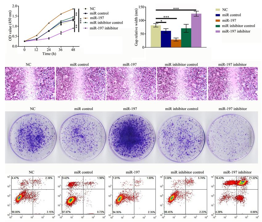
Ju et al.                                                                                     AbCD133@MSNs@GNR@miR-197 Inhibitor Affects Prostate Cance

                                            potential suppressor used in prostate cancer treatment. In short, our study found
                                            that miR-197 affected the development of prostate cancer by regulating ITGAV. The
                                            AbCD133@GNR@MSNs@miR-197 inhibitor prepared in this study could suppress the
                                            development and growth of PCSCs in vitro and in solid tumors not only by targeting the
                                            ITGAV but also through photothermal therapy. Our study not only provides a theoretical
                                            basis for the clinical treatment of prostate cancer but also provides a research scheme of
                                            drug loading and microRNA-based photothermal controlled therapy for prostate cancer.
                                            Keywords: prostate cancer, cancer stem cell, gold nanorod, miR-197, ITGAV

INTRODUCTION                                                                      These results suggest that miR-197 plays an important role in the
                                                                                  occurrence and development of PC. Therefore, miR-197-related
Prostate cancer (PC) is a major malignancy that occurs in middle-                 drugs could potentially be used to treat PC bone metastasis.
aged and elderly men, with an incidence rate of 25.3/10 million                   However, the molecular mechanism by which miR-197 exerts
worldwide. It is also the second most common cancer worldwide                     its function in bone metastasis of PC has not yet been reported
and the fifth leading cause of cancer death. In 2018 alone, there                 on. MiR-197 would need to efficiently enter cells for it to be
were nearly 1,300,000 PC cases and 359,000 new deaths as a result                 used for the treatment of PC. In addition, there are various
of PC (Aggarwal et al., 2015; Grozescu and Popa, 2017; Bray                       degradation mechanisms that lead to the degradation of miR-
et al., 2018; Taneja, 2020). PC is usually asymptomatic during the                197 in cells. At present, the clinical application of miR-197 lacks
early stage, and most patients are diagnosed during the middle                    efficiency, accuracy, and a targeted controlled-release delivery
and late stages. Endocrine therapy is the first choice of treatment               vector. Research conducted on tumor drug targeting carriers has
for advanced PC. However, endocrine therapy is not effective for                  found that the targeting property of a targeting agent is associated
advanced metastatic PC or castration-resistant PC patients (Shao                  with the targeting performance of the whole carrier, which is a
et al., 2012). At present, the exact molecular mechanism by which                 key factor for tumor targeting (Kaur et al., 2020; Prasher et al.,
PC develops and progresses is not clear. Therefore, it is of great                2020; Schneider-Rauber et al., 2020). Therefore, it is necessary to
significance to study the molecular mechanisms associated with                    identify drug delivery carriers with high transfection efficiency,
the occurrence and development of PC and develop new targeted                     good targeting performance, and controlled release. Mesoporous
drug carriers for the treatment of PC.                                            silica nanoparticles (MSNs) provide many advantages, including
    Cancer stem cells (CSCs) are a small number of cells that                     a simple process for synthesis, easy surface modification, and
drive the occurrence and development of tumor. CSCs can                           good biocompatibility. MSNs have become an inorganic carrier
maintain the stem cell characteristics of tumor cell growth and                   material that has been widely studied in the field of drug
can differentiate into the whole heterogeneous tumor (Bonnet                      carriers during recent years (Hargrove et al., 2020; Taweekarn
and Dick, 1997; Gratwohl et al., 2002; Orkin and Zon, 2008;                       et al., 2020; Wu et al., 2020). Gold nanorods (GNRs) are
O’Connor et al., 2014). Prostate CSCs (PCSCs) were first reported                 photothermal materials that can generate heat after near-
in 2007 (Miki et al., 2007). Studies have shown that PCSCs have                   infrared laser response. The surfactant cetyltrimethylammonium
self-renewal and unique tumorigenicity in vivo and that PCSCs                     bromide (CTAB) formed on the surface of solid GNRs has high
determine the development of PC; in addition, the resistance                      cytotoxicity, and it cannot load drugs. Coating the surface of
of PC to conventional chemotherapy and radiotherapy may be                        GNRs with mesoporous silica can not only reduce the toxicity
partly due to the existence of PCSCs (Hermann et al., 2007;                       of the material but also make use of the high specific surface
Huang et al., 2021; Navarro-Marchal et al., 2021; Zhang et al.,                   area that mesoporous silica will have for drug loading. GNRs can
2021). Therefore, research and development of drugs targeting                     melt phase change materials (PCMs), which block mesoporous
PCSCs is one of the important ways to treat PC.                                   silica after heating through near-infrared laser irradiation, so as
    MicroRNAs (miRNAs) are a type of endogenous non-                              to achieve photothermal controlled release of drugs (Das et al.,
coding RNAs found in eukaryotic cells, with a length of 18–25                     2020; Khelfa et al., 2020; Lebepe et al., 2020).
nucleotides. MiRNAs can bind to the 30 untranslated region                            In this study, we first studied the expression of miR-197
of target gene mRNA and can regulate the expression of                            in PC tissues and PC cells. Then, we predicted and verified
downstream target genes by changing the stability of the mRNA.                    the relationship between miR-197 and the target protein,
In recent years, abnormal levels of miRNA expression have been                    integrin subunit alpha V (ITGAV), through a bioinformatics
found in malignant tumors, which have been found to be closely                    analysis and double luciferase experiments. Further studies were
associated with the proliferation, differentiation, and apoptosis of              performed to study the molecular mechanism by which miR-197
tumor cells (Dimakakos et al., 2014; Brzeszczyńska et al., 2020;                 regulates the occurrence and development of PC by regulating
Paul et al., 2020). Studies have shown that the level of miR-                     the expression of ITGAV. Subsequently, we synthesized MSN-
197 expression in the serum of patients with PC is higher than                    coated GNRs, GNR@MSNs, which were loaded with the miR-
that of benign prostatic hyperplasia patients and healthy males,                  197 inhibitor. Then, the mesoporous GNR@MSNs were sealed
and the difference is statistically significant (Yuan et al., 2019).              using PCMs to create the GNR@MSNs@miR-197 inhibitor. The

Frontiers in Cell and Developmental Biology | www.frontiersin.org             2                                         June 2021 | Volume 9 | Article 646884
Ju et al.                                                                           AbCD133@MSNs@GNR@miR-197 Inhibitor Affects Prostate Cance

AbCD133@GNR@MSNs@miR-197 inhibitor was synthesized                       at 37◦ C in a humid atmosphere with 5% CO2 for 24 h. At the
by modifying the CD133 antibody on the surface of the                    end of the 48-h, 10 µl of MTT reagent obtained from Beenbio
GNR@MSNs@miR-197 inhibitor. Thereafter, we studied the                   Co., Ltd. (Shanghai, China) was added to the culture medium for
pharmacodynamic effect of the AbCD133@GNR@MSNs@miR-                      4 h of incubation. The cells were then suspended, and absorbance
197 inhibitor on PC in vivo and in vitro under near-infrared             was detected at 570 nm using an enzyme-linked immunosorbent
radiation. Overall, our study provides a theoretical basis for the       assay microplate reader (Sunrise Microplate Reader; TECAN,
clinical treatment of PC.                                                Männedorf, Switzerland).

                                                                         Luciferase Reporter Assay
MATERIALS AND METHODS                                                    ITGAV 30 -UTR fragments containing the predicted miR-197
                                                                         binding site were amplified using the cDNA of the PC3 cell.
Cell Culture                                                             Subsequently, the fragments were inserted into pGL3 luciferase
The DU145, PC3, and LNCaP cell lines were obtained from                  reporter vectors (Promega, Madison, WI, United States). For the
the Cell Bank of the Institute of Biochemistry and Cell Biology          luciferase reporter assay, PC3 cells were co-transfected with the
(Shanghai, China). RPMI-1640 (Thermo Fisher Scientific, Inc.,            pGL3 reporter and ITGAV UTR together with either NCO or
Waltham, MA, United States) culture medium was used for the              miR-197 mimics using a Lipofectamine 3000 system. Twenty-
                                                                                                                    R

DU145, PC3, LNCaP, and PCSCs, while the culture medium used              four hours after transfection, the cells were collected and lysed.
for RWPE-1 was supplied by Hunan Fenghui Biotechnology Co.,              Then, the luciferase activity was determined using a dual-
Ltd. All media were supplemented with 10% fetal bovine serum             luciferase reporter assay system (Promega), according to the
(FBS; Thermo Fisher Scientific, Inc.) and were kept at 37◦ C in a        manufacturer’s instructions.
humidified 5% CO2 incubator.
                                                                         Western Blotting Analysis
Prostate Cancer Stem Cell Screening                                      The treated cells were harvested and incubated in
PC3 cells of 108 were washed with phosphate-buffered saline              radioimmunoprecipitation assay (RIPA) lysis buffer (Cell
(PBS) three times; then cells were incubated with fluorescein            Signaling Technology Inc., Danvers, MA, United States) for
isothiocyanate (FITC)-labeled CD133 antibody in the dark; the            30 min at 4◦ C to isolate total proteins. The extracted proteins
cells were collected with centrifugation at 1,000 rpm for 5 min          were then separated using 10% sodium dodecyl sulfate–
and then washed with PBS two times; and the PCSCs was                    polyacrylamide gel electrophoresis (SDS-PAGE) and transferred
screened from PC3 cell by use of Aria SORP flow cytometry                onto a polyvinylidene fluoride membrane (Roche, Basel,
(BD, United States).                                                     Switzerland). Subsequently, the membranes were incubated
                                                                         with primary antibodies (Santa Cruz Biotechnology, Santa Cruz,
Cell Transfection                                                        CA, United States) at 4◦ C overnight and then incubated with
Human miR-197 mimics and negative control oligonucleotides               a secondary antibody (Santa Cruz Biotechnology) at room
(NCOs) were purchased from Beenbio Co., Ltd. (Shanghai,                  temperature for 2 h. The protein bands were visualized using an
China). Cells were seeded into six-well plates at a density of           enhanced chemiluminescence (ECL) western blotting substrate
4 × 10⁵ cells/well for plasmid transfection. Subsequently, these         (Thermo Fisher Scientific, Inc.).
RNA oligonucleotides or plasmids were transfected into cells
using a Lipofectamine 3000 system (Thermo Fisher Scientific,
                             R
                                                                         Flow Cytometry Analysis
Inc.) by following the manufacturer’s instructions.                      Cell apoptosis was detected using flow cytometry analysis.
                                                                         Annexin V-FITC and propidium iodide (PI) (BD Pharmingen,
Quantitative Real-Time Polymerase                                        San Diego, CA, United States) were used to calculate the number
Chain Reaction                                                           of Annexin V-positive cells.
Total RNA was extracted using Trizol reagent (Thermo Fisher
Scientific, Inc.). To obtain the cDNA, the extracted RNAs                Gold Nanorod Preparation
were reverse transcribed using a One Step PrimeScript miRNA              First, 5 ml of 0.5 mM HAuCl4 solution was mixed with 5 ml of
cDNA Synthesis Kit (TaKaRa, Dalian, China), according to                 0.2 M CTAB. Then, 0.6 ml of frozen 0.01 M NaBH4 was added,
the manufacturer’s instructions. qPCR was performed following            and the mixture was stirred for 2 min and allowed to stand for
standard protocol using a SYBR Green PCR kit (Toyobo,                    2 h at 25◦ C. Thereafter, 5 ml of 0.2 M CTAB, 5 ml of 1 mM
Japan). The following qPCR primer was used for miR-                      HAuCl4 , 0.5 ml of AgNO3 , and 0.07 ml of 0.1 M acetic acid (AA)
197: forward 50 -GTTCACCACCTTCTCCAC-30 ; reverse 50 -                    were added into the reaction vessel and stirred for 2 min. Finally,
GTGCAGGGTCCGAGGT-30 . U6 small nuclear RNA (snRNA                        0.012 ml of the seed solution was added into the growth solution,
U6) was used as the internal reference to determine the relative         stirred for 2 min, and allowed to stand at 28◦ C for 3 h to obtain
expression of miR-197 through the 2−11CT method.                         fully grown GNRs.

MTT Assay                                                                GNR@MSN Preparation
Cells were seeded onto a 96-well plate with 100 µl of culture            The prepared GNRs were washed twice with PBS, and
medium at a density of 5,000 cells/well. The cells were incubated        excess CTAB was removed through centrifugation and then

Frontiers in Cell and Developmental Biology | www.frontiersin.org    3                                      June 2021 | Volume 9 | Article 646884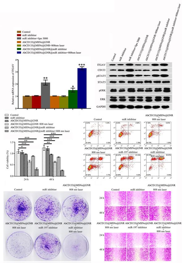
Ju et al.                                                                           AbCD133@MSNs@GNR@miR-197 Inhibitor Affects Prostate Cance

redistributed in 40 ml of PBS. Then, 50 µl of ammonia (25,               excess amount of hot water was added to the abovementioned
wt%) was added to adjust the GNR aqueous solution to pH 10,              preparation system to produce two phases that were obviously
and then 0.5 ml of 10 mM TEOS/ethanol solution was added at              incompatible with each other: the GNRs@MSN water phase
the rate of 3.5 ml/h. The reaction was carried out at 40◦ C for          with the PCM@miR-197 inhibitor and the chloroform phase
24 h. The reaction products were centrifuged several times with          without the PCM@miR-197 inhibitor encapsulation. The two
ethanol and water. To remove the CTAB residual molecules from            phases were rapidly separated, and the water phase was immersed
the mesoporous channels, 60 ml of ethanol/ammonium nitrate               in an ice water solution for cooling for 1 min and then
solution (10 mg/ml) was added to reflux for 6 h using the ion            centrifuged for 5 min at 12,000 rpm. The precipitates were
exchange method. Thereafter, the solution was centrifuged with           washed eight times with precooled deionized water and then
ethanol for purification. Finally, the GNR@MSN products were             freeze-dried for 24 h at −50◦ C and 0.8 mbar in a vacuum. The
dispersed in deionized water for preservation.                           GNRs@MSNs loaded with the PCM@miR-197 inhibitor were
                                                                         named the GNRs@MSNs@PCM@miR-197 inhibitor. In addition,
Characterization                                                         with the use of the same preparation method, the molecules
The nitrogen adsorption–desorption curve was created using               constructed that did not contain the miR-197 inhibitor were
a micrometrics Tristar 3000 system at 77 K. The sample                   named GNRs@MSNs@PCM.
was pre-dehydrated at 120◦ C for 6 h before determination.
The specific surface area and pore volume were calculated                Testing the Release Capacity of
using the Brunauer–Emmett–Teller (BET) formula and
                                                                         GNRs@MSNs@PCM@miR-197 Inhibitor
Barrett–Joyner–Halenda (BJH) model, respectively. The
                                                                         To test the miR-197 inhibitor release property of
transmission electron microscopy (TEM) image was
                                                                         GNRs@MSNs@PCM with or without the effect of an 808-
measured using a jem-2010f-hr electron microscope at a
                                                                         nm laser, the GNRs@MSNs@PCM@miR-197 inhibitor was
voltage of 200 kV. During sample preparation, the sample
                                                                         dispersed in 5 ml of PBS (pH 7.4) in semipermeable dialysis bags
was first refined, dispersed in ethanol solution, and then
                                                                         at 37◦ C by gentle shaking. Then, 1 ml of the released medium was
ultrasonically treated. Then, the suspension solution was
                                                                         removed, and 1 ml of fresh medium was added. The amount of
dropped onto a copper mesh coated with carbon film
                                                                         the miR-197 inhibitor released was measured using a NanoDrop
and was placed on the sample table for vacuumization
                                                                         3000 spectrophotometer.
and observation.

Detection of Heat Production in the                                      Cellular Uptake and Internalization
GNR@MSNs Under 808-nm Laser                                              A total of 2 × 10⁴ of PCSCs were seeded into glass-bottom plates
                                                                         (35 mm, Corning Incorporated) in Dulbecco’s modified Eagle’s
Irradiation                                                              medium (DMEM) with 10% FBS and were incubated at a final
The diluted GNR@MSN solution was irradiated with an 808-                 concentration of 50 µg/ml of FITC-labeled GNRs@MSNs@PCM
nm near-infrared laser for different durations and at different          and FITC-labeled AbCD133@GNRs@MSNs@PCM for 24 h. The
intensities (spot size: 5 mm). The temperature of the solution was       cells were incubated with 60 nM of LysoTracker Red DND-99
measured using an infrared temperature detector.                         (Beyotime Institute of Biotechnology, Haimen, China) for 1 h at
                                                                         37◦ C. After being washed with PBS, the cells were fixed with 4%
Fourier-Transform Infrared Analysis of                                   paraformaldehyde and stained with 10 µg/ml of 40 ,6-diamidino-
the GNR@MSNs                                                             2-phenylindole (DAPI; Sigma). The cells were then washed
GNR@MSNs were mixed with KBr powder, grinded to a uniform                three times with PBS and mounted. The micrographs were
size, and pressed onto a 1-mm-thick transparent sheet. Fourier-          first observed under a Nikon fluorescence microscope (Nikon
transform infrared (FTIR) analysis was performed using a nexus-          Eclipse Ti-S, CCD: Ri1) and then under a laser scanning confocal
470 infrared instrument.                                                 microscope (Leica TCS sp5, Germany) and were used for confocal
                                                                         luminescence imaging with a 63 × oil immersion objective lens.
MiR-197 Inhibitor Loading
Since the melting point of PCM tetradecanol is 38–39◦ C, a               Cytotoxicity of
sufficient amount of a miR-197 inhibitor and PCM needs to
be mixed for loading onto the GNRs@MSNs at a temperature
                                                                         GNRs@MSNs@PCM@miR-197 to
higher than the melting point. First, 100 mg of tetradecanol             Prostate Cancer Stem Cell Under
was heated at 60◦ C until it had melted fully. Then, 20 µg               Near-Infrared Laser Irradiation
of the miR-197 inhibitor was added and thoroughly mixed                  Prostate cancer stem cells at the logarithmic growth phase were
through magnetic stirring for 2 h. Then, 100 mg of the                   inoculated onto 96-well plates at a density of 5,000 cells/well,
GNRs@MSNs was mixed with a large amount of chloroform,                   1 day before the experiment, and the culture medium was
added into the PCM mixture solution, and stirred for 2 h at              discarded after 24 h of being inside the cell incubator, with
60◦ C. During this mixing process, through the volatilization of         different concentrations of AbCD133@GNRs@MSNs. The cells
trichloromethane, the PCM@miR-197 inhibitor mixture slowly               were cultured at 37◦ C for 4 h and then irradiated with a
infiltrated the mesopore cavities of the GNRs@MSNs. Finally, an          near-infrared laser (808 nm, 3 W/cm2 ). After 24 or 48 h of

Frontiers in Cell and Developmental Biology | www.frontiersin.org    4                                      June 2021 | Volume 9 | Article 646884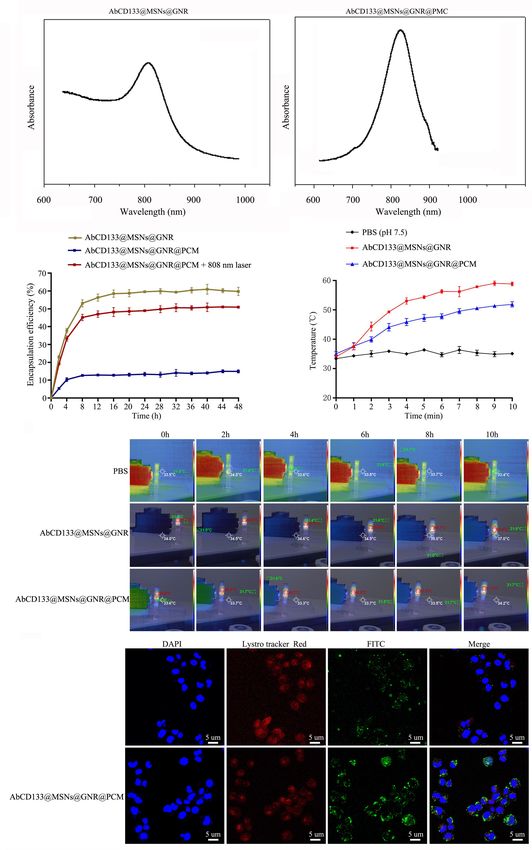
Ju et al.                                                                                      AbCD133@MSNs@GNR@miR-197 Inhibitor Affects Prostate Cance

incubation, Cell Counting Kit-8 (CCK-8) assay was used to                         AbCD133@MSNs@AuNP@miR-197 inhibitor + 808 nm laser
detect cell viability.                                                            exposure 10 min. Treatment was performed three times a week,
                                                                                  and tumor growth was measured every 5 days, while the tumor
                                                                                  volume was also calculated.
In vivo Antitumor Efficacy of Prostate
Cancer Stem Cell                                                                  Statistical Analysis
Eight-week-old nude mice with an average weight of 18 g                           SPSS 16.0 statistical software (IBM Corporation, Armonk, NY,
were used in this study. Each mouse was injected with 40 µl                       United States) was used to analyze the data. Non-paired t test was
of the anesthetic before operation. A PCSC suspension was                         used to estimate statistical differences between two groups, while
mixed with Matrigel at a ratio of 1:1. Each mouse was injected                    one-way analysis of variance (ANOVA) was applied to determine
with 1 × 10⁶ cells, and the total volume of the injection was                     differences among three or more groups. A P value of < 0.05 was
50 µl. The mice were reared under good laboratory conditions                      considered to indicate a statistically significant difference.
(temperature 25 ± 2◦ C; relative humidity 50 ± 20%) with
dark and light cycles (12/12 h) and access to standard balanced
diet. The mice were observed every day after operation. After
                                                                                  RESULTS
2 weeks, the growth of the solid tumors was observed; and
the length, width, and height were measured using a vernier
caliper to calculate the volume of the tumor. After about
                                                                                  MiR-197 Is Overexpressed in Prostate
4 weeks, therapy was started when the tumor volume was                            Cancer Tissues and Prostate Cancer
200 × 400 mm3 . Mice were randomized into four groups of five                     Cells
mice per group. The groups of animals were treated variously:                     qPCR assay was conducted to detect the expression level of
(1) control, (2) miR-197 inhibitor, (3) AbCD133@MSNs@AuNP,                        miR-197 in PC tissues and different PC cells. The results
(4) AbCD133@MSNs@AuNP + 808 nm laser exposure 10 min,                             indicated that miR-197 was significantly overexpressed in PC
(5) AbCD133@MSNs@AuNP@miR-197 inhibitor, and (6)                                  tissues (Figure 1A), compared with paired normal tissue, and

  FIGURE 1 | MiR-197 is overexpressed in prostate cancer tissues and prostate cancer cells. (A) qPCR assay was used to detect the expression of miR-197 in
  prostate cancer tissues and different prostate cancer cells. (B) qPCR assay was used to determine the expression of miR-197 in prostate cancer cells lines
  (RWPE-1, DU145, PC3, and LNCaP). (C) CD133 overexpressing PC3 cells were selected using flow cytometry. (D) qPCR assay was used to detect the expression
  of miR-197 in RWPE-1, PC3, and prostate cancer stem cells (PCSCs). *p < 0.05, **p < 0.01, ***p < 0.001.

Frontiers in Cell and Developmental Biology | www.frontiersin.org             5                                           June 2021 | Volume 9 | Article 646884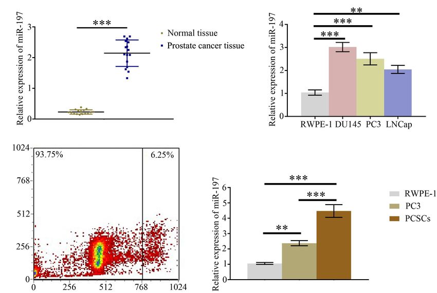
Ju et al. AbCD133@MSNs@GNR@miR-197 Inhibitor Affects Prostate Cance
that the expression of miR-197 in the PC cells lines (DU145, Synthesis and Characterization of
PC3, and LNCaP) was significantly increased than in the Mesoporous Silica-Coated Gold
normal prostate cell line, RWPE-1 (Figure 1B). To study
miR-197 expression in PCSCs, CD133-overexpressing PC3 cells Nanorods
were selected using flow cytometry (Figure 1C), and then It is necessary to develop nanocarriers with good
miR-197 expression was detected in the RWPE-1, PC3, and biocompatibility, loading capacity, cell internalization, and
PCSCs. The results showed that miR-197 expression in the release performance for use with miRNA-based methods of
PCSCs was significantly higher than in RWPE-1 and PC3 cancer treatment. In this study, we first synthesized mesoporous
cells (Figure 1D). silica-coated GNRs (GNR@MSNs) and then modified the
CD133 antibodies on the surface of the GNR@MSNs to
synthesize AbCD133@GNR@MSNs (Figure 4A). Mesoporous
MiR-197 Regulates ITGAV Expression in silica provides a high drug loading capacity and good release
the Prostate Cancer Tissues and performance, while GNR nanorods contribute photothermal
properties. The AbCD133 in AbCD133@GNR@MSNs can
Prostate Cancer Cells
target PCSCs, which are a small group of prostate tumor
An online bioinformatics analysis was conducted using the
cells that determine the proliferation, invasion, metastasis,
TargetScan 7.2 database to predict proteins that may be regulated
and drug resistance of PC. Figures 4B–E provide a TEM
by miR-197. The results showed that 1,362–1,369 of ITGAV
image of GNR@MSNs, while Figure 4F shows the mesoporous
30 -UTR might be the target of miR-197 (Figure 2A). qPCR
morphology and pore size distribution of the GNR@MSNs.
and western blotting assays were conducted to detect the
Then, CCK-8 assay was conducted to test the biocompatibility
expression of ITGAV in PC tissues and different PC cells. The
of AbCD133@GNR@MSNs to PCSCs. The result showed that
results showed that mRNA and protein expression levels of
a significant cytotoxicity effect was not produced when the
ITGAV were significantly decreased in PC tissues, compared
concentration was between 6.25 and 200 µg/ml (Figure 4G).
with normal tissues (Figures 2B,C), and that mRNA and protein
expression levels of ITGAV in PC cells (DU145, PC3, and
LNCaP) as well as PCSC were also significantly decreased, MicroRNA Loading Capacity, Release
compared with normal prostate cells (RWPE-1). Dual-luciferase Performance, Photothermal Properties,
reporter assay was conducted to study direct interactions
between miR-197 and ITGAV mRNA, and the relative luciferase and Cell Internalization of
value was found to have decreased significantly after miR-197 AbCD133@GNR@MSNs
transfection in the ITGAV wild-type (WT) group. However, To achieve controllable photothermal release, PCMs were
the relative luciferase value did not show a significant change used to seal the mesoporous AbCD133@GNR@MSNs. PCM
in the ITGAV mutant group (Figure 2F). Meanwhile, the can be welded under 808-nm near-infrared laser irradiation,
miR-197 mimic could significantly inhibit ITGAV and CD133 and AbCD133@GNR@MSNs will slowly release the miR-197
expression and significantly decrease the phosphorylation of inhibitor. NanoDrop 3000 was used to detect changes in miR-
STAT5 and ERK, while the miR-197 inhibitor produced an 197 inhibitor concentration before and after loading. The
opposite effect (Figures 2G–I). These results indicated that miR- results showed that 1 mg of AbCD133@GNR@MSNs could
197 directly regulates ITGAV/STAT5 pathway in PC tissues load 35.42 µg of the miR-197 inhibitor. Subsequently, a semi-
and PC cells and that miR-197 plays an important role permeable membrane release detection model and NanoDrop
in PC development. 3000 were used to study the AbCD133@GNR@MSNs release
properties of the miR-197 inhibitor with or without 808-nm laser
irradiation. According to the result shown in Figure 5A, both
MiR-197 Inhibitor Functions as a AbCD133@GNR@MSNs and AbCD133@GNR@MSNs@PCM
Potential Suppressor of Prostate Cancer have a maximum absorption peak at about 808 nm. The
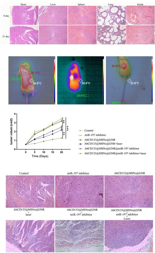
To study the effect of miR-197 on the development of results showed almost no miR-197 inhibitor release under
PC, PCSCs were transfected with either a miR-197 mimic 808-nm laser irradiation when the AbCD133@GNR@MSNs
or a miR-197 inhibitor. Then, CCK-8 assay was used to were sealed with PCM, while the AbCD133@GNR@MSNs
detect cell proliferation, cell scratch assay was used to without PCM showed a release performance that was similar
test the effect of the miRNA on PCSC migration, colony to that of AbCD133@GNR@MSNs@PCM under 808-nm laser
formation assay was used to observe cell growth features, irradiation. The results showed that there was a rapid release
and an Annexin V-FITC/PI apoptosis detection kit and of the miR-197 inhibitor at 0–8 h, and then the miR-197
flow cytometry were used to determine cell apoptosis. The inhibitor could be released stably to produce a long-term
results showed that the miR-197 inhibitor could significantly effect (Figure 5B). Figures 5C,D show the photothermal
decrease the proliferation and migration levels of the PCSCs properties of the AbCD133@GNR@MSNs. According to the
as well as significantly decrease the level of PCSC apoptosis results, the temperature of both the AbCD133@GNR@MSNs
(Figures 3A–E). Therefore, these results indicate that the miR- and AbCD133@GNR@MSNs@PCM can reach above 40◦ C
197 inhibitor is a potential suppressor of PC development after 3 min of 808-nm laser irradiation and can continue
and progression. to increase up to 60◦ C within 10 min. Specifically killing
Frontiers in Cell and Developmental Biology | www.frontiersin.org 6 June 2021 | Volume 9 | Article 646884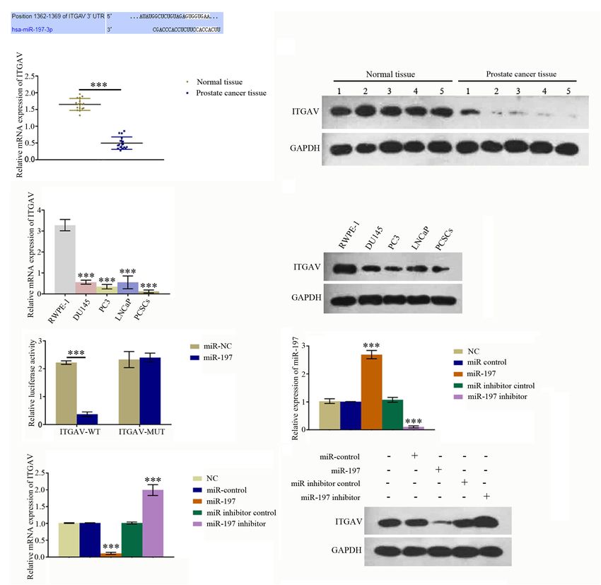
Ju et al.                                                                                         AbCD133@MSNs@GNR@miR-197 Inhibitor Affects Prostate Cance

  FIGURE 2 | MiR-197 regulated ITGAV expression in prostate cancer tissues and cells. (A) An online bioinformatics analysis was conducted using the TargetScan 7.2
  database to determine whether ITGAV could be regulated by miR-197 and 1362–1369 of ITGAV 30 -UTR was found to target miR-197. (B) qRT-PCR assay showed
  the expression of ITGAV mRNA was lower in prostate cancer tissues. (C) Western blotting assay showed the expression of ITGAV protein was lower in prostate
  cancer tissues. (D) qRT-PCR assay found the expression of ITGAV mRNA was lowest in prostate cancer stem cells (PCSCs). (E) Western blotting assay determined
  the protein expression levels of ITGAV was lower in PC3 and PCSCs. (F) Dual luciferase reporter assay confirmed the direct interaction between miR-197 and ITGAV
  mRNA. (G) qRT-PCR assay determined the expression of miR-197 in PCSCs cells after transfection with the miR control, miR-197 mimic, miR inhibitor control and
  miR-197 inhibitor. The results showed the expression of miR-197 was lowest in miR-197 inhibitor group. (H) qRT-PCR assay determined the expression of ITGAV
  mRNA in PCSCs cells after transfection with the miR control, miR-197 mimic, miR inhibitor control and miR-197 inhibitor. The results showed the expression of
  ITGAV mRNA was highest in miR-197 inhibitor group. (I) Western blotting assays was used to detect the expression of ITGAV, CD133 and the phosphorylation of
  STAT5 and ERK in PCSCs after transfection with the miR control, miR-197 mimic, miR inhibitor control and miR-197 inhibitor. *p < 0.05, **p < 0.01, ***p < 0.001.

Frontiers in Cell and Developmental Biology | www.frontiersin.org                7                                            June 2021 | Volume 9 | Article 646884
Ju et al.                                                                                          AbCD133@MSNs@GNR@miR-197 Inhibitor Affects Prostate Cance

  FIGURE 3 | MiR-197 inhibitor is a potential suppressor of prostate cancer. PC3 was transfected with miR-197 mimics or miR-197 inhibitor. (A) Cell Counting Kit-8
  (CCK-8) assay was conducted to detect the level of cell proliferation. (B,C) Cell scratch assay was conducted to determine the effect of miRNAs on prostate cancer
  stem cell (PCSC) migration. (D) Colony formation assay was conducted to observe PCSC growth features. (E) An Annexin V-FITC/PI apoptosis detection kit and
  flow cytometry were used to evaluate PCSC apoptosis. *p < 0.05, **p < 0.01, ***p < 0.001.

CSCs using targeted drug carriers is a novel strategy used                            and ITGAV-related proteins, and results showed that the
in cancer treatment. We modified the antibody of the PCSC                             AbCD133@GNR@MSNs@miR-197 inhibitor with 808-nm laser
marker, AbCD133, on the surface of the GNR@MSNs and then                              irradiation could significantly increase the expression of ITGAV
constructed FITC-labeled AbCD133@GNR@MSNs, while laser                                and CD133 and significantly increase the phosphorylation
confocal microscopy was used to detect PCSC internalization                           of STAT5 and ERK (Figures 6A,B). To test the effect
of the AbCD133@GNR@MSNs. The results showed that                                      of the AbCD133@GNR@MSNs@miR-197 inhibitor on the
CD133 antibody modification could significantly improve PCSC                          development of PCSCs, CCK-8 assay was conducted to determine
internalization of the GNR@MSNs. A significant increase                               cell viability, and the result showed that after irradiation
of FITC-labeled AbCD133@GNR@MSNs (green fluorescence),                                with an 808-nm laser, the AbCD133@GNR@MSNs@miR-197
compared with FITC-labeled GNR@MSNs, was observed in                                  inhibitor could significantly decrease the cell viability of the
PCSCs (Figure 5E).                                                                    PCSCs (Figure 6C). An Annexin V-FITC/PI apoptosis detection
                                                                                      kit was used to determine cell apoptosis, and the results
The Effect of                                                                         showed that the AbCD133@GNR@MSNs@miR-197 inhibitor
AbCD133@GNR@MSNs@miR-197                                                              with 808-nm laser irradiation could significantly induce PCSC
                                                                                      apoptosis (Figure 6D). Then, clone formation experiment
Inhibitor on the Development of Prostate                                              and cell scratch assay were conducted to determine the cell
Cancer Stem Cells Under 808-nm Laser                                                  invasion and infection abilities. According to the results, the
Irradiation                                                                           AbCD133@GNR@MSNs@miR-197 inhibitor with 808-nm laser
To test the effect of the AbCD133@GNR@MSNs@miR-197                                    irradiation could significantly decrease the clone formation rate
inhibitor on the ITGAV relative pathway, qPCR and western                             (Figure 6E) and significantly inhibit the scratch gap growth
blotting assay were used to test the expression of ITGAV                              rate (Figure 6F).

Frontiers in Cell and Developmental Biology | www.frontiersin.org                 8                                             June 2021 | Volume 9 | Article 646884
Ju et al. AbCD133@MSNs@GNR@miR-197 Inhibitor Affects Prostate Cance FIGURE 4 | Synthesis and characterization of mesoporous silica-coated gold nanorods. (A) GNR@MSNs and AbCD133@GNR@MSNs synthesis. (B–E) transmission electron microscopy (TEM) image of GNR@MSNs. (F) The mesoporous morphology and mesoporous distribution (inset) of the GNR@MSNs were determined using N2 adsorption–desorption isotherms. (G) Cell Counting Kit-8 (CCK-8) assay was conducted to test the biocompatibility of AbCD133@GNR@MSNs on prostate cancer stem cells (PCSCs). Frontiers in Cell and Developmental Biology | www.frontiersin.org 9 June 2021 | Volume 9 | Article 646884
Ju et al.                                                                                 AbCD133@MSNs@GNR@miR-197 Inhibitor Affects Prostate Cance

  FIGURE 5 | MicroRNA loading capacity, release performance, photothermal properties, and cell internalization of AbCD133@GNR@MSNs. (A) UV absorption
  spectrum detection of AbCD133@GNR@MSNs and AbCD133@GNR@MSNs@PCM. (B) Cell Counting Kit-8 (CCK-8) assay was used to test the biocompatibility of
  AbCD133@GNR@MSNs with or without 808-nm laser irradiation. (C,D) The photothermal properties of AbCD133@GNR@MSNs and
  AbCD133@GNR@MSNs@PCM determined under 808-nm laser irradiation. (E) Laser confocal microscopy was used to detect prostate cancer stem cell (PCSC)
  internalization of GNR@MSNs and AbCD133@GNR@MSNs.

Frontiers in Cell and Developmental Biology | www.frontiersin.org         10                                         June 2021 | Volume 9 | Article 646884
Ju et al.                                                                                          AbCD133@MSNs@GNR@miR-197 Inhibitor Affects Prostate Cance

  FIGURE 6 | The effect of AbCD133@GNR@MSNs@miR-197 inhibitor on the development of PC3 prostate cancer cells under 808-nm laser irradiation. (A) qPCR
  assay was used to test the mRNA expression of ITGAV. (B) Western blotting assay was used to test the expression of ITGAV and CD133 and the phosphorylation of
  STAT5, ERK, and ITGAV-related proteins. (C) Cell Counting Kit-8 (CCK-8) assay was used to test the cell viability of the prostate cancer stem cells (PCSCs). (D) An
  Annexin V-FITC/PI apoptosis detection kit was used to detect PCSC apoptosis. (E,F) Colony formation experiment and cell scratch assay were conducted to test
  the cell invasion and infection abilities of the PCSCs. *p < 0.05, **p < 0.01, ***p < 0.001.

Frontiers in Cell and Developmental Biology | www.frontiersin.org                 11                                             June 2021 | Volume 9 | Article 646884
Ju et al.                                                                             AbCD133@MSNs@GNR@miR-197 Inhibitor Affects Prostate Cance

The Effect of                                                             hotspot in this field (Li N. et al., 2020; Yue et al., 2020; An
AbCD133@GNR@MSNs@miR-197                                                  et al., 2021). MiRNAs can bind to the 30 untranslated region
                                                                          of the targeted gene mRNA and can regulate the expression
Inhibitor on the Prostate Cancer Stem                                     of downstream target genes. In recent years, many miRNAs
Cells in the Solid Tumor Model Under                                      associated with PC have been identified and have been confirmed
808-nm Laser Irradiation                                                  to be closely associated with the occurrence and development
We explored the effect of the AbCD133@GNR@MSNs@miR-                       of PC (Hou et al., 2020; Chen et al., 2021; Wang et al., 2021).
197 inhibitor on the PCSCs in solid tumor formation and                   Previous studies have reported that miR-197 is significantly
growth under 808-nm laser irradiation. Firstly, the acute toxicity        overexpressed in the serum of PC patients, which indicates
of AbCD133@GNR@MSNs on mice was evaluated, and the                        that miR-197 plays an important role in the occurrence and
results showed that AbCD133@GNR@MSNs have no effect on                    development of PC. However, the molecular mechanism of miR-
the weight increase of the mice, and H&E staining assay was               197 in the bone metastasis of PC has not as yet been reported
used to observe the effect of AbCD133@GNR@MSNs on the                     (Yuan et al., 2019).
heart, liver, spleen, lung, and kidneys of the mice (Figure 7A).              In this study, we first studied the molecular mechanism
Then, the effect of the AbCD133@GNR@MSNs@miR-197                          by which miR-197 affects the development of PC. Our study
inhibitor on the PCSC solid tumor growth was evaluated.                   verified that miR-197 is significantly overexpressed in the PC
Figure 7B shows that the AbCD133@GNR@MSNs@miR-197                         tissues and PC cells, compared with normal PC cells. These
inhibitor could increase the temperature of the PCSCs in                  results indicate that miR-197 may play an important role in
solid tumors with 808-nm laser irradiation to significantly               the proliferation, invasion, and metastasis of PC cells. Then,
inhibit the growth of the PCSC solid tumors (Figure 7C).                  we found and confirmed that miR-197 can regulate ITGAV
Then, H&E staining assay was conducted to observe the                     expression of PC cells by directly binding to the 30 -UTR of
effect of AbCD133@GNR@MSNs on PC3 PC cell solid tumors                    ITGAV. Previous studies have reported that ITGAV can regulate
under 808-nm laser irradiation. The results showed that                   the proliferation, invasion, and metastasis of several cancers, such
the destructive effect of the AbCD133@GNR@MSNs@miR-                       as gastric cancer, breast cancer, esophageal cancer, and colorectal
197 inhibitor under 808-nm laser irradiation was significantly            cancer (Waisberg et al., 2014; Ma et al., 2016; Wang et al., 2019;
higher on tumor tissue destruction than cells of the other                Cheuk et al., 2020; Li Q. et al., 2020). Our study showed that
groups (Figure 7D).                                                       miR-197 affects the proliferation, invasion, and metastasis of PC
                                                                          cells by regulating ITGAV expression. Together, these results
                                                                          indicated that the miR-197 inhibitor may potentially suppress the
DISCUSSION                                                                development and progression of PC, which can be used for the
                                                                          treatment of PC.
Prostate cancer is one of the most significant malignant                      For potential clinical application, many miR-197 inhibitors
tumors that affects males and has the second highest mortality            need to be introduced into PC cells, and they must avoid
rate of all tumors. The diagnosis and treatment of PC have                degradation caused by genetic drugs during and after entering
become an important research focus of modern medicine                     cancer cells. In addition, genetic drugs should be able to release a
(Mabuchi et al., 2020; Steiner et al., 2020). Current treatments          stable drug concentration after entering cancer cells. In this study,
for PC can be divided into endocrine therapy [bilateral                   we synthesized an AbCD133@GNR@MSNs@miR-197 inhibitor
orchiectomy, luteinizing hormone-releasing hormone (LHRH)                 drug carrier. The mesoporous structure of the MSNs could load
analog treatment, anti-androgen therapy, androgen combined                many miR-197 inhibitors (35.42 µg of miR-197 inhibitor/mg
blocking therapy, and intermittent anti-androgen therapy],                AbCD133@GNR@MSNs), and the miR-197 inhibitor showed
surgical treatment (radical prostatectomy and intracavitary               good release performance after entering the cells. The modified
surgery for adenocarcinoma), radiotherapy, and chemotherapy.              CD133 antibodies on the surface of the nano drug carriers can
However, the traditional methods of treatment for PC produce              specifically enrich drug carriers around the PCSCs and results in
obvious side effects and a high recurrence rate, which seriously          an increased quantity of drugs entering PCSCs. This increase in
affect the treatment effect and the quality of life of patients.          quantity leads to a more effective effect by selectively targeting
PCSCs are a small number of cells that drive the occurrence               and suppressing CSCs. GNR, the core of the drug carrier, not
and development of tumor. They play an important role                     only can release heat to kill cancer cells after being exposed
in the development, self-renewal, and drug resistance of PC               to near-infrared radiation but also can melt the PCM used to
(Gratwohl et al., 2002; Hermann et al., 2007; Miki et al.,                seal mesoporous structure to achieve photothermal controlled
2007; O’Connor et al., 2014; Zhang et al., 2021), Research and            release of the miR-197 inhibitor. The pharmacodynamic effect
development of drugs targeting PCSCs is one of the important              of the AbCD133@GNR@MSNs@miR-197 inhibitor on PCSCs
ways to treat PC.                                                         in vivo and in vitro under near-infrared radiation was studied.
    In recent years, the importance of gene therapy has been              The results showed that the AbCD133@GNR@MSNs@miR-
widely recognized in PC research due to it highly targeted                197 inhibitor could significantly decrease cell viability of the
effects and fewer side effects. Gene therapy can inhibit the              PCSCs, induce significant PCSCs apoptosis, and significantly
growth of PC cells by regulating the protein expression of                decrease the clone formation rate and invasion ability of the
targeted genes, and PC therapy based on miRNAs is a research              PCSCs. The AbCD133@GNR@MSNs@miR-197 inhibitor could

Frontiers in Cell and Developmental Biology | www.frontiersin.org    12                                       June 2021 | Volume 9 | Article 646884
Ju et al. AbCD133@MSNs@GNR@miR-197 Inhibitor Affects Prostate Cance FIGURE 7 | The effect of AbCD133@GNR@MSNs@miR-197 inhibitor on prostate cancer stem cells (PCSCs) in a solid tumor model under 808-nm laser irradiation. (A) The acute toxicity of AbCD133@GNR@MSNs on the mice was determined using H&E staining assay to observe the effect of AbCD133@GNR@MSNs on the heart, liver, spleen, lung, and kidneys of the mice. (B) AbCD133@GNR@MSNs@miR-197 inhibitor could increase the temperature of the PCSCs in the solid tumors under 808-nm laser irradiation. (C) AbCD133@GNR@MSNs@miR-197 inhibitor could significantly inhibit the growth of the PCSCs in the solid tumors. (D) H&E staining assay was conducted to observe the effect of AbCD133@GNR@MSNs on the PCSCs in solid tumors under 808-nm laser irradiation. *p < 0.05, **p < 0.01, ***p < 0.001. Frontiers in Cell and Developmental Biology | www.frontiersin.org 13 June 2021 | Volume 9 | Article 646884
Ju et al.                                                                                                   AbCD133@MSNs@GNR@miR-197 Inhibitor Affects Prostate Cance

also significantly inhibit the growth of PCSCs in solid tumors                               ETHICS STATEMENT
under 808-nm laser irradiation.
                                                                                             The experiments animals were performed under approval of the
                                                                                             Institutional Animal Care and Use Committee of Changzheng
CONCLUSION                                                                                   Hospital, Naval Medical University, Shanghai, China.

In summary, our study found that miR-197 affected the
proliferation, invasion, and metastasis of PC cells by regulating
ITGAV expression. The AbCD133@GNR@MSNs@miR-197                                               AUTHOR CONTRIBUTIONS
inhibitor prepared in this study could suppress the development
                                                                                             GJ designed research and performed the experiments. YZ and TD
and growth of PCSCs in vitro and in solid tumors not only by
                                                                                             performed the experiments. WC and JL interpreted results of the
targeting the ITGAV but also through photothermal therapy. Our
                                                                                             experiments. GJ and CL analyzed data and prepared figures. DX
study not only provides a theoretical basis and for the clinical
                                                                                             and ZW designed the experiments, provided the experimental
treatment of PC but also provides a research scheme of drug
                                                                                             insight, and edited the manuscript. All authors read and approved
loading and miRNA-based photothermal controlled therapy for
                                                                                             the final manuscript.
prostate cancer.

DATA AVAILABILITY STATEMENT                                                                  FUNDING
The original contributions presented in the study are included                               This work was supported by the Medical Guidance
in the article/supplementary material, further inquiries can be                              Project of Shanghai Science and Technology Committee
directed to the corresponding author/s.                                                      (No. 19411967600).

REFERENCES                                                                                   Hargrove, D., Liang, B., Kashfi-Sadabadi, R., Joshi, G., Gonzalez-Fajardo, L., Glass,
                                                                                                 S., et al. (2020). Tumor-mesoporous silica nanoparticle interactions following
Aggarwal, R., Beer, T., Weinberg, V., Higano, C., Taplin, M., Ryan, C., et al.                   intraperitoneal delivery for targeting peritoneal metastasis. J. Controll. Release
   (2015). Intermittent chemotherapy as a platform for testing novel agents in                   328, 846–858. doi: 10.1016/j.jconrel.2020.11.003
   patients with metastatic castration-resistant prostate cancer: a department of            Hermann, P., Huber, S., Herrler, T., Aicher, A., Ellwart, J., Guba, M., et al.
   defense prostate cancer clinical trials consortium randomized phase II trial of               (2007). Distinct populations of cancer stem cells determine tumor growth and
   intermittent docetaxel with prednisone with or without maintenance GM-CSF.                    metastatic activity in human pancreatic cancer. Cell Stem Cell 1, 313–323.
   Clin. Genitourin. Cancer 13, e191–e198.                                                       doi: 10.1016/j.stem.2007.06.002
An, G., Lu, F., Huang, S., Bai, J., He, L., Liu, Y., et al. (2021). Effects of miR-93        Hou, Z., Wang, Y., Xia, N., Lv, T., Yuan, X., and Song, Y. (2020). Pseudogene
   on epithelial-to-mesenchymal transition and vasculogenic mimicry in triple-                   KRT17P3 drives cisplatin resistance of human NSCLC cells by modulating
   negative breast cancer cells. Mol. Med. Rep. 23:30.                                           miR-497-5p/mTOR. Cancer Sci. 112, 275–286. doi: 10.1111/cas.14733
Bonnet, D., and Dick, J. (1997). Human acute myeloid leukemia is organized as                Huang, R., Guo, L., Gao, M., Li, J., and Xiang, S. (2021). Research trends and
   a hierarchy that originates from a primitive hematopoietic cell. Nat. Med. 3,                 regulation of CCL5 in prostate cancer. Onco Targets Ther. 14, 1417–1427. doi:
   730–737. doi: 10.1038/nm0797-730                                                              10.2147/ott.s279189
Bray, F., Ferlay, J., Soerjomataram, I., Siegel, R., Torre, L., and Jemal, A. (2018).        Kaur, L., Sohal, H., Kaur, M., Malhi, D., and Garg, S. (2020). A mini-review
   Global cancer statistics 2018: GLOBOCAN estimates of incidence and mortality                  on nano technology in the tumour targeting strategies: drug delivery to
   worldwide for 36 cancers in 185 countries. CA 68, 394–424. doi: 10.3322/caac.                 cancer cells. Anti Cancer Agents Med. Chem. 20, 2012–2024. doi: 10.2174/
   21492                                                                                         1871520620666200804103714
Brzeszczyńska, J., Brzeszczyński, F., Hamilton, D., McGregor, R., and Simpson, A.          Khelfa, A., Meng, J., Byun, C., Wang, G., Nelayah, J., Ricolleau, C., et al. (2020).
   (2020). Role of microRNA in muscle regeneration and diseases related to muscle                Selective shortening of gold nanorods: when surface functionalization dictates
   dysfunction in atrophy, cachexia, osteoporosis, and osteoarthritis. Bone Joint                the reactivity of nanostructures. Nanoscale 12, 22658–22667. doi: 10.1039/
   Res. 9, 798–807.                                                                              d0nr06326f
Chen, Y., Yang, J., Xue, Z., Cai, Q., Hou, C., Li, H., et al. (2021). Effects and            Lebepe, T., Parani, S., and Oluwafemi, O. (2020). Graphene oxide-coated gold
   mechanism of microRNA-218 against lung cancer. Mol. Med. Rep. 23:28.                          nanorods: synthesis and applications. Nanomaterials (Basel, Switzerland)
Cheuk, I., Siu, M., Ho, J., Chen, J., Shin, V., and Kwong, A. (2020). ITGAV targeting            10:2149. doi: 10.3390/nano10112149
   as a therapeutic approach for treatment of metastatic breast cancer. Am. J.               Li, N., Shen, F., Cai, Z., Pan, W., Yin, Y., Deng, X., et al. (2020). Target-
   cancer Res. 10, 211–223.                                                                      induced core-satellite nanostructure assembly strategy for dual-signal-on
Das, C., Guo, Y., Yang, G., Kang, L., Xu, G., Ho, H., et al. (2020). Gold nanorod                fluorescence imaging and raman quantification of intracellular MicroRNA
   assisted enhanced plasmonic detection scheme of COVID-19 SARS-CoV-2                           guided photothermal therapy. Small 16:e2005511.
   spike protein. Adv. Theory Simul. 3:2000185. doi: 10.1002/adts.202000185                  Li, Q., Peng, K., Chen, E., Jiang, H., Wang, Y., Yu, S., et al. (2020). IntegrinB5
Dimakakos, A., Armakolas, A., and Koutsilieris, M. (2014). Novel tools for prostate              upregulated by HER2 in gastric cancer: a promising biomarker for liver
   cancer prognosis, diagnosis, and follow-up. BioMed Res. Int. 2014:890697.                     metastasis. Ann. Trans. Med. 8:451. doi: 10.21037/atm.2020.03.184
Gratwohl, A., Brand, R., Apperley, J., Biezen Av, A., Bandini, G., Devergie, A.,             Ma, G., Jing, C., Li, L., Huang, F., Ding, F., Wang, B., et al. (2016). MicroRNA-92b
   et al. (2002). Graft-versus-host disease and outcome in HLA-identical sibling                 represses invasion-metastasis cascade of esophageal squamous cell carcinoma.
   transplantations for chronic myeloid leukemia. Blood 100, 3877–3886. doi:                     Oncotarget 7, 20209–20222. doi: 10.18632/oncotarget.7747
   10.1182/blood.v100.12.3877                                                                Mabuchi, K., Preston, D., Brenner, A., Sugiyama, H., Utada, M., Sakata, R., et al.
Grozescu, T., and Popa, F. (2017). Prostate cancer between prognosis and                         (2020). Risk of prostate cancer incidence among atomic bomb survivors:
   adequate/proper therapy. J. Med. Life 10, 5–12.                                               1958-2009. Radiat. Res. 195, 66–76.

Frontiers in Cell and Developmental Biology | www.frontiersin.org                       14                                                June 2021 | Volume 9 | Article 646884
Ju et al. AbCD133@MSNs@GNR@miR-197 Inhibitor Affects Prostate Cance Miki, J., Furusato, B., Li, H., Gu, Y., Takahashi, H., Egawa, S., et al. (2007). Waisberg, J., De Souza Viana, L., Affonso Junior, R., Silva, S., Denadai, M., Identification of putative stem cell markers, CD133 and CXCR4, in hTERT- Margeotto, F., et al. (2014). Overexpression of the ITGAV gene is associated immortalized primary nonmalignant and malignant tumor-derived human with progression and spread of colorectal cancer. Anti Cancer Res. 34, 5599– prostate epithelial cell lines and in prostate cancer specimens. Cancer Res. 67, 5607. 3153–3161. doi: 10.1158/0008-5472.can-06-4429 Wang, H., Chen, H., Jiang, Z., Lin, Y., Wang, X., Xiang, J., et al. (2019). Integrin Navarro-Marchal, S., Griñán-Lisón, C., Entrena, J., Ruiz-Alcalá, G., Tristán- subunit alpha V promotes growth, migration, and invasion of gastric cancer Manzano, M., Martin, F., et al. (2021). Anti-CD44-conjugated olive oil liquid cells. Pathol. Res. Pract. 215:152531. doi: 10.1016/j.prp.2019.152531 nanocapsules for targeting pancreatic cancer stem cells. Biomacromolecules 22, Wang, Y., Fu, J., Yang, L., and Liang, Z. (2021). Long non-coding RNA SNHG20 1374–1388. doi: 10.1021/acs.biomac.0c01546 promotes colorectal cancer cell proliferation, migration and invasion via miR- O’Connor, M., Xiang, D., Shigdar, S., Macdonald, J., Li, Y., Wang, T., et al. (2014). 495/STAT3 axis. Mol. Med. Rep. 23:31. Cancer stem cells: a contentious hypothesis now moving forward. Cancer Lett. Wu, M., Li, X., Guo, Q., Li, J., Xu, G., Li, G., et al. (2020). Magnetic mesoporous 344, 180–187. doi: 10.1016/j.canlet.2013.11.012 silica nanoparticles-aided dual MR/NIRF imaging to identify macrophage Orkin, S., and Zon, L. (2008). Hematopoiesis: an evolving paradigm for stem cell enrichment in atherosclerotic plaques. Nanomedicine 32:102330. doi: 10.1016/ biology. Cell 132, 631–644. doi: 10.1016/j.cell.2008.01.025 j.nano.2020.102330 Paul, S., Ruiz-Manriquez, L., Serrano-Cano, F., Estrada-Meza, C., Solorio-Diaz, K., Yuan, Y., Chen, X., and Huang, E. (2019). Upregulation of circular RNA Itchy E3 and Srivastava, A. (2020). Human microRNAs in host-parasite interaction: a ubiquitin protein ligase inhibits cell proliferation and promotes cell apoptosis review. 3 Biotech 10:510. through targeting MiR-197 in prostate cancer. Technol. Cancer Res. Treat. Prasher, P., Sharma, M., and Singh, S. (2020). Drug encapsulating polysaccharide- 18:1533033819886867. loaded metal nanoparticles: a perspective drug delivery system. Drug Dev. Res. Yue, S., Ye, X., Zhou, T., Gan, D., Qian, H., Fang, W., et al. (2020). PGRN 82, 145–148. doi: 10.1002/ddr.21754 TAMs-derived exosomes inhibit breast cancer cell invasion and migration Schneider-Rauber, G., Argenta, D., and Caon, T. (2020). Emerging technologies to and its mechanism exploration. Life Sci. 264:118687. doi: 10.1016/j.lfs.2020. target drug delivery to the skin – the role of crystals and carrier-based systems 118687 in the case study of dapsone. Pharm. Res. 37:240. Zhang, Y., He, L., Sadagopan, A., Ma, T., Dotti, G., Wang, Y., et al. (2021). Targeting Shao, Q., Ouyang, J., Fan, Y., Xie, J., Zhou, J., Wu, J., et al. (2012). Prostate cancer radiation-resistant prostate cancer stem cells by B7-H3 CAR T Cells. Mol. in the senior men from rural areas in east district of China: contemporary Cancer Therapeut. 20, 577–588. doi: 10.1158/1535-7163.mct-20-0446 management and 5-year outcomes at multi-institutional collaboration. Cancer Lett. 315, 170–177. doi: 10.1016/j.canlet.2011.09.035 Conflict of Interest: The authors declare that the research was conducted in the Steiner, D., Nagpal, K., Sayres, R., Foote, D., Wedin, B., Pearce, A., et al. absence of any commercial or financial relationships that could be construed as a (2020). Evaluation of the use of combined artificial intelligence and pathologist potential conflict of interest. assessment to review and grade prostate biopsies. JAMA Netw. Open 3, e2023267. doi: 10.1001/jamanetworkopen.2020.23267 Copyright © 2021 Ju, Zhu, Du, Cao, Lin, Li, Xu and Wang. This is an open-access Taneja, S. (2020). Urological oncology: prostate cancer. J. Ourol. 205, 303–306. article distributed under the terms of the Creative Commons Attribution License Taweekarn, T., Wongniramaikul, W., Limsakul, W., Sriprom, W., Phawachalotorn, (CC BY). The use, distribution or reproduction in other forums is permitted, provided C., and Choodum, A. (2020). A novel colorimetric sensor based on the original author(s) and the copyright owner(s) are credited and that the original modified mesoporous silica nanoparticles for rapid on-site detection of nitrite. publication in this journal is cited, in accordance with accepted academic practice. No Mikrochim. Acta 187:643. use, distribution or reproduction is permitted which does not comply with these terms. Frontiers in Cell and Developmental Biology | www.frontiersin.org 15 June 2021 | Volume 9 | Article 646884
You can also read