Cross Talk between Viruses and Insect Cells Cytoskeleton
←
→
Page content transcription
If your browser does not render page correctly, please read the page content below
viruses
Review
Cross Talk between Viruses and Insect Cells Cytoskeleton
Ayda Khorramnejad                  , Hugo D. Perdomo          , Umberto Palatini         , Mariangela Bonizzoni and Laila Gasmi *

                                          Department of Biology and Biotechnology, University of Pavia, Via Ferrata 9, 27100 Pavia, Italy;
                                          ayda.khorramnejad@unipv.it (A.K.); hugo.perdomo@unipv.it (H.D.P.);
                                          umberto.palatini01@universitadipavia.it (U.P.); mariangela.bonizzoni@unipv.it (M.B.)
                                          * Correspondence: laila.gasmi@unipv.it

                                          Abstract: Viruses are excellent manipulators of host cellular machinery, behavior, and life cycle,
                                          with the host cell cytoskeleton being a primordial viral target. Viruses infecting insects generally
                                          enter host cells through clathrin-mediated endocytosis or membrane fusion mechanisms followed by
                                          transport of the viral particles to the corresponding replication sites. After viral replication, the viral
                                          progeny egresses toward adjacent cells and reaches the different target tissues. Throughout all these
                                          steps, actin and tubulin re-arrangements are driven by viruses. The mechanisms used by viruses
                                          to manipulate the insect host cytoskeleton are well documented in the case of alphabaculoviruses
                                          infecting Lepidoptera hosts and plant viruses infecting Hemiptera vectors, but they are not well
                                          studied in case of other insect–virus systems such as arboviruses–mosquito vectors. Here, we
                                          summarize the available knowledge on how viruses manipulate the insect host cell cytoskeleton, and
                                          we emphasize the primordial role of cytoskeleton components in insect virus motility and the need
                                          to expand the study of this interaction.

                                          Keywords: cytoskeleton; virus motility; baculovirus; vector–virus interactions


Citation: Khorramnejad, A.;
Perdomo, H.D.; Palatini, U.;
Bonizzoni, M.; Gasmi, L. Cross Talk
                                          1. Introduction
between Viruses and Insect Cells                Insects are the most diverse and successful animal group on earth and host a huge
Cytoskeleton. Viruses 2021, 13, 1658.     diversity of viruses ranging from entomopathogenic viruses (EPVs), which cause lethal
https://doi.org/10.3390/v13081658         infections, to insect-specific viruses (ISVs), which cause non-lethal persistent infections. In
                                          addition, insects can act as vectors for most plant viruses and a wide range of veterinary
Academic Editor: Don Gammon               and public health-relevant pathogenic viruses. Viruses that establish persistent infections
                                          in insect hosts must overcome several barriers before being transmitted to a secondary host.
Received: 30 July 2021
                                          These barriers may include viral replication and/or dissemination in the host gut, trachea,
Accepted: 19 August 2021
                                          salivary glands, and/or ovaries. In general, viruses enter the host cells by fusion of their
Published: 20 August 2021
                                          viral envelope with the plasma membrane or via endocytosis followed by their subsequent
                                          escape from endosomes by membrane fusion or lysis, allowing the delivery of viral particles
Publisher’s Note: MDPI stays neutral
                                          to the corresponding replication sites [1]. The dynamics of viral dissemination from cells of
with regard to jurisdictional claims in
                                          the initially infected tissue are driven by re-arrangements of tubulin and/or actin filaments
published maps and institutional affil-
                                          that result in the regulation of cytoskeleton dynamics [2].
iations.
                                                The cytoskeleton and membrane systems of eukaryotic cells play key roles in the
                                          intracellular transport of vesicles, organelles, and macromolecules. These features are
                                          targeted by infecting viruses. As early as the 1970s, viral particles were shown to localize
                                          within cytoskeleton elements, and disruption of the cytoskeleton negatively affected viral
Copyright: © 2021 by the authors.
                                          replication [1]. Both the microtubule and actin cytoskeletons participate in active transport
Licensee MDPI, Basel, Switzerland.
                                          at multiple steps of the infection cycle of animal viruses, including the binding of virions to
This article is an open access article
                                          receptors, internalization, movement to the different cell compartments, genome replication,
distributed under the terms and
                                          virion assembly, and virus egress [3,4]. At different stages of the viral replication cycle,
conditions of the Creative Commons
                                          particle motility inside the cell and between cells is needed. How viruses manipulate the
Attribution (CC BY) license (https://
creativecommons.org/licenses/by/
                                          host cell cytoskeleton to move from cell to cell or within the cytoplasm is understood only in
4.0/).
                                          few virus–insect host systems, primarily regarding baculoviruses and their Lepidopteran

Viruses 2021, 13, 1658. https://doi.org/10.3390/v13081658                                                      https://www.mdpi.com/journal/viruses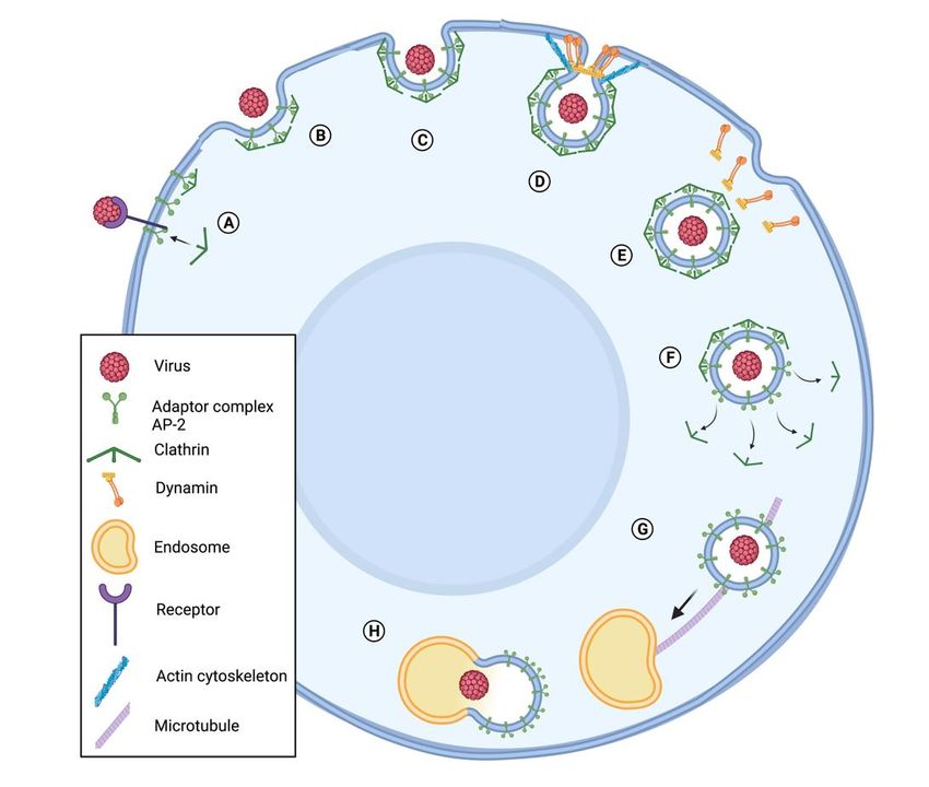
Viruses 2021, 13, 1658                                                                                             2 of 14

                         hosts. The same is true for processes allowing viral particles to enter the nucleus and
                         egress toward further target tissues. Herein, we summarize the current knowledge on viral
                         manipulation of the cytoskeleton of insect cells and highlight the need for further studies.

                         2. The Cytoskeleton in the Context of Animal–Virus Interactions
                         2.1. The Actin Cytoskeleton
                               The actin cytoskeleton is mainly composed of globular actin (G-actin), which is
                         monomeric actin able to self-assemble into filamentous actin (F-actin). The formation
                         of new actin filaments requires different actin regulators, one of which is the actin-related
                         protein 2/3 (Arp2/3) complex [5]. Changes in Arp2/3 affect the overall organization of the
                         cytoskeleton network [5]. The filamentous network formed by F-actin and actin-binding
                         proteins is the primary component of cytoskeleton microfilaments [6]. The actin cytoskele-
                         ton is subjected to alterations and organizations to promote cellular dynamic and particle
                         transport within and between cells. Eukaryotic cells polymerize actin filaments to provide
                         mechanical integrity and motility force for a wide range of cellular mechanisms [7].
                               Most if not all animal viruses studied so far manipulate the cellular actin from the
                         first steps of their infection cycle. The binding of virions to specific receptors and entry into
                         host cells is mediated by the cortical network of actin filaments, which are involved in the
                         internalization of the virions localized at the surface of the host cells [8–10]. The dynamic
                         polymerization of actin reshapes cellular membranes allowing viral internalization and
                         has a role in the formation and movement of endocytic vesicles in case of entry through
                         endocytosis mechanisms [11]. Host actin is also manipulated by viruses during the intra-
                         cellular movement of the viral genome and proteins, and the delivery of the viral particles
                         to the corresponding replication sites [12,13]. Finally, the actin cytoskeleton is required for
                         viral egress and dissemination of the virus to other host tissues [14–16].

                         2.2. Microtubule Cytoskeleton
                               Microtubules are formed through α-/β-tubulin heterodimers assembling into cylin-
                         dric filaments. The plus-ends of these filaments grow pointing towards the plasma mem-
                         brane into protrusions, while their minus ends are anchored at microtubule-organizing
                         centers (MTOCs) such as the centrosome [17,18]. This polarity allows selective directional
                         long-range cargo transport at the cell periphery [18].
                               The microtubule network is often used by viruses in the first steps of attachment
                         into specific sites of host cell receptors. The spatial reorganization of the receptors and
                         viral entry is in part controlled by this microtubule organization at the cell surface [19–21].
                         One of the most recognized and characterized viral internalization mechanisms is clathrin-
                         mediated endocytosis. Clathrin is a self-assembling protein that coats transport vesicles
                         by polymerizing into polyhedral lattice. These formations are called coating pits and
                         contribute to the organization and sorting of integral membrane proteins during receptor-
                         mediated endocytosis and organelle biogenesis and is involved in several cellular pathways
                         such as cargo transport into the cells, protein secretion from the trans-Golgi network, and
                         signal transduction [22–24]. Following viral particles’ attachment to the host cell surface,
                         these are delivered to pre-formed clathrin-coated pits (Figure 1). Then, these pits pinch
                         off from the plasma membrane through a mechanism controlled by cytoskeleton-specific
                         proteins, including actin [24] (Figure 1). Following internalization, clathrin pits gradually
                         disassemble, delivering viral particles to endosomes, which travel across the cytoplasm
                         through microtubule-related mechanisms (Figure 1). The trafficking of coated vesicles to
                         early endosomes, late endosomes, and lysosomes requires microtubules arrangement [25].
                         Following the endosomes escape, viruses that replicate in the nucleus usually manipulate
                         microtubule dynamics to facilitate the transit into the nucleus [25]. Microtubule-based
                         motor proteins, such as kinesin and dynein, transfer the cargoes along microtubules in
                         cells [26]. Dynein forms a complex with dynactin (actin-related filamentous protein) fa-
                         cilitating the cargo trafficking toward the cell center near the nucleus. Virions’ progeny
                         travel toward microtubule plus ends, from near the nucleus to the peripheral regions of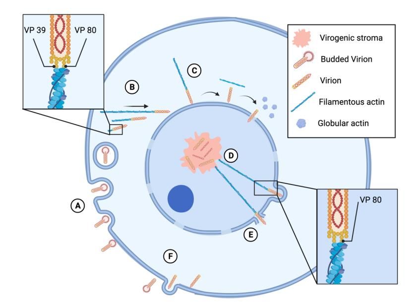
cells [26]. Dynein forms a complex with dynactin (actin-related filamentous protein
Viruses 2021, 13, 1658 itating the cargo trafficking toward the cell center near the nucleus. Virions’ 3 of 14 pr
travel toward microtubule plus ends, from near the nucleus to the peripheral regi
the cell, through kinetin-based microtubule [26,27]. Virions require actin and myos
shortthe cell, through kinetin-based microtubule [26,27]. Virions require actin and myosin for
transportation, while microtubules and dynein mediate the long-range trans
short transportation, while microtubules and dynein mediate the long-range transporta-
tion [10,26]. RNA
tion [10,26]. RNA viruses usedifferent
viruses use different cytoplasmic
cytoplasmic replication
replication structuresstructures and reorg
and reorganize
microtubules
microtubules to to
enhance theformation
enhance the formation of replication
of replication compartments
compartments [17]. [17].
.
Figure 1. Viral internalization in host cell through clathrin-mediated endocytosis. The virus binds
to a receptor, initiating a series of steps that lead to endocytosis. (A) The adaptor protein complex
FigureAP2
1. links
Viralclathrin
internalization in host
to the membrane, cellother
recruits through clathrin-mediated
accessory endocytosis.
proteins important for The virus
the endocytosis,
to a receptor, initiating a series of steps that lead to endocytosis. (A) The adaptor
and assembles a clathrin coat. (B) The clathrin coat grows, leading to invaginations in the membrane protein co
AP2 links
(C) andclathrin to the
later to the membrane,
formation recruits
of a vesicle. (D) Theother accessory
vesicle is pinchedproteins important
off by a mechanism for the endoc
mediated
by dynamin and the actin cytoskeleton. (E) After the vesicle detaches from the
and assembles a clathrin coat. (B) The clathrin coat grows, leading to invaginations in the mem cell membrane,
(F) later
(C) and it losesto
the clathrin
the coat (G)of
formation and
a travels
vesicle. to an
(D)endosome with the
The vesicle help of microtubules.
is pinched (H) The
off by a mechanism me
vesicle binds to an endosome and starts endosome maturation. Adapted from “Clathrin-Mediated
by dynamin and the actin cytoskeleton. (E) After the vesicle detaches from the cell membran
Endocytosis”, by BioRender.com (2021). The template of this figure was retrieved from https:
loses the clathrin coat (G) and travels to an endosome with the help of microtubules. (H) The
//app.biorender.com/biorender-templates (Publication License Agreement Number CN22UJ223Y).
binds to an endosome and starts endosome maturation. Adapted from “Clathrin-Mediated
3. Baculoviruses Hijack the Host Cells Cytoskeleton Actin to Replicate and Spread
cytosis”,
the by BioRender.com (2021). The template of this figure was retrieved from https://ap
Infection
render.com/biorender-templates (Publication License
Baculoviruses (Baculoviridae, double-stranded Agreement
(ds)DNA) constitute aNumber CN22UJ223Y)
unique group of
viruses that is specific to arthropods, mainly insects. Baculoviruses are the most studied
3. Baculoviruses
EPVs that causeHijack
epizooticthe HostinCells
diseases nature.Cytoskeleton
Baculoviruses areActin to Replicate
also commercialized as and
bio- Sprea
logical pesticides to control major forest and agricultural pests. They are enveloped viruses
Infection
with a circular, double-stranded DNA genome wrapped in rod-shaped nucleocapsids.
Baculoviruses
Baculoviruses exist(Baculoviridae, double-stranded
in two forms: occlusion-derived (ds)DNA)
viruses (ODVs) andconstitute a unique
budded viruses
(BVs). ODVs contain one or several nucleocapsids and are enveloped in an occlusion
of viruses that is specific to arthropods, mainly insects. Baculoviruses are the most st body
(OB) resistant in the environment. Once the insect ingests a baculovirus-contaminated
EPVsfoodthatand
cause epizootic diseases in nature. Baculoviruses are also commercializ
OBs reach the midgut, the alkaline pH dissolves the envelope. Released ODVs
biological pesticides to control major forest and agricultural pests. They are enve
viruses with a circular, double-stranded DNA genome wrapped in rod-shaped nucle
sids. Baculoviruses exist in two forms: occlusion-derived viruses (ODVs) and budd
ruses (BVs). ODVs contain one or several nucleocapsids and are enveloped in an occViruses 2021, 13, 1658 4 of 14
bind and fuse to the epithelial cells, releasing the nucleocapsids into the gut cells where
they replicate in the nucleus. The viral progeny bud out of the cells, forming BVs that are
single enveloped nucleocapsids. BVs spread the infection to adjacent tissues and therefore
are responsible for secondary infections. In the final stages of the infection cycle, the viral
nucleocapsids progeny gets occluded in a protein matrix to form OBs that are released in
the environment upon the death of the insect [28]. The expression of baculovirus proteins is
regulated throughout the replication cycle, and three groups of genes can be observed: early
genes, which are transcribed by the host RNA polymerase II early on in the infection [29],
late genes, and very late genes that are transcribed by the baculovirus RNA polymerase at
the late steps of the replication cycle [30]. The transition from early to late gene expression
is marked by the beginning of viral replication around 6 h post infection (hpi) [31].
Baculoviruses manipulate host cell properties, taking control of the host cellular
machinery throughout their infection cycle. Unlike other insect viruses, the impact of
baculovirus infection on the host cell cytoskeleton has been well characterized. Cytoskele-
ton re-arrangements happen in different steps of the infection and are crucial for bac-
ulovirus proper assembly and the encapsulation of newly produced virions [32]. These
interactions were mainly characterized for the baculovirus model Autographa californica
multiple nucleopolyhedrovirus (AcMNPV) infection in cell lines of the lepidopteran pests
Spodoptera frugiperda and Trichoplusia ni.
3.1. Baculovirus Entry and Transport through the Cytoplasm Depend on Re-Arrangement of Host
Cell Cytoskeleton
Baculovirus nucleocapsids enter their host cells through clathrin-dependent endo-
cytosis (Figure 1), although in some cases, direct fusion of virions with the host plasma
membrane has been described [33,34]. The first cytoskeleton arrangement induced by
baculovirus infection happens as soon as the viral particles enter the host cell. Upon
internalization of AcMNPV, viral nucleocapsids are released from the endosomes into the
cytoplasm (Figure 2). A few minutes post infection, filamentous actin cables form as a result
of the interaction between viral nucleocapsid proteins and host actin. The nucleocapsid
proteins VP39 and VP80, which have been identified in several baculoviruses such as AcM-
NPV, Helicoverpa armigera NPV (HaNPV), and Bombyx mori NPV (BmNPV), bind directly to
actin and promote F-actin polymerization [13,35,36] (Figure 2). The sequence of VP39 from
55 currently available baculovirus genomes was analyzed, leading to the identification of a
conserved glycine at position 276. Mutation of this Gly276 resulted in alterations of correct
nucleocapsid assembly, DNA packaging, and expression of late genes [37]. These results
altogether suggest that Gly276 is primordial for VP39 function and may be indispensable for
VP39 interaction with actin and therefore nucleocapsid movement toward the replication
sites of the virus. In AcMNPV-infected High Five cells (obtained from ovaries of T. ni),
viral nucleocapsids moved within the cytoplasm and were followed by actin comet tails at
very early time of infection (5–30 min post infection) (Figure 2). This movement was actin-
assembly dependent, since it was inhibited by an inhibitor of actin polymerization. The
nucleocapsid–actin interaction is promoted by the interaction of the nucleocapsid promotor
factor P78/83 and the Arp2/3 complex, as the inhibition of Arp2/3 decreased the velocity
of the viral movements and blocked the nuclear import of nucleocapsids [13,38]. Therefore,
P78/83 acts as a nucleation promotor factor (NPF) inducing actin polymerization. The
C-terminal part of P78/83 is conserved with other NPFs; meanwhile, the N-terminal part
contains a multifunctional regulatory sequence (MRS) and is unique to the baculovirus
P78/83 protein family. P78/83 stability is modulated by the MRS that prevents the host
cell proteolytic machinery from interacting with and degrading P78/83 [12].
Immediately following early gene expression, actin is re-arranged, resulting in actin
filaments aggregating at the plasma membrane. Actin cables localize at the cell surface
and transport the released nucleocapsids through the cytoplasm toward the nucleus.
This arrangement is a result of the interaction of viral actin re-arrangement-inducing
factor 1 (arif-1) with F-actin aggregates [39]. Arif-1 is expressed at the early stage of
baculovirus infection and co-localizes with F-actin at the plasma membrane until lateViruses 2021, 13, 1658 5 of 14
gene expression. It has been suggested that ARIF-1 induces the formation of invadosome-
like structures, leading to the formation of organized structures to enable systemic viral
spread [40]. The fast and early translocation of the nucleocapsids to the nucleus is critical
for baculovirus replication.
3.2. Baculovirus Entry into the Nucleus and Viral Progeny Egression Are Mediated by
Actin Re-Arrangements
Baculoviruses rapidly migrate to the nucleus within 1 hpi. Nucleocapsids collide
and are stuck with the nuclear envelope (Figure 2). AcMNPV nucleocapsids in Hi5 cells
remained docked at the nuclear periphery for 31 min after collision; then, they separated
from actin and entered the nucleus through the nuclear pores [13]. Entry of nucleocapsids
into the nucleus ended the actin re-arrangements on the nuclear periphery (Figure 2). When
nucleocapsids cross the nuclear membrane, P78/83 MRS most probably gets exposed to
the cellular proteolytic machinery, allowing the degradation of P78/83. Consequently,
the induction of F-actin polymerization at the nuclear periphery gets blocked, and those
filaments depolymerize [12]. Following entry into the nucleus, viral particles move to the
sites of uncoating and gene expression [13]. In the AcMNPV-infected S. frugiperda cell line
SF21, nuclear actin-based motility inside the nucleus had started by 12 ± 2 hpi and had
been enhanced by 16 ± 3 hpi. This period coincides with the start of development of viral
replication centers, which are called virogenic stroma [14].
Globular actin starts to accumulate in the nucleus since early viral gene expression,
while F-actin starts to polymerase with the expression of late viral genes. AcMNPV nucleo-
capsid protein VP80 in infected cells associates with the nuclear induced F-actin, forming a
three-dimensional network that connects the replication factories with the viral nucleocap-
sids [35]. At 12 hpi, VP80 spreads through the nucleus and then starts to co-localize with
DNA-containing areas in the virogenic stroma connecting the central area of the nucleus
and the nuclear periphery by 24 hpi. These observations suggest that VP80 is primordial for
progeny virions egress from the nucleus [35] (Figure 2). The sequences of both AcMNPV
and BmNPV VP80 proteins were characterized and compared to those of available protein
sequences in several databases [35]. A conserved domain encompassing GLy164 –Glu368
was identified as homologous to the insect paramyosin sequence Glu168 –Glu380 , which was
identified in B. mori and Aedes aegypti. In addition, a second conserved motif encompassing
Tyr575 –Phe597 was identified and aligned with the calmodulin-binding domain of eukary-
otic myosin-motor proteins, which was identified in several invertebrate and vertebrate
organisms such as Drosophila melanogaster and Arabidopsis thalina [35]. Paramyosin proteins
form filaments with myosin motor proteins and interact with actin-based filaments to
ensure filament-based motility. The presence in VP80 of a domain shared with paramyosin
suggests that VP80 is a paramyosin-like protein that mimics the host myosin-promotor
proteins, and it interacts with actomyosin filaments to facilitate active and directed nucleo-
capsid transport [35].
Following progeny nucleocapsids assembly, the nucleocapsids are transported to the
nuclear periphery becoming BVs, which bud through the cytoplasmic membrane (Figure 2).
The nucleocapsids, which are retained in the nucleus at very late time post infection, get
enveloped, are occluded into the envelope protein polyhedra, and form ODVs [41]. Another
event of actin re-arrangement favors AcMNPV virions exit from the nucleus. Actin-driven
viral motility is necessary to enter protrusions of the nuclear envelope as well as disruption
of nuclear envelope integrity during egress [14]. This actin-dependent disruption of the
nuclear envelope during the egression of viral progeny is unique to baculovirus.ruses 2021, 13, 1658
Viruses 2021, 13, 1658 6 of 14
.
Figure 2. Baculovirus replication cycle and its interaction with actin. (A) Budded virions bind to
the cell membrane and are endocyted through a clathrin-dependent process. After internalization,
Figure 2. Baculovirus replication cycle and its interaction with actin. (A) Budded virio
the nucleocapsids are released from the endosomes into the cytosol. (B) The nucleocapsid proteins
cell membrane
VP39 anddirectly
and VP80 bind are endocyted
to actin and through a clathrin-dependent
promote actin process.
polymerization. The viruses After intern
are moved
nucleocapsids
to the nucleus viaareanreleased from the endosomes
actin assembly-dependent mechanism,into the cytosol.
where (B)
they collide The
and nucleocapsid
stick to the
and VP80
nuclear bind directly
envelope. tocollision,
(C) After the actin and promote
the virions detachactin
from polymerization. The
the actin filaments, the viruses are
filaments
depolymerize into globular actin, and the virions enter the nucleus through
nucleus via an actin assembly-dependent mechanism, where they collide and stick nuclear pores. Inside
the nucleus, the virions move to the virogenic stroma. (D) After progeny virions are formed, they
envelope. (C) After the collision, the virions detach from the actin filaments, the filam
are transported to the nuclear periphery by filamentous actin as a result of actin binding to VP80.
merize into globular actin, and the virions enter the nucleus through nuclear pores.
(E) During the egress, baculovirus virions form protrusions in the nuclear envelope and disrupt it.
cleus, the the
(F) Finally, virions
virionsmove to the through
are transported virogenic stroma.and
the cytoplasm (D)become
Afterbudded
progeny virions
virions throughare form
transported
budding of the tocytoplasmic
the nuclear periphery
membrane. The by filamentous
figure was designedactin
using as a result(Publication
BioRender of actin bindin
During
License the egress,
Agreement baculovirus
Number virions form protrusions in the nuclear envelope and
BR22U9HSSA).
Finally, the virions are transported through the cytoplasm and become budded vi
3.3. Baculovirus Interactions with Microtubules
budding of the cytoplasmic membrane. The figure was designed using BioRender (P
While AcMNPV harnesses actin arrangements throughout all steps of the replication
cense
cycle,Agreement Number BR22U9HSSA).
its effect on microtubules have not been completely unraveled yet. AcMNPV infects
host cells via clathrin-mediated endocytosis, releasing nucleocapsids from early endosomes
3.3.
in aBaculovirus Interactions
microtubule-dependent with
manner [9].Microtubules
Additionally, baculoviruses express a nucleocapsid protein, EXON0, that co-localizes
withWhile
nuclearAcMNPV
microtubules, harnesses
suggesting actin arrangements
a possible throughout
interaction between microtubulesall steps
and of th
cycle, its effect on microtubules have not been completely unraveledSf9
nucleocapsids at late time post infection. Treating baculovirus-infected S. frugiperda yet. AcM
cells with microtubule inhibitors caused an 85% reduction in BV yield, suggesting that
host cells via clathrin-mediated endocytosis, releasing nucleocapsids from
microtubules are essential for BV egression from the nucleus [42]. BV yield reduction is
somes in a microtubule-dependent
time-dependent and relies on the nucleocapsid manner
form during[9]. trafficking [43]. P10 is another
Additionally,
baculovirus baculoviruses
protein that was suggested to express a nucleocapsid
interact with protein,
host cell microtubules. EXON0, tha
In AcMNPV-
infected Sf9 cells, P10 formed fibrous-like structures that extend from the cytoplasm to the
with nuclear microtubules, suggesting a possible interaction between micro
nucleus as a result of interaction with cellular microtubules. P10 fibrous-like structures
nucleocapsids at late time
result from the aggregation of P10post infection.
monomers Treating
that associates baculovirus-infected
with the microtubules subunit S. fr
cells with microtubule inhibitors caused an 85% reduction in BV yield, sug
α-tubilin, forming stronger microtubule bonds [44]. At a late infection time, these structures
fragmented in association with plasma membrane disruption and cellular lysis [45,46].
microtubules are essential for BV egression from the nucleus [42]. BV yield
time-dependent and relies on the nucleocapsid form during trafficking [43]. P
baculovirus protein that was suggested to interact with host cell microtub
MNPV-infected Sf9 cells, P10 formed fibrous-like structures that extend froViruses 2021, 13, 1658 7 of 14
4. Examples of Viruses Other Than Baculoviruses That Manipulate Host Cytoskeleton
for Successful Transmission
4.1. Plant Viruses Transmitted by Insect Vectors
Most plant pathogenic viruses rely on vectors, including insects, for survival and
transmission [47]. Viral acquisition, retention, and inoculation periods greatly vary, influ-
encing virus–host interactions [48,49]. Some plant viruses, such as the Cucumber mosaic
virus (Bromoviridae, positive-sense single-stranded (ss)RNA) and the Lettuce infectious
yellows virus (Closteroviridae, positive-sense ssRNA) vectored by the aphid Myzus persicae
and the whitefly Bemicia tabaci, respectively, are only retained by the insect vector on the
surface of their feeding apparatus and are transmitted to the next plant [50,51]. Mean-
while, other viruses, such as the Rice dwarf virus (RDV, Reoviridae, dsRNA) and the Rice
black-streaked dwarf virus (Reoviridae) transmitted by the leafhopper Nephotettix cincticeps
and the planthopper Sogatella furcifera, respectively, replicate and disseminate in the host
establishing a persistent infection before being transmitted to a new host [52,53]. Persis-
tent plant viruses have complex relationships with their host: they circulate sequentially
through the insect stylet, esophagus, digestive tract, and gut epithelial cells to reach the
hemolymph and circulate toward the salivary glands and ovaries [49]. Throughout this
process, plant viruses exploit the host cellular machinery to propagate, move, and com-
plete their life cycle. Breaching through the different physical barriers of the insect vector
(i.e., gut, ovaries, salivary glands) is shown to be dependent on re-arrangements of the
host cytoskeleton [54,55]. Re-arrangement of the host cytoskeleton has been mostly studied
for reoviruses, which are non-enveloped segmented double-strand RNA viruses that are
vectored by leafhoppers or planthoppers (Hemiptera: Auchenorrhyncha) [56].
4.1.1. Plant Virus Entry, Transmission, and Breaching through the Host Intestinal Barrier Is
Mediated by Viral Interaction with the Host Cytoskeleton
RDVs penetrate the vector N. cincticeps cells via clathrin-mediated endocytosis [57]
(Figure 3). The early endosomes directly move to the plasma membrane through the tubular
network and release viral particles into the cytoplasm [57,58]. Microscopical observations of
RDV motility after internalization showed that the non-structural viral protein Pns10 forms
tubule structures through interaction with myosin-motor proteins [59]. The Pns10 tubules
directly interact with actin and actin-binding proteins such as myosin, tropomodulin,
and vitellogenin [15]. As a result, actin filaments are recruited to enclose tubules of
approximately 85 nm in diameter. These tubules protrude from the surface of leafhopper
cells and are surrounded by an extended plasma membrane. The virion containing tubules
pass through the microvilli of the intestine into the lumen where they associated with
actin-based filopodia, and this facilitates viral spread to adjacent tissues [59,60] (Figure 3).
Moreover, the inhibition of actin filaments abrogated the extension of Pns10 tubules from
the surface of RDV in infected N. cincticeps cells. This negatively affected the intercellular
spread of RDV, confirming the importance of actin protrusions in the efficient spread of
Pns10 tubules from the surface of infected cells to non-infected neighboring cells [60].
The interaction between Pns10 and actin is specific to the natural vector N. cincticeps and
involves a putative α-helical transmembrane domain encompassing amino acids 98 to
119 of Pns10. If this domain is deleted, it impairs the protrusion of Pns10 tubules from
infected cells, thus preventing penetration to neighboring cells [59,60]. There was no
interaction between Pns10 and actin of Recilia dorsalis, a leafhopper from the same family
that is inefficient in transmitting RDV [61]. In a natural host, transmission of RDV from the
microvilli of epithelial cells to the visceral muscle tissues of the digestive canal is a result
of RDV manipulation of actin-based cellular protrusions (Figure 3). The actin-associated
protein tropomodulin positively controls the transmission of RDV tubules by regulating the
length and stability of the actin-tropomyosin filament [15]. These data altogether suggest
actin-based cellular machinery and actin-associated proteins enable RDV efficient spread
from cell to cell and from the gut to adjacent tissues.Viruses 2021, 13, 1658 8 of 14
Similar viral spread mechanisms have been described for other plant viruses. The
rice gall dwarf virus (RGDV: Reoviridae), which is transmitted by the leafhopper R. dorsalis,
forms viral tubules from the non-structural protein Pns11. In host cells, persistent cell-
to-cell spread of RGDV occurs through the trafficking of viral tubules along actin-based
cellular protrusions of infected cells to neighboring uninfected cells [62]. The southern
rice black-streaked dwarf virus (SRBSDV: Reoviridae), which is transmitted by the white-
backed planthopper S. furcifera, also encodes a non-structural protein, P7-1, which arranges
virus-containing tubules. The self-interaction of P7-1 monomers leads to the formation of
homodimers or oligomers [63], resulting in the formation of the helical structure of viral
tubules. These tubules travel along the basal lamina of the intestinal epithelium and the
intestine circular muscle fibers by interacting with the actin-based cellular protrusions. As
a result, SRBSDV disseminates across the epithelium toward the gut visceral muscle [64].
The beet western yellow virus (BWYV: Solemoviridae, positive-sense ssRNA), which is
transmitted by aphids, can bind to different host proteins, including actin. BWYV crosses
the aphid intestine, hemocoel, and the salivary glands through transcytosis mediated by
interaction of the virus with cytoskeleton elements [16].
A different mechanism of interaction with the host cytoskeleton was described for the
rice stripe virus (RSV: Phenuiviridae, negative-sense ssRNA), which is transmitted by the
small brown planthopper Laodelphax striatellus. RSV forms filamentous ribonucleoprotein
particles (RNPs). RNPs consist of viral nucleoprotein, RNAs, and RNA-dependent RNA
polymerase, and they associate with the non-structural protein NS4 to form inclusion
bodies. These inclusion bodies interact with the microvilli of the host midgut epithelium,
which are formed by actin, resulting in viral entry and propagation to midgut visceral
muscles, alimentary tract, salivary glands, and reproductive system [65].
The rice yellow stunt virus (RYSV: Rhabdoviridae, negative-sense ssRNA), which is
persistently transmitted by the leafhopper N. cincticeps, invades host nervous system and
exploits axonal transport to disseminate throughout the leafhopper body. The neuron
cytoskeleton structures consist of microtubules, actin filaments, and neuro-filaments. The
interaction of RYSV with microtubule-based neurofilaments leads to the rapid delivery of
virions along the axons, resulting in efficient viral dissemination [66].
4.1.2. Horizontal Transmission of Plant Viruses through the Salivary Glands
Salivary glands are the ultimate transmission barrier for plant viruses before their
transmission to a new host. RDV deficient in Pns10 failed to be transmitted to the plant sec-
ondary host by the vector N. cincticeps [67]. The RGDV tubules composed of Pns11 proteins
interact directly with the actin-based apical plasmalemma of the salivary glands of R. dor-
salis and release into the salivary cavities in an exocytosis-like process [68] (Figure 3). These
results suggest that the interaction of viral non-structural proteins with the insect actin is
primordial to breach through the salivary glands and be transmitted to a secondary host.
4.1.3. Vertical Transmission of Plant Viruses
Vertical or transovarial transmission helps viral spread and persistence within vector
populations. Vertical transmission requires that viral particles pass through the ovary
follicular cells and enter the host oocyte [69]. The plasma membrane of follicular cells is
connected with the oocyte through actin-based microvilli [70]. Some plant viruses use these
actin-based junctions of the follicular cells to enter the oocyte. For instance, RGDV can
enter oocytes of the R. dorsalis leafhopper through the actin-based microvilli of follicular
cells as well as the actin-based junctions between the cells. Upon crossing the junctions
between follicular cells, virions spread and propagate in the cytoplasm of the oocyte [71]
(Figure 3).
Transovarial transmission of plant viruses can also be mediated by vitellogenin
(Figure 3). Vitellogenin is synthesized in the fat body and transported into the grow-
ing oocytes through receptor-mediated endocytosis [72]. In the ovaries of the small brown
planthopper, RSV expresses a nucleocapsid protein that binds to the vitellogenin. TheViruses 2021, 13, 1658
Viruses 2021, 13, 1658
planthopper, RSV expresses a nucleocapsid protein that binds to9the of 14
vitello
tellogenin-nucleocapsid complex moves along the follicular cells throug
junctions to enter the oocyte [72]. In addition, Pns10 and Pns11, which are n
vitellogenin-nucleocapsid complex moves along the follicular cells through actin-based
proteins
junctions toof RSV
enter theand RGDV,
oocyte respectively,
[72]. In addition, were
Pns10 and found
Pns11, which to
areinteract with vitel
non-structural
ovaries ofRSV
proteins of theand
respective vectors.were
RGDV, respectively, These interactions
found arevitellogenin
to interact with involvedininthethe vert
ovaries of the respective vectors. These interactions are involved in the vertical trans-
sion of RSV and RGDV to the host progeny via receptor-mediated endocy
mission of RSV and RGDV to the host progeny via receptor-mediated endocytosis into
cytes
oocytescytoplasm [69,71].
cytoplasm [69,71].
.
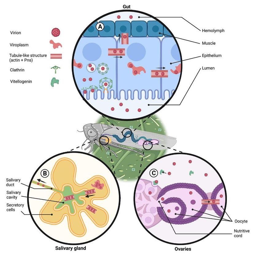
Figure 3. Reovirus infection cycle in the insect vector. Virions should breach three major tissue
Figure
barriers: 3. Reovirus
insect infection
gut for their cyclethroughout
dissemination in the insect vector.
the insect bodyVirions should
(A), salivary glandsbreach
for their three m
riers: insect
horizontal gut for (B),
transmission theiranddissemination throughout
ovaries for their vertical the insect
transmission (C). (A) body
Gut. The(A),virus
salivary g
enters the guttransmission
horizontal epithelial cells via
(B),clathrin-mediated
and ovaries for endocytosis. Insidetransmission
their vertical the cell, the virus forms
(C). (A) Gut. T
viroplasms, which are subcellular structures where viral accumulation, the synthesis of viral proteins,
the gut epithelial cells via clathrin-mediated endocytosis. Inside the cell, the virus for
and replication take place. One of the viral non-structural proteins (Pns) interacts with the insect actin
which are subcellular structures where viral accumulation, the synthesis of viral pro
and forms tubule-like structures, which facilitate the infection of neighboring cells and eventual viral
lication
release to take place. One
the hemolymph andof the
viral viral non-structural
dissemination. proteins
(B) Salivary glands. (Pns) interacts
Tubule-like with the i
structures are
forms tubule-like structures, which facilitate the infection of neighboring
formed by the interaction of actin and Pns in infected secretory cells. These structures are used by the cells and
release to the
virus to egress hemolymph
from andand
the secretory cells viral
reachdissemination. (B)
the salivary cavities. Salivary
Virions presentglands. Tubule-lik
in the cavities
are excreted when the insect feeds. (C) Ovaries. Virions bind to vitellogenin
formed by the interaction of actin and Pns in infected secretory cells. These in the hemolymph and structu
enter the ovaries through vitellogenin-receptor mediated endocytosis. The virus can spread to the
the virus to egress from the secretory cells and reach the salivary cavities. Virions
cytosol of the oocytes though the nutritive cord or through the tubule like structures, which are
cavities are excreted when the insect feeds. (C) Ovaries. Virions bind to vitellogen
formed by interactions of viral proteins and actin that cross the junctions between follicular cells. The
lymph
figure wasand enterusing
designed theBioRender
ovaries (Publication
through vitellogenin-receptor
License Agreement Number mediatedTH22QJKFSB). endocytosis
spread to the cytosol of the oocytes though the nutritive cord or through the tubule
which are formed by interactions of viral proteins and actin that cross the junctions
ular cells. The figure was designed using BioRender (Publication License Agree
TH22QJKFSB).
4.2. ArbovirusesViruses 2021, 13, 1658 10 of 14
4.2. Arboviruses
Arboviruses represent an increasing threat to global veterinary and human health.
Mosquitoes of the Aedes genus are the primary worldwide vectors of epidemic viruses such
as Dengue (DENV: Flaviviridae, positive-sense ssRNA), Chikungunya (CHIKV: Togaviridae,
positive-sense ssRNA), Zika (ZIKV: Flaviviridae) and yellow fever (YFV: Flaviviridae) viruses.
Following a blood meal from an infected human, arboviruses replicate in the female midgut
and further disseminate to reach the salivary glands. Once infection has been established
in the salivary glands, it lasts throughout the mosquito lifespan, and arboviruses can be
transmitted to subsequent hosts [73]. Seldomly, arboviruses can reach female ovaries and
be vertically transmitted [74].
At present, most if not all our knowledge on arboviral interaction with the cytoskeleton
of mosquito cells comes from descriptive transcriptomic and proteomic studies regard-
ing the Aedes albopictus cell line C6/36 or Aedes aegypti females infected with either the
alphavirus chikungunya virus (CHIKV; positive sense single-strand RNA virus) or fla-
viviruses (positive sense single-strand RNA viruses). These viruses share the fact that they
enter host cells by a clathrin-dependent endocytosis pathway [75–77].
4.2.1. Examples of Flavivirus Interaction with Insect Cytoskeleton
Dengue virus-labeled particles were detected within early endosomes 5 min post
infection (mpi) and near lysosomes 0.5 hpi in C6/36 cells [75]. The disruption of host
microtubule and microfilaments reduced viral infection by more than 80%, suggesting an
implication of the cell cytoskeleton in the early steps of viral entry. In addition, 10 mpi with
DENV, the co-localization of small groups of viral particles with host actin was detected,
suggesting that actin filaments may favor movement of the viral particles [75]. At 48 hpi,
transcription of actin and tubulin genes decreases to increase again at 96 hpi, suggesting
a viral-mediated host shutoff effect followed by the activation of mechanisms that main-
tain homeostasis and reorganization of cytoskeleton during virus infection [78]. Further
evidence on DENV manipulation of the host cell cytoskeleton comes from proteomic and
transcriptomic analyses of different developmental stages of the mosquito Ae. aegypti.
A total of 49 Ae. aegypti proteins were identified that interact with the DENV NS5 pro-
tein [79]. The highest enriched domains among these interacting proteins were found to be
myosin-related [79]. In addition, several transcriptomic studies have shown that flavivirus
infections alter the expression of genes in the cytoskeleton network such as actin, tubulin,
and microtubule-associated proteins [77,80,81].
Similarly, west Nile virus (WNV) entry into C6/36 cells is drastically affected by chemical
drugs that inhibit clathrin-mediated endocytosis or disrupt the microtubule network [77].
Additionally, the WNV non-structural protein NS2A and the envelope protein E were found
to bind to cellular beta-tubulins, which build a network of microtubules [77]. However, the
disruption of actin filaments did not affect the WNV entry into C6/36 cells [76].
4.2.2. Examples of CHIKV Interaction with Insect Cytoskeleton
Proteomic analyses showed that the cytoskeleton-associated protein, syntenin, is
induced as soon as 30 min after CHIKV infection in Ae. aegypti gut [82]. Syntenin is a
membrane binding protein that regulates the architecture of the cell membrane, cellular
trafficking, and is involved in actin polymerization [83,84]. In counterpart, actin and
twinfilin, an actin-binding protein that regulates cytoskeleton dynamics, were repressed
30 mpi. However, the F-actin capping protein beta subunit, an actin-binding protein that
regulates actin polymerization, was induced 24 hpi [82]. These results suggest a regu-
lation of the actin polymerization dynamics at early time post CHIKV infection (0.5 to
24 hpi). Simultaneously with these observations, vesicle trafficking to the cellular vac-
uole was induced 0.5 hpi and was attenuated 24 hpi. Additionally, 24 hpi, endocytosis,
vesicle formation and intracellular trafficking were reduced [82]. These results support
the hypothesis that CHIKV manipulates the Ae. aegypti cytoskeleton by regulating actin
polymerization/depolymerization. In addition to the interruption of endocytosis andViruses 2021, 13, 1658 11 of 14
cellular trafficking mechanisms after 24 hpi, tubulin and cytoskeleton motor proteins such
as myosin were significantly downregulated in the mosquito gut [82]. This indicates a
host response to restrain further de novo infection once the virus is spread throughout the
mosquito tissues.
5. Conclusions
The currently available data on the manipulation of host cytoskeleton by viruses
that infect insects suggest a primordial role of actin and microtubule cytoskeletons in
viral internalization, replication, and dissemination. Common traits of cytoskeleton ma-
nipulation by insect viruses described in this review include the entry of viral particles
through clathrin-mediated endocytosis, the direct interaction of viral proteins with actin,
and possibly regulation of the expression of genes coding for components of the host
cytoskeleton. Although the precise mechanisms of the interaction between insect viruses
and host cytoskeleton are not well understood, current data suggest similar mechanisms
as used by animal viruses. At least in the case of baculoviruses and plant reoviruses
transmitted by insects, successful viral motility and dissemination depend on interactions
of viral proteins with cytoskeleton proteins. Interestingly, amino acids and protein motifs
intervening in the interaction with host cytoskeleton were identified, in primis, in the
baculovirus protein family VP80 that shares common domains with host myosin-promotor
proteins. Another common feature between these viruses is the formation during infection
of tubular structures that interact with the actomyosin complex to transport the viral genetic
material. In the case of arboviruses, several transcriptomic studies showed attenuation of
the expression of cytoskeleton-related genes at different points post infection. Even though
little is understood on whether and how insect viruses regulate the expression of host
cytoskeleton-related genes, it is plausible to suggest that tight regulation of gene expression
occurs simultaneously with the virus manipulation of actin and microtubule cytoskeletons.
A deeper understanding of the cellular and molecular mechanisms regulating cy-
toskeleton reorganization during viral infection of insect cells can have applicative out-
comes. Depending on the insect–virus system and the application aims, cytoskeleton
regulation might be enhanced or altered so that the viral infection is enhanced or blocked.
In the case of insect pests, the knowledge of how infecting viruses hijack host cytoskeleton
could be used to enhance viral pathogenesis toward more efficient biological pesticides
that can compete with and replace chemical pesticides. A better understanding of the
interaction between plant viruses and/or arboviruses and insect vector cytoskeleton may
enable interfering with viral acquisition, retention, and transmission by the insect vectors,
thus limiting or abrogating viral transmission to plants and/or animals/humans. As a
result of the primordial role of cytoskeleton in cell function, it would be ideal to not di-
rectly target the host cytoskeleton but rather viral proteins that interact with or manipulate
host cytoskeleton.
Author Contributions: Conceptualization L.G.; writing—original draft preparation, A.K., H.D.P.,
U.P., M.B., L.G.; writing—review and editing, A.K., H.D.P., U.P., M.B., L.G.; supervision, L.G., M.B.
All authors have read and agreed to the published version of the manuscript.
Funding: M.B. received funding from the Italian Ministry of Education, University and Research,
project R1623HZAH5 and a European Research Council Consolidator Grant under the European
Union’s Horizon 2020 Programme (Grant N. ERC-CoG 682394). L.G. was supported by a fellowship
within the Italian Ministry of Education, University and Research: Dipartimenti Eccellenza Program
(2018-2022) to the Dept. of Biology and Biotechnology “L. Spallanzani”, University of Pavia.
Acknowledgments: The authors acknowledge the online tool BioRender (BioRender.com) used to
create Figures 1–3.
Conflicts of Interest: The authors declare no conflict of interest. The funders had no role in the
writing or in the decision to publish the review.Viruses 2021, 13, 1658 12 of 14
References
1. Ploubidou, A.; Way, M. Viral transport and the cytoskeleton. Curr. Opin. Cell Biol. 2001, 13, 97–105. [CrossRef]
2. Wei, T.; Li, Y. Rice Reoviruses in Insect Vectors. Annu. Rev. Phytopathol. 2016, 54, 99–120. [CrossRef] [PubMed]
3. Harries, P.A.; Schoelz, J.E.; Nelson, R.S. Intracellular transport of viruses and their components: Utilizing the cytoskeleton and
membrane highways. Mol. Plant-Microbe Interact. 2010, 23, 1381–1393. [CrossRef]
4. Stidwill, R.P.; Greber, U.F. Lntracellular virus trafficking reveals physiological characteristics of the cytoskeleton. News Physiol.
Sci. 2000, 15, 67–71. [CrossRef] [PubMed]
5. Liman, J.; Bueno, C.; Eliaz, Y.; Schafer, N.P.; Waxham, M.N.; Wolynes, P.G.; Levine, H.; Cheung, M.S. The role of the Arp2/3
complex in shaping the dynamics and structures of branched actomyosin networks. Proc. Natl. Acad. Sci. USA 2020, 117,
10825–10831. [CrossRef] [PubMed]
6. Goley, E.D.; Ohkawa, T.; Mancuso, J.; Woodruff, J.B.; D’Alessio, J.A.; Cande, W.Z.; Volkman, L.E.; Welch, M.D. Dynamic nuclear
actin assembly by Arp2/3 complex and a baculovirus WASP-like protein. Science 2006, 314, 464–467. [CrossRef]
7. Pollard, T.D.; Cooper, J.A. Actin, a central player in cell shape and movement. Science 2009, 326, 1208–1212. [CrossRef]
8. Mercer, J.; Schelhaas, M.; Helenius, A. Virus entry by endocytosis. Annu. Rev. Biochem. 2010, 79, 803–833. [CrossRef]
9. Qin, F.; Xu, C.; Hu, J.; Lei, C.; Zheng, Z.; Peng, K.; Wang, H.; Sun, X. Dissecting the Cell Entry Pathway of Baculovirus by
Single-Particle Tracking and Quantitative Electron Microscopic Analysis. J. Virol. 2019, 93, 1–25. [CrossRef]
10. Taylor, M.P.; Koyuncu, O.O.; Enquist, L.W. Subversion of the actin cytoskeleton during viral infection. Nat. Rev. Microbiol. 2011,
9, 427–439. [CrossRef] [PubMed]
11. Kaksonen, M.; Toret, C.P.; Drubin, D.G. Harnessing actin dynamics for clathrin-mediated endocytosis. Nat. Rev. Mol. Cell Biol.
2006, 7, 404–414. [CrossRef]
12. Wang, Y.; Zhang, Y.; Han, S.; Hu, X.; Zhou, Y.; Mu, J.; Pei, R.; Wu, C.; Chen, X. Identification of a novel regulatory sequence of
actin nucleation promoting factor encoded by Autographa californica multiple nucleopolyhedrovirus. J. Biol. Chem. 2015, 290,
9533–9541. [CrossRef]
13. Ohkawa, T.; Volkman, L.E.; Welch, M.D. Actin-based motility drives baculovirus transit to the nucleus and cell surface. J. Cell Biol.
2010, 190, 187–195. [CrossRef] [PubMed]
14. Ohkawa, T.; Welch, M.D. Baculovirus Actin-Based Motility Drives Nuclear Envelope Disruption and Nuclear Egress. Curr. Biol.
2018, 28, 2153–2159.e4. [CrossRef] [PubMed]
15. Chen, Q.; Zhang, L.; Zhang, Y.; Mao, Q.; Wei, T. Tubules of plant reoviruses exploit tropomodulin to regulate actin-based tubule
motility in insect vector. Sci. Rep. 2017, 7, 1–12. [CrossRef]
16. Seddas, P.; Boissinot, S.; Strub, J.M.; Van Dorsselaer, A.; Van Regenmortel, M.H.V.; Pattus, F. Rack-1, GAPDH3, and actin: Proteins
of Myzus persicae potentially involved in the transcytosis of beet western yellows virus particles in the aphid. Virology 2004,
325, 399–412. [CrossRef]
17. Naghavi, M.H.; Walsh, D. Microtubule Regulation and Function during Virus Infection. J. Virol. 2017, 91. [CrossRef]
18. Walsh, D.; Naghavi, M.H. Exploitation of Cytoskeletal Networks during Early Viral Infection. Trends Microbiol. 2019, 27, 39–50.
[CrossRef]
19. Simpson, C.; Yamauchi, Y. Microtubules in influenza virus entry and egress. Viruses 2020, 12, 117. [CrossRef] [PubMed]
20. Meng, G.; Wei, X.; Wu, X.; Sellers, M.T.; Decker, J.M.; Moldoveanu, Z.; Orenstein, J.M.; Graham, M.F.; Kappes, J.C.; Mestecky,
J.; et al. Primary intestinal epithelial cells selectively transfer R5 HIV-1 to CCR5+ cells. Nat. Med. 2002, 8, 150–156. [CrossRef]
21. Marozin, S.; Prank, U.; Sodeik, B. Herpes simplex virus type 1 infection of polarized epithelial cells requires microtubules and
access to receptors present at cell-cell contact sites. J. Gen. Virol. 2004, 85, 775–786. [CrossRef]
22. Brodsky, F.M. Clathrin and Clathrin-Dependent Endocytosis. Encycl. Cell Biol. 2016, 2, 384–393. [CrossRef]
23. McMahon, H.T.; Boucrot, E. Molecular mechanism and physiological functions of clathrin-mediated endocytosis. Nat. Rev. Mol.
Cell Biol. 2011, 12, 517–533. [CrossRef] [PubMed]
24. Kaksonen, M.; Roux, A. Mechanisms of clathrin-mediated endocytosis. Nat. Rev. Mol. Cell Biol. 2018, 19, 313–326. [CrossRef]
[PubMed]
25. Liu, H.; Liu, Y.; Liu, S.; Pang, D.-W.; Xiao, G. Clathrin-Mediated Endocytosis in Living Host Cells Visualized through Quantum
Dot Labeling of Infectious Hematopoietic Necrosis Virus. J. Virol. 2011, 85, 6252–6262. [CrossRef] [PubMed]
26. Hsieh, M.J.; White, P.J.; Pouton, C.W. Interaction of viruses with host cell molecular motors. Curr. Opin. Biotechnol. 2010,
21, 633–639. [CrossRef] [PubMed]
27. Leopold, P.L.; Pfister, K.K. Viral strategies for intracellular trafficking: Motors and microtubules. Traffic 2006, 7, 516–523. [CrossRef]
[PubMed]
28. Ahmad, I.; Ahmad, F.; Pichtel, J. Microbes and Microbial Technology: Agricultural and Environmental Applications; Springer: New
York, NY, USA, 2011; Chapter 16.
29. Hoopes, R.R.; Rohrmann, G.F. In vitro transcription of baculovirus immediate early genes: Accurate mRNA initiation by nuclear
extracts from both insect and human cells. Proc. Natl. Acad. Sci. USA 1991, 88, 4513–4517. [CrossRef] [PubMed]
30. Glocker, B.; Hoopes, R.R.; Hodges, L.; Rohrmann, G.F. In vitro transcription from baculovirus late gene promoters: Accurate
mRNA initiation by nuclear extracts prepared from infected Spodoptera frugiperda cells. J. Virol. 1993, 67, 3771–3776. [CrossRef]
31. Huh, N.E.; Weaver, R.F. Identifying the RNA polymerases that synthesize specific transcripts of the Autographa californica
nuclear polyhedrosis virus. J. Gen. Virol. 1990, 71, 195–201. [CrossRef]Viruses 2021, 13, 1658 13 of 14
32. Gasmi, L.; Jakubowska, A.K.; Herrero, S. Gasmin (BV2-5), a polydnaviral-acquired gene in Spodoptera exigua. Trade-off in the
defense against bacterial and viral infections. Dev. Comp. Immunol. 2016, 56, 37–45. [CrossRef] [PubMed]
33. Qin, F.; Xu, C.; Lei, C.; Hu, J.; Sun, X. Autographa californica multiple nucleopolyhedrovirus enters host cells via clathrin-mediated
endocytosis and direct fusion with the plasma membrane. Viruses 2018, 10, 632. [CrossRef] [PubMed]
34. Dong, S.; Wang, M.; Qiu, Z.; Deng, F.; Vlak, J.M.; Hu, Z.; Wang, H. Autographa californica Multicapsid Nucleopolyhedrovirus
Efficiently Infects Sf9 Cells and Transduces Mammalian Cells via Direct Fusion with the Plasma Membrane at Low pH. J. Virol.
2010, 84, 5351–5359. [CrossRef]
35. Marek, M.; Merten, O.-W.; Galibert, L.; Vlak, J.M.; van Oers, M.M. Baculovirus VP80 Protein and the F-Actin Cytoskeleton Interact
and Connect the Viral Replication Factory with the Nuclear Periphery. J. Virol. 2011, 85, 5350–5362. [CrossRef] [PubMed]
36. Xu, H.; Yao, L.; Lu, S.; Qi, Y. Host filamentous actin is associated with Heliothis armigera single nucleopolyhedrosis virus
(HaSNPV) nucleocapsid transport to the host nucleus. Curr. Microbiol. 2007, 54, 199–206. [CrossRef]
37. Katsuma, S.; Kokusho, R. A Conserved Glycine Residue Is Required for Proper Functioning of a Baculovirus VP39 Protein. J. Virol.
2017, 91. [CrossRef] [PubMed]
38. Au, S.; Wu, W.; Zhou, L.; Theilmann, D.A.; Panté, N. A new mechanism for nuclear import by actin-based propulsion used by a
baculovirus nucleocapsid. J. Cell Sci. 2016, 129, 2905–2911. [CrossRef]
39. Dreschers, S.; Roncarati, R.; Knebel-Mörsdorf, D. Actin Rearrangement-Inducing Factor of Baculoviruses Is Tyrosine Phosphory-
lated and Colocalizes to F-Actin at the Plasma Membrane. J. Virol. 2001, 75, 3771–3778. [CrossRef] [PubMed]
40. Lauko, D.I.; Ohkawa, T.; Mares, S.E.; Welch, M.D. Baculovirus actin-rearrangement-inducing factor ARIF-1 induces the formation
of dynamic invadosome clusters. Mol. Biol. Cell 2021, 3200. [CrossRef]
41. Williams, G.V.; Faulkner, P. Cytological Changes and Viral Morphogenesis during Baculovirus Infection. Baculoviruses 1997, 61–107.
[CrossRef]
42. Fang, M.; Nie, Y.; Theilmann, D.A. AcMNPV EXON0 (AC141) which is required for the efficient egress of budded virus
nucleocapsids interacts with β-tubulin. Virology 2009, 385, 496–504. [CrossRef]
43. Danquah, J.O.; Botchway, S.; Jeshtadi, A.; King, L.A. Direct Interaction of Baculovirus Capsid Proteins VP39 and EXON0 with
Kinesin-1 in Insect Cells Determined by Fluorescence Resonance Energy Transfer-Fluorescence Lifetime Imaging Microscopy.
J. Virol. 2012, 86, 844–853. [CrossRef]
44. van Oers, M.M.; Flipsen, J.T.; Reusken, C.B.; Vlak, J.M. Specificity of Baculovirus p10 Functions. Virology 1994, 200, 513–523.
[CrossRef]
45. Patmanidi, A.L.; Possee, R.D.; King, L.A. Formation of P10 tubular structures during AcMNPV infection depends on the integrity
of host-cell microtubules. Virology 2003, 317, 308–320. [CrossRef]
46. Carpentier, D.C.J.; Griffiths, C.M.; King, L.A. The baculovirus P10 protein of Autographa californica nucleopolyhedrovirus
forms two distinct cytoskeletal-like structures and associates with polyhedral occlusion bodies during infection. Virology 2008,
371, 278–291. [CrossRef] [PubMed]
47. Whitfield, A.E.; Falk, B.W.; Rotenberg, D. Insect vector-mediated transmission of plant viruses. Virology 2015, 479–480, 278–289.
[CrossRef] [PubMed]
48. Bragard, C.; Caciagli, P.; Lemaire, O.; Lopez-Moya, J.J.; Macfarlane, S.; Peters, D.; Susi, P.; Torrance, L. Status and prospects of
plant virus control through interference with vector transmission. Annu. Rev. Phytopathol. 2013, 51, 177–201. [CrossRef]
49. Whitfield, A.E.; Rotenberg, D. Disruption of insect transmission of plant viruses. Curr. Opin. Insect Sci. 2015, 8, 79–87. [CrossRef]
50. Pirone, T.P.; Megahed, E.S. Aphid transmissibility of some purified viruses and viral RNA’s. Virology 1966, 30, 631–637. [CrossRef]
51. Chen, A.Y.S.; Walker, G.P.; Carter, D.; Ng, J.C.K. A virus capsid component mediates virion retention and transmission by its
insect vector. Proc. Natl. Acad. Sci. USA 2011, 108, 16777–16782. [CrossRef]
52. Chen, H.; Chen, Q.; Omura, T.; Uehara-Ichiki, T.; Wei, T. Sequential infection of Rice dwarf virus in the internal organs of its
insect vector after ingestion of virus. Virus Res. 2011, 160, 389–394. [CrossRef]
53. Ng, J.C.K.; Falk, B.W. Virus-vector interactions mediating nonpersistent and semipersistent transmission of plant viruses. Annu.
Rev. Phytopathol. 2006, 44, 183–212. [CrossRef]
54. DeRosier, D.J.; Tilney, L.G. F-actin bundles are derivatives of microvilli: What does this tell us about how bundles might form?
J. Cell Biol. 2000, 148, 1–6. [CrossRef]
55. Pitzalis, N.; Heinlein, M. The roles of membranes and associated cytoskeleton in plant virus replication and cell-to-cell movement.
J. Exp. Bot. 2017, 69, 117–132. [CrossRef] [PubMed]
56. Wilson, M.R. A handbook of leafhopper and planthopper vectors of plant disease. Bull. Insectol. 2007, 60, 175–176.
57. Wei, T.; Chen, H.; Ichiki-Uehara, T.; Hibino, H.; Omura, T. Entry of Rice Dwarf Virus into Cultured Cells of Its Insect Vector
Involves Clathrin-Mediated Endocytosis. J. Virol. 2007, 81, 7811–7815. [CrossRef] [PubMed]
58. Conner, S.D.; Schmid, S.L. Regulated portals of entry into the cell. Nature 2003, 422, 37–44. [CrossRef]
59. Wei, T.; Shimizu, T.; Omura, T. Endomembranes and myosin mediate assembly into tubules of Pns10 of Rice dwarf virus and
intercellular spreading of the virus in cultured insect vector cells. Virology 2008, 372, 349–356. [CrossRef]
60. Wei, T.; Kikuchi, A.; Moriyasu, Y.; Suzuki, N.; Shimizu, T.; Hagiwara, K.; Chen, H.; Takahashi, M.; Ichiki-Uehara, T.; Omura, T.
The Spread of Rice Dwarf Virus among Cells of Its Insect Vector Exploits Virus-Induced Tubular Structures. J. Virol. 2006, 80,
8593–8602. [CrossRef] [PubMed]You can also read