The HIV-1 Capsid: From Structural Component to Key Factor for Host Nuclear Invasion - MDPI
←
→
Page content transcription
If your browser does not render page correctly, please read the page content below
viruses
Review
The HIV-1 Capsid: From Structural Component to Key Factor
for Host Nuclear Invasion
Viviana Scoca 1,2 and Francesca Di Nunzio 1, *

                                          1   Advanced Molecular Virology and Retroviral Dynamics Group, Department of Virology Pasteur Institute,
                                              75015 Paris, France; viviana.scoca@pasteur.fr
                                          2   BioSPC Doctoral School, Universitè de Paris, 75015 Paris, France
                                          *   Correspondence: dinunzio@pasteur.fr

                                          Abstract: Since the discovery of HIV-1, the viral capsid has been recognized to have an important role
                                          as a structural protein that holds the viral genome, together with viral proteins essential for viral life
                                          cycle, such as the reverse transcriptase (RT) and the integrase (IN). The reverse transcription process
                                          takes place between the cytoplasm and the nucleus of the host cell, thus the Reverse Transcription
                                          Complexes (RTCs)/Pre-integration Complexes (PICs) are hosted in intact or partial cores. Early
                                          biochemical assays failed to identify the viral CA associated to the RTC/PIC, possibly due to the
                                          stringent detergent conditions used to fractionate the cells or to isolate the viral complexes. More
                                          recently, it has been observed that some host partners of capsid, such as Nup153 and CPSF6, can
                                          only bind multimeric CA proteins organized in hexamers. Those host factors are mainly located in
                                          the nuclear compartment, suggesting the entrance of the viral CA as multimeric structure inside
                                          the nucleus. Recent data show CA complexes within the nucleus having a different morphology
                                from the cytoplasmic ones, clearly highlighting the remodeling of the viral cores during nuclear

                                          translocation. Thus, the multimeric CA complexes lead the viral genome into the host nuclear
Citation: Scoca, V.; Di Nunzio, F. The    compartment, piloting the intranuclear journey of HIV-1 in order to successfully replicate. The aim
HIV-1 Capsid: From Structural             of this review is to discuss and analyze the main discoveries to date that uncover the viral capsid as a
Component to Key Factor for Host          key player in the reverse transcription and PIC maturation until the viral DNA integration into the
Nuclear Invasion. Viruses 2021, 13,       host genome.
273. https://doi.org/10.3390/
v13020273
                                          Keywords: HIV-1; capsid; nucleus; PIC; RTC; MLO

Academic Editors: Duane
P. Grandgenett and
Luis Menéndez-Arias
                                          1. Introduction
Received: 29 December 2020
Accepted: 5 February 2021                       Human immunodeficiency virus 1 (HIV-1) is part of the lentiviruses subfamily that
Published: 10 February 2021               disseminated around humans starting from the twentieth century; nonetheless, the virus
                                          was isolated only in 1983 [1]. The main outcome of HIV-1 infection is the deep depletion of
Publisher’s Note: MDPI stays neutral      CD4+ T lymphocytes; however, the count decrease is just transient during the first weeks
with regard to jurisdictional claims in   of infection, making complex the early diagnosis. The T count reduction slowly worsens
published maps and institutional affil-   over the years, yielding to the Acquired Immune Deficiency Syndrome (AIDS) and the
iations.                                  related consequences.
                                                The key feature of lentiviruses consists in the ability to reverse transcribe their
                                          RNA genome into double-stranded DNA with subsequent integration into the host chro-
                                          matin [2,3]. Usually, HIV-1 integration step targets active host genes to ensure the release
Copyright: © 2021 by the authors.         of its own progeny, but some not yet clear conditions favor the persistence of silent viral
Licensee MDPI, Basel, Switzerland.        genomes (a process known as latency) [4]. Indeed, the virus survives silently in apparently
This article is an open access article    healthy cells, making it difficult to cure AIDS. For their importance in the viral life cycle,
distributed under the terms and           the reverse transcriptase (RT) and the integrase (IN) have always been in the spotlight as
conditions of the Creative Commons        crucial partners of the reverse-transcribed DNA and as therapeutic targets [5,6]. However,
Attribution (CC BY) license (https://     from the past years, the viral capsid progressively gained relevance both in reverse tran-
creativecommons.org/licenses/by/          scription and post-nuclear entry steps [7–12], but also as a target for new anti-retroviral
4.0/).

Viruses 2021, 13, 273. https://doi.org/10.3390/v13020273                                                   https://www.mdpi.com/journal/viruses
Viruses 2021, 13, 273 2 of 11
treatments [13,14]. Indeed, the scientific community is abandoning the early and abso-
lutistic view of the immediate uncoating, likely arisen from the difficulties in studying
the association of viral capsid with the Reverse Transcription Complexes/Pre-integration
Complexes (RTC/PIC) by biochemical essays [15,16]. Thanks to new cutting-edge tech-
nologies to study the fate of the capsid in infected cells, it has now been put forward the
idea of a more tightly regulated uncoating process [11], in which the core shell is preserved
until the nuclear translocation step [17]. Importantly, the progressive uncoating ensures
protection of the viral complexes from the cytoplasmic environment, and it plays a key
direct or indirect role in DNA synthesis, nuclear import, and integration. On one side,
in vitro studies are essential for the characterization of the RTC and PIC, being that these
transient and heterogeneous viral structures are very difficult to study in cells. On the
other side, in vitro studies as biochemical approaches of purification of RTC/PIC [15,16,18]
were unable to unravel the role of the capsid in HIV-1 life cycle. These results were mainly
due to the cell-free experimental context or for the usage of strong detergents. In recent
years, the advent of new technologies, particularly in the imaging field, allowed to develop
a better overview of the role of the viral capsid, not only for its structural importance, but
also as a key viral component for the RTC/PIC dynamics in the cells. In this review, we
aim to describe the new studies which are allowing to uncover the importance of HIV-1
capsid in viral early life cycle steps, besides its structural role.
2. Analysis of HIV-1 Core Dynamics along the Reverse Transcription Process in
Cell-Free Conditions
Both the discovery of the human oncogenic retrovirus, HTLV-1 [19] and of HIV-1 [1]
were based on the detection of RT activity in cells derived from infected patients. From that
discovery, several groups aimed to unravel the composition of the RTC and its functional
dynamics in vitro and in cells [20]. First hints about the impact of the core proteins in the
reverse transcription process were found through the employment of CA N-term domain
mutants implied in low- or high-core stability [21–23]. Indeed, the premature uncoating
or any impairment of the CA multimeric lattice highly affects viral early life cycle steps,
in particular the initiation of the DNA synthesis. These observations mainly derive from
studies showing the interplay between the viral CA and host co-factors, such as IP6 [24,25]
and Cyclophilin A (CypA) [26–28] or restriction factors, like TRIM5α [29], and synthetic
molecules, such as PF74 [30,31], which all specifically target HIV-1 capsid and affect the
outcome of reverse transcription. It has been observed that a premature uncoating inhibits
the reverse transcription process [29], thus the stability of the viral core has a direct impact
on reverse transcription efficiency [23], disproving the model of immediate uncoating.
Nonetheless, the cores evidently remodel during the journey to the nucleus, not only due to
the cellular environment, but also due to the reverse transcription process. Cosnefroy et al.
proposed that this type of remodeling starts specifically after the first strand transfer [32].
Experiments of in vitro reverse transcription paired to time-lapse atomic force microscopy
show how the newly synthetized DNA increases the pressure inside the core, which triggers
the disassembly [33]. In summary, the capsid is a permeable and dynamic structure that
persists along the whole reverse transcription process as intact or partial core. In support
of this hypothesis, very advanced results in a cell-free system show that a partial rupture of
the core is caused by the dsDNA synthesis, but the majority of the DNA genome is packed
inside [34].
3. HIV-1 Capsid as Crucial Nuclear Import Partner of the RTC/PIC
During the early steps of the viral life cycle, the HIV genome begins to be reverse tran-
scribed to generate both episomal forms and a mature PIC, the latter contains the essential
components for viral integration: the fully reverse-transcribed DNA accompanied by the
viral IN. In the last few years, biochemical and cellular studies have contributed to reveal
the detailed RTC/PIC structure with the identification of viral [18,35–38] and host factors,
such as lens-epithelium-derived growth factor (LEDGF/p75), barrier-to-autointegration
factor (BAF), high-mobility group proteins (HMGs) [39–44]. The role of capsid in PICViruses 2021, 13, 273 3 of 11
dynamics became clear with its relevance in HIV-1 DNA nuclear import, especially along
the search of determinants of non-dividing cells infection. Pioneer studies had highlighted
only the matrix (MA) and Vpr as viral partners of the PIC for the nuclear import [15,45–47]
and, later on, the central cis-acting DNA flap [48]. The replacement of HIV-1 capsid with
the one from Murine Leukemia Virus (MLV) [49] clearly revealed a functional and unique
role of HIV-1 capsid in PIC nuclear entry. This replacement impairs nuclear import in non-
mitotic cells [49], indeed the infection of non-mitotic cells is a peculiarity of lentiviruses,
thus HIV-1 capsid evolved to successfully infect this type of cells [7,9].
HIV-1 capsid is indeed the main intermediator between the RTC/PIC complex and the
cellular factors involved in nuclear import, like the nucleoporins Nup358/RanBP2 [50,51]
and Nup153 [52–54], which are critical host factors for viral nuclear invasion. More
specifically, Nup358/RanBP2 directly interacts with the Cyp-like domain of the viral CA
allowing the docking of the RTC/PIC to the cytoplasmic side of the Nuclear Pore Complex
(NPC) [50]. Then, Nup153, which is exclusively part of the nuclear basket of the pore,
binds to multimeric CA in its hydrophobic pocket and aids the RTC/PIC to translocate
through the NPC channel [52–56]. Interestingly, Nup153 is also involved in the nuclear
import of the genome of other HIV-unrelated viruses. The yeast nucleoporin, Nup124p,
ortholog of the human Nup153, binds the Tf1 Gag protein enhancing the nuclear import of
the retrotransposon Tf1 [57]. Nup153 also interacts with the HBV capsid [58], indicating
that Nup153 may be a common partner among some viruses that exploit the nuclear
compartment.
Other host factors like TNPO3 have been found to be indirectly involved in HIV-1
nuclear import [59–63] affecting the localization of CPSF6 (Cleavage and Polyadenylation
Specific Factor 6) [64–66], a polyadenylation factor, whose role in HIV-1 early steps is
still under investigation. CPSF6 directly binds the viral CA and it is suggested that this
interaction facilitates the translocation of HIV-1 PIC in the nucleus [55,67–69]. Additionally,
it has been demonstrated that the depletion of CPSF6 or the infection with HIV-1 CA
mutants, defective for CPSF6 binding, alter viral integration site selection [69–71]. However,
in some kinds of cells depleted for CPSF6, the viral infectivity was not reduced, thus the
relevance and the mechanism behind the CPSF6 role require further studies. The relevance
of CA/CPSF6 binding has been investigated in detail by Yamashita laboratory [72]. They
exploited HIV-1 CA carrying the point mutation A77V that showed a reduction in CPSF6
binding, while this single amino acid mutation does not affect the late steps of viral life
cycle. Using animal models, they found that HIV-1 CA mutants reverted to the wild type
CA. This result highlights that, although CA/CPSF6 binding is not essential for HIV-1
replication, this host–viral interaction confers a significant advantage to the viral fitness [72].
On the other hand, ex vivo experiments published by Kräusslich group showed that the
depletion of CPSF6 in macrophages led to accumulation of viral complexes at the nuclear
envelope followed by a reduced infectivity [56]. Controversially, the group of Melikyan
has described in a recent manuscript that the CA/CPSF6 interaction is largely dispensable
for HIV-1 infection in macrophages, yet the lack of this host–viral interaction excludes
viral genomes from nuclear speckles (NSs), leading the viral integration in non-canonical
sites [73]. All the aforementioned studies implied the investigation and the visualization of
the viral IN or CA, often using surrogated viruses, or by labeling the reverse-transcribed
genome with approaches not compatible with single-molecule visualization.
New technologies will provide more detailed and less artificial information about
the viral life cycle in both dividing and non-dividing cells, with the aim of imaging
RTC/PIC complexes during HIV-1 infection. To this purpose, a bipartite system derived
from a bacterial ParABS chromosome segregation machinery has been recently adapted to
visualize HIV-1 DNA. This system is known as HIV-1 ANCHOR [17]. The HIV-1 genome
has been modified to carry a limited number of nucleation parS sites (ANCH sequence)
to which modified ParB proteins (OR) bind and then spread onto adjacent DNA through
a mechanism of protein–protein interaction [74–76], which amplifies the signal allowing
single-DNA detection [77]. Through the coupling of HIV-1 ANCHOR technology [17,77] toViruses 2021, 13, 273 4 of 11
track the HIV-1 DNA, and the electron microscopy for the detection of the CA (Correlative
Light-Electron Microscopy (CLEM)), Blanco et al. pinpointed the PIC crossing the nuclear
pore containing multiple CA proteins [17] in a quasi-wild type HIV-1 infection context. In
particular, immunogold-labelled CA structures were revealed thanks to the exposure of
epitopes on the sides of the analyzed sections. Interestingly, a different distribution of gold
particles between the cytoplasm and the nucleus has been observed. This difference reveals
the presence of divergent CA shapes in the cytoplasm compared to the nucleus, indicating
a core remodeling during viral nuclear entry [17].
This observation merges the two concepts of the core remodeling, necessary to cross
the NPC channel, and the capsid relevance in nuclear transport, in single-cell.
4. HIV-1 CA Involvement in Post-Nuclear Entry Steps
The ability of HIV-1 to cross the NPC is addressed to the interplays between CA
proteins and host factors at the cytoplasmic/nuclear interface, both in mitotic and non-
mitotic cells. However, what happens to the capsid right after nuclear entry? Is the role of
capsid limited to PIC transport? Is the viral capsid associated to the viral genome inside
the nucleus? HIV-1 nuclear import is tightly bridged to integration [52,78,79]; thus, the
capsid could be directly or indirectly involved in the steps that follow the NPC passage.
4.1. The Role of the HIV-1 CA in the Nucleus
The presence of HIV-1 CA in the nucleus was firstly suggested by the in vitro in-
teraction of assembled cores with Nup153, exclusively located in the nuclear side of the
NPC [52,53,78] and by experiments on the impairment of nuclear import conditions [62,80].
Next, thanks to the employment of surrogate viruses, it was possible to detect and live-track
nuclear CA proteins, in different cell types [73,81–83]. However, to decipher whether the
CA proteins can drive the viral genome through the NPC, it has been essential the coupling
of the CA detection to a reliable DNA labelling technology, to study CA-PIC structures
during and after nuclear import. This has been recently demonstrated by Blanco et al., who
exhibited ultrastructural imaging data, showing CA complexes in the nucleus associated
to the viral DNA [17]. These results were obtained by the coupling of the immunogold
labeling to fluorescence microscopy by CLEM [17]. In addition, experiments of double-
gold labeling indicated the intra-nuclear presence of complexes formed by CA proteins
associated to the viral DNA [17]. On the other hand, confocal microscopy also revealed
HIV-1 CA association with the viral DNA detected by EdU labelling, which is based on
a metabolic labeling of DNA with “clickable” nucleoside analogs [84]. EdU labeling rep-
resents an important strategy to track down newly synthetized DNA, in non-dividing
cells or mitotically blocked cells [85]. Using this DNA labeling technique, Peng et al. were
able to study by imaging RTCs/PICs carrying HIV-1 DNA labelled with EdU. A major
difference was highlighted between HeLa and monocyte-derived-macrophages (MDMs)
in the detection of nuclear CA, which was much stronger in macrophages [86]. Indeed,
studies using imaging technologies with a resolution higher than that imposed by the
diffraction limit of light, such as super-resolution structured illumination microscopy (SIM)
or photoactivable localization microscopy (PALM) [87], had confirmed the low CA amount
in HeLa cells nuclei [82]. Proliferating cells, like HeLa and activated CD4+T lymphocytes,
may be more prone to lose nuclear capsid multimers than macrophage cells, because of
cellular division or because of a faster HIV-1 replication. Moreover, it is likely that this cell
type-dependency is given by differences in cellular determinants that are involved in HIV-1
core stability [88], as well as by the reverse-transcription dynamics in dividing and non-
dividing cells [85,89]. A recent study from Kräusslich group suggested that CPSF6 shields
CA epitopes, explaining the difficulties of CA detection in the nucleus of CD4+ T cells by
immunofluorescence [90]. However, Chin et al. were able to fluorescently label the viral CA
in dividing and non-dividing cells, like HeLa, U2OS and MDM cells, in co-localization with
the HIV-1 DNA labeled with ViewHIV technology in fixed-cells [67]. Burdick et al. showed
that a large viral CA signal derived from viruses incorporating enhanced green fluorescentViruses 2021, 13, 273 5 of 11
protein (eGFP)-tagged capsid (CA-eGFP) proteins [91] can be detected in the nucleus only
before viral transcription, suggesting that an intranuclear uncoating occurs near HIV-1 inte-
gration sites [81]. Newly published data show that multiple viruses accumulate, conveyed
by CPSF6, in speckle organelles in macrophages [73,85], which contain a major amount
of viral nuclear CA in comparison to T cells, facilitating the nuclear CA visualization by
confocal microscopy. More research is required to assess if dividing cells concentrate less
CA-positive viral complexes in the nucleus, compared to terminally differentiated ones.
The heterogeneity of the data on capsid nuclear detection has been also addressed to the
differences in antibodies used [67]. This issue can be unraveled by electron microscopy
studies; indeed, using this technology, it has been shown viral CA complexes in the nucleus
regardless of the employed antibody, in HeLa cells and CD4+ T lymphocytes [17]. Cryo-EM
studies have also been performed in infected macrophages to reveal viral CA structures in
the nucleus [90].
New insights into the functionality of HIV-1 CA in the nucleus were only proposed
during 2020, and it is slowly becoming possible to solve the puzzle of HIV-1 early steps
triptych: reverse transcription, uncoating, and integration. An interesting biochemical
study confirms the presence of HIV-1 CA oligomers in the nucleus using hyperstable cores
allowing reverse transcription, showing that the majority of reverse-transcribed intermedi-
ate products are enriched in the nuclear fractions [27]. Along the same lines, recent studies
highlighted that the process of reverse transcription can occur in the host nucleus. Newly
synthetized viral DNAs cluster in the nucleus, colocalizing with incoming viral RNA [85].
Indeed, the viral RNA accumulates in the nucleus upon block of RT through a reversible in-
hibitor, the Nevirapine. When the drug is washed out, the reverse transcription is restored,
generating newly synthesized viral DNA, highlighting the occurrence of nuclear reverse
transcription [85]. Another recent study, exploiting NPC blockade, showed that HIV-1
crosses the nuclear envelope (NE) in less than 5 h post infection [92] and supports that
completion of both reverse transcription and uncoating follow the nuclear import. Taken
together, all these results imply that partial or intact cores containing the viral genome can
reach the nuclear compartment and synthetize DNA inside the nucleus.
4.2. The Role of the HIV-1 CA in the Viral Integration
A role of the CA in HIV-1 integration has been envisaged as the depletion of the
Nups capable of binding the viral CA hexamers results in a change in the distribution of
integration sites [52]. However, it is still not clear whether the viral CA plays a direct role in
the HIV-1 integration. Probably, nuclear uncoating could represent an advantage for HIV-1
nuclear steps. Viral CA complexes can protect the viral genome from antiviral sensors and
escort the mature PIC to the vicinity of active gene regions for efficient viral integration.
Following in live HIV-1 labelled CA in HeLa cells, it is possible to see the disappearance
of CA signal at the nuclear location where an HIV-1 transcriptional focus appears [81].
Then, how does HIV-1 CA know where to locate the viral genetic material? CPSF6-CA
interaction [68] seems to dictate the nuclear location of HIV-1 genomes [73]. Importantly,
through the direct detection of HIV-1 DNA, it was possible to visually confirm the relevance
of this interaction, which might have a role in HIV integration sites distribution [69–71,93].
New results demonstrate that EdU-labelled viral DNA [85], as well as viral IN [73,77],
accumulate in CPSF6 clusters, which are retained in SC35-positive nuclear speckles [73,77].
Therefore, CPSF6 seems to be not only responsible for HIV-1 CA shuttling, but also to
escort the RTC/PIC in nuclear speckles (NS) regions where the viral DNA labeled with
EdU has been found [73,85]. Of note, the NS regions are known to be interchromatin
granules [85,94]; therefore, they unlikely can be sites of viral integration. However, NSs
have been indicated as HIV-1 integration sites because of the detection in these nuclear
organelles components of the P-TEFb complex [73], which are usually recruited to the
transcribing viral genome [95]. On the other hand, these factors were found by other
authors in the vicinity of NSs [94]. NSs are also considered nuclear storage sites, rather
than sites of functional processes [94,96]. However, what determines the specific subset ofViruses 2021, 13, 273 6 of 11
transcription factors localized to nuclear speckles is still unclear. Mainly, NSs are important
for the assembly of higher-order complexes and/or for the state or accessibility of splicing
and transcription factors [94]. Indeed, NS-neighboring chromatin regions that contain
active chromatin [97] represent favorable chromatin loci for viral integration [77] (Figure 1).
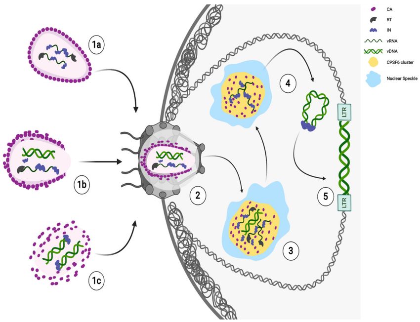
Figure 1. Ongoing model of HIV-1 early steps. HIV-1 cores are released in the cytoplasm after membrane fusion. Likely,
the core state reflects different stages of reverse transcription. Different potential core states can reach the nucleus: nearly
intact (1a), remodeled (1b), or partially uncoated (1c). Next, HIV-1 core remodels to translocate through the nuclear pore
complex (NPC) channel and CA proteins interact with Nucleoporins (Nup358/RanBP2, Nup153) for the translocation of
RTCs/PICs (2). The completion of reverse transcription and maturation of functional PICs (3) occur in HIV-1 MLOs, thanks
to the CPSF6 clusters formation in SC35 nuclear speckles (NSs). HIV-1 mature PICs separate from HIV-1 MLOs (4); as long
as incoming viral RNA is retained inside, viral components, like the IN proteins remain accumulated in the clusters (4).
Integration may occur in active chromatin regions in proximity of the nuclear speckles and of the nuclear envelope (5).
Created with BioRender.com.
Recent findings show late HIV-1 reverse-transcribed DNA separated from IN foci [77,90]
in the nucleus. The majority of intranuclear-detected INs are retained in CPSF6 clusters,
while the integration of the viral DNA occurs in proximity, but not inside these nuclear
condensates [77] (Figure 1). Thus, the HIV-induced CPSF6/SC35 membraneless organelles
(HIV-1 MLOs) might be safe nuclear sites to complete reverse transcription and uncoating.
Again, the accumulation of RTC in CPSF6/NS appears to precede the completion or, possi-
bly, the beginning of DNA synthesis. Concentration of forming PICs in NS may promote
integration of HIV-1 into euchromatin regions located outside, but not far from NS, for
optimal replication [77].Viruses 2021, 13, 273 7 of 11
5. Conclusions
The knowledge of HIV-1 capsid function deeply evolved from the mere structural-
shield protein. Recent exciting results about nuclear uncoating and nuclear reverse tran-
scription completely change our view on HIV-1 early phases of the life cycle. Apparently,
nuclear import precedes the complete uncoating and the reverse transcription can occur
in nuclear HIV-specific membraneless organelles (HIV-1 MLOs), at least in macrophages.
Does the nuclear reverse transcription represent an advantage for HIV? Is it correlated to
an efficient replication? All these questions open new frontiers of research based on the
role of HIV-1 MLOs on viral persistence and rebound, which represent the major obstacle
to cure AIDS.
Possibly, the nuclear CA can trigger nuclear antiviral pathways [98], but a late uncoat-
ing keeps safe the viral genetic material until integration. Future studies are required to
clarify whether the viral CA may play a direct role in choosing integration sites or whether
viral CA is acting behind the scenes to direct virus integration. Surely, new single-cell
cutting-edge technologies are allowing and will continue to allow to build a new model of
HIV-1 early steps (Figure 1).
Author Contributions: Conceptualization, V.S. and F.D.N.; writing—original draft preparation and
figure, V.S.; writing—review and editing, F.D.N.; supervision, F.D.N. All authors have read and
agreed to the published version of the manuscript.
Funding: The authors were supported by the Pasteur Institute and by the ANRS (Agence Nationale
de Recherche sur le SIDA) grant ECTZ88162 with a nominative PhD student fellowship ECTZ88177
for Viviana Scoca.
Conflicts of Interest: The authors declare no conflict of interest.
References
1. Barre-Sinoussi, F.; Chermann, J.C.; Rey, F.; Nugeyre, M.T.; Chamaret, S.; Gruest, J.; Dauguet, C.; Axler-Blin, C.; Vezinet-Brun, F.;
Rouzioux, C.; et al. Isolation of a T-lymphotropic retrovirus from a patient at risk for acquired immune deficiency syndrome
(AIDS). Science 1983, 220, 868–871. [CrossRef]
2. Temin, H.M.; Mizutani, S. Viral RNA-dependent DNA Polymerase: RNA-dependent DNA Polymerase in Virions of Rous
Sarcoma Virus. Nat. Cell Biol. 1970, 226, 1211–1213. [CrossRef] [PubMed]
3. Baltimore, D. Viral RNA-dependent DNA Polymerase: RNA-dependent DNA Polymerase in Virions of RNA Tumour Viruses.
Nat. Cell Biol. 1970, 226, 1209–1211. [CrossRef]
4. Maldarelli, F.; Wu, X.; Su, L.; Simonetti, F.R.; Shao, W.; Hill, S.; Spindler, J.; Ferris, A.L.; Mellors, J.W.; Kearney, M.F.; et al. Specific
HIV integration sites are linked to clonal expansion and persistence of infected cells. Science 2014, 345, 179–183. [CrossRef]
[PubMed]
5. Ezzell, C. AZT given the green light for clinical treatment of AIDS. Nat. Cell Biol. 1987, 326, 430. [CrossRef]
6. Hazuda, D.J.; Felock, P.; Witmer, M.; Wolfe, A.; Stillmock, K.; Grobler, J.A.; Espeseth, A.; Gabryelski, L.; Schleif, W.; Blau, C.; et al.
Inhibitors of Strand Transfer That Prevent Integration and Inhibit HIV-1 Replication in Cells. Science 2000, 287, 646–650. [CrossRef]
[PubMed]
7. Di Nunzio, F. New insights in the role of nucleoporins: A bridge leading to concerted steps from HIV-1 nuclear entry until
integration. Virus Res. 2013, 178, 187–196. [CrossRef]
8. Berry, F.; Khalfi, P.; Maillot, F.; Seigneres, P.; Sid Ahmed, S.; Di Nunzio, F. Host nuclear pore factors: Team players of HIV-1 nuclear
translocation and integration. Med. Sci. 2018, 34, 512–515.
9. Fassati, A. Multiple roles of the capsid protein in the early steps of HIV-1 infection. Virus Res. 2012, 170, 15–24. [CrossRef]
[PubMed]
10. Ambrose, Z.; Aiken, C. HIV-1 uncoating: Connection to nuclear entry and regulation by host proteins. Virology 2014, 455, 371–379.
[CrossRef]
11. Campbell, E.M.; Hope, T.J. HIV-1 capsid: The multifaceted key player in HIV-1 infection. Nat. Rev. Genet. 2015, 13, 471–483.
[CrossRef]
12. Novikova, M.; Zhang, Y.; Freed, E.O.; Peng, K. Multiple Roles of HIV-1 Capsid during the Virus Replication Cycle. Virol. Sin.
2019, 34, 119–134. [CrossRef]
13. Yant, S.R.; Mulato, A.; Hansen, D.; Tse, W.C.; Niedziela-Majka, A.; Zhang, J.R.; Stepan, G.J.; Jin, D.; Wong, M.H.; Perreira,
J.M.; et al. A highly potent long-acting small-molecule HIV-1 capsid inhibitor with efficacy in a humanized mouse model. Nat.
Med. 2019, 25, 1377–1384. [CrossRef]Viruses 2021, 13, 273 8 of 11
14. Link, J.O.; Rhee, M.S.; Tse, W.C.; Zheng, J.; Somoza, J.R.; Rowe, W.; Begley, R.; Chiu, A.; Mulato, A.; Hansen, D.; et al. Clinical
targeting of HIV capsid protein with a long-acting small molecule. Nat. Cell Biol. 2020, 584, 614–618. [CrossRef]
15. Bukrinsky, M.I.; Sharova, N.; McDonald, T.L.; Pushkarskaya, T.; Tarpley, W.G.; Stevenson, M. Association of integrase, matrix,
and reverse transcriptase antigens of human immunodeficiency virus type 1 with viral nucleic acids following acute infection.
Proc. Natl. Acad. Sci. USA 1993, 90, 6125–6129. [CrossRef]
16. Farnet, C.M.; Haseltine, W.A. Determination of viral proteins present in the human immuno-deficiency virus type 1 preintegration
complex. J. Virol. 1991, 65, 1910–1915. [CrossRef] [PubMed]
17. Blanco-Rodriguez, G.; Gazi, A.; Monel, B.; Frabetti, S.; Scoca, V.; Mueller, F.; Schwartz, O.; Krijnse-Locker, J.; Charneau, P.; Di
Nunzio, F. Remodeling of the Core Leads HIV-1 Preintegration Complex into the Nucleus of Human Lymphocytes. J. Virol. 2020,
94. [CrossRef] [PubMed]
18. Fassati, A.; Goff, S.P. Characterization of Intracellular Reverse Transcription Complexes of Human Immunodeficiency Virus Type
1. J. Virol. 2001, 75, 3626–3635. [CrossRef] [PubMed]
19. Poiesz, B.J.; Ruscetti, F.W.; Gazdar, A.F.; Bunn, P.A.; Minna, J.D.; Gallo, R.C. Detection and isolation of type C retrovirus particles
from fresh and cultured lymphocytes of a patient with cutaneous T-cell lymphoma. Proc. Natl. Acad. Sci. USA 1980, 77, 7415–7419.
[CrossRef]
20. Coffin, J.M.; Fan, H. The Discovery of Reverse Transcriptase. Annu. Rev. Virol. 2016, 3, 29–51. [CrossRef]
21. Fitzon, T.; Leschonsky, B.; Bieler, K.; Paulus, C.; Schröder, J.; Wolf, H.; Wagner, R. Proline Residues in the HIV-1 NH2-Terminal
Capsid Domain: Structure Determinants for Proper Core Assembly and Subsequent Steps of Early Replication. Virology 2000, 268,
294–307. [CrossRef]
22. Tang, S.; Murakami, T.; Agresta, B.E.; Campbell, S.; Freed, E.O.; Levin, J.G. Human immunodeficiency virus type 1 N-terminal
capsid mutants that exhibit aberrant core morphology and are blocked in initiation of reverse transcription in infected cells. J.
Virol. 2001, 75, 9357–9366. [CrossRef]
23. Forshey, B.M.; von Schwedler, U.; Sundquist, W.I.; Aiken, C. Formation of a human immunodeficiency virus type 1 core of
optimal stability is crucial for viral replication. J. Virol. 2002, 76, 5667–5677. [CrossRef]
24. Mallery, D.L.; Marquez, C.L.; McEwan, W.A.; Dickson, C.F.; Jacques, D.A.; Anandapadamanaban, M.; Bichel, K.; Towers, G.J.;
Saiardi, A.; Bocking, T.; et al. IP6 is an HIV pocket factor that prevents capsid collapse and promotes DNA synthesis. eLife 2018, 7,
e35335. [CrossRef]
25. Dick, R.A.; Mallery, D.L.; Vogt, V.M.; James, L.C. IP6 Regulation of HIV Capsid Assembly, Stability, and Uncoating. Viruses 2018,
10, 640. [CrossRef]
26. Sayah, D.M.; Sokolskaja, E.; Berthoux, L.; Luban, J. Cyclophilin A retrotransposition into TRIM5 explains owl monkey resistance
to HIV-1. Nat. Cell Biol. 2004, 430, 569–573. [CrossRef] [PubMed]
27. Selyutina, A.; Persaud, M.; Simons, L.M.; Bulnes-Ramos, A.; Buffone, C.; Martinez-Lopez, A.; Scoca, V.; Di Nunzio, F.; Hiatt, J.;
Marson, A.; et al. Cyclophilin A Pre-vents HIV-1 Restriction in Lymphocytes by Blocking Human TRIM5alpha Binding to the
Viral Core. Cell Rep. 2020, 30, 3766–3777. [CrossRef]
28. Kim, K.; Dauphin, A.; Komurlu, S.; McCauley, S.M.; Yurkovetskiy, L.; Carbone, C.; Diehl, W.E.; Strambio De Castillia, C.;
Campbell, E.M.; Luban, J. Cyclophilin A protects HIV-1 from restriction by human TRIM5α. Nat. Microbiol. 2019, 4, 2044–2051.
[CrossRef] [PubMed]
29. Stremlau, M.; Owens, C.M.; Perron, M.J.; Kiessling, M.K.H.; Autissier, P.; Sodroski, J. The cytoplasmic body component TRIM5α
restricts HIV-1 infection in Old World monkeys. Nat. Cell Biol. 2004, 427, 848–853. [CrossRef] [PubMed]
30. Shi, J.; Zhou, J.; Shah, V.B.; Aiken, C.; Whitby, K. Small-molecule inhibition of human immuno-deficiency virus type 1 infection by
virus capsid destabilization. J. Virol. 2011, 85, 542–549. [CrossRef] [PubMed]
31. Rankovic, S.; Ramalho, R.; Aiken, C.; Rousso, I. PF74 Reinforces the HIV-1 Capsid to Impair Reverse Transcription-Induced
Uncoating. J. Virol. 2018, 92. [CrossRef] [PubMed]
32. Cosnefroy, O.; Murray, P.J.; Bishop, K.N. HIV-1 capsid uncoating initiates after the first strand transfer of reverse transcription.
Retrovirology 2016, 13, 1–17. [CrossRef] [PubMed]
33. Rankovic, S.; Varadarajan, J.; Ramalho, R.; Aiken, C.; Rousso, I. Reverse Transcription Mechanically Initiates HIV-1 Capsid
Disassembly. J. Virol. 2017, 91. [CrossRef] [PubMed]
34. Christensen, D.E.; Ganser-Pornillos, B.K.; Johnson, J.S.; Pornillos, O.; Sundquist, W.I. Reconstitution and visualization of HIV-1
capsid-dependent replication and integration in vitro. Science 2020, 370, eabc8420. [CrossRef]
35. Miller, M.D.; Farnet, C.M.; Bushman, F.D. Human immunodeficiency virus type 1 preintegration complexes: Studies of organiza-
tion and composition. J. Virol. 1997, 71, 5382–5390. [CrossRef]
36. Karageorgos, L.; Li, P.; Burrell, C. Characterization of HIV Replication Complexes Early after Cell-to-Cell Infection. AIDS Res.
Hum. Retrovir. 1993, 9, 817–823. [CrossRef] [PubMed]
37. McDonald, D.; Vodicka, M.A.; Lucero, G.; Svitkina, T.M.; Borisy, G.G.; Emerman, M.; Hope, T.J. Visualization of the intracellular
behavior of HIV in living cells. J. Cell. Biol. 2002, 159, 441–452. [CrossRef] [PubMed]
38. Iordanskiy, S.; Berro, R.; Altieri, M.; Kashanchi, F.; Bukrinsky, M. Intracytoplasmic maturation of the human immunodeficiency
virus type 1 reverse transcription complexes determines their capacity to integrate into chromatin. Retrovirology 2006, 3, 4.
[CrossRef]Viruses 2021, 13, 273 9 of 11
39. Farnet, C.M.; Bushman, F.D. HIV-1 cDNA Integration: Requirement of HMG I(Y) Protein for Function of Preintegration Complexes
In Vitro. Cell 1997, 88, 483–492. [CrossRef]
40. Li, L.; Yoder, K.; Hansen, M.S.T.; Olvera, J.; Miller, M.D.; Bushman, F.D. Retroviral cDNA Integration: Stimulation by HMG I
Family Proteins. J. Virol. 2000, 74, 10965–10974. [CrossRef]
41. Lin, C.-W.; Engelman, A.N. The Barrier-to-Autointegration Factor Is a Component of Functional Human Immunodeficiency Virus
Type 1 Preintegration Complexes. J. Virol. 2003, 77, 5030–5036. [CrossRef] [PubMed]
42. Llano, M.; Saenz, D.T.; Meehan, A.; Wongthida, P.; Peretz, M.; Walker, W.H.; Teo, W.; Poeschla, E.M. An Essential Role for
LEDGF/p75 in HIV Integration. Science 2006, 314, 461–464. [CrossRef] [PubMed]
43. Emiliani, S.; Mousnier, A.; Busschots, K.; Maroun, M.; Van Maele, B.; Tempe, D.; Vandekerckhove, L.; Moisant, F.; Ben-Slama, L.;
Witvrouw, M.; et al. Integrase mutants defective for interaction with LEDGF/p75 are impaired in chromosome tethering and
HIV-1 replication. J. Biol. Chem. 2005, 280, 25517–25523. [CrossRef]
44. Ciuffi, A.; Llano, M.; Poeschla, E.; Hoffmann, C.; Leipzig, J.; Shinn, P.; Ecker, J.R.; Bushman, F. A role for LEDGF/p75 in targeting
HIV DNA integration. Nat. Med. 2005, 11, 1287–1289. [CrossRef] [PubMed]
45. Gallay, P.; Swingler, S.; Aiken, C.; Trono, D. HIV-1 infection of nondividing cells: C-terminal tyrosine phosphorylation of the viral
matrix protein is a key regulator. Cell 1995, 80, 379–388. [CrossRef]
46. Heinzinger, N.K.; Bukinsky, M.I.; Haggerty, S.A.; Ragland, A.M.; KewalRamani, V.; Lee, M.A.; Gendelman, H.E.; Ratner, L.;
Stevenson, M.; Emerman, M. The Vpr protein of human immunodeficiency virus type 1 influences nuclear localization of viral
nucleic acids in nondividing host cells. Proc. Natl. Acad. Sci. USA 1994, 91, 7311–7315. [CrossRef]
47. Popov, S.; Rexach, M.; Zybarth, G.; Reiling, N.; Lee, M.; Ratner, L.; Lane, C.M.; Moore, M.S.; Blobel, G.; Bukrinsky, M. Viral protein
R regulates nuclear import of the HIV-1 pre-integration complex. EMBO J. 1998, 17, 909–917. [CrossRef]
48. Zennou, V.; Petit, C.; Guetard, D.; Nerhbass, U.; Montagnier, L.; Charneau, P. HIV-1 Genome Nuclear Import Is Mediated by a
Central DNA Flap. Cell 2000, 101, 173–185. [CrossRef]
49. Yamashita, M.; Emerman, M. Capsid Is a Dominant Determinant of Retrovirus Infectivity in Nondividing Cells. J. Virol. 2004, 78,
5670–5678. [CrossRef]
50. Di Nunzio, F.; Danckaert, A.; Fricke, T.; Perez, P.; Fernandez, J.; Perret, E.; Roux, P.; Shorte, S.; Charneau, P.; Diaz-Griffero, F.; et al.
Human nucleoporins promote HIV-1 docking at the nuclear pore, nuclear import and integration. PLoS ONE 2012, 7, e46037.
[CrossRef]
51. Schaller, T.; Ocwieja, K.E.; Rasaiyaah, J.; Price, A.J.; Brady, T.L.; Roth, S.L.; Hué, S.; Fletcher, A.J.; Lee, K.; KewalRamani, V.N.; et al.
HIV-1 Capsid-Cyclophilin Interactions Determine Nuclear Import Pathway, Integration Targeting and Replication Efficiency.
PLoS Pathog. 2011, 7, e1002439. [CrossRef]
52. Di Nunzio, F.; Fricke, T.; Miccio, A.; Valle-Casuso, J.C.; Perez, P.; Souque, P.; Rizzi, E.; Severgnini, M.; Mavilio, F.; Charneau,
P.; et al. Nup153 and Nup98 bind the HIV-1 core and contribute to the early steps of HIV-1 replication. Virology 2013, 440, 8–18.
[CrossRef]
53. Matreyek, K.A.; Yücel, S.S.; Li, X.; Engelman, A.N. Nucleoporin NUP153 Phenylalanine-Glycine Motifs Engage a Common
Binding Pocket within the HIV-1 Capsid Protein to Mediate Lentiviral Infectivity. PLoS Pathog. 2013, 9, e1003693. [CrossRef]
54. Lelek, M.; Casartelli, N.; Pellin, D.; Rizzi, E.; Souque, P.; Severgnini, M.; Di Serio, C.; Fricke, T.; Diaz-Griffero, F.; Zimmer, C.; et al.
Chromatin organization at the nuclear pore favours HIV replication. Nat. Commun. 2015, 6, 6483. [CrossRef] [PubMed]
55. Price, A.J.; Jacques, D.A.; McEwan, W.A.; Fletcher, A.J.; Essig, S.; Chin, J.W.; Halambage, U.D.; Aiken, C.; James, L.C. Host
Cofactors and Pharmacologic Ligands Share an Essential Interface in HIV-1 Capsid That Is Lost upon Disassembly. PLoS Pathog.
2014, 10, e1004459. [CrossRef]
56. Bejarano, D.A.; Peng, K.; Laketa, V.; Borner, K.; Jost, K.L.; Lucic, B.; Glass, B.; Lusic, M.; Muller, B.; Krausslich, H.G. HIV-1 nuclear
import in macrophages is regulated by CPSF6-capsid interactions at the nuclear pore complex. eLife 2019, 8, e41800. [CrossRef]
[PubMed]
57. Balasundaram, D.; Benedik, M.J.; Morphew, M.; Dang, V.D.; Levin, H.L. Nup124p is a nuclear pore factor of Schizosaccharomyces
pombe that is important for nuclear import and activity of retrotransposon Tf1. Mol. Cell. Biol. 1999, 19, 5768–5784. [CrossRef]
[PubMed]
58. Schmitz, A.; Schwarz, A.; Foss, M.; Zhou, L.; Rabe, B.; Hoellenriegel, J.; Stoeber, M.; Panté, N.; Kann, M. Nucleoporin 153 Arrests
the Nuclear Import of Hepatitis B Virus Capsids in the Nuclear Basket. PLoS Pathog. 2010, 6, e1000741. [CrossRef]
59. Brass, A.L.; Dykxhoorn, D.M.; Benita, Y.; Yan, N.; Engelman, A.; Xavier, R.J.; Lieberman, J.; Elledge, S.J. Identification of Host
Proteins Required for HIV Infection Through a Functional Genomic Screen. Science 2008, 319, 921–926. [CrossRef]
60. Rodríguez-Mora, S.; De Wit, F.; García-Perez, J.; Bermejo, M.; López-Huertas, M.R.; Mateos, E.; Martí, P.; Rocha, S.; Vigón, L.;
Christ, F.; et al. The mutation of Transportin 3 gene that causes limb girdle muscular dystrophy 1F induces protection against
HIV-1 infection. PLoS Pathog. 2019, 15, e1007958. [CrossRef]
61. Christ, F.; Thys, W.; De Rijck, J.; Gijsbers, R.; Albanese, A.; Arosio, D.; Emiliani, S.; Rain, J.-C.; Benarous, R.; Cereseto, A.; et al.
Transportin-SR2 Imports HIV into the Nucleus. Curr. Biol. 2008, 18, 1192–1202. [CrossRef]
62. Zhou, L.; Sokolskaja, E.; Jolly, C.; James, W.; Cowley, S.A.; Fassati, A. Transportin 3 Promotes a Nuclear Maturation Step Required
for Efficient HIV-1 Integration. PLoS Pathog. 2011, 7, e1002194. [CrossRef]Viruses 2021, 13, 273 10 of 11
63. Valle-Casuso, J.C.; Di Nunzio, F.; Yang, Y.; Reszka, N.; Lienlaf, M.; Arhel, N.; Perez, P.; Brass, A.L.; Di-az-Griffero, F. TNPO3 is
required for HIV-1 replication after nuclear import but prior to integration and binds the HIV-1 core. J. Virol. 2012, 86, 5931–5936.
[CrossRef]
64. De Iaco, A.; Santoni, F.; Vannier, A.; Guipponi, M.; Antonarakis, S.; Luban, J. TNPO3 protects HIV-1 replication from CPSF6-
mediated capsid stabilization in the host cell cytoplasm. Retrovirology 2013, 10, 20. [CrossRef]
65. Fricke, T.; Valle-Casuso, J.C.; White, T.E.; Brandariz-Nuñez, A.; Bosche, W.J.; Reszka, N.; Gorelick, R.J.; Diaz-Griffero, F. The ability
of TNPO3-depleted cells to inhibit HIV-1 infection requires CPSF6. Retrovirology 2013, 10, 46. [CrossRef] [PubMed]
66. Maertens, G.N.; Cook, N.J.; Wang, W.; Hare, S.; Gupta, S.S.; Öztop, I.; Lee, K.; Pye, V.E.; Cosnefroy, O.; Snijders, A.P.; et al.
Structural basis for nuclear import of splicing factors by human Transportin 3. Proc. Natl. Acad. Sci. USA 2014, 111, 2728–2733.
[CrossRef]
67. Chin, C.R.; Perreira, J.M.; Savidis, G.; Portmann, J.M.; Aker, A.M.; Feeley, E.M.; Smith, M.C.; Brass, A.L. Direct Visualization of
HIV-1 Replication Intermediates Shows that Capsid and CPSF6 Modulate HIV-1 Intra-nuclear Invasion and Integration. Cell Rep.
2015, 13, 1717–1731. [CrossRef] [PubMed]
68. Lee, K.; Ambrose, Z.; Martin, T.D.; Oztop, I.; Mulky, A.; Julias, J.G.; Vandegraaff, N.; Baumann, J.G.; Wang, R.; Yuen, W.; et al.
Flexible Use of Nuclear Import Pathways by HIV-1. Cell Host Microbe 2010, 7, 221–233. [CrossRef] [PubMed]
69. Buffone, C.; Martinez-Lopez, A.; Fricke, T.; Opp, S.; Severgnini, M.; Cifola, I.; Petiti, L.; Frabetti, S.; Skorupka, K.; Zadrozny,
K.K.; et al. Nup153 Unlocks the Nuclear Pore Complex for HIV-1 Nuclear Translocation in Nondividing Cells. J. Virol. 2018, 92.
[CrossRef]
70. Achuthan, V.; Perreira, J.M.; Sowd, G.A.; Puray-Chavez, M.; McDougall, W.M.; Paulucci-Holthauzen, A.; Wu, X.; Fadel, H.J.;
Poeschla, E.M.; Multani, A.S.; et al. Capsid-CPSF6 Interaction Licenses Nuclear HIV-1 Trafficking to Sites of Viral DNA Integration.
Cell Host Microbe 2018, 24, 392–404. [CrossRef] [PubMed]
71. Sowd, G.A.; Serrao, E.; Wang, H.; Wang, W.; Fadel, H.J.; Poeschla, E.M.; Engelman, A.N. A critical role for alternative polyadeny-
lation factor CPSF6 in targeting HIV-1 integration to transcriptionally active chromatin. Proc. Natl. Acad. Sci. USA 2016, 113,
E1054–E1063. [CrossRef]
72. Saito, A.; Henning, M.S.; Serrao, E.; DuBose, B.N.; Teng, S.; Huang, J.; Li, X.; Saito, N.; Roy, S.P.; Siddiqui, M.A.; et al. Capsid-
CPSF6 Interaction Is Dispensable for HIV-1 Replication in Primary Cells but Is Selected during Virus Passage In Vivo. J. Virol.
2016, 90, 6918–6935. [CrossRef] [PubMed]
73. Francis, A.C.; Marin, M.; Singh, P.K.; Achuthan, V.; Prellberg, M.J.; Palermino-Rowland, K.; Lan, S.; Tedbury, P.R.; Sarafianos, S.G.;
Engelman, A.N.; et al. HIV-1 replication complexes accumulate in nuclear speckles and integrate into speckle-associated genomic
domains. Nat. Commun. 2020, 11, 3505. [CrossRef]
74. Graham, T.G.W.; Wang, X.; Song, D.; Etson, C.M.; Van Oijen, A.M.; Rudner, D.Z.; Loparo, J.J. ParB spreading requires DNA
bridging. Genes Dev. 2014, 28, 1228–1238. [CrossRef] [PubMed]
75. Sanchez, A.; Cattoni, D.I.; Walter, J.-C.; Rech, J.; Parmeggiani, A.; Nollmann, M.; Bouet, J.-Y. Stochastic Self-Assembly of ParB
Proteins Builds the Bacterial DNA Segregation Apparatus. Cell Syst. 2015, 1, 163–173. [CrossRef] [PubMed]
76. Mariame, B.; Kappler-Gratias, S.; Kappler, M.; Balor, S.; Gallardo, F.; Bystricky, K. Real-Time Visualization and Quantification of
Human Cytomegalovirus Replication in Living Cells Using the ANCHOR DNA Labeling Technology. J. Virol. 2018, 92. [CrossRef]
77. Scoca, V.; Louveaux, M.; Morin, R.; Ershov, D.; Tinevez, J.-Y.; Di Nunzio, F. Direct tracking of single proviruses reveals HIV-
1/LEDGF complexes excluded from virus-induced membraneless organelles. bioRxiv 2020. [CrossRef]
78. Di Nunzio, F. Nuclear Pore Complexes, Genome Organization and HIV-1 Infection. In Nuclear Pore Complexes in Genome
Organization, Function and Maintenance; D’Angelo, M., Ed.; Springer: Cham, Switzerland, 2018; pp. 183–199.
79. Marini, B.; Kertesz-Farkas, A.; Ali, H.; Lucic, B.; Lisek, K.; Manganaro, L.; Pongor, S.; Luzzati, R.; Recchia, A.; Mavilio, F.; et al.
Nuclear architecture dictates HIV-1 integration site selection. Nat. Cell Biol. 2015, 521, 227–231. [CrossRef]
80. Dismuke, D.J.; Aiken, C. Evidence for a functional link between uncoating of the human immunodeficiency virus type 1 core and
nuclear import of the viral preintegration complex. J. Virol. 2006, 80, 3712–3720. [CrossRef] [PubMed]
81. Burdick, R.C.; Li, C.; Munshi, M.; Rawson, J.M.O.; Nagashima, K.; Hu, W.S.; Pathak, V.K. HIV-1 un-coats in the nucleus near sites
of integration. Proc. Natl. Acad. Sci. USA 2020, 117, 5486–5493. [CrossRef]
82. Hulme, A.E.; Kelley, Z.L.; Foley, D.; Hope, T.J. Complementary Assays Reveal a Low Level of CA Associated with Viral Complexes
in the Nuclei of HIV-1-Infected Cells. J. Virol. 2015, 89, 5350–5361. [CrossRef]
83. Francis, A.C.; Marin, M.; Shi, J.; Aiken, C.; Melikyan, G.B. Time-Resolved Imaging of Single HIV-1 Uncoating In Vitro and in
Living Cells. PLoS Pathog. 2016, 12, e1005709. [CrossRef]
84. Kolb, H.C.; Finn, M.G.; Sharpless, K.B. Click Chemistry: Diverse Chemical Function from a Few Good Reactions. Angew. Chem.
Int. Ed. Engl. 2001, 40, 2004–2021. [CrossRef]
85. Rensen, E.; Mueller, F.; Scoca, V.; Parmar, J.J.; Souque, P.; Zimmer, C.; Di Nunzio, F. Clustering and reverse transcription of HIV-1
genomes in nuclear niches of macrophages. EMBO J. 2021, 40, e105247. [CrossRef]
86. Peng, K.; Muranyi, W.; Glass, B.; Laketa, V.; Yant, S.R.; Tsai, L.; Cihlar, T.; Muller, B.; Krausslich, H.G. Quantitative microscopy of
functional HIV post-entry complexes reveals association of replication with the viral capsid. eLife 2014, 3, e04114. [CrossRef]
87. Lelek, M.; Di Nunzio, F.; Henriques, R.; Charneau, P.; Arhel, N.; Zimmer, C. Superresolution imaging of HIV in infected cells with
FlAsH-PALM. Proc. Natl. Acad. Sci. USA 2012, 109, 8564–8569. [CrossRef]Viruses 2021, 13, 273 11 of 11
88. Francis, A.; Marin, M.; Prellberg, M.; Palermino-Rowland, K.; Melikyan, G. HIV-1 Uncoating and Nuclear Import Precede the
Completion of Reverse Transcription in Cell Lines and in Primary Macrophages. Viruses 2020, 12, 1234. [CrossRef] [PubMed]
89. Arfi, V.; Riviere, L.; Jarrosson-Wuilleme, L.; Goujon, C.; Rigal, D.; Darlix, J.L.; Cimarelli, A. Characterization of the early steps
of infection of primary blood monocytes by human immunodeficiency virus type 1. J. Virol. 2008, 82, 6557–6565. [CrossRef]
[PubMed]
90. Müller, T.G.; Zila, V.; Peters, K.; Schifferdecker, S.; Stanic, M.; Lucic, B.; Laketa, V.; Lusic, M.; Müller, B.; Kräusslich, H.-G. HIV-1
uncoating by release of viral cDNA from capsid-like structures in the nucleus of infected cells. bioRxiv 2020. [CrossRef]
91. Zurnic Bonisch, I.; Dirix, L.; Lemmens, V.; Borrenberghs, D.; De Wit, F.; Vernaillen, F.; Rocha, S.; Christ, F.; Hendrix, J.; Hofkens,
J.; et al. Capsid-Labelled HIV To Investigate the Role of Capsid during Nuclear Import and Integration. J. Virol. 2020, 94.
[CrossRef] [PubMed]
92. Dharan, A.; Bachmann, N.; Talley, S.; Zwikelmaier, V.; Campbell, E.M. Nuclear pore blockade reveals that HIV-1 completes
reverse transcription and uncoating in the nucleus. Nat. Microbiol. 2020, 5, 1–8. [CrossRef]
93. Koh, Y.; Wu, X.; Ferris, A.L.; Matreyek, K.A.; Smith, S.J.; Lee, K.; KewalRamani, V.N.; Hughes, S.H.; Engelman, A.N. Differential
Effects of Human Immunodeficiency Virus Type 1 Capsid and Cellular Factors Nucleoporin 153 and LEDGF/p75 on the Efficiency
and Specificity of Viral DNA Integration. J. Virol. 2012, 87, 648–658. [CrossRef]
94. Lamond, A.I.; Spector, D.L. Nuclear speckles: A model for nuclear organelles. Nat. Rev. Mol. Cell Biol. 2003, 4, 605–612. [CrossRef]
[PubMed]
95. Peterlin, B.M.; Price, D.H. Controlling the Elongation Phase of Transcription with P-TEFb. Mol. Cell 2006, 23, 297–305. [CrossRef]
96. Huang, S.; Spector, D.L. Intron-dependent recruitment of pre-mRNA splicing factors to sites of transcription. J. Cell. Biol. 1996,
133, 719–732. [CrossRef] [PubMed]
97. Kim, J.; Venkata, N.C.; Gonzalez, G.A.H.; Khanna, N.; Belmont, A.S. Gene expression amplification by nuclear speckle association.
J. Cell Biol. 2019, 219. [CrossRef]
98. Lahaye, X.; Gentili, M.; Silvin, A.; Conrad, C.; Picard, L.; Jouve, M.; Zueva, E.; Maurin, M.; Nadalin, F.; Knott, G.J.; et al. NONO
Detects the Nuclear HIV Capsid to Promote cGAS-Mediated Innate Immune Activation. Cell 2018, 175, 488–501. [CrossRef]
[PubMed]You can also read