High-Frequency Stimulation of Ventral CA1 Neurons Reduces Amygdala Activity and Inhibits Fear
←
→
Page content transcription
If your browser does not render page correctly, please read the page content below
ORIGINAL RESEARCH
                                                                                                                                              published: 09 March 2021
                                                                                                                                       doi: 10.3389/fnbeh.2021.595049

                                            High-Frequency Stimulation of
                                            Ventral CA1 Neurons Reduces
                           Edited by:       Amygdala Activity and Inhibits Fear
                      Jeansok J. Kim,
            University of Washington,       Jalina Graham 1† , Alexa F. D’Ambra 1 , Se Jung Jung 1 , Yusuke Teratani-Ota 1,2 ,
                        United States       Nina Vishwakarma 3 , Rasika Venkatesh 4 , Abhijna Parigi 1† , Evan G. Antzoulatos 1,3 ,
                       Reviewed by:         Diasynou Fioravante 1,3‡ and Brian J. Wiltgen 1,2* ‡
                  Fred J. Helmstetter,
                                            1
  University of Wisconsin–Milwaukee,         Center for Neuroscience, University of California, Davis, Davis, CA, United States, 2 Department of Psychology, University
                        United States       of California, Davis, Davis, CA, United States, 3 Department of Neurobiology, Physiology and Behavior, University
                       Michael Drew,        of California, Davis, Davis, CA, United States, 4 Department of Plant Sciences, University of California, Davis, Davis, CA,
        University of Texas at Austin,      United States
                        United States
                  *Correspondence:          The hippocampus can be divided into distinct segments that make unique contributions
                       Brian J. Wiltgen
               bjwiltgen@ucdavis.edu
                                            to learning and memory. The dorsal segment supports cognitive processes like spatial
                   † Present  address:
                                            learning and navigation while the ventral hippocampus regulates emotional behaviors
                        Jalina Graham,      related to fear, anxiety and reward. In the current study, we determined how pyramidal
        Department of Psychological
                                            cells in ventral CA1 respond to spatial cues and aversive stimulation during a context
      and Brain Sciences, Dartmouth
 College, Hanover, NH, United States        fear conditioning task. We also examined the effects of high and low frequency
                         Abhijna Parigi,    stimulation of these neurons on defensive behavior. Similar to previous work in the
       School of Veterinary Medicine,
 University of California, Davis, Davis,
                                            dorsal hippocampus, we found that cells in ventral CA1 expressed high-levels of c-Fos in
                     CA, United States      response to a novel spatial environment. Surprisingly, however, the number of activated
         ‡ These   authors share senior     neurons did not increase when the environment was paired with footshock. This was
                            authorship
                                            true even in the subpopulation of ventral CA1 pyramidal cells that send direct projections
                   Specialty section:       to the amygdala. When these cells were stimulated at high-frequencies (20 Hz) we
         This article was submitted to      observed feedforward inhibition of basal amygdala neurons and impaired expression
                Learning and Memory,
                a section of the journal
                                            of context fear. In contrast, low-frequency stimulation (4 Hz) did not inhibit principal cells
 Frontiers in Behavioral Neuroscience       in the basal amygdala and produced an increase in fear generalization. Similar results
         Received: 14 August 2020           have been reported in dorsal CA1. Therefore, despite clear differences between the
        Accepted: 10 February 2021          dorsal and ventral hippocampus, CA1 neurons in each segment appear to make similar
         Published: 09 March 2021
                                            contributions to context fear conditioning.
                            Citation:
    Graham J, D’Ambra AF, Jung SJ,          Keywords: learning, memory, optogenetics, context fear, mice, hippocampus
      Teratani-Ota Y, Vishwakarma N,
                Venkatesh R, Parigi A,
    Antzoulatos EG, Fioravante D and
   Wiltgen BJ (2021) High-Frequency
                                           INTRODUCTION
  Stimulation of Ventral CA1 Neurons
          Reduces Amygdala Activity
                                           The hippocampus can be divided into distinct segments that make unique contributions to
                    and Inhibits Fear.     learning and memory (Fanselow and Dong, 2010). The dorsal segment supports cognitive processes
  Front. Behav. Neurosci. 15:595049.       like spatial learning and navigation via interactions with the entorhinal, parahippocampal,
    doi: 10.3389/fnbeh.2021.595049         and retrosplenial cortices (Moser and Moser, 1998; Cenquizca and Swanson, 2006, 2007;

Frontiers in Behavioral Neuroscience | www.frontiersin.org                         1                                            March 2021 | Volume 15 | Article 595049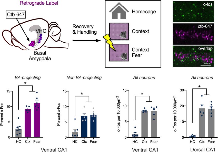
Graham et al. vCA1 Stimulation and Context Fear
Strange et al., 2014; Moser et al., 2017). The ventral hippocampus RESULTS
(VH), in contrast, regulates emotional behavior through
its connections with the amygdala, nucleus accumbens, Context Learning Activates vCA1
lateral hypothalamus, BNST and medial prefrontal cortex
(Cenquizca and Swanson, 2006, 2007; Hoover and Vertes, 2007;
Neurons That Project to the Basal
Jimenez et al., 2018). Despite these differences, the dorsal and Amygdala
ventral hippocampus share some important properties. They The DH responds to spatial and contextual cues while amygdala
have the same basic architecture and intrinsic organization neurons respond strongly to emotional stimuli like footshock
(tri-synaptic loop) and neurons in both regions respond to (O’Keefe and Dostrovsky, 1971; O’Keefe and Speakman, 1987;
spatial cues (e.g., place cells) (Ishizuka et al., 1990; Kjelstrup et al., Radulovic et al., 1998; Pelletier et al., 2005; Barot et al., 2009;
2008; Strange et al., 2014). These parallels suggest that similar Wolff et al., 2014; Beyeler et al., 2018; Tanaka et al., 2018).
computations may be carried out in the DH and VH during The current experiment determined how vCA1 neurons respond
cognitive and emotional learning. to these stimuli using c-Fos as a proxy for neural activity and
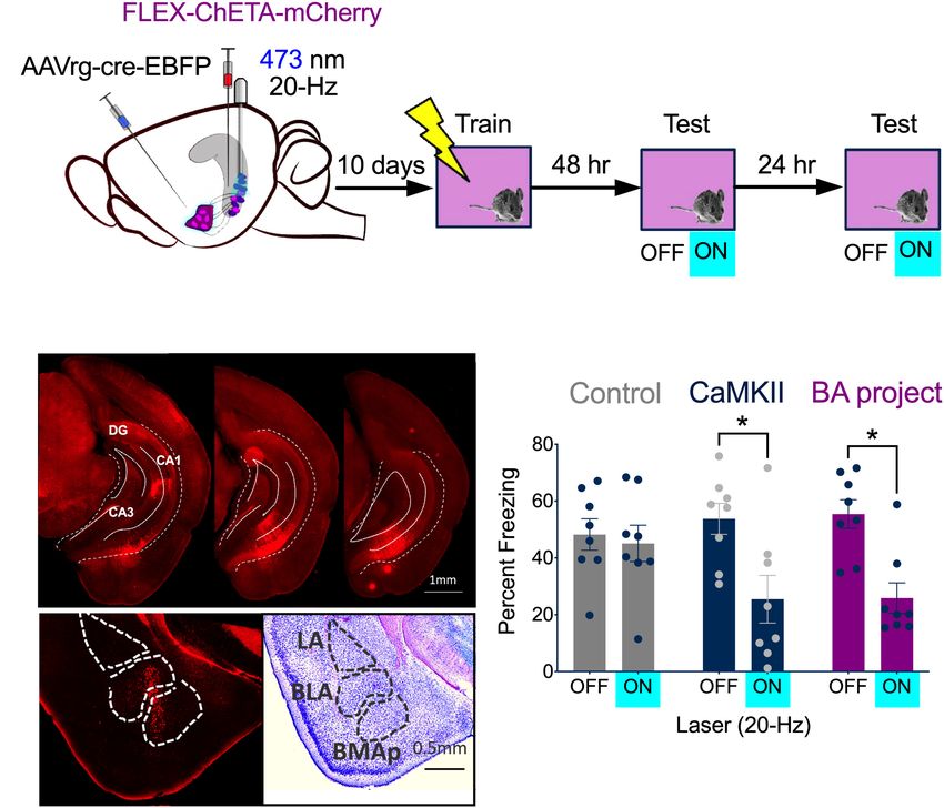
The integration of spatial and emotional information depends plasticity. To do this, we compared changes in c-Fos expression
on interactions between the DH and VH. For example, during after mice were exposed to a novel environment or underwent
context fear conditioning, animals learn to associate a novel context fear conditioning. Expression was quantified in neurons
environment with aversive footshock. Encoding this relationship that send direct projections to the BA and those that do not.
requires spatial information from the DH to be transmitted to To identify the former, the retrograde tracer ctb-647 was infused
the basal nucleus of the amygdala (BA) via the VH (Fanselow into the BA prior to conditioning (Figure 1A). On the training
and Dong, 2010; Xu et al., 2016). However, neurons in the day, control mice were left in their home cages (HC, n = 7).
VH do not act as passive relays; their activity is strongly The context group (Ctx, n = 5) explored a novel environment
modulated by emotional states like fear and anxiety, which is not for 5 min and the context + shock group underwent contextual
typically the case in the DH (Ciocchi et al., 2015; Jimenez et al., fear conditioning (Fear, n = 6) (Figure 1B). Fear conditioning
2018). Consistent with this fact, lesions of the VH reduce stress consisted of two footshocks (2 s, 0.3 mA, separated by 1
hormone release and anxiety-related behaviors while damage min) that were delivered after a 3 min exploration period.
to the DH does not (Kjelstrup et al., 2002). Place cells in Ninety-minutes after training, the animals were sacrificed, and
the VH are also distinct; they have large, overlapping place their brains fixed for c-Fos immunohistochemistry (Figure 1C).
fields that encode behaviorally-relevant contexts as opposed Compared to the control group, there was an increase in the
to precise spatial locations (Komorowski et al., 2013). Based number of c-Fos positive vCA1 neurons in mice that explored
on these findings, we hypothesized that dorsal (dCA1) and the novel environment or underwent context fear conditioning.
ventral (vCA1) CA1 neurons will respond to different stimuli The size of this increase was similar for the experimental groups
during context fear conditioning. Specifically, we predicted and observed both in vCA1 neurons that project to the BA
that neurons in dCA1 would primarily respond to the spatial (Ctb + neurons) as well as those that do not (Ctb- neurons).
context while cells in vCA1 would be more responsive to Interestingly, a higher percentage of Ctb + cells expressed c-Fos
footshock. than Ctb- cells in all groups (Figures 1D,E) [Repeated Measures
To examine our hypothesis, we quantified immediate-early ANOVA, Main effect of Group, F(2, 15) = 42.99, p < 0.0001;
gene expression (IEG) in vCA1 neurons after spatial exploration Main effect of Cell type, F(1, 15) = 22.93, p = 0.0002; No Group
or emotional learning. For the former, mice were exposed × Cell type interaction F(2, 15) = 3.115, p = 0.0739; Bonferroni
to a novel environment and for the latter, mice underwent post hoc tests, Control vs. Context (p < 0.0001), Control vs.
context fear conditioning. We found that vCA1 neurons were Context + Shock (p < 0.0001), Context vs. Context + shock
strongly activated by the novel environment but, surprisingly, (p = 0.5778)].
c-Fos expression did not increase further when the context While the expression of c-Fos did not increase significantly
was paired with shock. Neurons in dCA1 have been shown to in the fear group compared to the context group, there was a
respond in the same way under similar conditions (Radulovic trend in this direction. The lack of a difference could be due to
et al., 1998; Lovett-Barron et al., 2014). Next, we stimulated the fact that we used F1 hybrids (C57BL/6 × 129S6) rather than
vCA1 neurons that project to the BA to determine if defensive the more commonly used C57BL/6 strain. Hybrids acquire more
behaviors could be induced after context fear conditioning. fear than C57s and can be trained with lower shock intensities
We found that high-frequency stimulation (20 Hz) disrupted and fewer trials (Owen et al., 1997; Balogh and Wehner, 2003).
freezing and led to feed-forward inhibition of principal cells To determine if this difference affected our results, we fear
in the BA. In contrast, low frequency stimulation (4 Hz) conditioned a group of C57BL/6 mice with three, 0.75 mA shocks
increased fear generalization and did not inhibit the BA. Similar (Fear, n = 7) and compared them to animals that only explored
results have been reported when dCA1 neurons are stimulated the context (Ctx, n = 9) Ctb was once again infused into the
at low frequencies (Ryan et al., 2015). These data suggest BA to label vCA1 neurons that project to this region. Similar to
that dorsal and ventral CA1 make similar contributions to the data collected in hybrid mice, c-Fos expression was similar
context fear conditioning despite the functional differences in fear conditioned animals and those that explored the context.
between these regions. This was true both in Ctb + neurons (Ctx mean = 11.17%,
Frontiers in Behavioral Neuroscience | www.frontiersin.org 2 March 2021 | Volume 15 | Article 595049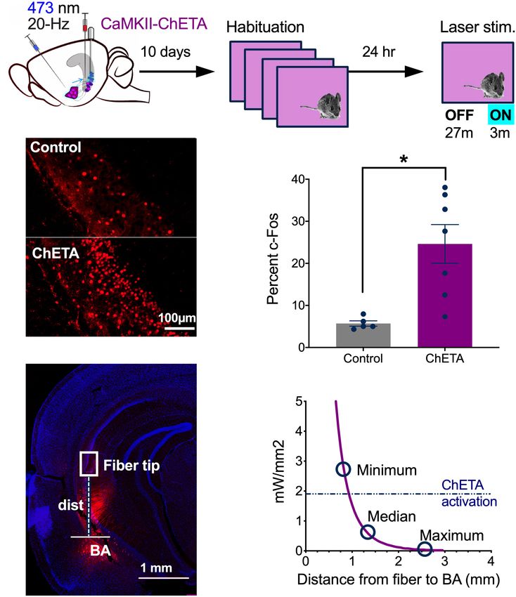
Graham et al.                                                                                                                     vCA1 Stimulation and Context Fear

  FIGURE 1 | Context learning activates vCA1 neurons that project to the basal amygdala. (A–C) Experimental design. (A) Ctb (magenta) was injected into the BA.
  (B) Mice were sacrificed 90 min after exploring a novel context (Ctx) or undergoing context fear conditioning (Fear). Control animals remained in their homecages
  (HC). (C) Example histology showing c-Fos staining in green, Ctb labeling in magenta and overlap in BA-projecting neurons (right). (D,E) The percentage of
  BA-projecting (Ctb+) and non-projecting (Ctb−) vCA1 neurons that expressed c-Fos in HC, Ctx and Fear groups. (F,G) The number of neurons in ventral and dorsal
  CA1 that expressed c-Fos in HC, Ctx and Fear groups. Data are presented as mean ± SEM. *p < 0.05.

SEM = 0.7387; Fear mean = 11.57%, SEM = 0.9617) and in Ctb-                           The results obtained with this methodology were similar to the
cells (Ctx mean = 7.932%, SEM = 0.9590; Fear mean = 6.113%,                           vCA1 data described above; c-Fos expression increased in the
SEM = 0.5846). In addition, the overall amount of c-Fos                               context and fear conditioning groups compared to homecage
expression was once again higher in vCA1 neurons that project                         controls and these conditions did not differ from one another.
to the BA (Ctb + mean = 11.37%, SEM = 0.850) compared to                              The same pattern was found in dCA1, although in this region,
those that do not (Ctb- mean = 7.02%, SEM = 0.771) [Repeated                          the total number of c-Fos + cells was higher than we observed in
Measures ANOVA, Main effect of cell type (Ctb + /Ctb−), F(1,                          vCA1 (Figures 1F,G) [Repeated Measures ANOVA, Main effect
14) = 16.55, p < 0.0012; No effect of group, F(1, 14) = 1.698,                        of Group F(2, 15) = 120.3, p < 0.0001; Main effect of Region
p = 0.2135; No group × cell type interaction F(1, 14) = 1.077,                        F(1, 15) = 55.64, p < 0.0001; Group × Region interaction F(2,
p = 0.3170] (Data not shown).                                                         15) = 16.88, p = 0.0001; Bonferroni post hoc tests, in both dCA1
   Together with previous work, these results demonstrate that                        and vCA1, HC vs. Context (p < 0.0001), HC vs. Fear (p < 0.0001)
novel environments strongly activate pyramidal neurons in                             Context vs. Fear (p > 0.9999); dCA1 vs. vCA1, HC vs. HC
dorsal and ventral CA1. Pairing the environment with shock                            (p > 0.9999), Fear vs. Fear (p < 0.0001) Context vs. Context
does not further increase activity in either of these subregions                      (p > 0.9999)].
(measured via c-Fos), as it does in subcortical areas like the                           We should note that vCA1 neurons activated during context
amygdala (Milanovic et al., 1998; Radulovic et al., 1998; Barot                       exploration may also respond to footshock. If that were the
et al., 2009). To ensure we could replicate the results of prior                      case, it could be difficult to find differences in c-Fos expression
studies done in dCA1, we quantified c-Fos expression in this                          between fear conditioned animals and mice that were exposed
region and compared it to vCA1 in the same animals. For these                         to the context. This issue could be addressed in future studies
analyses, single scan planes were taken from each area and the                        by labeling context responsive cells and footshock activated
number of c-Fos + neurons were counted per 10,000 um2 .                               neurons with different IEGs (Barot et al., 2009). Single-unit

Frontiers in Behavioral Neuroscience | www.frontiersin.org                        3                                          March 2021 | Volume 15 | Article 595049
Graham et al.                                                                                                                         vCA1 Stimulation and Context Fear

recordings and Ca2+ imaging could also be used to examine                               stimulated at 50 Hz, they responded reliably only to the first
the activity of individual vCA1 neurons during exploration and                          light pulse (average spike prob. ± SEM: 1.0 ± 0.00). The firing
fear conditioning. An advantage of these tools is that precise                          probability to subsequent stimuli progressively decreased and was
firing patterns can be obtained and compared across different                           significantly reduced by pulse 4 (Figure 2D) (permutation test for
experimental conditions.                                                                pulses 2–5 compared to pulse 1: 50 Hz: p = 0.17, p = 0.09, p = 0.01,
                                                                                        p = 0.001).
In vitro Stimulation of vCA1 Neurons
With ChETA                                                                              In vivo Stimulation of vCA1 Neurons With
High-frequency stimulation (20 Hz) in dCA1 does not induce                              ChETA
freezing after fear conditioning like it does in the dentate gyrus                      Next, we confirmed that 20 Hz laser stimulation activated vCA1
(DG) and CA3 (Ramirez et al., 2013; Ryan et al., 2015; Oishi et al.,                    pyramidal neurons in vivo (Figure 3). Mice received infusions
2019). This may be the case because dCA1 does not send a direct                         of CaMKII-ChETA-EYFP (n = 7) or a control virus (CaMKII-
projection to the ventral segment of the hippocampus like DG                            EYFP, n = 5) and 10 days later were habituated to a novel
and CA3 (Fricke and Cowan, 1978; Swanson et al., 1978; Ishizuka                         environment for 4 days (30 min each day) to reduce c-Fos
et al., 1990). To examine this idea, we used ChETA to stimulate                         expression (Hess et al., 1995). On Day 5, they were returned
pyramidal neurons in vCA1 after context fear conditioning. We                           to the same context and received 3 min of laser stimulation
first identified optimal stimulation parameters in hippocampal                          after a 27 min baseline period (Figure 3A). Ninety-minutes
slices by infusing AAV5-ChETA-EYFP into the VH and recording                            after this session, the animals were sacrificed, and their brains
from vCA1 neurons 2–3 weeks later. Recordings were performed                            fixed for c-Fos immunohistochemistry. We found a significant
using a cell-attached patch configuration (see section “Materials                       increase in the number of c-Fos + neurons in the ChETA group
and Methods” for details) while stimulating with 488 nm light at                        compared to control animals, indicating that 20 Hz stimulation
10, 20, or 50 Hz (Figure 2). As observed in dCA1, pyramidal cells                       strongly activated vCA1 neurons (Figures 3B,C) (Two-tailed
in vCA1 could easily follow 10 and 20 Hz optogenetic stimulation                        unpaired t-test, p = 0.0066, t = 3 df = 10). While the majority
across multiple trials (Figure 2D). At these frequencies, the                           of ChETA + neurons were found in vCA1, we also observed
spike probability for light pulses 2–5 was close to 1 and not                           some expression in the BA. To determine if light delivered to
significantly different from the spike probability for the first pulse                  vCA1 could activate these cells directly, we measured the distance
(permutation test for pulses 2–5 compared to pulse 1: 10 Hz:                            between each of our fiber tips and the BA (Figure 3D). These data
p = 0.82, p = 0.82, p = 0.82, p = 0.20; 20 Hz: p = 0.89, p = 0.88,                      (minimum, median, and maximum distances) were then plotted
p = 0.62, p = 0.43). In contrast, when the same neurons were                            against the predicted decay in laser power observed when light

  FIGURE 2 | In vitro stimulation of vCA1 neurons with ChETA. (A) Expression of ChETA-EYFP in ventral/intermediate CA1. (B) Ex vivo cell-attached recording from a
  representative ChETA-expressing vCA1 pyramidal neuron in response to five 10 ms light pulses at 20 Hz (blue lines). (B1) Single example trial. (B2) Raster plot of 5
  trials. (C) Average instantaneous firing rate (in spikes/s) for vCA1 pyramidal neurons (N = 10; bins: 50 ms, black: average firing rate, red: SEM). (D) Average spike
  probability for 3 photostimulation frequencies (10, 20, and 50 Hz). Data are presented as mean ± SEM.

Frontiers in Behavioral Neuroscience | www.frontiersin.org                          4                                           March 2021 | Volume 15 | Article 595049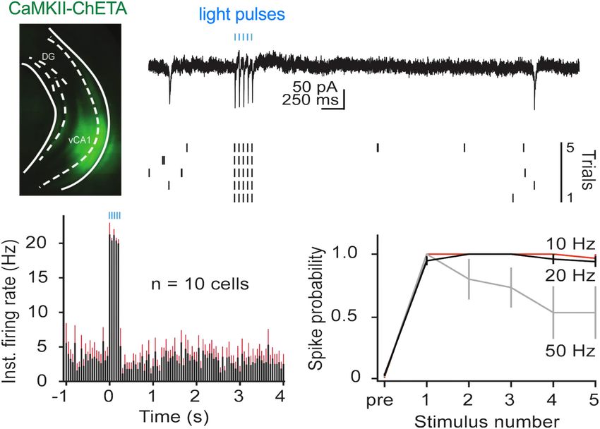
Graham et al.                                                                                                                       vCA1 Stimulation and Context Fear

  FIGURE 3 | In vivo stimulation of vCA1 neurons with ChETA. (A) Experimental design. (B) Examples of c-fos expression in a control animal (top) and a mouse that
  had vCA1 pyramidal neurons stimulated with ChETA (bottom). (C) The percentage of c-Fos positive vCA1 neurons in Control and ChETA groups. (D) Image showing
  an example of AAVrg-ChETA-mCherry expression in vCA1 and BA and how distance was measured between the optic fiber tip and the BA. (E) Predicted irradiance
  values were plotted against distance from the optic fiber tip. The minimum, median and maximum fiber tip to BA distance values are marked on the curve. The
  dashed line indicates the threshold at which ChETA responds to light stimulation with less than 10% fidelity (Berndt et al, 2011). Data are presented as mean ± SEM.
  *p < 0.05.

passes through tissue (Figure 3E) (Stanford predicted irradiance                       conditioning. Our initial plan was to activate neurons that
tool), (Yizhar et al., 2011). The dashed line on this figure (0.90–                    expressed c-Fos during training (i.e., engram/memory cells)
0.95 mm) indicates the distance at which blue light stimulation                        using TetTag mice from Jackson labs (stock no. 008344).
(10 mW, 20 Hz) fails to produce an action potential >90%                               However, we observed a significant amount of non-specific
of the time in ChETA + neurons (Berndt et al, 2011). We                                labeling in these mice compared to our original fos-tTA line
found that 94% of our fibers fell beneath this line, making                            (Tayler et al., 2013; Tanaka et al., 2014; Nakazawa et al., 2016;
it unlikely that light stimulation in vCA1 would activate BA                           Wilmot et al., 2018; Crestani et al., 2019). Therefore, instead
neurons directly.                                                                      of targeting c-Fos + cells, we stimulated vCA1 neurons that
                                                                                       project to the BA. To do this, AAVrg-EBFP-Cre was injected
                                                                                       into the BA and FLEX-ChETA-mCherry virus was infused
High-Frequency Stimulation of vCA1                                                     into the VH (BA project, n = 8) (Figure 4A, left). Bilateral
Neurons Impairs the Expression of                                                      optic fibers were implanted directly over vCA1. Histological
Context Fear                                                                           analyses confirmed that ChETA-mCherry was expressed in
Based on our recording and c-Fos results, we decided to                                vCA1 neurons (Figure 4B, top) and Cre expression was
stimulate vCA1 pyramidal neurons at 20 Hz after context fear                           restricted to cells in the BA (Figure 4B, bottom). A second

Frontiers in Behavioral Neuroscience | www.frontiersin.org                         5                                           March 2021 | Volume 15 | Article 595049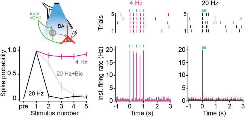
Graham et al. vCA1 Stimulation and Context Fear
group received infusions of CaMKII-ChETA-EYFP into vCA1 (p = 0.0003)]. The size of this decrease was similar whether
(All vCA1 n = 8) to examine the effects of non-selective all vCA1 pyramidal neurons were stimulated or just those that
stimulation on freezing. Control groups received infusions of project to the BA [No group x laser interaction, F(1, 14) =
AAV-CaMKII-EYFP into vCA1 (n = 4) or combined injections 0.01590, p = 0.9014]. These results demonstrate that context fear
of AAVrg-EBFP-Cre into BA and FLEX-tdTomato in vCA1 is not enhanced by high-frequency stimulation of vCA1 neurons.
(n = 4). Therefore, the inability of 20 Hz stimulation to induce freezing
Following recovery from surgery, animals were handled and in dCA1 is not due to the fact that this region lacks direct
habituated to the optic fiber cable for 5 days and then were projections to the VH or the amygdala (Ramirez et al., 2013;
trained on context fear conditioning. Training consisted of a 3 Wilmot et al., 2019; Krueger et al., 2020). To explain our results,
min baseline period followed by 2 shocks (0.3 mA, 2 s duration) we next examined the effects of vCA1 stimulation on the activity
delivered 1 min apart. Two days later, the mice were placed back of principal cells in the BA.
in the training environment to assess context fear memory. The
test began with a 3 min baseline period that was followed by 3
min of stimulation with blue light (473 nm, 10 mW, 20 Hz).
High Frequency Stimulation of vCA1
Mice received an identical test 24 h later (Figure 4A, right). Terminals Inhibits Principal Cells in the
During the baseline period, freezing was similar for all groups BA
(Figure 4C). When 20 Hz laser stimulation was delivered to Stimulation of vCA1 neurons at high-frequencies can produce
vCA1, freezing decreased significantly in the ChETA groups, feed-forward inhibition of principal cells in the BA (Hübner
but not in control animals [Repeated measures 2-way ANOVA, et al., 2014; Bazelot et al., 2015). To examine this possibility,
Stimulation × Group interaction F(2, 21) = 5.88, p = 0.0093; we recorded from BA neurons while stimulating vCA1 terminals
Bonferroni post hoc tests, Control On vs. Off (p > 0.9999), All at high (20 Hz) or low (4 Hz) frequencies (Figure 5A). AAV5-
vCA1 On vs. Off (p = 0.0005) BA- projecting vCA1 On vs. Off CaMKII-ChETA was infused into the VH and coronal slices were
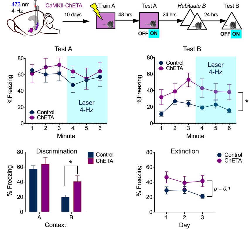
FIGURE 4 | High-frequency stimulation of vCA1 neurons impairs the expression of context fear. (A) Experimental design. Stimulation groups received infusions of
CaMKII-ChETA-EYFP or FLEX-ChETA-EYFP into the VH. Control groups received infusions of CaMKII-EYFP or FLEX-EYFP into the VH. To target amygdala
projecting vCA1 neurons, mice that received FLEX viruses also had AAVrg-Cre-EBFP infused into the BA. (B) Expression of FLEX-ChETA-mCherry in vCA1 (top);
Immunostaining for Cre in the BA (bottom left) and an adjacent Nissl section (bottom right). (C) Average freezing during baseline (OFF) and 20 Hz stimulation epochs
(ON) during the memory retrieval tests. Data are presented as mean ± SEM. *p < 0.05.
Frontiers in Behavioral Neuroscience | www.frontiersin.org 6 March 2021 | Volume 15 | Article 595049Graham et al. vCA1 Stimulation and Context Fear
FIGURE 5 | High frequency stimulation of vCA1 terminals inhibits principal cells in the BA. (A) Schematic of the experimental configuration: vCA1 axons expressing
ChETA-EYFP were optically stimulated in the BA while recording on-cell from pyramidal neurons (PN). vCA1 axons also synapse on interneurons (IN) in the BA. (B,C)
Example raster plots (B1,C1) and average instantaneous firing rates (B2,C2) for BA PNs in response to five, 10-ms light pulses delivered at (B) 4 Hz or (C) 20 Hz
(bins: 50ms; N = 11 cells). (D) Average spike probability for 4 Hz, 20 Hz, and 20 Hz in the presence of the GABAAR antagonist bicuculine (Bic). Data are presented
as mean ± SEM.
taken from the BA 2–3 weeks later. BA neurons were excited by (n = 6) or AAV-CaMKII-EYFP (n = 5) into the VH and optic
4 Hz stimulation of vCA1 terminals and fired action potentials fibers were implanted above vCA1. Two weeks later, they were
to every light pulse in the stimulus train (Figures 5B1,B2) trained on context fear conditioning as described above. Memory
(average spike prob ± SEM: pulse 1: 0.97 ± 0.02; pulse 2: was tested 48 h after training and vCA1 neurons were stimulated
0.88 ± 0.06, pulse 3: 0.86 ± 0.05, pulse 4: 0.86 ± 0.05, pulse at 4 Hz (473 nm, 10 mW, 15 ms pulses) during the last 3 min of
5: 0.91 ± 0.04). In contrast, 20-Hz stimulation only produced the session (Figure 6A, left). Unlike high-frequency stimulation,
a single action potential and suppressed responding to all this manipulation did not disrupt the expression of context
subsequent pulses (Figures 5C1,C2) (average spike prob. ± SEM: fear (Figure 6B) [2-way ANOVA, no laser × virus interaction
pulse 1: 0.95 ± 0.02; pulse 2: 0.06 ± 0.03, pulse 3: 0.01 ± 0.01, F = 0.05282, p = 0.824, no main effect of laser F(1, 9) = 0.005282,
pulse 4: 0.05 ± 0.04, pulse 5: 0.04 ± 0.03) (permutation test, p = 0.8234].
pulses 1–5, 4 Hz vs. 20 Hz: p = 0.09, p = 0.00, p = 0.00, p = 0.00,
p = 0.00). To determine if this suppression was caused by local Fear Generalization Increases After
inhibition, we stimulated vCA1 terminals at 20 Hz in the presence Low-Frequency Stimulation of vCA1
of the GABAA receptor antagonist bicuculine. This manipulation
partially rescued the activity of BA neurons (Figure 5D) (average Neurons
spike prob. ± SEM: pulse 1: 0.96 ± 0.04; pulse 2: 0.72 ± 0.08, We next determined if 4 Hz stimulation could induce freezing
pulse 3: 0.25 ± 0.11, pulse 4: 0.09 ± 0.09, pulse 5: 0.05 ± 0.05) in a novel environment. To do this, mice from the previous
(permutation test, pulses 1–5, 20 Hz vs. 20 Hz + Bic: p = 0.59, experiment were first exposed to context B for 2 days to reduce
p = 0.001, p = 0.18, p = 0.38, p = 0.48), suggesting that feed- any generalized fear (Figure 6A, right). On day 3, the animals
forward inhibition plays a role in suppressing excitatory activity were put back in context B and vCA1 neurons were stimulated
when vCA1 neurons are stimulated at high frequencies (Hübner at 4 Hz after a 3 min baseline period. This manipulation
et al., 2014; Bazelot et al., 2015). However, given that firing was did not increase freezing in ChETA mice relative to controls
not completely rescued, other factors like synaptic depression (Figure 6C) [2-way repeated measures ANOVA, no Group ×
likely contribute to this effect as well. Laser interaction, F(1, 9) = 0.001, p = 0.968]. However, ChETA
mice did exhibit an overall increase in freezing in context B,
which suggests that prior stimulation altered their behavior
Low Frequency Stimulation of vCA1 [Main effect of Group, F(1, 9) = 5.50, p = 0.043]. To determine
Does Not Disrupt the Expression of if vCA1 stimulation increased fear generalization, we compared
Context Fear freezing levels during the first exposure session in context B to
During aversive learning, activity in the hippocampus, amygdala that observed during the test in context A (Figure 5D). Both
and prefrontal cortex becomes synchronized to 4-Hz oscillations ChETA mice and controls froze more in the training context
(Seidenbecher et al., 2003; Narayanan et al., 2007; Lesting et al., (A) than the novel environment (B), indicating they could
2011; Padilla-Coreano et al., 2016). Given that BA neurons are discriminate between these places [Two-way ANOVA main effect
able to follow 4 Hz stimulation of vCA1 terminals, we examined of context F(1, 9) = 76.30, p < 0.0001]. Nonetheless, ChETA
the impact of this manipulation on context fear expression. mice showed significantly more fear in context B than control
Mice received bilateral infusions of AAV-CaMKII-ChETA-EYFP animals (Bonferroni post hoc test, p < 0.05), suggesting that
Frontiers in Behavioral Neuroscience | www.frontiersin.org 7 March 2021 | Volume 15 | Article 595049Graham et al. vCA1 Stimulation and Context Fear FIGURE 6 | Low frequency stimulation of vCA1 does not disrupt the expression of context fear and enhances generalization. (A) Experimental design. (B) Percent freezing over time (minutes) during baseline and laser stimulation in the training context (Test A). (C) Percent freezing over time (minutes) during baseline and laser stimulation in a novel environment (Test B). (D) Percent freezing during the first 3 min of the test in context A and the exposure session in context B 24 h later. (E) Percent freezing in control and ChETA groups during the first 3 min of the context B exposure days. All data are presented as the mean ± SEM. *p < 0.05. vCA1 stimulation increased generalization. However, additional DISCUSSION studies will be needed to confirm the selectivity of this effect, as the group × context interaction did not quite reach statistical During context fear conditioning, spatial information is thought significance [Two-way repeated measures ANOVA, No group × to be transmitted from dorsal to ventral hippocampus where it context interaction F(1, 9) = 4.04, p = 0.075; no effect of group can be relayed to the amygdala and associated with shock (Maren F(1, 9) = 1.94, p = 0.19]. and Fanselow, 1995; Wiltgen et al., 2006; Sutherland et al., 2008; Finally, to determine if vCA1 stimulation altered the Xu et al., 2016; Jimenez et al., 2018; Kim and Cho, 2020). In the extinction of generalized fear, we analyzed freezing during the current study, we examined the contribution of vCA1 neurons 3 exposure sessions in context B (Figure 6E). Analyses were to the expression of context fear. Similar to previous results restricted to the baseline period of each session so we could obtained in the DH, we found that c-Fos expression increased in include the data from day 3. We found that freezing levels vCA1 neurons after exposure to a novel environment (Radulovic decreased slightly across days in both groups, but this change et al., 1998). However, the addition of shock did not further was not statistically significant. This suggests our exposure increase the number of labeled cells as it does in subcortical sessions were not long enough to induce robust extinction [2- structures like the amygdala (Milanovic et al., 1998; Radulovic way repeated measures ANOVA, No Group × Session interaction et al., 1998; Barot et al., 2009). This result was surprising given F(2, 18) = 1.38, p = 0.27; No effect of Group F(1, 9) = 3.31, the role of the VH in learned fear and anxiety and the fact that p = 0.1; No effect of session, F(2, 18) = 1.81, p = 0.19]. However, it it communicates with subcortical regions involved in emotion should be noted that the current experiments were not designed (Cenquizca and Swanson, 2006, 2007; Hoover and Vertes, 2007; to detect small/moderate differences in fear generalization or Jimenez et al., 2018). Nonetheless, it remains possible that extinction. Future work will need to use behavioral protocols that footshock activated many of the same cells that responded to the are optimized to study these processes in order to determine how context, making it difficult for us to find a difference between they are affected by low-frequency stimulation of vCA1 neurons. these groups. Consistent with this idea, a recent study showed Frontiers in Behavioral Neuroscience | www.frontiersin.org 8 March 2021 | Volume 15 | Article 595049
Graham et al.                                                                                                      vCA1 Stimulation and Context Fear

that vCA1 neurons activated during exploration (c-Fos +) are                of activated neurons does not increase further if the context is
the same cells that strengthen their connections with BA neurons            paired with shock. In addition, the expression of fear is impaired
after the environment is paired with shock (Kim and Cho, 2020).             when neurons in dorsal or ventral CA1 are stimulated at 20
    High-frequency stimulation (20 Hz) of engram cells (c-Fos +)            Hz. Low-frequency stimulation, in contrast, increases freezing
in dorsal DG and CA3 has been shown to increase freezing                    and enhances fear generalization in dorsal and ventral CA1,
after context fear conditioning (Ramirez et al., 2013; Ryan et al.,         respectively. Additional research will be required to determine if
2015; Oishi et al., 2019). However, the same manipulation does              more robust changes in defensive behavior can be induced when
not drive freezing when performed in dorsal CA1 (Ryan et al.,               firing patterns are coordinated in the mPFC, BA, and vCA1.
2015). We hypothesized that this may be the case because dCA1
neurons do not project to the VH or the amygdala (Fricke and
Cowan, 1978; Swanson et al., 1978; Ishizuka et al., 1990). If so,           MATERIALS AND METHODS
stimulating vCA1 neurons at high frequencies should be able to
increase fear. Inconsistent with this prediction, we found that 20          Subjects
Hz stimulation of BA-projecting vCA1 neurons impaired freezing              Experiments were performed in 2–5-month-old male and female
rather than enhancing it. A similar effect was observed in a                F1 hybrid mice (C57BL/6NT x 129S6/SvEv) from Taconic
previous study when vCA1 terminals in the BA were stimulated                (B6129F1) or C57BL/6J mice from Jackson Labs (Stock Number
at 10 Hz (Jimenez et al., 2018). To determine why high-frequency            #000664). Animals were maintained on a 12 h light/12 h
stimulation produced impairments in freezing, we recorded from              dark cycle with ad libitum access to food and water. All
principal cells in the BA while activating terminals from vCA1.             experiments were performed during the light portion (7am–
We found that 20 Hz stimulation inhibited excitatory responses              7pm) of the light/dark cycle. Mice were group housed until
in the BA while 4 Hz did not. The inhibitory effect of 20 Hz                surgery, at which point they were single housed for the
stimulation could be reduced with a GABAA -receptor antagonist,             remainder of the experiment. All experiments were reviewed and
indicating that it was due, in part, to feed-forward inhibition.            approved by the UC Davis Institutional Animal Care and Use
    Interestingly, the medial prefrontal cortex (mPFC) has been             Committee (IACUC).
shown to disinhibit principal cells in the BA and allow them
to respond to strong inputs from vCA1 (Hübner et al., 2014).                Surgeries
A circuit like this could function to rapidly select adaptive               Stereotaxic surgery was performed 2–3 weeks before behavioral
responses in different situations. For example, when animals                experiments began. Mice were anesthetized with isoflurane (5%
come across a novel environment place cell activity in vCA1                 induction, 2% maintenance) and placed in a stereotaxic frame
could inhibit BA neurons and promote exploration. If a threat               (Kopf Instruments). An incision was made in the scalp and the
was subsequently encountered in this same place, input from                 skull was adjusted to place bregma and lambda in the same
the mPFC could quickly disinhibit BA neurons and allow                      horizontal plane. Small holes were drilled above the injection sites
vCA1 to drive defensive behaviors like freezing. We plan to                 for each brain region and virus or tracer was injected through a
examine these ideas in future experiments by simultaneously                 glass pipette with a tip diameter between 25 and 40 µm using
manipulating inputs from vCA1 and the mPFC to the BA after                  a microsyringe pump (UMP3, World Precision Instruments)
fear conditioning.                                                          at 2 nl/s. In the tracing experiment (Figure 1), Ctb-647 was
    Unlike 20 Hz, stimulation of vCA1 neurons at 4 Hz                       infused into the BA (50 nl) at each of the following 4 sites
did not inhibit principal cells in the BA. Given that the                   (AP, −1.55 mm; ML, ± 2.85; DV, −5 mm and −4.8 mm from
hippocampus, amygdala and mPFC oscillate around 4 Hz during                 dura). In the optogenetic behavioral experiments (Figures 3, 4,
fear expression, we hypothesized that low-frequency stimulation             6), AAV-CaMKII ChETA-EYFP, AAV-DIO-ChETA-mCherry or
may enhance freezing rather than impair it (Seidenbecher et al.,            a control virus (AAV-CaMKII-EYFP or AAV-FLEX-TdTomato)
2003; Narayanan et al., 2007; Lesting et al., 2011; Courtin et al.,         were infused into vCA1 (250 nl) at the following 4 sites (AP,
2014; Karalis et al., 2016; Padilla-Coreano et al., 2016). Consistent       −3 mm; ML, ± 3.5 mm; DV −3.9 and −3.5 mm from dura).
with this idea, 4-Hz stimulation increased fear generalization              In Figure 4, AAVrg was also infused into the BA (37 nl) at the
and did not disrupt freezing in the training context. However,              following 4 sites (AP, −1.55 mm; ML, ± 2.85; DV, −5.0 mm
freezing did not increase during laser stimulation itself, as has           and −4.9 mm from dura). We waited 3–5 min after each
been observed when dCA1 engram cells are stimulated at this                 infusion before withdrawing the pipette. Optic fiber cannulas
frequency (Ryan et al., 2015). It is possible that our behavioral           (0.39 NA, 200 µm diameter, Thorlabs) were manufactured
effects would have been larger if we were able to selectively               as previously described (Sparta et al., 2012) and implanted
stimulate engram/memory cells in vCA1. In addition, high-                   bilaterally above the virus injection sites in ventral CA1 (AP, –
frequency stimulation of BA-projecting neurons in vCA1 may be               3 mm; ML, ± 3.75 mm, length 3.4 mm). The fibers were secured
able induce freezing if it co-occurs with a disinhibitory input from        to the skull using 3 screws and dental cement (Harry J. Bosworth
the mPFC (Hübner et al., 2014; Karalis et al., 2016).                       Company). Three- to four-week-old mice were used in the
    To summarize, our results suggest that dorsal and ventral               electrophysiology experiments (Figures 2, 5), so the stereotaxic
CA1 neurons respond similarly to context exploration and fear               coordinates were adjusted for body size. AAV-ChETA-EYFP (350
conditioning. Many cells in each region express c-Fos when                  nl) was infused into the VHC at the following 2 sites (AP,
animals are exposed to a novel environment, and the number                  −2.8 mm; ML, ± 3.6 mm; DV, −2.8 mm from brain surface).

Frontiers in Behavioral Neuroscience | www.frontiersin.org              9                                     March 2021 | Volume 15 | Article 595049
Graham et al. vCA1 Stimulation and Context Fear
Contextual Fear Conditioning and ABE457, RRID:AB_2631318) suspended in blocking buffer. The
Optogenetic Stimulation next day, tissue was washed 3x with 0.1M PBS and then incubated
with a solution containing 1:500 Biotin-SP-conjugated Donkey
Stereotaxic surgery was performed on day 1. On days 8–12, all
anti-rabbit secondary antibody (Jackson ImmunoResearch Labs
animals were handled for 5 min a day (either in the vivarium or
Cat# 711-065-152, RRID: AB_2340593). After washing, the
in a room adjacent to the fear conditioning chambers). Animals
antibodies were detected using Streptavidin-conjugated Cy3
in optogenetic experiments had their implants attached to a 1
(1:500), (Jackson ImmunoResearch Labs Cat# 016-160-084,
m split optic patch cable (0.22 NA, 200 µm diameter) during
RRID: AB_2337244) or Cy5 (1:250), (Jackson ImmunoResearch
handling. All mice were trained 24 h after the last handling
Labs Cat# 016-170-084, RRID: AB_2337245). Finally, sections
session. Training consisted of 3 min of context exploration
were counterstained with DAPI (1:10,000, Life Technologies)
and either 2 shocks (0.3 mA, 1 min ITI, Taconic hybrids) or
for 15 min and mounted on slides (Vectashield mounting
3 shocks (0.6 mA, 1 min ITI, C57s). Testing consisted of 5-
medium). Slides were imaged using an Olympus fluorescence
10 min in the training context or a novel context (context B).
virtual slide scanning microscope. For c-Fos quantification, 35
Mice were sacrificed after training or their final testing session
µm z-stacks were acquired at 20x magnification. ROIs were
(depending on the experiment) and c-Fos was quantified as
chosen in vCA1 either beneath the optic fiber tip (optogenetic
described below. For optogenetic experiments, a 473 nm, 300mW
experiments) or in sections that contained AMY projecting
DPSS laser system (OptoEngine) was coupled to the branched
neurons (ctb experiment). Fluorescent images were imported
optic cable and implant through a rotating comutator fixed
into FIJI, converted to grayscale and separated by channel.
above the conditioning chamber. Laser output was adjusted to
Fluorescent label was marked on each channel independently
obtain 10 mW from the optic fiber tip measured with an optical
using the FIJI cell counter tool and the macro metamorph
power meter (Thorlabs) before each experiment. Doric’s OptG4
emulator (©2005 Fabrice P. Cordelières). Overlap (as in ctb
software was used to control laser pulse frequency and a Med
experiment) was determined by superimposing the markers
Associates SG-231 28V DC-TTL adapter was used to control
from one channel onto another and counting the number of
onset and offset of laser pulses during behavioral sessions. Laser
overlapping markers. For any experiments estimating the percent
stimulation consisted of 3 min epochs with 15 ms pulses at
of cells expressing label out of the total number of cells per area,
20 Hz. Mice were trained and/or tested in Med Associates fear
the 3D Objects Counter tool in FIJI was used to estimate the
conditioning chambers (30.5 × 24.1 × 21.0 cm) that were housed
number of DAPI stained nuclei in each area by dividing the
in sound-attenuating boxes containing overhead LED lights and
obtained volume by the average single nucleus volume for the
a scanning, charge-coupled video camera. Context A was lit with
animal/area. For quantification of c-fos in all dorsal and ventral
white light, cleaned with 70% EtOH and contained a stainless-
CA1 neurons (Figures 1F,G), 35 µm single-plane images were
steel grid floor. Context B was lit with infrared light, cleaned with
acquired at 20x magnification. Images were cropped to contain
Sani Wipes and contained a smooth plastic insert for the floor
approximately 10,000 µm2 of area CA1 in both the dorsal and
(covered with a small amount of corn cob bedding) as well as a
ventral hippocampus. After acquisition, images were converted
curved plastic insert for the walls.
to grayscale and c-Fos positive cells were quantified using the FIJI
cell counter tool.
Immunohistochemistry and Microscopic
Imaging Virus Constructs
Animals were sacrificed 90 min after training, testing, or final The following constructs (AAV2, serotype 5) were packaged by
laser stimulation. Mice were deeply anesthetized using 5% the Vector Core at the University of North Carolina: AAV5 -
isoflurane mixed with O2 and then transcardially perfused CaMKII-hChR2(E123T/T159C)-p2A-EYFP-WPRE had a titer of
with 0.1 M Phosphate buffered saline (PBS) followed by 4% 3.6 × 10¹² – 4.1 × 10¹² viral particles/ml. AAV5- EF1a-DIO-
paraformaldehyde (PFA). Brains were extracted and left in PFA hChR2 (E123T/T159C) p2A-mCherry had a titer of 4.10e12 virus
for 24–48 h and then sliced into 40 µm coronal sections molecules/ml. AAV5-CAG-FLEX-tdTomato had a titer of 4.8e12
using a Leica VT-1000 vibratome. To visualize virus spread and viral particles/ml. The AAVrg-cre-EBFP plasmid was purchased
locate injection sites and fiber optic tips, two separate series from Addgene (catalog# 51507) and packaged by the UC Davis
of slices were taken that spanned the anterior-posterior axis Vector Core with a titer of 7.6312 GC/ml.
(every 5th and 6th slice). For Ctb injection site localization,
one series was Nissl-stained and amygdala nuclei were identified
using the online Allen interactive mouse reference atlas. The Slice Preparation for
adjacent sections in the other series were stained with DAPI Electrophysiological Recordings
and the location of the injection site was mapped onto the Mice (postnatal week 6-7; both sexes) were anesthetized through
identified Amygdala nuclei. Slices 1–4 were stored for later intraperitoneal injection of an anesthetic cocktail (ketamine:
c-Fos immunohistochemistry. Three to four sections per area 10 mg/kg; xylazine: 1 mg/kg; acepromazine: 0.1 mg/kg) and
were randomly chosen for c-Fos quantification. Slices were transcardially perfused with ice-cold artificial CSF (aCSF; in mM:
incubated in blocking buffer (2% normal Donkey serum, 0.2% 127 NaCl, 2.5 KCl, 1.25 NaH2 PO4 , 25 NaHCO3 , 1 MgCl2 , 2
Triton-X 100 in 0.1 M PBS) for 15 min followed by overnight CaCl2 , 25 glucose; supplemented with 0.4 sodium ascorbate and
incubation in primary antibody at 1:5,000 (Millipore Cat# 2 sodium pyruvate; ∼310 mOsm). Brains were rapidly removed,
Frontiers in Behavioral Neuroscience | www.frontiersin.org 10 March 2021 | Volume 15 | Article 595049Graham et al. vCA1 Stimulation and Context Fear
blocked, and placed in choline slurry (110 choline chloride, from pulse onset (the longer time window was used to account for
25 NaHCO3, 25 glucose, 2.5 KCl, 1.25 NaH2 PO4 , 7 MgCl2 , synaptic delays). Baseline spike probability was quantified as the
0.5 CaCl2 , 11.6 sodium ascorbate, 3.1 sodium pyruvate; ∼310 average probability of an action potential within 500 randomly
mOsm). Coronal sections (250 µm) containing vCA1 or BA selected time windows (10 ms for vCA1; 15 ms for BA) during
were cut on a vibratome (Leica VT1200S) and transferred to the 3-s pre-stimulus baseline. For peri-stimulus time histograms
an incubation chamber containing aCSF at 32◦ C for 25 min (PSTH), action potentials were counted in 50-ms bins, with time
before moving to room temperature until used for recordings. referenced to the start of light pulses.
All solutions were bubbled with 95% O2 –5% CO2 continuously. Permutation tests were used for statistical comparisons
Chemicals were from Sigma. of average spike probabilities between conditions (Odén and
Wedel, 1975). Data were randomly shuffled between conditions
Patch-Clamp Recordings 1,000 times, while maintaining the original sample sizes,
For recordings, slices were mounted onto glass coveslips coated and the differences between the group averages of observed
with poly-l-lysine and placed in a submersion chamber perfused spike probabilities were compared against the corresponding
with aCSF (2 ml/min) at 30–32◦ C. Loose on-cell patch-clamp differences between the group averages of random permutations.
recordings were made from visually identified cells in vCA1 or The reported p-values indicate the probability that a difference
BAusing borosilicate glass pipettes (3–5 M) filled with 150 mM between average spike probabilities equal to or greater than the
NaCl. This configuration does not perturb the intracellular milieu observed difference could have arisen by chance alone (i.e., due
of the recording cell. vCA1 pyramidal neurons were identified to random sampling).
based on position and shape and were selected for ChETA-EYFP
expression. BA primary neurons (PNs) were identified based on
size (>15 µm) and firing rate (Graham et al. vCA1 Stimulation and Context Fear
REFERENCES Kjelstrup, K. B., Solstad, T., Brun, V. H., Hafting, T., Leutgeb, S., Witter, M. P.,
et al., (2008). Finite scale of spatial representation in the hippocampus. Science
Balogh, S. A., and Wehner, J. A. (2003). Inbred mouse strain differences in the 321, 140–143. doi: 10.1126/science.1157086
establishment of long-term fear memory. Behav. Brain Res. 140, 97–106. doi: Kjelstrup, K. G., Tuvnes, F. A., Steffenach, H., Murison, R., Moser, E. I., and Moser,
10.1016/s0166-4328(02)00279-6 M. B. (2002). Reduced fear expression after lesions of the ventral hippocampus.
Barot, S. K., Chung, A., Kim, J. J., and Bernstein, I. L. (2009). Functional Proc. Natl. Acad. Sci. U.S.A. 99, 10825–10830. doi: 10.1073/pnas.152112
imaging of stimulus convergence in amygdalar neurons during pavlovian fear 399
conditioning. PLoS One 4:e6156. doi: 10.1371/journal.pone.0006156 Komorowski, R. W., Garcia, C. G., Wilson, A., Hattori, S., Howard, M. W., and
Bazelot, M., Bocchio, M., Kasugai, Y., Fischer, D., Dodson, P. D., Ferraguti, F., et al., Eichenbaum, H. (2013). Ventral hippocampal neurons are shaped by experience
(2015). Hippocampal theta input to the amygdala shapes feedforward inhibition to represent behaviorally relevant contexts. J. Neurosci. 33, 8079–8087. doi:
to gate heterosynaptic plasticity. Neuron 87, 1290–1303. doi: 10.1016/j.neuron. 10.1523/JNEUROSCI.5458-12.2013
2015.08.024 Krueger, J. N., Wilmot, J. H., Teratani-Ota, Y., Puhger, K. R., Nemes, S. E., Crestani,
Berndt, A., Schoenenberger, P., Mattis, J., Tye, K. M., Deisseroth, K., Hegemann, P., A. P., et al. (2020). Amnesia for context fear is caused by widespread disruption
et al., (2011). High-efficiency channelrhodopsins for fast neuronal stimulation of hippocampal activity. Neurobiol. Learn. Mem. 175:107295. doi: 10.1016/j.
at low light levels. PNAS 108, 7595–7600. nlm.2020.107295
Beyeler, A., Chang, C., Silvestre, M., Leveque, C., Namburi, P., Wildes, C. P., et al., Lesting, J., Narayanan, R. T., Kluge, C., Sangha, S., Seidenbecher, T., and Pape, H. C.
(2018). Organization of valence-encoding and projection-defined neurons in (2011). Patterns of coupled theta activity in amygdala-hippocampal-prefrontal
the basolateral amygdala. Cell Rep. 22, 905–918. doi: 10.1016/j.celrep.2017.12. cortical circuits during fear extinction. PLoS One 6:e21714. doi: 10.1371/journal.
097 pone.0021714
Cenquizca, L. A., and Swanson, L. W. (2006). Analysis of direct hippocampal Lin, J. Y. (2011). A user’s guide to channelrhodopsin variants: features, limitations
cortical field ca1 axonal projections to diencephalon in the rat. J. Comp. Neurol. and future developments. Exp. Physiol. 96, 19–25. doi: 10.1113/expphysiol.2009.
497, 101–114. doi: 10.1002/cne.20985 051961
Cenquizca, L. A., and Swanson, L. W. (2007). Spatial organization of direct Lovett-Barron, M., Kaifosh, P., Kheirbek, M. A., Danielson, N., Zaremba, J. D.,
hippocampal field CA1 axonal projections to the rest of the cerebral cortex. Reardon, T. R., et al., (2014). Dendritic inhibition in the hippocampus suports
Brain Res. Rev. 56, 1–26. doi: 10.1016/j.brainresrev.2007.05.002 fear learning. Science 343, 857–864. doi: 10.1126/science.1247485
Ciocchi, S., Passecker, J., Malagon-Vina, H., Mikus, N., and Klausberger, T. (2015). Maren, S., and Fanselow, M. S. (1995). Synaptic plasticity in the basolateral
Selective information routing by ventral hippocampal ca1 projection neurons. amygdala induced by hippocampal formation stimulation in vivo. J. Neurosci.
Science 348, 560–563. doi: 10.1126/science.aaa3245 15, 7548–7564.
Courtin, J., Chaudun, F., Rozeske, R. R., Karalis, N., Gonzalez-Campo, C., Wurtz, Milanovic, S., Radulovic, J., Laban, O., Stiedl, O., Henn, F., and Spiess, J. (1998).
H., et al., (2014). Prefrontal parvalbumin interneurons shape neuronal activity Production of the fos protein after contextual fear conditioning of C57BL/6N
to drive fear expression. Nature 505, 92–96. doi: 10.1038/nature12755 mice. Brain Res. 784, 37–47.
Crestani, A. P., Krueger, J. N., Barragan, E. V., Nakazawa, Y., Nemes, S. E., Moser, E. I., Moser, M. B., and McNaughton, B. L. (2017). Spatial representation
Quillfeldt, J. A., et al. (2019). Metaplasticity contributes to memory formation in the hippocampal formation: a history. Nat. Neurosci. 20, 1448–1464. doi:
in the hippocampus. Neuropsychopharmacology 44, 408–414. doi: 10.1038/ 10.1038/nn.4653
s41386-018-0096-7 Moser, M. B., and Moser, E. I. (1998). Functional differentiation in the
Fanselow, M. S., and Dong, H. W. (2010). Are the dorsal and ventral hippocampus hippocampus. Hippocampus 8, 608–619. doi: 10.1002/(SICI)1098-106319988:
functionally distinct structures? Neuron 65, 7–19. doi: 10.1016/j.conb.2013.11. 6Graham et al. vCA1 Stimulation and Context Fear Perkins, K. L. (2006). Cell-attached voltage-clamp and current-clamp recording Tayler, K. K., Tanaka, K. Z., Reijmers, L. G., and Wiltgen, B. J. (2013). Reactivation and stimulation techniques in brain slices. J. Neurosci. Methods 154, 1–18. of neural ensembles during the retrieval of recent and remote memory. Curr. doi: 10.1016/j.jneumeth.2006.02.010 Biol. 23, 99–106. doi: 10.1016/j.cub.2012.11.019 Radulovic, J., Kammermeier, J., and Joachim, S. (1998). Relationship between fos Wilmot, J. H., Graham, J. A., LaFreniere, M. M., Puhger, K., and Wiltgen, B. J. production and classical fear conditioning: effects of novelty, latent inhibition, (2018). “Altered immediate early gene expression in fos-tTA transgenic mice,” and unconditioned stimulus preexposure. J. Neurosci. 18, 7452–7461. in Proceedings of the Society for Neuroscience Meeting, San Diego, CA. Program Ramirez, S., Liu, X., Lin, P. A., Suh, J., Pignatelli, M., Redondo, R. L., et al., No. (331.26), Session No. (HHH49). (2013). Creating a false memory in the hippocampus. Science 341, 387–391. Wilmot, J. H., Puhger, K., and Wiltgen, B. J. (2019). Acute disruption doi: 10.1126/science.1239073 of the dorsal hippocampus impairs the encoding and retrieval of trace Ryan, T. J., Roy, D. S., Pignatelli, M., Arons, A., and Tonegawa, S. (2015). Engram fear memories. Front. Behav. Neurosci. 13:116. doi: 10.3389/fnbeh.2019. cells retain memory under retrograde amnesia. Science 348, 1007–1014. 00116 Seidenbecher, T., Laxmi, R. T., Stork, O., and Pape, H. C. (2003). Amygdalar and Wiltgen, B. J., Sanders, M. J., Anagnostaras, S. G., Sage, J. R., and Fanselow, M. S. hippocampal theta rhythm synchronization. Science 846, 846–851. doi: 10.1126/ (2006). Context fear learning in the absence of the hippocampus. J. Neurosci. science.1085818 26, 5484–5491. doi: 10.1523/JNEUROSCI.2685-05.2006 Sosulina, L., Meis, S., Seifert, G., Steinhauser, C., and Pape, H. C. (2006). Wolff, S. B. E., Gründemann, J., Tovote, P., Krabbe, S., Jacobson, G. A., Classification of projection neurons and interneurons in the rat lateral Müller, C., et al., (2014). Amygdala interneuron subtypes control fear amygdala based upon cluster analysis. Mol. Cell Neurosci. 33, 57–67. doi: 10. learning through disinhibition. Nature 509, 453–458. doi: 10.1038/nature1 1016/j.mcn.2006.06.005 3258 Sparta, D. R., Stamatakis, A. M., Phillips, J. L., Hovelsø, N., van Zessen, R., and Xu, C., Krabbe, S., Gründemann, J., Botta, P., Fadok, J. P., Osakada, F., Stuber, G. D. (2012). Construction of implantable optical fibers for long-term et al., (2016). Distinct hippocampal pathways mediate dissociable roles of optogenetic manipulation of neural circuits. Nat. Protoc. 7, 12–23. doi: 10.1038/ context in memory retrieval. Cell 167:961-972.e16. doi: 10.1016/j.cell.2016.09. nprot.2011.413 051 Strange, B. A., Witter, M. P., Lein, E. S., and Moser, E. I. (2014). Functional Yizhar, O., Fenno, L. E., Davidson, T. J., Mogri, M., and Deisseroth, K. (2011). organization of the hippocampal longitudinal axis. Nat. Publ. Group 15, 655– Optogenetics in neural systems. Neuron 71, 9–34. doi: 10.1016/j.neuron.2011. 669. doi: 10.1038/nrn3785 06.004 Sutherland, R. J., O’Brien, J., and Lehmann, H. (2008). Absence of systems consolidation of fear memories after dorsal, ventral, or complete hippocampal Conflict of Interest: The authors declare that the research was conducted in the damage. Hippocampus 18, 710–718. doi: 10.1002/hipo.20431 absence of any commercial or financial relationships that could be construed as a Swanson, L. W., Wyss, J. M., and Cowan, W. M. (1978). An autoradiographic potential conflict of interest. study of the organization of intrahippocampal association pathways in the rat. J. Comp. Neurol. 181, 681–715. doi: 10.1002/cne.901810402 Copyright © 2021 Graham, D’Ambra, Jung, Teratani-Ota, Vishwakarma, Venkatesh, Tanaka, K. Z., Pevzner, A., Hamidi, A. B., Nakazawa, Y., Graham, J., and Wiltgen, Parigi, Antzoulatos, Fioravante and Wiltgen. This is an open-access article B. J. (2014). Cortical representations are reinstated by the hippocampus during distributed under the terms of the Creative Commons Attribution License (CC BY). memory retrieval. Neuron 84, 347–357. doi: 10.1016/j.neuron.2014.09.037 The use, distribution or reproduction in other forums is permitted, provided the Tanaka, K. Z., He, H., Anupratap, T., Niisato, K., Huang, A. J. Y., and McHugh, original author(s) and the copyright owner(s) are credited and that the original T. J. (2018). The hippocampal engram maps experience but not place. Science publication in this journal is cited, in accordance with accepted academic practice. No 361, 392–397. doi: 10.1126/science.aat5397 use, distribution or reproduction is permitted which does not comply with these terms. Frontiers in Behavioral Neuroscience | www.frontiersin.org 13 March 2021 | Volume 15 | Article 595049
You can also read