Autophagy facilitates type I collagen synthesis in periodontal ligament cells - Nature
←
→
Page content transcription
If your browser does not render page correctly, please read the page content below
www.nature.com/scientificreports
OPEN Autophagy facilitates type I
collagen synthesis in periodontal
ligament cells
Tomomi Nakamura, Motozo Yamashita*, Kuniko Ikegami, Mio Suzuki, Manabu Yanagita,
Jirouta Kitagaki, Masahiro Kitamura & Shinya Murakami
Autophagy is a lysosomal protein degradation system in which the cell self-digests its intracellular
protein components and organelles. Defects in autophagy contribute to the pathogenesis of age-
related chronic diseases, such as myocardial infarction and rheumatoid arthritis, through defects in
the extracellular matrix (ECM). However, little is known about autophagy in periodontal diseases
characterised by the breakdown of periodontal tissue. Tooth-supportive periodontal ligament (PDL)
tissue contains PDL cells that produce various ECM proteins such as collagen to maintain homeostasis
in periodontal tissue. In this study, we aimed to clarify the physiological role of autophagy in
periodontal tissue. We found that autophagy regulated type I collagen synthesis by elimination
of misfolded proteins in human PDL (HPDL) cells. Inhibition of autophagy by E-64d and pepstatin
A (PSA) or siATG5 treatment suppressed collagen production in HPDL cells at mRNA and protein
levels. Immunoelectron microscopy revealed collagen fragments in autolysosomes. Accumulation
of misfolded collagen in HPDL cells was confirmed by sodium dodecyl sulfate–polyacrylamide gel
electrophoresis. E-64d and PSA treatment suppressed and rapamycin treatment accelerated the
hard tissue-forming ability of HPDL cells. Our findings suggest that autophagy is a crucial regulatory
process that facilitates type I collagen synthesis and partly regulates osteoblastic differentiation of
PDL cells.
Periodontal ligament (PDL) tissue is a connective tissue consisting of rich collagenous fibres with elastic prop-
erties, which is located between the cementum and alveolar bone. PDL serves as a biological barrier that pro-
tects against invasive stimuli such as mechanical stress and bacterial i nfection1. PDL cells are the main cellular
components of PDL tissue and can differentiate into cementoblasts, osteoblasts, and gingival fibroblastic cells
during wound healing. Studies in recent decades have revealed that multiple mesenchymal stem cells are present
in PDL, which indicates their central roles in wound healing and regeneration of periodontal tissue through
proliferation and differentiation2–4. PDL cells produce various extracellular matrix (ECM) proteins. For example,
they synthesise considerable quantities of collagen, fibronectin, and proteoglycan species to sustain the space
and elastic properties of the periodontium in a quiescent state5. Collagen fibres are components of the ECM,
which support tissues and cell structures in various connective tissues throughout the body. Type I and III col-
lagens are expressed in bone, skin, blood vessels, muscles, and t endons6. In periodontal tissue, type I collagen
is particularly abundant in PDL, bone, and dentin in teeth7. Accordingly, genetic mutations of collagen genes
have been found in patients with osteogenesis imperfecta and Ehlers-Danlos syndrome, who exhibit irregular
connective tissue in periodontal d iseases8. Comprehensive gene analysis of PDL from extracted human teeth has
revealed constitutive expression of type I and III collagens, as well as ribosomal function-related genes involved
in protein translation in PDL c ells9. These findings strongly indicate that ECM proteins are actively synthesised
in PDL, as well as in chondrocytes and tendon tissue.
Autophagy is a major intracellular protein degradation system in which the cell self-digests its cytoplasmic
components by engulfing them within lipid double membrane-coated organelles (i.e., autophagosomes) that are
then delivered to the lysosome for degradation10. Nutritional starvation with respect to the surrounding environ-
ment promptly activates autophagy. Thus, cells presumably acquired this autonomous survival system during
the process of biological evolution from yeasts to mammals11. Autophagy serves as an environmental adaptation
system against bacterial infection and external stress in cells, as well as at organ and systemic levels12,13. Cur-
rently, canonical autophagy is referred to as m acroautophagy14 that differs from other types of autophagy such
Department of Periodontology, Graduate School of Dentistry, Osaka University, 1‑8, Yamadaoka, Suita,
Osaka 565‑0871, Japan. *email: yamashim@dent.osaka‑u.ac.jp
Scientific Reports | (2021) 11:1291 | https://doi.org/10.1038/s41598-020-80275-4 1
Vol.:(0123456789)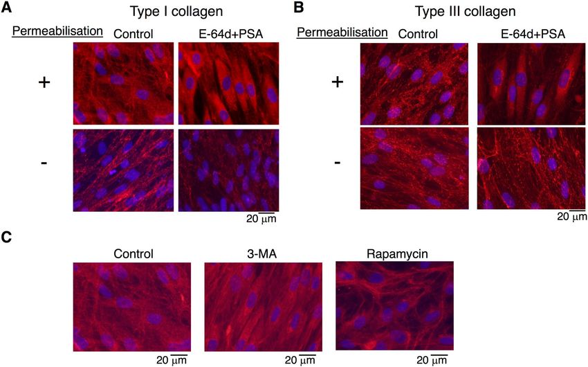
www.nature.com/scientificreports/
as microautophagy15, chaperone-mediated autophagy16, and xenophagy17. Among them, macroautophagy is the
most important system for cellular clearance of damaged organelles and misfolded p roteins10. Macroautophagy
(hereafter referred to as autophagy) is the lysosomal protein degradation system in which the cell self-digests
its protein components and organelles to maintain cellular homeostasis. Clearance of damaged organelles is the
most important function of autophagy in mammalian cells. There is increasing evidence from epidemiological
studies and research in animal models which indicates that defects in autophagy contribute to the pathogenesis
of age-related degenerative diseases with chronic inflammation, such as diabetes, myocardial infarction, rheu-
matoid arthritis, and neurodegenerative d iseases18–21.
Periodontal disease is caused by the establishment of dental biofilms, while accumulating environmental
stress affects the onset and subsequent progression of the disease through inflammation and impaired immune
responses. Interestingly, epidemiology and diseased animal models have suggested mutual relationships between
age-related lifestyle diseases and periodontal diseases22–24. Therefore, the roles of autophagy in periodontal tissue
and the pathogenesis of periodontal disease have been a research focus.
In this study, we aimed to clarify the physiological role of autophagy in periodontal tissue. Tooth-supportive
periodontal ligament tissue contains PDL cells that produce various ECM proteins. ECM proteins provide a
scaffold for cells, which regulate bona fide biological processes in PDL, such as cell differentiation, wound heal-
ing, and development. Balanced synthesis of ECM proteins and elimination of misfolded ECM proteins are
required for proper ECM production in PDL cells at the cellular level. Therefore, autophagy machinery might
be important for wound healing and tissue regeneration by accelerating the hard tissue-forming ability of PDL
cells through collagen synthesis.
Results
Induction of autophagy in HPDL cells. While the role of autophagy is well-known in a variety of cell
types, its function in HPDL cells is not fully elucidated. Autophagy has been reported to protect against envi-
ronmental stress in mammalian cells10,11. PDL encounters multiple cellular stresses, such as bacterial infection,
pH change, and mechanical force. We first examined autophagy in HPDL cells under fetal bovine serum (FBS)/
essential amino acids-deprived culture conditions in vitro, because nutrient starvation is a major stressful sig-
nal and could threaten the fate of affected cells25. Transmission electron microscopy (TEM) analysis revealed
the induction of microvacuole formation in HPDL cells after 4 h of serum starvation in culture with Hank’s
balanced saline solution (HBSS) (Fig. 1A). Enlarged images of microvacuoles revealed a double membrane-
coated vacuole structure, which is characteristic of the autophagosome. Furthermore, single membrane-coated
vacuoles with various cytosolic protein aggregates were present around the autophagosome, which may have
originated from fusion of the autophagosome and lysosome, these fused vacuoles are known as autolysosomes26.
The presence of these distinct vacuole types suggested that autophagy machinery is maintained in HPDL cells
(Fig. 1B). Next, we examined the induction of autophagy in HPDL cells at the biochemical level. Microtubule-
associated protein 1 light chain 3 (hereafter, LC3) is distributed in cytosolic spaces in its LC3-I form. LC3-I is
converted to LC3-II after binding to membrane lipids of autophagosomes and autolysosomes during the process
of autophagy27. p62 is a specific substrate for LC3-II28, therefore, the p62/LC3-II complex initiates degradation
in the progression to autolysosomes. Western blotting analysis revealed changes in the LC3-I/II protein ratio
and reduction of the p62 level in HPDL cells during starvation culture with HBSS (Fig. 2A). Endogenous LC3-II
was increased after 30 min of starvation treatment, whereas LC3-I and p62 both gradually decreased after 1 h of
starvation treatment. Moreover, we examined expression of other autophagy-related genes and turnover of pro-
teins (Supplementary Fig. S1). These results suggest that autophagy functions correctly in HPDL cells in a cell-
autonomous manner. To confirm this, we transduced green fluorescent protein (GFP)-LC3 into HPDL cells to
determine the localisation of LC3 in HPDL cells. GFP-LC3 was dispersed in the cytosol of GFP-LC3 HPDL cells
without aggregates, whereas starvation treatment for 4 h induced GFP-LC3 puncta in HPDL cells (Fig. 2B). To
characterise LC-3 localisation in organelles in HPDL cells, we labelled the lysosomes with LysoTracker (Molecu-
lar Probes, Eugene, OR, USA). Our results showed that some GFP-LC3 puncta exhibited colocalisation with
lysosomes (Fig. 2C).
Inhibition of autophagy induces deposition of collagen in HPDL cells. The periodontium, located
between the tooth and alveolar bone, is supported by PDL surrounding the cementum on the tooth root. Of all
cell species involved in periodontal tissue, PDL cells exhibit the most vigorous production of ECM proteins (e.g.,
type I collagen alpha-1) for the maintenance of periodontal tissue. To determine how autophagy regulates PDL
function, we focused on the role of autophagy in the production of ECM (especially highly macromolecular
and complex structured peptides, such as collagen species) from HPDL cells. First, we stained HPDL cells with
picrosirius red to determine the distribution of collagen fibres in HPDL cells (Fig. 3A). Next, we treated HPDL
cells with a combination of E-64d and pepstatin A at 10 µM each to interfere with lysosomal protein degrada-
tion by inhibiting lysosomal cysteine proteases (i.e., cathepsins B, D, and L) at low doses29. As shown in Fig. 3A,
an intensely red-stained area (stained by picrosirius red) in HPDL cells was dramatically enhanced after E-64d
and pepstatin A treatment for 6 days; in contrast, treatment with 250 nM rapamycin, a well-known activator of
autophagy, did not induce the formation of a red-stained area, compared with control. Densitometric analysis
of the picrosirius red-stained area revealed > fivefold enhancement of HPDL cells after E-64d and pepstatin A
treatment (Fig. 3B). To characterise the features of the picrosirius red-stained area, we performed microscopic
analysis at a magnification of × 200; the intensity of redness was stronger in the cytosolic area than in the extra-
cellular spaces of HPDL cells after E-64d and pepstatin A treatment (Fig. 3C). Next, we examined the immuno-
histochemical staining of HPDL cells with types I or III collagen-specific antibodies to clarify the localisation of
each collagen in the cytosolic area, because picrosirius red can stain both types I and III collagen equally. With-
Scientific Reports | (2021) 11:1291 | https://doi.org/10.1038/s41598-020-80275-4 2
Vol:.(1234567890)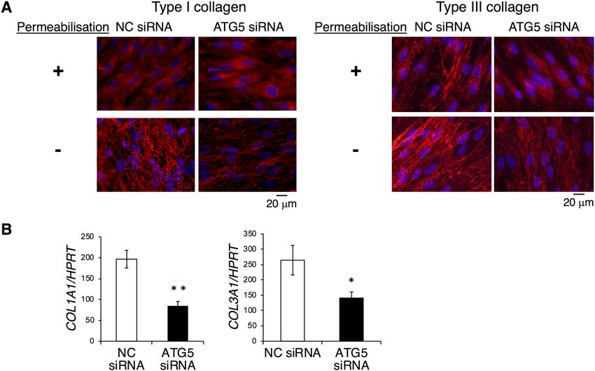
www.nature.com/scientificreports/
Figure 1. Autophagy is induced in human periodontal ligament (HPDL) cells in vitro. (A) Transmission
electron microscopy showed the induction of double membrane coated vacuoles after treatment with no-serum
Hank’s balanced saline solution (HBSS) for 4 h (1870 ×) (right). Enlarged image of the bold square in the left
panel (4780 ×). (B) Typical image of autophagosome after treatment with no-serum HBSS for 4 h is shown in
upper panel (31,800 ×). AP; Autophagosome AL; Autolysosome Mt; Mitochondria.
out permeabilisation treatment, type I collagen was mainly detected in extracellular regions around HPDL cells,
whereas the distribution of type I collagen in extracellular regions was dramatically reduced after E-64d and
pepstatin A treatment for 4 days. Intriguingly, the localisation of type I collagen in cytosolic regions appeared
to increase after E-64d and pepstatin A treatment for 4 days with concurrent permeabilisation treatment. These
results suggest that combination treatment by E-64d and pepstatin A may induce collagen accumulation in the
cytosol, thereby inhibiting collagen secretion from HPDL cells (Fig. 4A). To confirm the effect of combination
treatment by E-64d and pepstatin A on collagen species, we also examined the distribution of type III collagen
in HPDL cells. Similar to type I collagen, type III collagen showed more intense staining in cytosolic regions of
HPDL cells after E-64d and pepstatin A treatment for 4 days with permeabilisation (Fig. 4B).
Moreover, we examined another autophagy inhibitor, 3-methyladenine (3-MA), to confirm the above find-
ings. Treatment with 1 mM 3-MA, an inhibitor of class III phosphoinositide 3-kinase that is known to inhibit
the growth of isolation membrane, also inhibited the deposition of type I collagen in the extracellular space of
HPDL cells. Conversely, treatment with rapamycin, the mammalian target of rapamycin inhibitor that activates
the autophagy pathway, induced type I collagen expression in extracellular regions around HPDL cells (Fig. 4C).
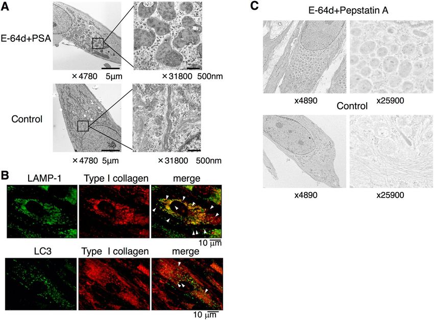
Inhibition of autophagy induces accumulation of collagen in lysosomes in HPDL cells. To
clarify the mechanism of induced collagen accumulation in the cytosol of HPDL cells after treatment with an
inhibitor of autophagy, we conducted TEM analysis. Comparable numbers of enlarged vacuoles were observed
after E-64d and pepstatin A treatment for 4 days (Fig. 5A). Magnified images of enlarged vacuoles revealed that
most vacuoles were coated with a single membrane and were filled with fibril-like structures. In control cells, the
formation of small vacuoles (i.e., without fibril-like structures) was observed. Next, we examined whether type I
collagen was localised in lysosomes by performing immunohistochemical staining for LC3 and lysosomal-asso-
ciated membrane protein (LAMP)-1, which are specific markers for lysosomes. After 6 days of treatment with an
inhibitor of autophagy, comparable numbers of punctae were clearly observed in HPDL cells. Intriguingly, the
colocalisation of type I collagen with LC3 was reduced at this time point (Fig. 5B). To confirm the above finding,
we performed immunogold staining with TEM analysis to characterise the type I collagen present in autolys-
osomes. Enlarged vacuole formation was induced after E-64d and pepstatin A treatment for 6 days. We subse-
quently found a comparable number of dot spots indicating the existence of type I collagen, which was labelled
Scientific Reports | (2021) 11:1291 | https://doi.org/10.1038/s41598-020-80275-4 3
Vol.:(0123456789)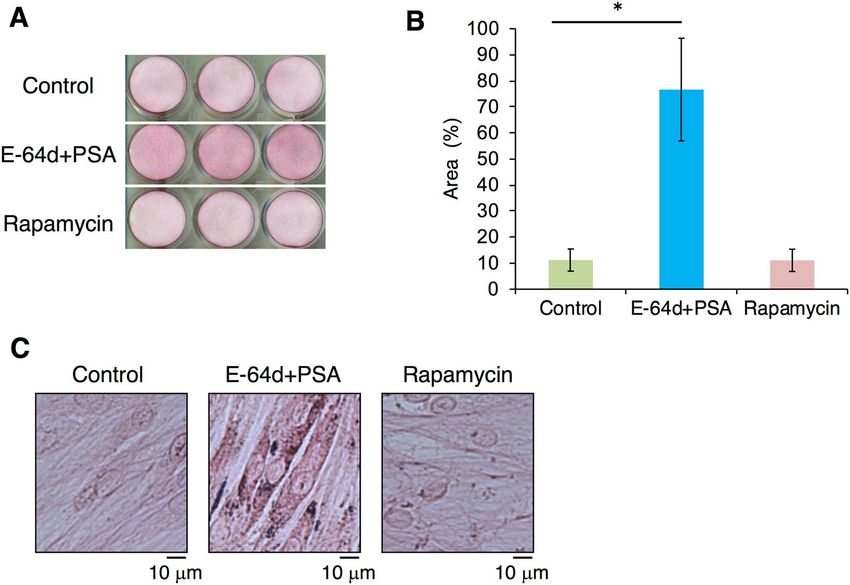
www.nature.com/scientificreports/
Figure 2. Autophagosome formation is stimulated by nutrient deprivation in human periodontal ligament
(HPDL) cells. (A) Western blotting analysis of LC3 and p62 in HPDL cells. HPDL cells were treated with
no-serum Hank’s balanced saline solution (HBSS) for the indicated period. Upper bands indicate LC3-I and
lower bands indicate LC3-II for LC3 blotting. Full length blots are presented in Supplementary Fig. S5. (B)
GFP-LC3-transduced HPDL cells were treated with no-serum HBSS for 4 h. LC3 dot formation was detected
by phase-contrast fluorescence microscopy. (C) Localisation of LC3 was determined by confocal microscopy
with lysosome labelled GFP-LC3 transduced HPDL cells. HPDL cells were treated with no-serum HBSS for
4 h. Lysosomes were labelled with Lysotracker (red) Arrow indicates the localisation of LC3 in lysosomes.
Representative data from three independent experiments are shown.
with immunogold-conjugated antibodies in enlarged vacuoles (Fig. 5C). Thus, a subset of the fibril-like products
in enlarged vacuoles were likely to be type I collagen or its derivatives, which had accumulated in lysosomes.
Involvement of autophagy in production of type I collagen in HPDL cells. To investigate the
role of autophagy in collagen secretion from HPDL cells, we examined the role of autophagy in type I collagen
expression at the mRNA level. In contrast to the results of immunohistochemical staining, autophagy inhibitor
treatment (e.g., with E-64d and pepstatin A, or chloroquine and 3-MA), significantly inhibited type I collagen
alpha-1 mRNA expression. Rapamycin treatment enhanced type I collagen alpha-1 mRNA expression (Fig. 6A).
Furthermore, we quantified the production of type I collagen from HPDL cells by enzyme-linked immunosorb-
ent assay. The mature form of type I collagen in culture supernatants significantly decreased after treatment with
various autophagy inhibitors for 6 days (Fig. 6B).
To determine how autophagy regulates type I collagen production, we performed western blotting analysis
to investigate protein synthesis in HPDL cells. In SDS PAGE analysis, autophagy inhibitor treatment reduced
the expression of type I pro-collagen bands around 150 kDa. Intriguingly, around 120 kDa, distinct molecular
sizes of type I collagen were induced by E-64d and pepstatin A treatment. Under native-PAGE conditions, the
mature form of type I collagen was reduced after autophagy inhibitor treatment (Fig. 6C). In the rapamycin-
treated group, there were fewer pro-collagen bands under reducing conditions, whereas the protein expression
level of mature type I collagen was increased. Thus, the condensed band expressed around 120 kDa represents
aggregated or misfolded type I collagen, which was less soluble in denatured or reducing conditions.
Effects of ATG5 knockdown on expression of type I collagen in HPDL cells. In this study, we used
various autophagy inhibitors, which differentially affect each step of autophagy, to clarify the role of autophagy
in HPDL cells. To exclude the possible side effects of the chemical treatment, we repeated the same set of experi-
ments using siRNA against ATG5 that triggers the process of autophagic membrane formation30,31 to confirm
the results. Induction of ATG5 RNAi in HPDL cells revealed nearly 70% inhibition of ATG5 expression at the
mRNA level (Supplementary Fig. 3A) and 80% inhibition of the form of ATG5 that covalently binds with ATG12
at the protein level (Supplementary Fig. S3B). Results from ATG5 RNAi treatment in HPDL cells were similar to
Scientific Reports | (2021) 11:1291 | https://doi.org/10.1038/s41598-020-80275-4 4
Vol:.(1234567890)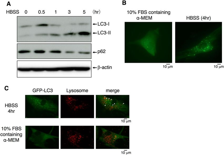
www.nature.com/scientificreports/
Figure 3. Suppression of autophagy increases the deposition of collagen in human periodontal ligament
(HPDL) cells. (A) Collagen production was evaluated with picrosirius red staining after ossification-inducing
culture for 6 days in the presence of autophagy inhibitors, E-64d plus pepstatin A (10 µg/ml) or autophagy
activators, rapamycin (250 nM) in the presence of ascorbic acids (AA, 50 mg/mL) plus β-glycerophosphate
(β-GP, 50 mM). (B) Quantification of the collagen fibre formation in HPDL cells with E-64d plus pepstatin A or
rapamycin treatment. Densitometric analysis was applied to the scanned culture plate images at day 6. Positive
scores were calculated by multiplying the stained area by density of picrosirius red. **p < 0.01 vs the control.
Values are means ± SD. Representative data from three independent experiments are shown. PSA; pepstatin A.
(C) Magnified images are shown.
those obtained in the prior inhibitor treatment analyses in this study. Indeed, ATG5 RNAi-treated HPDL cells
showed reduced types I and III collagen distributions in extracellular regions around HPDL cells without per-
meabilisation treatment (Fig. 7A). Moreover, types I and III collagen mRNA expression levels were significantly
reduced in ATG5 RNAi-treated HPDL cells, similar to the results of E-64d and pepstatin A treatment (Fig. 7B).
Requirement of autophagy for hard tissue‑forming ability of HPDL cells. It has been reported
that HPDL cells possess the ability to differentiate into cementoblastic or osteoblastic cells, which are hard tissue-
forming cells2. To clarify the effects of autophagy on the differentiation from HPDL cells into hard tissue-forming
cells, we analysed the alkaline phosphatase (ALPase) activity and mRNA expression levels of ossification-related
genes in long-term ossification culture for HPDL cells. We found that ALPase activity was significantly reduced
after treatment with various autophagy inhibitors or ATG5 RNAi (Fig. 8A). Moreover, mRNA expression of
ALPase was significantly reduced after treatment with various autophagy inhibitors or ATG5 RNAi (Fig. 8B).
Intriguingly, the effects on the mRNA expression levels of SP7 and RUNX2 were not as strong as the effects on
ALPase activity and mRNA expression. We also evaluated the hard tissue formation ability of HPDL cells by
Alizarin Red S staining. We found that rapamycin accelerated hard tissue formation and E-64d and pepstatin A
treatment suppressed hard tissue formation of HPDL cells at day 17 (Fig. 8C). Thus, autophagy may also affect
the cytodifferentiation of HPDL cells into hard tissue-forming cells.
Discussion
Bacterial species can induce disease by escape from cells or by inhibition of autophagy10. Additionally, a peri-
odontopathic bacteria (Porphyromonas gingivalis) evades host immune defences by using intracellular trafficking
pathways designed for autophagosomes and lysosomes. Thus, autophagy has been proposed to play multiple roles
in the regulation of inflammation and the immune system in periodontal tissue32. However, the physiological
role of autophagy in periodontal tissue is largely unknown. In this study, we investigated the role of autophagy in
homeostasis of PDL tissues by using primary HPDL cells in vitro. PDL cells are located between the tooth root
and alveolar bones; they produce a variety of ECM proteins in the extracellular space, which maintain the elastic
stiffness of the periodontium. Moreover, PDL cells can differentiate into various types of cells (e.g., osteoblasts,
cementoblasts, and fibroblasts) to regenerate periodontal tissue. Therefore, PDL cells are presumed to play key
Scientific Reports | (2021) 11:1291 | https://doi.org/10.1038/s41598-020-80275-4 5
Vol.:(0123456789)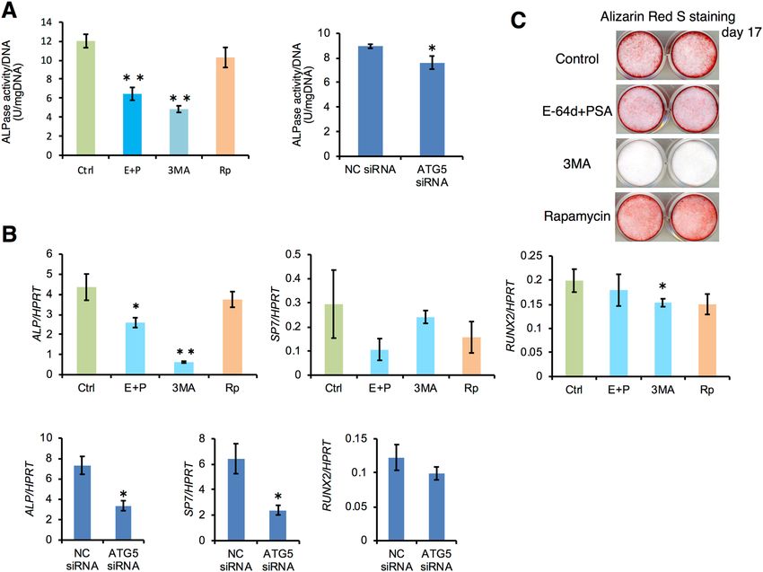
www.nature.com/scientificreports/
Figure 4. Suppression of autophagy increases the deposition of type I and type III human periodontal ligament
(HPDL) cells. (A) Immunofluorescence staining for type I collagen showed the accumulation of collagen
after ossification-inducing culture for 6 days in the presence of autophagy inhibitors, E-64d plus pepstatin
A (10 µg/ml). (B) Immunofluorescence staining for type III collagen showed the accumulation of collagen
after ossification-inducing culture for 6 days in the presence of autophagy inhibitors, E-64d plus pepstatin A
(10 µg/ml). (C) Immunofluorescence staining for type I collagen showed the accumulation of collagen after
ossification-inducing culture for 6 days in the presence of autophagy inhibitors, E-64d plus pepstatin A (10 µg/
ml), 3-methyladenine (3-MA, 10 µM) or rapamycin (250 nM). Representative data from three independent
experiments are shown.
roles during wound healing and tissue regeneration in periodontal tissue. In this study, we first examined whether
autophagy is induced in HPDL cells under physiological conditions such as nutrient starvation. Three distinct
lines of evidence (anatomical analysis by TEM, imaging by confocal microscopy, and biochemical analysis)
strongly suggested the presence of autophagy machinery in HPDL cells to protect against cellular stress (Figs. 1
and 2). Then, we examined the role of autophagy in ECM production, especially with regard to collagen; subse-
quently, we examined the ability of HPDL cells to differentiate into hard tissue-forming cells.
Collagen, a major ECM protein, is the most abundant protein in skeletal tissue. PDL is almost entirely
composed of collagen; gingival connective tissue, bone, and cementum around the tooth root are also rich in
collagen9. Notably, type I collagen has been reported to undergo protein degradation by an autophagy-dependent
system in mammalian c ells33. Because collagen metabolism is more rapid in PDL cells than in other fibroblastic
cells34, we hypothesised that autophagy was the prominent system for the control of collagen in HPDL cells. To
determine the effect of autophagy on collagen synthesis, we used different approaches to inhibit autophagy in
HPDL cells: inhibition of autolysosomal protein degradation with E-64d and pepstatin A treatment, as well as
ATG5 knockdown by specific siRNA (Figs. 4 and 5A). Our results showed that inhibition of autophagy induced
the accumulation of collagen in the cytosol and caused reduction of collagen production from HPDL cells (Figs. 3
and 4). The observation by TEM suggested that the enlarged vacuoles might constitute autolysosomes prior to
autophagic degradation (Fig. 5A). Moreover, immunohistochemical staining of LC3 and LAMP-1 indicated the
existence of type I collagen or its derivatives in lysosomes within HPDL cells (Fig. 5B). Furthermore, the reduc-
tion of LC3-II suggests the process of lysosome-dependent degradation, because expression of LC3-II decreases
after transfer to the lysosome m embrane26.
These results suggest that autophagy prevents accumulation of an excess amount of collagen in HPDL cells in
the ground state. Colocalisation of type I collagen with LC3 or with LAMP-1 was induced by E-64d and pepstatin
A treatment. Thus, our data suggest that autophagy spontaneously participates in the intracellular degradation
pathway for impaired type I collagen synthesis in HPDL cells. Notably, TEM analysis revealed the accumulation
of undegraded protein components in autolysosomes in HPDL cells that had been treated with E-64d and pep-
statin A (Fig. 5A). Taken together, our current results strongly suggest that type I collagen and its related protein
Scientific Reports | (2021) 11:1291 | https://doi.org/10.1038/s41598-020-80275-4 6
Vol:.(1234567890)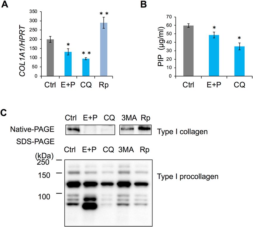
www.nature.com/scientificreports/

                                  Figure 5.  Transmission electron microscopy indicates the accumulation of collagen I in the autophagosome
                                  in human periodontal ligament (HPDL) cells. (A) Transmission electron microscopy showed the induction of
                                  the enlarged autophagosome in the presence of autophagy inhibitors, E-64d plus pepstatin A (10 µg/ml) after
                                  treatment with no-serum Hank’s balanced saline solution (HBSS) for 4 days. Magnified image of the bold square
                                  in was showed in right panels (× 30,600). AP; Autophagosome AL; Autolysosome. (B) Immunofluorescence
                                  staining for the collagen I and LAMP-1 showed the accumulation of collagen after the ossification inducing
                                  culture for 6 days in the presence of autophagy inhibitors, E-64d plus pepstatin A (10 µg/ml). White square
                                  indicates the colocalisation of collagen I and LAMP-1 positive dots. Representative data from three independent
                                  experiments are shown. (C) Immune electron microscopy of autophagosome in aged HPDL cells showed the
                                  collagen I in enlarged autophagosomes following immune-gold labelling for type I collagen. HPDL cells were
                                  treated in the presence of autophagy inhibitors, E-64d plus pepstatin A (10 µg/ml) for 4 days.

                                  products are actively degraded through the lysosome-dependent degradation system in autophagy machinery
                                  of HPDL cells under physiological conditions.
                                      Because E-64d and pepstatin A are inhibitors for enzymes in autolysosomes (i.e., enzymes active late in the
                                  process of autophagy), we speculated that some undegraded protein components in the autolysosomes might
                                  originate from aggregated protein components (e.g., damaged collagen or its products). Consistent with this
                                  hypothesis, immunoelectron microscopic analysis using gold-labelled type I collagen antibody indicated the
                                  involvement of type I collagen or related products in enlarged autolysosomes after E-64d and pepstatin A treat-
                                  ment (Fig. 5C). Notably, mRNA transcription and collagen secretion from HPDL cells were suppressed (Fig. 6);
                                  in contrast, the accumulation of collagen in the cytosol was enhanced (Figs. 4 and 7A). These results suggest a
                                  negative feedback system whereby accumulation of misfolded collagen downregulates mRNA transcription for
                                  collagen synthesis in HPDL cells. Alternatively, HPDL cells themselves may exhibit a cell autonomous safety
                                  system; for instance, they may use an endoplasmic reticulum (ER) sensor mechanism for misfolded or damaged
                                  protein aggregates. Moreover, E-64d and pepstatin A treatment induced the expression of an aberrant mature
                                  collagen band, with a distinct size when separated in denaturing conditions. Concomitantly, the expression level
                                  of soluble mature collagen was reduced in native conditions (Fig. 6C). This condensed band may indicate the
                                  existence of aggregated collagen protein in autophagosomes, which are resistant to reducing conditions. These
                                  data suggest that autophagy may regulate collagen turnover through the clearance of detergent-insoluble, mis-
                                  folded, or non-folded collagen in HPDL cells. In mammalian cells, ER-to-Golgi transport is the first step leading
                                  to the secretory pathway. Collagen is synthesised and packed in vesicles at the ER; these collagen-containing

Scientific Reports |   (2021) 11:1291 |               https://doi.org/10.1038/s41598-020-80275-4                                                 7

                                                                                                                                            Vol.:(0123456789)
www.nature.com/scientificreports/
Figure 6. Autophagy regulates the type I collagen synthesis in human periodontal ligament (HPDL) cells.
(A) Autophagy was required for the mRNA synthesis of type I collagen. HPDL cells after ossification-inducing
culture for 3 days in the presence of autophagy inhibitors, E-64d plus pepstatin A (10 µg/ml), chloroquine (CQ,
10 µM) or autophagy activators rapamycin (10 µM). The relative level of type I collagen mRNA in HPDL cells
was quantified by RT-qPCR. Quantitative mRNA values were normalised to the amount of HPRT mRNA. B,
Inhibition of autophagy suppressed the collagen type I production in culture supernatants. Collagen type I
production in culture supernatants was measured by enzyme-linked immunosorbent assay. **p < 0.01, *p < 0.05
vs the control. Values are the means ± SD. (C) Inhibition of autophagy induced misfolded type I collagen in cell
lysates. Western blotting analysis of collagen type I protein under reducing or native conditions. Cell lysates were
obtained from HPDL cells after ossification-inducing culture for 6 days in the presence of autophagy inhibitors,
E-64d plus pepstatin A (10 µg/ml), chloroquine (CQ, 10 µM), 3-MA (10 µM) or rapamycin (250 nM).
Representative data from three independent experiments are shown.
vesicles are then transported to the Golgi apparatus. After protein modification at the Golgi apparatus, procol-
lagen is exported to the extracellular space for secretion. In general, misfolded proteins are degraded in the ER
associated-degradation (ERAD)-dependent ubiquitin–proteasome s ystem35, whereas high molecular weight,
misfolded collagen (i.e., with a triple helix structure) is degraded in the autophagic s ystem33. In this study, we
could not detect misfolded protein-like products at the ER during TEM analysis (Fig. 5A). Further analysis of
the misfolded collagen in each organelle is needed to determine the fate of misfolded or non-folded collagen
in HPDL cells.
The ER stress response has been reported to accelerate bone formation through osteoblast a ctivity36,37. In
ERAD response-related gene-targeted mice, mRNA transcription of bone substrates and type I collagen was
upregulated in mesenchymal stem cells. In our study, the inhibition of autophagy induced a significant reduc-
tion in ALPase activity and in the expression levels of ossification-related genes during ossification-inducing
culture in HPDL cells (Fig. 8A,B). In contrast, autophagy activation enhanced hard tissue formation of HPDL
cells (Fig. 8C). Thus, cytodifferentiation of HPDL cells into hard tissue-forming cells may not involve an ERAD-
dependent cell system; otherwise, moderate ER stress might be required for hard tissue formation.
Recently, it was reported that autophagy plays an important role in controlling myofibroblast differentiation
in human periodontal soft tissues during wound healing38. It was demonstrated that gingival fibroblasts express
low levels of α-SMA and Col1A1, and that autophagy is not activated in wound healing. This report suggests
Scientific Reports | (2021) 11:1291 | https://doi.org/10.1038/s41598-020-80275-4 8
Vol:.(1234567890)www.nature.com/scientificreports/
Figure 7. The siATG5 treatment increases the accumulation of collagen in human periodontal ligament
(HPDL) cells. (A) Immunofluorescence staining for type I and III collagen showed the accumulation of collagen
after ossification-inducing culture for 6 days in the presence of siATG5 treatment. (B) The relative levels of
type I and type III collagen mRNA in HPDL cells were quantified by RT-qPCR. Quantitative mRNA values
were normalised to the amount of HPRT mRNA. **p < 0.01, *p < 0.05 vs the control. Values are the means ± SD.
Representative data from three independent experiments are shown.
that autophagy activity varies in each cell type of periodontal tissue. In our study, we showed that autophagy is
an important process to maintain homeostasis of the periodontal ligament and hard tissue formation ability of
PDL cells through the synthesis of collagens.
This study had some limitations. It was designed and carried out by using primary HPDL cells in vitro. There-
fore, it is conceivable that experimental periodontitis could be induced in a small animal study with autophagy-
related gene targeting, perhaps involving ATG5 or ATG7. However, in general, autophagy-related gene knockout
mice fail to survive39–43. Thus, the next research focus involves the use of novel gene targeting strategies for
autophagy-related genes, or evaluation systems for autophagy in each organ, to confirm the physiological role
of autophagy in periodontal tissue in vivo. Additionally, we performed most experiments at least three times to
evaluate their reproducibility and accuracy.
Impaired autophagy has been identified in the pathogenesis of various chronic diseases, such as diabetes
mellitus, myocardial infarction, and neurodegenerative diseases18,20,21. Numerous epidemiological and biological
studies have indicated strong correlations between aging-dependent systemic diseases, which are characterised by
chronic inflammation, and periodontal disease. Intriguingly, functional defects of autophagy have been reported
in various aged a nimals44.Therefore, aging-dependent reduction of autophagy may affect biological resistance
against environmental stresses, including bacterial infection and traumatic mechanical stress in periodontal
tissue. Moreover, intrinsic defects of autophagy may accelerate the susceptibility and severity of periodontal
diseases by diminishing HPDL cellular functions and immune responses. To the best of our knowledge, this is
the first study to demonstrate that autophagy regulates collagen production and osteoblastic differentiation of
HPDL cells at the molecular level.
In conclusion, autophagy participates in the regulation of collagen biosynthesis in HPDL cells. Accumulation
of damaged collagen induced by defects in autophagy may result in failed ECM synthesis, thereby inhibiting
osteoblastic differentiation of HPDL cells. The present findings suggest that autophagy machinery is indispensable
for homeostasis in PDL tissue and imply that the autophagy system may serve as a therapeutic target of peri-
odontal diseases. Thus, amelioration of reduced autophagy may accelerate wound healing and tissue regeneration
in periodontal tissue of elderly populations.
Scientific Reports | (2021) 11:1291 | https://doi.org/10.1038/s41598-020-80275-4 9
Vol.:(0123456789)www.nature.com/scientificreports/
Figure 8. Suppression of autophagy inhibits the ALPase activity in human periodontal ligament (HPDL) cells.
(A) Quantification of ALPase activity after 6 days of HPDL cell culture in mineralisation-inducing medium
in the presence of autophagy inhibitors E-64d plus pepstatin A (10 µg/ml), 3-MA (10 µM), or rapamycin
(250 nM), or siATG5 treatment. ALPase activity was determined as described in the Methods section. Activity
in units/mg protein for the cell lysates is shown. **p < 0.01 vs control, ctrl: control; E + P: E-64d plus pepstatin A;
3MA: 3-MA; Rp: rapamycin. (B) Relative quantification of ALP, SP7, and RUNX2 mRNA expression levels was
performed after 6 days of HPDL cell culture in mineralisation-inducing medium in the presence of autophagy
inhibitors, E-64d plus pepstatin A (10 µg/ml), 3-MA (10 µM) or rapamycin (250 nM) or si ATG5 treatment. The
relative level of collagen type I mRNA in human periodontal ligament (HPDL) cells was quantified by RT-qPCR.
Quantitative mRNA values were normalised to the amount of HPRT mRNA. Values are the means ± SD.
*p < 0.05 vs control. **p < 0.01 vs control, ctrl: control; E + P: E-64d plus pepstatin A; 3MA: 3-MA; Rp:
rapamycin; NC: negative control. Representative data from three independent experiments are shown. (C) Hard
tissue formation ability of human periodontal ligament (HPDL) cells was evaluated by Alizarin Red S staining
after ossification-inducing culture in the presence of autophagy inhibitors E-64d plus pepstatin A (10 µg/ml) or
autophagy activator rapamycin (250 nM) in the presence of ascorbic acid (50 mg/mL) plus β-glycerophosphate
(50 mM) at day 17.
Methods
Reagents. E-64D, pepstatin A, Chloroquine, rapamycin, 3-MA, ascorbic acid, or β-glycerophosphate
(Sigma-Aldrich, St. Louis, MO, USA) were applied to cell culture media to treat cells at the indicated concen-
trations and for the indicated periods of time. Hydrogen peroxide was purchased from Wako (Osaka, Japan).
LysoTracker, MitoTracker, and Mito-TEMPO were obtained from Santa Cruz Biotechnology (Dallas, TX, USA).
Establishment of GFP‑LC3 HPDL cells. Analysis of the GFP-LC3 distribution is a widely used method
to monitor the process of autophagy45. HPDL cells were transfected with GFP-LC3 plasmids (Cell Biolabs, San
Diego, CA, USA), using the Neon Transfection system (Thermo Fisher Scientific, Waltham, MA, USA), in
accordance with the manufacturer’s instructions. Because transiently overexpressed GFP-LC3 is likely to mis-
localise with aggregated proteins in cells without autophagy, we established stable GFP-LC3 HPDL cells by drug
selection (Supplementary Fig. S2). GFP-LC3-positive HPDL cells were selected with G418 (Invitrogen, Carlsbad,
CA, USA) treatment for 15 days; GFP-LC3 HPDL cells were then used in experiments.
Scientific Reports | (2021) 11:1291 | https://doi.org/10.1038/s41598-020-80275-4 10
Vol:.(1234567890)www.nature.com/scientificreports/
Transfection. HPDL cells were transfected with siRNAs for ATG-5 using Lipofectamine 2000 (Thermo
Fisher Scientific), in accordance with the manufacturer’s instructions.
RNAi. RNAi for the ATG-5 gene was performed by transfection of siRNA oligos using the Lipofectamine 2000
transfection reagent (Invitrogen), in accordance with the manufacturer’s instructions. The siRNA sequences for
ATG-5 and control were provided by Invitrogen.
Cell culture. Primary HPDL cells (ScienCell Research Laboratories, Carlsbad, CA, USA) at passages lower
than 8 were used in this study. HPDL cells were cultured in α-Minimum Essential Medium (MEM; Wako) sup-
plemented with 10% FBS (Life Technologies, Carlsbad, CA, USA). HBSS was used as the nutrient starvation
medium for HPDL cells. α-MEM without phenol red was used for immunofluorescence analysis of GFP-LC3
HPDL cells.
Quantification of type I collagen in supernatants of HPDL cells. After ossification induction,
supernatants of HPDL cells were harvested on the indicated days and centrifuged at 1000×g for 5 min at 4 °C to
remove debris. Enzyme-linked immunosorbent assay analysis was performed in accordance with the instruc-
tions of the human PIP collagen Kit (PerkinElmer, Waltham, MA, USA).
Picrosirius red staining. HPDL cells were cultured in α-MEM with 2% FBS for 6 days, then fixed with
100% ethanol for 10 min at room temperature. Collagen bundle staining was performed in accordance with
the instructions of the Picrosirius Red Stain Kit (Polysciences, Warrington, PA, USA). Picrosirius red dye stains
both type I and III collagen fibres as bright red, which enables observation of the distribution of collagen fibres
by optical microscopy46. The stained area was observed by optical microscopy (Nikon, Tokyo, Japan). Densito-
metric analysis of the images was performed using the imaging analysis tool WinROOF (Mitani, Fukui, Japan).
Confocal fluorescence microscopy and colocalisation analysis. HPDL cells were grown to subcon-
fluency on glass bottom dishes and then fluorescence microscopy and colocalisation analysis were performed as
described previously47.
Cells were fixed in 4% paraformaldehyde for 15 min at room temperature. After cells had been permeabilised
by incubation with or without 0.25% Triton X-100 or 100 µg/ml digitonin, they were blocked in 1.5% FBS at
room temperature for 1 h. Subsequently, HPDL cells were incubated with primary and secondary antibodies,
then stained with DAPI. Rabbit anti-human type I collagen (Cat. No. ab34710, 1:500, Abcam, Cambridge, MA,
USA), rabbit anti-human type III collagen (Cat. No. ab7778, 1:500, Abcam), mouse anti-human LC3 (Cat. No.
PM036, 1:500, MBL, Woburn, MA, USA), mouse anti-human LAMP1 (Cat. No. ab25245, 1:200, Abcam), Alexa
Fluor 594 Donkey anti-rabbit IgG (Cat. No. A32754, 1:500, Invitrogen), and Alexa Fluor 488 Goat anti-mouse IgG
antibodies (Cat. No. A11001, 1:500, Invitrogen) were used for 30 min at room temperature. Immunofluorescence
microscopy and quantitative image analysis were acquired with a SP8 microscope (Leica, Wetzlar, Germany),
using a 63 × oil immersion lens with a numerical aperture (NA) of 1.4. After acquisition, images were processed
with the Airyscan (Zen software; Carl Zeiss, Oberkochen, Germany). For conventional confocal microscopy, a
confocal laser microscope (Zeiss LSM 510 confocal microscope systems; Carl Zeiss) with a 63 × oil immersion
lens with NA of 1.4. was used.
TEM analysis. TEM analysis and immunogold labelling of HPDL cells have been described previously48.
Briefly, HPDL cells were fixed with 2% paraformaldehyde and 2% glutaraldehyde in 0.1 M phosphate buffer (pH
7.4) at 37 °C, then refrigerated for 30 min to reach a temperature of < 4 °C. Thereafter, the cells were fixed with
2% glutaraldehyde in 0.1 M phosphate buffer at 4 °C overnight. Subsequently, cells were rinsed three times in
0.1 M phosphate buffer for 30 min each; they then underwent post-fixation with 2% osmium tetroxide in 0.1 M
phosphate buffer at 4 °C for 1 h. HPDL cells were dehydrated through a series of graded ethanol incubations. For
embedding and polymerisation, HPDL cells were transferred to a resin (Quetol-812; Nisshin EM, Tokyo, Japan)
and polymerised at 60 °C for 48 h. The blocks were ultra-thin sectioned at 70 nm with a diamond knife using an
ultramicrotome (ULTRACUTUCT; Leica) and sections were placed on copper grids. They were stained with
2% uranyl acetate at room temperature for 15 min, rinsed with distilled water, and then stained with lead stain
solution (Sigma-Aldrich) at room temperature for 3 min. The grids were observed using a transmission electron
microscope (JEM-1200EX; JEOL, Tokyo, Japan) at an acceleration voltage of 80 kV. Digital images (2048 × 2048
pixels) were captured with a charge-coupled device camera (VELETA; Olympus Soft Imaging Solution, Münster,
Germany).
TEM analysis of immunogold labelling. For rapid-freezing, sandwiched samples with gold disks were
frozen in liquid propane at − 175 °C. For freeze-substitution, frozen samples were freeze substituted with 0.2%
glutaraldehyde in acetone and 2% distilled water at − 80 °C for 24 h. Subsequently, samples were transferred to
− 20 °C for 3 h, then warmed to 4 °C for 90 min. For dehydration, samples were dehydrated by incubation in
anhydrous ethanol, three times for 30 min each. For infiltration, samples were infiltrated with a 50:50 mixture
of ethanol and resin (LR White; London Resin, Berkshire, UK) at 4 °C for 60 min. Subsequently, samples under-
went three changes of 100% LR white at 4 °C for 60 min each. For embedding and polymerisation, samples were
transferred to a fresh 100% resin, then polymerised at 50 °C overnight. For ultra-thin sections, blocks were
ultra-thin sectioned at 80 nm with a diamond knife using an ultramicrotome and sections were placed on nickel
grids. For immunostaining, grids were incubated with the primary antibody (rabbit anti-collagen I pAb, Cat. No.
Scientific Reports | (2021) 11:1291 | https://doi.org/10.1038/s41598-020-80275-4 11
Vol.:(0123456789)www.nature.com/scientificreports/
ab34710, 1:100, Abcam) in 1% bovine serum albumin (Wako) and PBS at 4 °C overnight, then washed with 1%
bovine serum albumin and PBS three times for 1 min. Grids were subsequently incubated with the secondary
antibody conjugated to 15-nm gold particles (goat anti-rabbit IgG H&L pAb, Cat. No. ab27236, 1:100, Abcam)
for 90 min at room temperature. After grids had been washed with PBS, they were placed in 2% glutaraldehyde
in 0.1 M phosphate buffer at 4 °C overnight. Subsequently, the grids were dried and stained with 1% uranyl
acetate for 15 min, then stained with lead stain solution (Sigma-Aldrich) at room temperature for 3 min. For
observation and imaging, grids were observed using a transmission electron microscope (JEM-1400Plus; JEOL)
at an acceleration voltage of 80 kV. Digital images (2048 × 2048 pixels) were taken with a charge-coupled device
camera (VELETA; Olympus Soft Imaging Solution).
Slices preparation and imaging analysis were performed according to the protocols of Tokai Electron Micros-
copy (Aichi, Japan).
RT‑qPCR. RT-qPCR was performed according to previously described protocols49.
Total RNA was extracted from cultured cells using a RNeasy kit (Qiagen, Hilden, Germany), in accordance
with the manufacturer’s instructions. Reverse transcription was performed using the High Capacity RNA-to-
cDNA Kit (Applied Biosystems, Foster City, CA, USA). Quantitative real time PCR was performed using an
ABI 7300 Fast Real-Time PCR System with Power SYBR Green PCR Master Mix (Applied Biosystems) and
gene-specific primers (Supplementary Table S1), in accordance with the manufacturer’s instructions. The rela-
tive expression was determined, following normalisation to hypoxanthine phosphoribosyltransferase (HPRT)
expression. The PCR primer sequences are listed in Supplementary Table S1.
Statistical analysis. The data are representative of three independent experiments. All experiments other
than TEM analysis with immune gold labelling were performed at least three times. All quantitative data are
presented as the mean and standard deviation of triplicate assays. Differences between two means were assessed
using Student’s t-tests for two-sample comparisons or one-way analysis of variance for multiple comparisons
with Bonferroni post-hoc tests. P values < 0.05 were considered to indicate significance.
Data availability
All data supporting the findings of this study are available within the article and its supplementary materials.
Received: 20 June 2020; Accepted: 18 December 2020
References
1. Picton, D. C. The periodontal enigma: eruption versus tooth support. Eur. J. Orthod. 11, 430–439. https://doi.org/10.1093/oxfor
djournals.ejo.a036015 (1989).
2. Seo, B. M. et al. Investigation of multipotent postnatal stem cells from human periodontal ligament. Lancet 364, 149–155. https
://doi.org/10.1016/s0140-6736(04)16627-0 (2004).
3. Lekic, P. & McCulloch, C. A. Periodontal ligament cell population: the central role of fibroblasts in creating a unique tissue. Anat.
Rec. 245, 327–341. https://doi.org/10.1002/(SICI)1097-0185(199606)245:2%3c327::AID-AR15%3e3.0.CO;2-R (1996).
4. Lekic, P. C., Rajshankar, D., Chen, H., Tenenbaum, H. & McCulloch, C. A. Transplantation of labeled periodontal ligament cells
promotes regeneration of alveolar bone. Anat. Rec. 262, 193–202. https: //doi.org/10.1002/1097-0185(200102 01)262:2%3c193: :AID-
AR1028%3e3.0.CO;2-7 (2001).
5. Beertsen, W., McCulloch, C. A. & Sodek, J. The periodontal ligament: A unique, multifunctional connective tissue. Periodontology
2000(13), 20–40. https://doi.org/10.1111/j.1600-0757.1997.tb00094.x (1997).
6. Halper, J. & Kjaer, M. Basic components of connective tissues and extracellular matrix: Elastin, fibrillin, fibulins, fibrinogen,
fibronectin, laminin, tenascins and thrombospondins. Adv. Exp. Med. Biol. 802, 31–47. https://doi.org/10.1007/978-94-007-7893-
1_3 (2014).
7. Nohutcu, R. M., McCauley, L. K., Koh, A. J. & Somerman, M. J. Expression of extracellular matrix proteins in human periodontal
ligament cells during mineralization in vitro. J. Periodontol. 68, 320–327. https://doi.org/10.1902/jop.1997.68.4.320 (1997).
8. Reinstein, E. et al. Ehlers-Danlos syndrome type VIII is clinically heterogeneous disorder associated primarily with periodontal
disease, and variable connective tissue features. Eur. J. Hum. Genet. 21, 233–236. https://doi.org/10.1038/ejhg.2012.132 (2013).
9. Yamada, S. et al. Expression profile of active genes in human periodontal ligament and isolation of PLAP-1, a novel SLRP family
gene. Gene 275, 279–286. https://doi.org/10.1016/s0378-1119(01)00683-7 (2001).
10. Mizushima, N. Autophagy: process and function. Genes Dev. 21, 2861–2873. https://doi.org/10.1101/gad.1599207 (2007).
11. Iwata, A., Riley, B. E., Johnston, J. A. & Kopito, R. R. HDAC6 and microtubules are required for autophagic degradation of aggre-
gated huntingtin. J. Biol. Chem. 280, 40282–40292. https://doi.org/10.1074/jbc.M508786200 (2005).
12. Levine, B., Mizushima, N. & Virgin, H. W. Autophagy in immunity and inflammation. Nature 469, 323–335. https://doi.
org/10.1038/nature09782 (2011).
13. He, C. & Klionsky, D. J. Regulation mechanisms and signaling pathways of autophagy. Annu. Rev. Genet. 43, 67–93. https://doi.
org/10.1146/annurev-genet-102808-114910 (2009).
14. Yang, Z. & Klionsky, D. J. Eaten alive: A history of macroautophagy. Nat. Cell. Biol. 12, 814–822. https://doi.org/10.1038/ncb09
10-814 (2010).
15. Farré, J. C. & Subramani, S. Peroxisome turnover by micropexophagy: an autophagy-related process. Trends Cell. Biol. 14, 515–523.
https://doi.org/10.1016/j.tcb.2004.07.014 (2004).
16. Massey, A. C., Kaushik, S., Sovak, G., Kiffin, R. & Cuervo, A. M. Consequences of the selective blockage of chaperone-mediated
autophagy. Proc. Natl. Acad. Sci. USA 103, 5805–5810. https://doi.org/10.1073/pnas.0507436103 (2006).
17. Huang, J. & Brumell, J. H. Bacteria-autophagy interplay: A battle for survival. Nat. Rev. Microbiol. 12, 101–114. https://doi.
org/10.1038/nrmicro3160 (2014).
18. Nakai, A. et al. The role of autophagy in cardiomyocytes in the basal state and in response to hemodynamic stress. Nat. Med. 13,
619–624. https://doi.org/10.1038/nm1574 (2007).
19. Lin, N. Y. et al. Autophagy regulates TNFalpha-mediated joint destruction in experimental arthritis. Ann. Rheum. Dis. 72, 761–768.
https://doi.org/10.1136/annrheumdis-2012-201671 (2013).
Scientific Reports | (2021) 11:1291 | https://doi.org/10.1038/s41598-020-80275-4 12
Vol:.(1234567890)www.nature.com/scientificreports/
20. Ebato, C. et al. Autophagy is important in islet homeostasis and compensatory increase of beta cell mass in response to high-fat
diet. Cell Metab. 8, 325–332. https://doi.org/10.1016/j.cmet.2008.08.009 (2008).
21. Narendra, D., Tanaka, A., Suen, D. F. & Youle, R. J. Parkin is recruited selectively to impaired mitochondria and promotes their
autophagy. J. Cell. Biol. 183, 795–803. https://doi.org/10.1083/jcb.200809125 (2008).
22. Nassar, H., Kantarci, A. & van Dyke, T. E. Diabetic periodontitis: A model for activated innate immunity and impaired resolution
of inflammation. Periodontology 2000(43), 233–244. https://doi.org/10.1111/j.1600-0757.2006.00168.x (2007).
23. Paraskevas, S., Huizinga, J. D. & Loos, B. G. A systematic review and meta-analyses on C-reactive protein in relation to periodon-
titis. J. Clin. Periodontol. 35, 277–290. https://doi.org/10.1111/j.1600-051X.2007.01173.x (2008).
24. de Pablo, P., Chapple, I. L., Buckley, C. D. & Dietrich, T. Periodontitis in systemic rheumatic diseases. Nat. Rev. Rheumatol. 5,
218–224. https://doi.org/10.1038/nrrheum.2009.28 (2009).
25. Kuma, A. & Mizushima, N. Physiological role of autophagy as an intracellular recycling system: with an emphasis on nutrient
metabolism. Semin. Cell. Dev. Biol. 21, 683–690. https://doi.org/10.1016/j.semcdb.2010.03.002 (2010).
26. Klionsky, D. J. et al. Guidelines for the use and interpretation of assays for monitoring autophagy (3rd edition). Autophagy 12,
1–222. https://doi.org/10.1080/15548627.2015.1100356 (2016).
27. Kabeya, Y. et al. LC3, a mammalian homologue of yeast Apg8p, is localized in autophagosome membranes after processing. Embo
J. 19, 5720–5728. https://doi.org/10.1093/emboj/19.21.5720 (2000).
28. Bjorkoy, G. et al. Monitoring autophagic degradation of p62/SQSTM1. Methods Enzymol. 452, 181–197. https://doi.org/10.1016/
s0076-6879(08)03612-4 (2009).
29. Mizushima, N. & Yoshimori, T. How to interpret LC3 immunoblotting. Autophagy 3, 542–545. https://doi.org/10.4161/auto.4600
(2007).
30. Mizushima, N. et al. Dissection of autophagosome formation using Apg5-deficient mouse embryonic stem cells. J. Cell. Biol. 152,
657–668 (2001).
31. Matsushita, M. et al. Structure of Atg5.Atg16, a complex essential for autophagy. J. Biol. Chem. 282, 6763–6772. https://doi.
org/10.1074/jbc.M609876200 (2007).
32. Takeuchi, H., Furuta, N., Morisaki, I. & Amano, A. Exit of intracellular Porphyromonas gingivalis from gingival epithelial cells is
mediated by endocytic recycling pathway. Cell. Microbiol. 13, 677–691. https://doi.org/10.1111/j.1462-5822.2010.01564.x (2011).
33. Ishida, Y. et al. Autophagic elimination of misfolded procollagen aggregates in the endoplasmic reticulum as a means of cell protec-
tion. Mol. Biol. Cell 20, 2744–2754. https://doi.org/10.1091/mbc.e08-11-1092 (2009).
34. Sodek, J. A comparison of the rates of synthesis and turnover of collagen and non-collagen proteins in adult rat periodontal tissues
and skin using a microassay. Arch. Oral. Biol. 22, 655–665. https://doi.org/10.1016/0003-9969(77)90095-4 (1977).
35. Nakatsukasa, K. & Brodsky, J. L. The recognition and retrotranslocation of misfolded proteins from the endoplasmic reticulum.
Traffic 9, 861–870. https://doi.org/10.1111/j.1600-0854.2008.00729.x (2008).
36. Saito, A. et al. Endoplasmic reticulum stress response mediated by the PERK-eIF2(alpha)-ATF4 pathway is involved in osteoblast
differentiation induced by BMP2. J. Biol. Chem. 286, 4809–4818. https://doi.org/10.1074/jbc.M110.152900 (2011).
37. Tohmonda, T. et al. The IRE1alpha-XBP1 pathway is essential for osteoblast differentiation through promoting transcription of
Osterix. EMBO Rep. 12, 451–457. https://doi.org/10.1038/embor.2011.34 (2011).
38. Vescarelli, E. et al. Autophagy activation is required for myofibroblast differentiation during healing of oral mucosa. J. Clin. Peri-
odontol. 44, 1039–1050. https://doi.org/10.1111/jcpe.12767 (2017).
39. Sou, Y. S. et al. The Atg8 conjugation system is indispensable for proper development of autophagic isolation membranes in mice.
Mol. Biol. Cell 19, 4762–4775. https://doi.org/10.1091/mbc.E08-03-0309 (2008).
40. Kuma, A. et al. The role of autophagy during the early neonatal starvation period. Nature 432, 1032–1036. https: //doi.org/10.1038/
nature03029 (2004).
41. Komatsu, M. et al. Impairment of starvation-induced and constitutive autophagy in Atg7-deficient mice. J. Cell. Biol. 169, 425–434.
https://doi.org/10.1083/jcb.200412022 (2005).
42. Saitoh, T. et al. Loss of the autophagy protein Atg16L1 enhances endotoxin-induced IL-1beta production. Nature 456, 264–268.
https://doi.org/10.1038/nature07383 (2008).
43. Saitoh, T. et al. Atg9a controls dsDNA-driven dynamic translocation of STING and the innate immune response. Proc. Natl. Acad.
Sci. USA 106, 20842–20846. https://doi.org/10.1073/pnas.0911267106 (2009).
44. Mizushima, N. A brief history of autophagy from cell biology to physiology and disease. Nat. Cell. Biol. 20, 521–527. https://doi.
org/10.1038/s41556-018-0092-5 (2018).
45. Kuma, A., Matsui, M. & Mizushima, N. LC3, an autophagosome marker, can be incorporated into protein aggregates independent
of autophagy: Caution in the interpretation of LC3 localization. Autophagy 3, 323–328. https://doi.org/10.4161/auto.4012 (2007).
46. Junqueira, L. C., Bignolas, G. & Brentani, R. R. Picrosirius staining plus polarization microscopy, a specific method for collagen
detection in tissue sections. Histochem. J. 11, 447–455. https://doi.org/10.1007/BF01002772 (1979).
47. De Leo, M. G. et al. Autophagosome-lysosome fusion triggers a lysosomal response mediated by TLR9 and controlled by OCRL.
Nat. Cell. Biol. 18, 839–850. https://doi.org/10.1038/ncb3386 (2016).
48. Polishchuk, E. V., Polishchuk, R. S. & Luini, A. Correlative light-electron microscopy as a tool to study in vivo dynamics and
ultrastructure of intracellular structures. Methods Mol. Biol. 931, 413–422. https://doi.org/10.1007/978-1-62703-056-4_20 (2013).
49. Kawahara, T. et al. TGF-beta negatively regulates the BMP2-dependent early commitment of periodontal ligament cells into hard
tissue forming cells. PLoS ONE 10, e0125590. https://doi.org/10.1371/journal.pone.0125590 (2015).
50. Bessey, O. A., Lowry, O. H. & Brock, M. J. A method for the rapid determination of alkaline phosphates with five cubic millimeters
of serum. J. Biol. Chem. 164, 321–329 (1946).
51. Bradford, M. M. A rapid and sensitive method for the quantitation of microgram quantities of protein utilizing the principle of
protein-dye binding. Anal. Biochem. 72, 248–254. https://doi.org/10.1006/abio.1976.9999 (1976).
52. Laemmli, U. K. Cleavage of structural proteins during the assembly of the head of bacteriophage T4. Nature 227, 680–685. https
://doi.org/10.1038/227680a0 (1970).
Acknowledgements
This study was supported by Grants-in-Aid for Scientific Research (18K17063, 23659976, 17H04416, 20K18532)
by the Japan Society for the Promotion of Science. We thank Ryan Chastain-Gross, Ph.D., and Mitchell Arico
from Edanz Group (https://en-author-services.edanzgroup.com/ac) for editing a draft of this manuscript.
Author contributions
T.N., M.Y., K.I., M.S., M.Y., J.K., M.K., and S.M. conceived experiments; T.N., M.Y., K.I., and M.S. performed
the experiments; T.N., M.Y., and S.M. analysed the data; T.N., M.Y., and S.M. wrote the manuscript; all authors
reviewed the manuscript.
Scientific Reports | (2021) 11:1291 | https://doi.org/10.1038/s41598-020-80275-4 13
Vol.:(0123456789)You can also read