Tryptophan, a non-canonical melanin precursor: new L-tryptophan based melanin production by Rubrivivax benzoatilyticus JA2 - Nature
←
→
Page content transcription
If your browser does not render page correctly, please read the page content below
www.nature.com/scientificreports

            OPEN           Tryptophan, a non-canonical
                           melanin precursor: New
                           L-tryptophan based melanin
                           production by Rubrivivax
                           benzoatilyticus JA2
                           Shabbir Ahmad1, Mujahid Mohammed1,3, Lakshmi Prasuna Mekala1,4, Sasikala Chintalapati2
                           & Venkata Ramana Chintalapati1 ✉
                           Melanins are chemically diverse ubiquitous pigments found across the life forms synthesized via
                           different biochemical pathways mainly from L-tyrosine or acetyl CoA. Though few reports suggest the
                           possibility of tryptophan-based melanin synthesis, however, such tryptophan-based melanin and its
                           biosynthesis remained a biochemical riddle. Here we report tryptophan-based melanin production
                           by bacterium, Rubrivivax benzoatilyticus JA2. Aerobic cultures of strain JA2 produced brown pigment
                           when grown on L-tryptophan-containing media. Purified pigment showed typical physico-chemical
                           properties of melanin. Further, extensive spectroscopic studies revealed that pigment is an amorphous,
                           indole-type polymer with stable free radical centers. Further, hydrolysis of the brown pigment revealed
                           the presence of indole moiety, confirming the indolic nature of the pigment. Demonstration of in vitro
                           and in vivo pigment synthesis directly from L-tryptophan or hydroxytryptophan confirms tryptophan-
                           based melanin synthesis in strain JA2. Interestingly, canonical melanin biosynthetic inhibitors did not
                           affect the pigment synthesis indicating possible non-canonical tryptophan-based melanin biosynthesis
                           in strain JA2. Further, the exometabolite profiling and precursor feeding studies suggests that
                           L-tryptophan converted to hydroxytryptophan/hydroxyindoles and their subsequent polymerization
                           lead to the formation of melanin. The current study sheds light on biosynthetic diversity of melanins
                           and L-tryptophan can be a potential precursor for melanin synthesis in life forms.

                           Microorganisms are prolific producers of a diverse array of pigments and these pigments are produced under dif-
                           ferent physiological conditions. The pigments are structurally diverse ranging from simple low molecular organic
                           compound to complex macromolecules such as melanins1. Melanins are structurally complex, ubiquitous heter-
                           ogenous polymeric pigments found in all biological systems2–5. Melanins are enigmatic biopolymers formed by
                           oxidative polymerization of phenolic and hydroxyindole derivatives3,6. Melanins display different colors such as
                           black, brown to yellow and are highly hydrophobic amorphous polymers2,3,6. Melanins are negatively charged3
                           and contain stable free radicals, a characteristic feature of melanins and are highly resistant to degradation4,5,7.
                               Melanins display remarkable chemical diversity and are classified as eumelanin, pheomelanin, pyomelanin,
                           allomelanin, neuromelanin and DHN melanin based on the color and chemical composition of the melanin3,8,9.
                           Eumelanin, pheomelanin, pyomelanin, and neuromelanin are synthesized from L-tyrosine3,8 whereas, DHN mel-
                           anins are synthesized from acetyl CoA or malonyl CoA as a precursor7. Eumelanins contain nitrogen, pheom-
                           elanin contains both nitrogen and sulfur while allomelanin or DHN melanin contains neither of them3,8. Diverse
                           life forms synthesize different melanins, for example, several groups of fungi produce mainly nitrogen-free
                           DHN melanin as well as nitrogenous DOPA melanin6,7,10. Eumelanins are mainly found in all animals imparting

                           1
                            Department of Plant Sciences, School of Life Sciences, University of Hyderabad, Hyderabad, 500046, India.
                           2
                            Centre for Environment, IST, JNT University, Hyderabad, 500 085, India. 3Present address: Department of Botany,
                           Bharathidasan Government College for Women, Puducherry, U.T. – 605003, India. 4Present address: Department
                           of Plant Sciences, Avvaiyar Government College for Women, Karaikal, Puducherry, U.T 609602, India. ✉e-mail:
                           cvramana449@gmail.com

Scientific Reports |   (2020) 10:8925 | https://doi.org/10.1038/s41598-020-65803-6                                                           1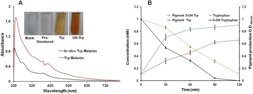
www.nature.com/scientificreports/                                                                www.nature.com/scientificreports

                           Figure 1. Growth, tryptophan utilization and brown pigment production by Rubrivivax benzoatilyticus JA2
                           under aerobic conditions. Values are the mean ± standard deviation of two biological replicates.

                           different colors and few microorganisms also produce eumelanin2,3,8. Except for the DHN melanin, all other types
                           of melanins are synthesized from, tyrosine/phenylalanine. On the other hand, DHN melanins are synthesized
                           mainly from acetyl CoA/malonyl CoA using the polyketide synthase enzyme system7,8,10. Though melanins are
                           chemically diverse their biosynthetic origin is fairly simple starts from tyrosine or acetyl CoA as a precursor8,11.
                           However, a recent study revealed a non-canonical Asp-melanin biosynthesis in Aspergillus terreus indicating mel-
                           anin biosynthetic diversity in life forms12. Similarly, few in vivo studies in animal models13,14 and in vitro oxidation
                           studies suggested tryptophan can be a potential precursor for melanin synthesis14,15. However, tryptophan-based
                           (named as Trp-melanin) melanins are neither characterized nor their biosynthetic pathway is identified and more
                           so there are no reports of Trp-melanins in microorganisms.
                               Anoxygenic photosynthetic bacteria are metabolically versatile yet less explored group of bacteria capable of
                           producing different biomolecules such as carotenoid pigments16,17 and melanin using aromatic amino acids as
                           precursors18. Rubrivivax benzoatilyticus JA2 is one such a photosynthetic bacterium with remarkable aromatic
                           compound biotransformation abililty19–22. Studies on aromatic compound metabolism by strain JA2 revealed the
                           production of several value-added compounds22,23 and multiple catabolic pathways22,24. Recently we reported
                           pyomelanin production by aerobic cultures of strain JA2 and genomic and metabolic insights revealed pyomela-
                           nin biosynthetic pathway in strain JA218 Employing the newly developed metabolite-centric approach we identi-
                           fied anthocyanin-like pigment production in phenylalanine-amended aerobic cultures of strain JA219. Our recent
                           studies on aerobic aromatic metabolism of strain JA2 revealed new biomolecules and metabolic pathways18,19,24.
                           Similarly, while working on aerobic L-tryptophan metabolism in strain JA2 surprisingly we found melanin-like
                           pigment synthesis in aerobic tryptophan amended cultures. In the present study, we report a tryptophan-based
                           melanin production for the first time in a microorganism and characterized the novel melanin produced by
                           strain JA2. The study also suggests a possible non-canonical route of tryptophan-based melanin (Trp-melanin)
                           synthesis.

                           Results
                           Growth, L-Tryptophan utilization, and pigment production. Strain JA2 could grow on L-tryptophan
                           as a nitrogen source under aerobic conditions and utilized 90% of L-tryptophan within 12 h of incubation (Fig. 1).
                           Strain JA2 produced brown pigment with concomitant utilization of L-tryptophan and pigment production was
                           higher at 12 h wherein the maximum amount of tryptophan was utilized (Fig. 1). The pigment was produced
                           only in tryptophan-containing aerobic cultures while no pigment was observed in L-tryptophan-amended
                           anaerobic as well as control (without tryptophan) aerobic cultures (Fig. S1). The pigment produced only in
                           L-tryptophan-containing media inoculated with strain JA2 and pigment was not formed in un-inoculated tryp-
                           tophan-containing media. The pigment produced by strain JA2 was purified from acidified culture supernatants
                           of L-tryptophan-amended aerobic cultures and upon acidification, the pigment settled as a brown precipitate
                           (Fig. 2A). The dried pure pigment appeared as dark brown (Fig. 2B) and the yield of the pigment was 33 ± 3 mg
                           dry weight per 0.5 liters.

                           Physicochemical properties of purified brown pigment.                  The purified pigment was soluble only in
                           alkaline solution (1 M NaOH) and insoluble in organic solvents (hexane, chloroform, acetone, ethyl acetate, eth-
                           anol, benzene) as well as water. Pigment is insoluble in neutral buffers and sparingly soluble in alkaline buffers
                           (pH:10–12) (Table S1). The pigment readily precipitated in acidic conditions (5 M HCl), bleached when treated
                           with oxidizing agents like H2O2 and NaOCl (Table S1). The pigment gave a positive reaction to the polyphenol
                           test by forming flocculent brown precipitate reacting with FeCl3 (Table S1). Pigment gave positive to ammonical
                           silver nitrate test and Na2S2O4 addition decolorized the pigment, upon adding potassium ferricyanide colorless

Scientific Reports |   (2020) 10:8925 | https://doi.org/10.1038/s41598-020-65803-6                                                               2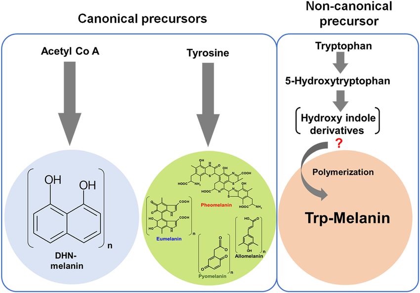
www.nature.com/scientificreports/ www.nature.com/scientificreports
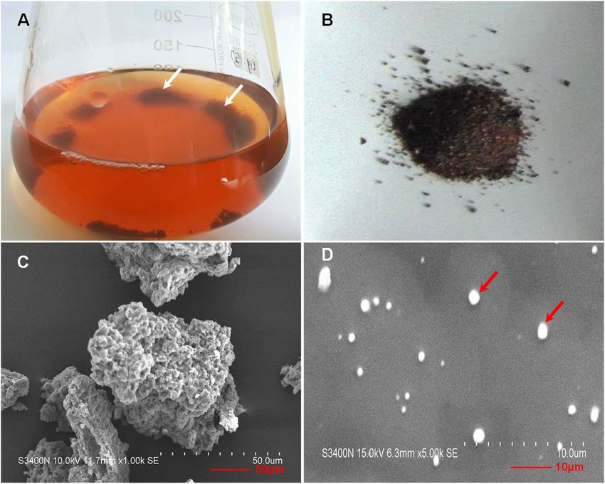
Figure 2. Image showing the brown pigment in acidified culture supernatant obtained from tryptophan-
amended aerobic culture of strain JA2 (A), Dry purified pigment (B), SEM micrograph showing aggregated
granules of pigments (C) and dispersed individual spherical granules of pigment (D).
pigment turned brown. The purified brown pigment was positive to all the characteristic chemical tests used to
identify melanins25 (Table S1).
Characterization of brown pigment revealed that the pigment is melanin. Scanning electron
micrographs of the purified brown pigment revealed unorganized aggregated spherical granules of pigment
(Fig. 2C) while the SEM micrograph of the sonicated sample showed dispersed typical sphere-like structures with
the homogenous smooth surface characteristic of typical melanin26 (Fig. 2D). The UV-Visible spectrum of puri-
fied pigment showed a broad absorption spectrum from UV to the visible region and the absorption was higher in
the UV compared to visible region (Fig. 3A). Absorption of brown pigment decreased progressively with increas-
ing wavelength showing a linear correlation between absorption and wavelength (Fig. S2), a characteristic feature
of melanins26,27. Elemental analysis of purified brown pigment revealed the presence of C, H, N, O, and no sulfur
was detected. Pigment consisted of carbon-46.73%-, hydrogen-5.26%, nitrogen-9.16% and oxygen-38.38% and
high C/H ratio (8.9%) of brown pigment indicates the presence of many fused aromatic structures in the pig-
ment26,28. Presence of high nitrogen content (9%) and absence of sulfur moiety indicates that brown pigment is a
nitrogen-containing pigment.
FTIR spectrum of the purified pigment exhibited the absorption bands typical to a functional moiety of mel-
anin. The broad band at 3276 cm−1 can be assigned to the –OH and –NH stretching vibrations and the bands at
2928–2802 cm−1 ascribed to CH3, -CH2 stretching (Fig. 3B). The band at 1723 cm−1 assigned to C = O stretching
vibrations from carbonyl, carboxyl, ketone or quinone groups present in the pigment29. Strong absorption at 1626
cm−1 can be assigned to C = C stretching vibrations of aromatic/pyrrole moiety29 (Fig. 3B). The band at 1526 cm−1
can be designated to the N-H bending along with the band at 1414 cm−1 (C-N stretching) suggests the presence
of indole/pyrrolic functional groups in the structure of the pigment27,29. Absorption at 1220 cm−1, indicative of
–C-OH group of a phenolic or indolic moiety of pigment30 (Fig. 3B). The band at 1048 cm−1 can be ascribed to
the CH- in-plane or CH- out plane deformations and abruption below 700 cm−1 corresponds to out of plane
carbon-hydrogen bending of the aromatic moiety30. The IR spectroscopic properties of the pigment of strain JA2
closely correlated with eumelanin2,5,30.
Electron Spin Resonance (ESR) analysis of purified brown pigment revealed the singlet ESR signal at 336.46
mT corresponding to a g-value of 2.00634 (Fig. 3C). Singlet ESR signal observed in the ESR spectrum of brown
pigment indicates the presence of stable free-radicals (paramagnetic) a characteristic feature used to identify mel-
anins7,27. X-ray diffraction analysis of the purified pigment showed a broad diffraction peak around 2θ = 10–50°
suggestive of the amorphous nature of the pigment (Fig. 3D) and this in agreement with previous reports on
Scientific Reports | (2020) 10:8925 | https://doi.org/10.1038/s41598-020-65803-6 3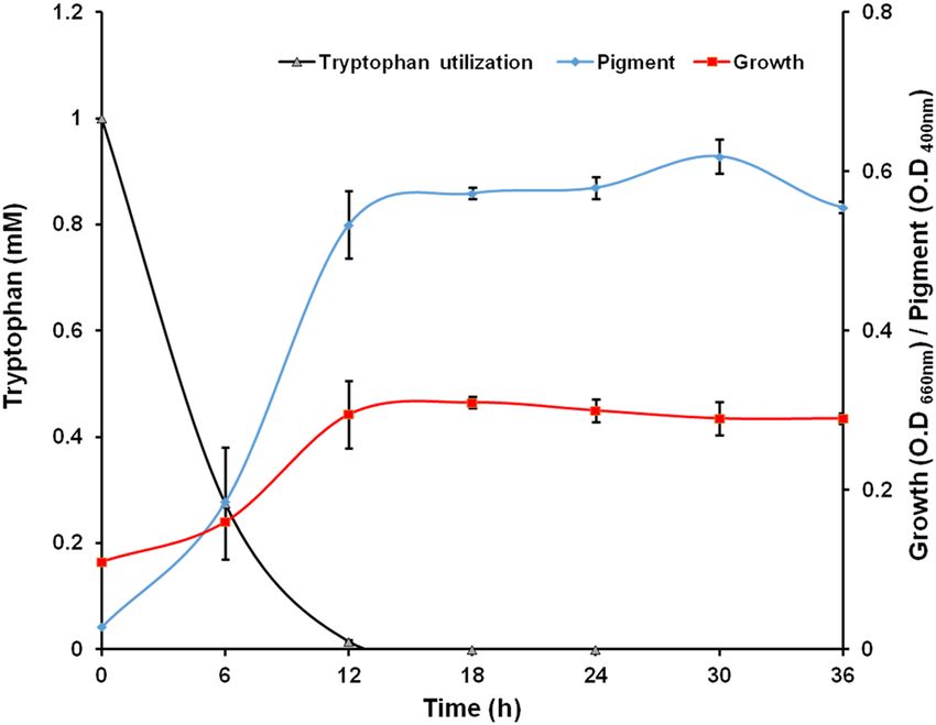
www.nature.com/scientificreports/                                                              www.nature.com/scientificreports

                           Figure 3. Spectroscopic characterization of purified brown pigment. (A) UV-Visible spectrum; (B) FTIR
                           spectrum; (C) ESR spectrum; (D) X-Ray Diffraction spectrum.

                           melanins18. Amorphous compounds like melanins which do not have any regular repeating units display broad
                           diffraction features18.

                           13
                             C and 15N solid-state NMR studies. The 13C solid-state NMR of purified brown pigment showed the
                           intense signal at 160–180 ppm in carboxyl region is usually designated as carbonyl carbon of carboxylates, and
                           quinones possibly associated with a pigment (Fig. 4A)2,31,32. Chemical shifts from 110–150 ppm correspond to
                           aromatic carbons and 100–134 ppm with a broad and weak signal at 126, 110 ppm designated to aromatic car-
                           bons of indole/pyrrole moiety2,31,32. Peak around 156 ppm indicates the presence of C-NH- group, possibly of
                           pyrrole/indole moiety2. The intense signal from 10–45 ppm is due to side-chain aliphatic carbons correspond to
                           methyl, methylene and methine groups (Fig. 4A)2,31,32. Chemical shifts range from 45–60 ppm can be assigned to
                           backbone carbon of α-C, and β-carbons. The 13C solid NMR spectrum of the brown pigment is similar to that of
                           eumelanin spectrum2,31,32.
                               The solid-state 15N NMR CP/MAS spectral studies were carried out to further confirm the presence of the
                           nitrogen in the brown pigment. The 15N NMR spectrum showed an intense broad chemical shift ranging from
                           110–140 ppm which is consistent with a chemical shift of the nitrogen atom of indole/pyrrole moiety31,33 (Fig. 4B).
                           The 15N spectrum of brown pigment is similar to that of the 15N spectrum of indole-type Sepia and human hair
                           melanin31.

                           Effect of canonical melanin inhibitors on brown pigment formation. Pigment production by strain
                           JA2 was monitored in the presence of different inhibitors of known melanin biosynthetic pathways. Glyphosate,
                           an inhibitor of tyrosine dependent melanin synthesis11, as well as quercetin, kojic acid, a eumelanin inhibitor2 has
                           no effect on pigment production (Fig. 5A). Similarly, pigment production was unaffected in the presence of sul-
                           cotrione (visual observation), a pyomelanin inhibitor11,18. Interestingly sodium-azide, a laccase specific inhibitor
                           significantly inhibited the pigment production (Fig. 5A).

                           HRMS based exometabolite profiling of Trp-fed aerobic cultures. LCMS based exometabolite pro-
                           filing was carried out to identify the indole derivatives of aerobic tryptophan metabolism and potential immediate
                           precursor/intermediates of pigment synthesis. Typical total ion chromatogram of methanolic extracts of aero-
                           bic Trp-amended culture supernatants represented here showing the elution profile of the tryptophan-derived
                           metabolites (Fig. S3). LCMS analysis of a methanolic extract of aerobic Trp culture supernatants revealed metab-
                           olite showing identical UV-Visible profile, Rt 7.9 mins having molecular ion mass of 219.0778 [M-] correspond-
                           ing to 5-hydroxytryptophan (Table 1). Metabolite with Rt 14.3 mins also showed identical UV-visible spectrum
                           and molecular ion mass of 190.0509 [M-] correspond to 5-hydroxyindole-3-acetic acid (Table 1). Subsequently,
                           metabolites with Rt 7.9 and 14.3 mins were confirmed as 5-hydroxytryptophan and 5-hydroxyindole-3-acetic acid
                           by co-eluting with authentic standards. Metabolites with Rt 10.9, 15.5 and 16.3 mins having molecular ion masses

Scientific Reports |   (2020) 10:8925 | https://doi.org/10.1038/s41598-020-65803-6                                                           4
www.nature.com/scientificreports/ www.nature.com/scientificreports
Figure 4. Nuclear Magnetic Resonance spectra of purified brown pigment. (A) Solid-state CPMAS 13C and (B)
15
N NMR spectra.
of 206.04, 218.04, and 147.06[M-], were tentatively identified as 5,6-dihydroxyindole-3-acetic acid (DHIAA),
5-hydroxyindole-3-pyruvate (HIPy), and 5-hydroxy-methyl-indole (HMI) respectively based on mass spectra
search against database hit and UV-visible profiles.
Brown pigment formation from L-tryptophan and 5-hydroxytryptophan. As we found
5-hydroxytryptophan (OH-Trp) in culture supernatants of Trp-amended cultures, we checked the pigment pro-
duction in the presence of Trp and OH-Trp in strain JA2. Interestingly, strain JA2 produced intense brown pig-
ment in OH-Trp containing media (Fig. 5B) while no pigment was formed in uninoculated OH-Trp containing
media. The pigment production was significantly higher in the presence of OH-Trp compared to tryptophan
(Fig. 5B). Pigment purified from OH-Trp-containing cultures showed similar physicochemical properties and
UV-Visible profile with that of pigment obtained from Trp cultures (data not shown).
In vitro pigment formation by strain JA2. Strain JA2 showed in vivo pigment formation from Trp as well
as OH-Trp containing media under aerobic conditions. Here we tried to replicate the pigment formation in vitro
by exogenously supplying L-Trp and OH-Trp to cell-free extracts obtained from tryptophan supplied aerobic
cultures. In vitro study revealed the pigment formation in L-Trp and OH-Trp supplied cell-free reactions while
pigment formation was not observed in pre-denatured cell-free extracts or reaction mixture without Trp/OH-Trp
(Fig. 6A). Pigment intensity was high in OH-Trp compared to L-Trp supplemented in vitro assays and purified
in vitro pigment displayed similar UV-Visible profile (Fig. 6A). In vitro studies on pigment formation revealed
simultaneous consumption of L-Trp or OH-Trp and formation of pigment over a period of time. Further, the
pigment content was high in case of OH-Trp compared to L-Trp (Fig. 6B).
Brown pigment hydrolysis and chemical oxidation confirms the indolic nature of the poly-
mer. Chemical hydrolysis of the brown pigment was performed to confirm the presence of indole derivatives
in brown pigment. TLC analysis and indole specific staining of hydrolyzed products obtained from tryptophan
(Trp) and hydroxytryptophan (OH-Trp) brown pigments revealed the identical staining pattern in the form of
two purple bands indicative of indole moiety (Fig. S4). While staining was not observed in case of unhydrolyzed
fractions (methanolic washout) of Trp and OH-Trp pigments indicating that indoles were indeed derived from
hydrolyzed brown pigments (Fig. S4). Further chemical oxidation of brown pigment and subsequent HR-LCMS
analysis indicated presence of molecular ion mass of 198.0 [M-H] and 241.8 [M-H] corresponding to the
pyrrole-2,3,5-tricarboxylic acid(PTCA) and pyrrole-2,3,4,5-tetracarboxylic acid (PTeCA), respectively (Fig. S5).
Further PTCA and PTeCA, were confirmed by mass spectral fragmentation pattern, UV spectra and co-elution
Scientific Reports | (2020) 10:8925 | https://doi.org/10.1038/s41598-020-65803-6 5www.nature.com/scientificreports/ www.nature.com/scientificreports
Figure 5. Brown pigment production by strain JA2 in the presence of various inhibitors and substrates.
(A) Effect of canonical melanin specific biosynthetic pathway inhibitors on brown pigment production under
tryptophan-amended aerobic conditions. (B) Brown pigment production by strain JA2 grown in the presence of
tryptophan and 5-hydroxytryptophan. Values are the mean ± standard deviation of three biological replicates.
**P- valuewww.nature.com/scientificreports/ www.nature.com/scientificreports
Figure 6. In vitro formation of brown pigment in the presence of tryptophan and 5-hydroxytryptophan. (A) In
vitro pigment formation. (B) Time-dependent in vitro pigment formation. Figure shows UV spectra of pigment
obtained from in vivo and in vitro assays from Trp, Inserts shows in vitro pigment formation.
of new biomolecules16,35,36, catabolic pathways19,20,22–24,35,37, and enzymes reported24,36. Recently we designed a new
metabolite-centric secondary metabolite mining strategy and employing this tool we successfully demonstrated
novel anthocyanin–like pigment production in phenylalanine-amended aerobic cultures of strain JA219. Similarly,
while working on aerobic metabolism of L-tryptophan, serendipitously we found brown pigment production in
strain JA2. Interestingly, only Trp-amended aerobic cultures of strain JA2 produced brown pigment with con-
comitant utilization of Trp and this suggests that pigment synthesis is Trp-dependent oxidative metabolic process.
Surprisingly, the purified pigment displayed the characteristic properties of melanin such as alkaline solubility,
insoluble in organic solvents, positive to polyphenol test and decolorizing to oxidizing agent25 indicating that the
brown pigment is melanin (Table S1). Furthermore purified brown pigment showed typical UV-Visible profile of
melanin; higher UV absorption and absorbing all the wavelengths in the visible region further confirms that the
pigment is melanin (Fig. 3A). Melanins are amorphous polymers with stable free radical centers7,27, in agreement
with this, broad diffraction peak and singlet ESR signal displayed by the brown pigment (Fig. 3C,D) of strain JA2
strongly suggests it is an amorphous pigment having free radical centers. Physico-chemical and spectroscopic
studies unequivocally confirm that the brown pigment is indeed a melanin. Melanins are known to be synthesized
during the oxidative metabolism of phenylalanine/tyrosine8,18,24,33, in contrast, here we report oxidative metabo-
lism of tryptophan leading to Trp-melanin formation in strain JA2. Further, the high nitrogen content (9%) and
C/H ratio (8.9%) (typical of aromatic polymers)26,28 revealed by elemental analysis of brown pigment indicate that
the pigment is a nitrogen-containing aromatic polymer.
This is further supported by IR, NMR analysis wherein brown pigment showed characteristic signals of
hydroxy, indolic/pyrrolic and aliphatic moieties. Moreover, a broad signal in 15N NMR of brown pigment con-
firms the presence of pyrrole/indolic nitrogen atom (Fig. 4B) and similar 15N chemical shift was displayed by
indole-based eumelanins31,33. In addition, TLC analysis of the degradation products of brown pigment revealed
the presence of indole moiety (Fig. S4) confirming the indolic nature of the brown pigment. Presence of high
nitrogen and absence of sulfur content further confirms that brown pigment is indole-type melanin and this is in
agreement with the previous reports26 and also rules out the possibility of brown pigment being DHN or pheom-
elanin. Interestingly, brown pigment showed similar spectroscopic features of eumelanin (indole-type melanin)
derived from tyrosine. In contrast, here for the first time, we identified and characterized indole-type melanin
derived from L-tryptophan in a bacterium.
Tyrosine or acetyl CoA derived melanin biosynthesis is well established while Trp-based melanin biosynthe-
sis is a mystery13–15. To gain the insights into the Trp-melanin biosynthesis, first, the genome of strain JA2 was
mined for the genes of canonical melanin biosynthesis. Interestingly, strain JA2 genome lacks candidate genes for
synthesis of DHN, DOPA, and eumelanin except for the pyomelanin pathway genes. Furthermore, biosynthetic
inhibitors of DOPA-melanin/eumelanin (Kojic acid)4, pyomelanin (Sulcotrione)18 and de novo tyrosine-based
melanin inhibitor glyphosate did not inhibit the Trp-melanin synthesis in strain JA2 (Fig. 5A) suggesting that
the Trp-melanin is not synthesized from canonical tyrosine-based melanin biosynthetic process. However, pig-
ment synthesis is inhibited by sodium azide, a laccase inhibitor4 and this implies the possible role of laccase in
Trp-melanin synthesis. Role of laccase in melanin synthesis is reported in few microorganisms4,8, although we did
not find gene coding for laccase, we speculate laccase-like enzyme role in Trp-melanin synthesis by strain JA2 as
laccase-like enzyme role is reported in melanin synthesis in few bacteria38,39.
Eumelanin is a polymer of 5,6-dihydroxyindole (DHI) and 5,6-dihydroxyindole-2-carboxylic acid (DHICA),
synthesized from tyrosine via DOPA, DOPA-chrome finally to DHI, DHICA, and their polymerization lead to
eumelanin formation3,8. In contrast, for Trp-melanin, tryptophan acts as a precursor and its oxidative metabolism
possibly leading to the formation of hydroxyindoles and their subsequent polymerization to melanin (Fig. 7). In
support of this, we found OH-Trp and other hydroxyindole derivatives in the exometabolome of Trp-amended
aerobic cultures of strain JA2 (Table 1). Hydroxyindole derivatives and brown pigment formation were not
found in anaerobic cultures and on the other hand correlation between hydroxyindoles and pigment formation
in aerobic cultures indicates possible role of hydroxyindoles in pigment formation. Furthermore, significantly
higher brown pigment production in the presence of OH-Trp compared to Trp (Fig. 5B) suggests that OH-Trp
Scientific Reports | (2020) 10:8925 | https://doi.org/10.1038/s41598-020-65803-6 7www.nature.com/scientificreports/ www.nature.com/scientificreports
Figure 7. Schematic representation showing melanin synthesis from canonical precursors and non-canonical
tryptophan-based melanin synthesis in strain JA2. Predicted trp-based melanin biosynthetic pathway in strain
JA2. Metabolites in parenthesis are unknown hydroxyindoles involved in polymerization.
may have readily converted to hydroxyindoles and thereby leading to higher pigment production in strain JA2.
Taken together these findings suggest oxidative metabolism of Trp leading to the formation of hydroxyindole
derivatives and thereby Trp-melanin formation. Similarly, Vekey et al. 1992, reported tryptophan-based melanin
formation during chemical oxidation of Trp using perchloric acid and study revealed that polymer consists of
hydroxyindoles40.
However, our study demonstrates oxidative metabolism of Trp leading to Trp-melanin formation in a biologi-
cal system. Melanins are natural polymers of phenols or hydroxyindoles mostly derived from tyrosine2,3 however
in our study hydroxyindole derivatives are derived directly from Trp. Interestingly, alkaline chemical oxidation of
Trp-melanin revealed presence of PTCA and PTeCA, which are oxidativate products of 5,6-dihydroxyindole-2-c
arboxylic acid (DHICA)5,9,34, thus we speculate that DHICA could be one of the monomer of Trp-melanin.
Alternatively, detection of eumelanin markers (PTCA and PTeCA) suggest co-exsitance of eumelanin along with
Trp-melanin. Whether DHICA is a involved in Trp-melanin formation needs further investigation, however, the
possible role of other hydroxyindole derivatives role in Trp-melanin formation can not be ruled out.
In vitro melanin formation by strain JA2 from Trp or its derivative OH-Trp (Fig. 6) and significantly higher
pigment synthesis from OH-Trp suggests (Figs. 5B and 6) that OH-Trp is a possible immediate precursor of
pigment synthesis. Higher pigment content in the presence of OH-Trp is possible because of availability of high
levels of hydroxyindoles derived from OH-Trp and their subsequent polymerization may have resulted in higher
melanin synthesis. Previous study on Planaria larval eye melanogenesis demonstrated that formation of OH-Trp
is essential for pigment synthesis and suggested possible role of OH-Trp in melanogenesis13. Similarly, in vitro
peroxidase, mediated OH-Trp based melanogenesis is reported elsewhere suggesting the role of OH-Trp in mel-
anogenesis14,15. Based on findings from the current study we hypothesize Trp-melanin synthesis, wherein during
oxidative metabolism, tryptophan, is possibly converted to OH-Trp, thereafter into hydroxyindoles and these
subsequently polymerized to melanin (Fig. 7). Although few chemical oxidation and in vitro studies revealed
Trp-melanin formation, for the first time, our study revealed in vivo Trp-melanin synthesis, characterized the
melanin and study provided important clues on the possible biochemical route of Trp-melanin synthesis.
In conclusion, our study revealed tryptophan-based melanin synthesis by aerobic cultures of photosynthetic
bacterium, Rubrivivax benzoatilyticus JA2. Traditionally indole-type melanins (eumelanin) are known to synthe-
size from tyrosine, however, our study demonstrated novel indole-type melanin (named as Trp-melanin) synthe-
sis from a non-canonical precursor, Trp possibly through an alternative route in strain JA2. Future investigations
on the identification of monomeric constituents and transcriptome studies would reveal chemical diversity and
possible candidate genes involved in Trp-melanin synthesis in strain JA2. Findings from our study strongly sup-
ports the view that tryptophan can be a physiological precursor for melanin synthesis and organisms may have
evolved mechanisms to use non-canonical precursors for melanin synthesis based on their physiological condi-
tions. Our study indicates that enigmatic melanin pigment chemical and biosynthetic diversity are far from over
and suggests possible occurrence of non-canonical melanin biosynthetic process in other life forms.
Scientific Reports | (2020) 10:8925 | https://doi.org/10.1038/s41598-020-65803-6 8www.nature.com/scientificreports/ www.nature.com/scientificreports
Methods
Organism, growth conditions and pigment production. Rubrivivax benzoatilyticus JA2 (ATCC BAA-
35) was used as a model organism for all the experiments and pure culture was maintained and grown photohet-
erotrophically (under anaerobic condition, pH 6.8, 30 ± 2 °C; light 2,400 lux) on minimal media containing
22 mM of malate (carbon source) and 7 mM of ammonium chloride as a nitrogen source in completely filled
culture bottle (250 ml). Photoheterotrophically grown mid-log phase culture (O.D- 0.4 at 660 nm) was used as
inoculum(10%) and culture was grown aerobically in 500/1000 ml conical flasks and incubated at 30±1 °C,
180 rpm in shaker incubator (Innova Eppendorf) for 48 h. Ammonium chloride was replaced with 1 mM of
L-tryptophan as a nitrogen source for induction of pigment synthesis. The growth was measured as a change in
optical density (OD) at 660 nm, while pigment production was measured spectrophotometrically against uninoc-
ulated media as a blank at 400 nm. To check the effect of different melanin inhibitors on brown pigment produc-
tion by strain JA2, glyphosate (1 mM), sodium azide (1 mM), Kojic acid (0.5 mM) and quercetin (0.15 mM) were
added to the aerobic cultures containing L-tryptophan. All the inhibitor studies were carried out with mid-log
phase aerobic cultures of strain JA2. Whenever required 1 mM L-tryptophan replaced with 5-hydroxytrypto-
phan(1 mM; Himedia chemicals) as a nitrogen source to check the pigment production.
Extraction and purification of brown pigment. L-tryptophan amended aerobic cultures of strain JA2
was used to isolate the pigment and after 48 h of incubation, culture was harvested by centrifugation (10,000 rpm
for 10 mins at 4 °C). Later the supernatant was collected and acidified to pH 2.0 using 5 M HCl and acidified
supernatant was stored at 4 °C for 2–4 days. The brown precipitate settled at the bottom of the flask was collected
by centrifugation at 10,000 rpm for 10 mins at 4 °C. The brown pigment was further purified by serial washings
using different organic solvents (10 ml of hexane, acetonitrile, acetone, ethyl acetate, ethanol, and methanol) and
finally with Milli-Q water. The purified pigment was dried under freeze drier (Labcanco, USA) and dried pigment
was used for physicochemical and spectroscopic studies.
Scanning Electron Microscopic (SEM) and UV visible spectroscopic analysis. SEM analysis of
purified pigment was carried out according to Prasuna et al.24 with slight modifications, In brief, 1 mg of pigment
was suspended in 1 ml of acetonitrile, 100 µl of suspended solution or sonicated solution was mounted on a glass
slide and fixed on to the stubs. Samples were subjected to gold sputtering and examined using SEM (Philips XL30
series). UV-Visible spectroscopic studies of purified pigment were done on a spectrophotometer (SHIMADZU,
Japan) and the spectra were recorded from 200–800 nm using 1 M NaOH as blank.
FTIR, ESR and X-ray diffraction analysis. FTIR, the spectrum of brown pigment was recorded on alpha
ATR-FTIR spectrometer (ALPHA T model, Bruker Optics, Germany) Dried sample was loaded on diamond
crystal and the spectrum was recorded from the range of 400–4000 cm−1 at resolution 4 cm−1 with 20–30 scans
in transmittance mode. ESR and XRD analysis of purified pigment was done according to Prasuna et al.18. Briefly,
dried pigment (20 mg) was loaded into the ESR quartz tube and spectra were recorded on JEOL X-band ESR
spectrometer at room temperature. X-ray Diffraction spectrum of purified pigment was recorded using Bruker
D8 Advance, Germany X-Ray Diffractometer. Spectrum was recorded by scanning the pigment using the CuKα
radiation at wavelength 1.5406 A° with a step size of 0.04°, voltage 40 kV, current 40 mA and 2θ range 10–90°.
Solid-state 13C and 15N NMR analysis. The Solid-state 13C and 15N spectra of purified brown pigment
were acquired on an Avance III Bruker Spectro spinning as 800 MHz spectrometers operated using as a tri-
ple resonance probe with 5 mm. Purified melanin packed (20–30 mg) in 13C and 15N probe Bruker operating as
resonance probe with the operating conditions; spinning speed −7 kHz: 1 H 90°; pulse length was 2.83 μs and
the 13C 180° pulse length was 6.52 μs. The contact time −2000 μs, acquisition time was 33 ms and the recycle
delay-8 s with total 10000 scans. The 13C chemical shifts were calibrated relative to tetramethylsilane (0 ppm).
The solid-state 15N NMR CP/MAS spectrum was obtained with the same instrument using N-15 probe Bruker
polarization depends on the 15N—H dipole interaction.
Brown pigment hydrolysis, alkaline oxidation and TLC analysis. The brown pigment degradation
products were identified by hydrolysis of the pigment as described by Ellis and Griffiths 197441 with slight modifi-
cations. Five milligrams of the dried pure brown pigment was added to 0.5 g of KOH in a 5 ml tightly sealed screw
cap glass tube. The mixture was brought to boil on hot water bath (1 h) and the dark color residue thus formed was
allowed to cool. To the dried residue 1.5 ml, distilled water was added and acidified (pH 2) with concentrated HCl
and metabolites were extracted with diethyl ether and dried under rota evaporator (Heidolph, Germany) finally
dissolved in HPLC grade methanol. To detect the indoles, the hydrolyzed fraction was run on TLC (Merk, Silica
gel 60 F254, 20×10 cm, 0.2 mm,) using a mixture of Chloroform: Methanol: Glacial acetic acid (9: 0.95: 0.05 v/v)
as a solvent system and TLC plate was developed using indole-specific TLC reagent prepared as described by
Ehmann et al.42. Alkaline hydrogen peroxide oxidation of brown pigment was carriedout according to Ito et al.
2011 and LCMS analysis was performed as described in HRLC-MS.
Extraction of metabolites and HRLC-MS analysis. Metabolites were extracted from the
tryptophan-amended aerobic cultures of strain JA2 and culture was harvested after 48 h of incubation by centrif-
ugation (10,000 rpm, 4 °C, 10 mins). The supernatant was collected and, the supernatant was dried under vacuum
using rotary flash evaporator (Heidolph, Germany). The dried supernatant was dissolved in 5 ml of HPLC grade
methanol and centrifuged (12,000 rpm, 5 mins), the clear methanolic fraction was collected and freeze dried
under vacuum. Finally, the dried methanolic extract was redissolved in 1 ml MS grade methanol and used for
HPLC and LCMS analysis.
Scientific Reports | (2020) 10:8925 | https://doi.org/10.1038/s41598-020-65803-6 9www.nature.com/scientificreports/ www.nature.com/scientificreports
HRLC-MS ESI analysis was performed according to Prasuna et al.24, Briefly, analysis was performed on 6520
Accurate-Mass Q-TOF LC-MS system(Agilent Technologies) equipped with Agilent 1200 series HPLC with a
photodiode array detector; an auto sampler was used to inject (2 µl) the sample and chromatographic separation
was carried out at 25 °C. Reverse phase column (Phenomenex) C-18 (Luna, 5 µm, 150×4.6 mm) was used to
separate the metabolites using a constant flow rate 0.8 ml/minute of mobile phase consisting of 0.1% glacial acetic
acid in water (v/v) (eluent A) and acetonitrile (eluent B). The gradient program was used to elute the metabo-
lite (60 mins): starting with 1% eluent B followed by 55% within 48 min then step gradient to 100% within 55 min
and held for 5 minutes. The separated analytes were infused into the ESI ion source under full-spectrum scan
mode (50–1000 m/z). Data were collected under negative and positive modes in independent runs. Mass Hunter
Qualitative software (version 6.0, Agilent Technologies) was used to analyze the raw data. The standards PTCA
and PDCA were generously gifted by Prof. Shosuke Ito and Prof. Kazumasa Wakamatsu, Fujita Health University,
Japan. 5-hydroxyindole-3-acetic acid standard was purchased from Sigma-Aldrich (Sigma-Aldrich, H8876).
In vitro melanin production by strain JA2. Log phase aerobic cultures grown on L-tryptophan con-
taining media were used for in vitro melanin production studies and cell-free extracts were prepared according
to Prasuna et al.18. Briefly, the culture was harvested by centrifugation at 4 °C for 10 min 10,000 rpm, collected
pellet was washed thrice with 50 mM Tris-buffer and resuspend in 4 ml of 50 mM (pH 7.5) Tris-HCl buffer. The
cell lysate was prepared by sonicating the cells using sonicator (BANDELIN, Germany 8 cycles, 55% amplitude)
and cell lysate was centrifuged at 16,000 rpm for 20 minutes. The pellet was discarded, the clear supernatant was
collected and was used as an enzyme source for in vitro pigment production. The in vitro assay was carried out
in 4 ml final volume consisting of 1 mM L-tryptophan or OH-Trp as substrate and appropriate amount cell-free
extract and reaction mixture was incubated at 180 rpm, 37 °C for 1 hour in a incubator shaker (Innova). The
reaction was terminated by adding the 300 µl of concentrated HCl and pre-denatured enzyme (with conc. HCl)
and reaction mixture without cell-free extract were used as blanks. After the reaction was terminated the brown
pigment was purified as described in material and methods earlier. For time-dependent in vitro pigment produc-
tion studies, same procedure was followed as described above except, 1 ml sample was withdrawn periodically
(at 30 mins). Samples were centrifuged and the supernatants were analyzed for Trp or OH-Trp using HPLC.The
pigment produced was analyzed spectrophotometrically at 400 nm.
Data availability
Melanin samples and spectral data available from the corresponding author on request.
Received: 1 February 2020; Accepted: 8 May 2020;
Published: xx xx xxxx
References
1. Narsing Rao, M. P., Xiao, M. & Li, W. J. Fungal and bacterial pigments: Secondary metabolites with wide applications. Front
Microbiol 8, 1113, https://doi.org/10.3389/fmicb.2017.01113 (2017).
2. Banerjee, A., Supakar, S. & Banerjee, R. Melanin from the nitrogen-fixing bacterium Azotobacter chroococcum: a spectroscopic
characterization. PLoS One 9, e84574, https://doi.org/10.1371/journal.pone.0084574 (2014).
3. El-Naggar, N. E. & El-Ewasy, S. M. Bioproduction, characterization, anticancer and antioxidant activities of extracellular melanin
pigment produced by newly isolated microbial cell factories Streptomyces glaucescens NEAE-H. Sci Rep 7, 42129, https://doi.
org/10.1038/srep42129 (2017).
4. Drewnowska, J. M., Zambrzycka, M., Kalska-Szostko, B., Fiedoruk, K. & Swiecicka, I. Melanin-like pigment synthesis by soil Bacillus
weihenstephanensis isolates from Northeastern Poland. PLoS One 10, e0125428, https://doi.org/10.1371/journal.
pone.0125428PONE-D-15-00099[pii] (2015).
5. Glass, K. et al. Direct chemical evidence for eumelanin pigment from the Jurassic period. Proc Natl Acad Sci USA 109, 10218–10223,
https://doi.org/10.1073/pnas.1118448109 (2012).
6. d’Ischia, M. et al. Melanins and melanogenesis: from pigment cells to human health and technological applications. Pigment Cell
Melanoma Res 28, 520–544, https://doi.org/10.1111/pcmr.12393 (2015).
7. Eisenman, H. C. & Casadevall, A. Synthesis and assembly of fungal melanin. Appl Microbiol Biotechnol 93, 931–940, https://doi.
org/10.1007/s00253-011-3777-2 (2012).
8. Plonka, P. M. & Grabacka, M. Melanin synthesis in microorganisms–biotechnological and medical aspects. Acta Biochim Pol 53,
429–443, 20061329[pii] (2006).
9. d’Ischia, M. et al. Melanins and melanogenesis: methods, standards, protocols. Pigment Cell Melanoma Res 26, 616–633, https://doi.
org/10.1111/pcmr.12121 (2013).
10. Langfelder, K., Streibel, M., Jahn, B., Haase, G. & Brakhage, A. A. Biosynthesis of fungal melanins and their importance for human
pathogenic fungi. Fungal Genet Biol 38, 143–158 (2003).
11. Almeida-Paes, R. et al. Biosynthesis and functions of a melanoid pigment produced by species of the sporothrix complex in the
presence of L-tyrosine. Appl Environ Microbiol 78, 8623–8630, https://doi.org/10.1128/AEM.02414-12 (2012).
12. Geib, E. et al. A Non-canonical melanin biosynthesis pathway protects Aspergillus terreus conidia from environmental stress. Cell
Chem Biol 23, 587–597, https://doi.org/10.1016/j.chembiol.2016.03.014 (2016).
13. Lambrus, B. G. et al. Tryptophan hydroxylase is required for eye melanogenesis in the Planarian Schmidtea mediterranea. PLoS One
10, e0127074, https://doi.org/10.1371/journal.pone.0127074PONE-D-15-04567[pii] (2015).
14. Vogliardi, S. et al. An investigation on the role of 5-hydroxytryptophan in the biosynthesis of melanins. J Mass Spectrom 37,
1292–1296, https://doi.org/10.1002/jms.383 (2002).
15. Allegri, G. et al. Involvement of 5-hydroxytryptophan in melanogenesis. Adv Exp Med Biol 527, 723–730, https://doi.
org/10.1007/978-1-4615-0135-0_85 (2003).
16. Mohammed, M. et al. Genome sequence of the phototrophic betaproteobacterium Rubrivivax benzoatilyticus strain JA2T. J
Bacteriol 193, 2898–2899, https://doi.org/10.1128/JB.00379-11 (2011).
17. Ouchane, S., Picaud, M., Vernotte, C., Reiss-Husson, F. & Astier, C. Pleiotropic effects of puf interposon mutagenesis on carotenoid
biosynthesis in Rubrivivax gelatinosus. A new gene organization in purple bacteria. J Biol Chem 272, 1670–1676, https://doi.
org/10.1074/jbc.272.3.1670 (1997).
Scientific Reports | (2020) 10:8925 | https://doi.org/10.1038/s41598-020-65803-6 10www.nature.com/scientificreports/ www.nature.com/scientificreports
18. Mekala, L. P., Mohammed, M., Chinthalapati, S. & Chinthalapati, V. R. Pyomelanin production: Insights into the incomplete aerobic
l-phenylalanine catabolism of a photosynthetic bacterium, Rubrivivax benzoatilyticus JA2. Int J Biol Macromol 126, 755–764,
https://doi.org/10.1016/j.ijbiomac.2018.12.142 (2019).
19. Mekala, L. P., Mohammed, M., Chintalapati, S. & Chintalapati, V. R. Precursor-feeding and altered-growth conditions reveal novel
blue pigment production by Rubrivivax benzoatilyticus JA2. Biotechnol Lett 41, 813–822, https://doi.org/10.1007/s10529-019-
02682-610.1007/s10529-019-02682-6[pii] (2019).
20. Mujahid, M., Sasikala, C. & Ramana, C. V. Aniline-induced tryptophan production and identification of indole derivatives from
three purple bacteria. Curr Microbiol 61, 285–290, https://doi.org/10.1007/s00284-010-9609-2 (2010).
21. Kumavath, R. N., Ramana, C. V. & Sasikala, C. L-Tryptophan catabolism by Rubrivivax benzoatilyticus JA2 occurs through indole
3-pyruvic acid pathway. Biodegradation 21, 825–832, https://doi.org/10.1007/s10532-010-9347-y (2010).
22. Mujahid, M., Sasikala, C. & Ramana, C. V. Production of indole-3-acetic acid and related indole derivatives from L-tryptophan by
Rubrivivax benzoatilyticus JA2. Appl Microbiol Biotechnol 89, 1001–1008, https://doi.org/10.1007/s00253-010-2951-2 (2011).
23. Prasuna, M. L., Mujahid, M., Sasikala, C. & Ramana, C. V. L-Phenylalanine catabolism and L-phenyllactic acid production by a
phototrophic bacterium, Rubrivivax benzoatilyticus JA2. Microbiol Res 167, 526–531, https://doi.org/10.1016/j.micres.2012.03.001
(2012).
24. Mekala, L. P., Mohammed, M., Chintalapati, S. & Chintalapati, V. R. Stable isotope-assisted metabolic profiling reveals growth mode
dependent differential metabolism and multiple catabolic pathways of l-phenylalanine in Rubrivivax benzoatilyticus JA2. J Proteome
Res 17, 189–202, https://doi.org/10.1021/acs.jproteome.7b00500 (2018).
25. Ellis, D. H. & Griffith, D. A. The location and analysis of melanins in the cell walls of some soil fungi. Canadian Journal of
Microbiology 20, 1379–1386 (1974).
26. Saini, A. S. & Melo, J. S. One-pot green synthesis of eumelanin: process optimization and its characterization. RSC Advances 5,
47671–47680, https://doi.org/10.1039/C5RA01962A (2015).
27. Goncalves, R. C., Lisboa, H. C. & Pombeiro-Sponchiado, S. R. Characterization of melanin pigment produced by Aspergillus
nidulans. World J Microbiol Biotechnol 28, 1467–1474, https://doi.org/10.1007/s11274-011-0948-3 (2012).
28. Xun Hu et al. High yields of solid carbonaceous materials from biomass. Green Chemistry 21, 1128–1140 (2019).
29. Di Capua, R. et al. Eumelanin Graphene-like integration: The impact on physical properties and electrical conductivity. Front Chem
7, 121, https://doi.org/10.3389/fchem.2019.00121 (2019).
30. Silvia, A. Centeno & Shamir, J. Surface enhanced raman scattering (SERS) and FTIR characterization of the sepia melanin pigment
used in works of art. Journal of Molecular Structure 873, 149–159 (2008).
31. Bhavin, B. et al. Solid-state cross-polarization magic angle spinning 13C and 15N NMR characterization of Sepia melanin, Sepia
melanin free acid and human hair melanin in comparison with several model compounds. Magnetic Resonance in Chemistry 41,
466–474 (2003).
32. Xiao, M. et al. Elucidation of the hierarchical structure of natural eumelanins. J R Soc Interface 15, https://doi.org/10.1098/
rsif.2018.0045 (2018).
33. Chatterjee, S. et al. Demonstration of a common indole-based aromatic core in natural and synthetic eumelanins by solid-state
NMR. Org Biomol Chem 12, 6730–6736, https://doi.org/10.1039/c4ob01066c (2014).
34. Ito, S. et al. Usefulness of alkaline hydrogen peroxide oxidation to analyze eumelanin and pheomelanin in various tissue samples:
application to chemical analysis of human hair melanins. Pigment Cell Melanoma Res 24, 605–613, https://doi.
org/10.1111/j.1755-148X.2011.00864.x (2011).
35. Kumavath, R. N. & Ch Sasikala, C. V. R. Rubrivivaxin, a new cytotoxic and cyclooxygenase-I inhibitory metabolite from Rubrivivax
benzoatilyticus JA2. World Journal of Microbiology and Biotechnology 27, 11–16 (2011).
36. Kumavath, R. N. et al. Isolation and Characterization of L-tryptophan ammonia lyase from Rubrivivax benzoatilyticus strain JA2.
Curr Protein Pept Sci 16, 775–781 (2015).
37. Mujahid, M., Sasikala, C. & Ramana, C. V. Carbon catabolite repression-independent and pH-dependent production of indoles by
Rubrivivax benzoatilyticus JA2. Curr Microbiol 67, 399–405, https://doi.org/10.1007/s00284-013-0378-6 (2013).
38. Castro-Sowinski, S., Martinez-Drets, G. & Okon, Y. Laccase activity in melanin-producing strains of Sinorhizobium meliloti. FEMS
Microbiol Lett 209, 119–125, https://doi.org/10.1111/j.1574-6968.2002.tb11119.x (2002).
39. Lucas-Elio, P., Solano, F. & Sanchez-Amat, A. Regulation of polyphenol oxidase activities and melanin synthesis in Marinomonas
mediterranea: identification of ppoS, a gene encoding a sensor histidine kinase. Microbiology 148, 2457–2466, https://doi.
org/10.1099/00221287-148-8-2457 (2002).
40. Karoly Vèkey, J. T., Somogyi, A., Bertazzo, A., Costa, C. & Allegri, G. Roberta Seraglia, Piero Traldi. Studies on structure
characterization of tryptophan melanin: Comparison between filament and curie-point pyrolysis gas chromatography/mass
spectrometry. Journal of Mass Spectromerty 27, 1216–1219 (1992).
41. D. H. ELLIS, D. A. G. The location and analysis of melanins in the cell walls of some soil fungi. Canadian Journal of Microbiology 20,
1379–1386 (1974).
42. Ehmann, A. The van urk-Salkowski reagent–a sensitive and specific chromogenic reagent for silica gel thin-layer chromatographic
detection and identification of indole derivatives. J Chromatogr 132, 267–276, https://doi.org/10.1016/s0021-9673(00)89300-0
(1977).
Acknowledgements
Shabbir Ahmad acknowledges University Grant Commission, Govt. of India for Fellowship (File No:F1–
17.1/ 201415/ MANF-2014-15-MUS- JHA-35979). Ramana thank the Department of Biotechnology (Tata
Innovative Fellowship- BT/HRD/35/01/02/2015) and CSIR, Government of India for the award project funding
(CSIR/04/0404/2018/01247).We are greatful to Prof. Shosuke Ito and Prof. Kazumasa Wakamatsu, Fujita Health
University, Japan for generously gifting the PTCA and PDCA standards.
Author contributions
S.A., L.P.M., M.M. and V.R.C., conceived and designed the research. S.A. performed the experiments. S.A, L.P.M.,
M.M and V.R.C discussed and analysed the data. S.A and M.M. drafted the manuscript. L.P.M., M.M., V.R.C., and
S.C., edited the manuscript. All authors have read and approved the manuscript.
Competing interests
The authors declare no competing interests.
Scientific Reports | (2020) 10:8925 | https://doi.org/10.1038/s41598-020-65803-6 11www.nature.com/scientificreports/ www.nature.com/scientificreports
Additional information
Supplementary information is available for this paper at https://doi.org/10.1038/s41598-020-65803-6.
Correspondence and requests for materials should be addressed to V.R.C.
Reprints and permissions information is available at www.nature.com/reprints.
Publisher’s note Springer Nature remains neutral with regard to jurisdictional claims in published maps and
institutional affiliations.
Open Access This article is licensed under a Creative Commons Attribution 4.0 International
License, which permits use, sharing, adaptation, distribution and reproduction in any medium or
format, as long as you give appropriate credit to the original author(s) and the source, provide a link to the Cre-
ative Commons license, and indicate if changes were made. The images or other third party material in this
article are included in the article’s Creative Commons license, unless indicated otherwise in a credit line to the
material. If material is not included in the article’s Creative Commons license and your intended use is not per-
mitted by statutory regulation or exceeds the permitted use, you will need to obtain permission directly from the
copyright holder. To view a copy of this license, visit http://creativecommons.org/licenses/by/4.0/.
© The Author(s) 2020
Scientific Reports | (2020) 10:8925 | https://doi.org/10.1038/s41598-020-65803-6 12You can also read