Cyr61 promotes Schwann cell proliferation and migration via αvβ3 integrin
←
→
Page content transcription
If your browser does not render page correctly, please read the page content below
Cheng et al. BMC Molecular and Cell Biology             (2021) 22:21
https://doi.org/10.1186/s12860-021-00360-y
                                                                                                                                      BMC Molecular and
                                                                                                                                      Cell Biology

 RESEARCH ARTICLE                                                                                                                                   Open Access

Cyr61 promotes Schwann cell proliferation
and migration via αvβ3 integrin
Zhenghui Cheng1†, Yawen Zhang1†, Yinchao Tian1, Yuhan Chen1, Fei Ding1,2, Han Wu3, Yuhua Ji1* and
Mi Shen1,2*

  Abstract
  Background: Schwann cells (SCs) play a crucial role in the repair of peripheral nerves. This is due to their ability to
  proliferate, migrate, and provide trophic support to axon regrowth. During peripheral nerve injury, SCs de-
  differentiate and reprogram to gain the ability to repair nerves. Cysteine-rich 61 (Cyr61/CCN1) is a member of the
  CCN family of matrix cell proteins and have been reported to be abundant in the secretome of repair mediating
  SCs. In this study we investigate the function of Cyr61 in SCs.
  Results: We observed Cyr61 was expressed both in vivo and in vitro. The promoting effect of Cyr61 on SC
  proliferation and migration was through autocrine and paracrine mechanisms. SCs expressed αvβ3 integrin and the
  effect of Cyr61 on SC proliferation and migration could be blocked via αvβ3 integrin. Cyr61 could influence c-Jun
  protein expression in cultured SCs.
  Conclusions: In this study, we found that Cyr61 promotes SC proliferation and migration via αvβ3 integrin and
  regulates c-Jun expression. Our study contributes to the understanding of cellular and molecular mechanisms
  underlying SC’s function during nerve injury, and thus, may facilitate the regeneration of peripheral nerves after
  injury.
  Keywords: Schwann cells, Cyr61, Proliferation, Migration

Background                                                                             mechanisms of peripheral nerve injury will be helpful for
Peripheral nerve injury is a common clinical problem. It                               the clinical treatment of peripheral nerve injury.
seriously affects the quality of life in patients and results                            Compared to peripheral nerve injury in the central
in social and economic burdens. The treatment for per-                                 nervous system (CNS), SCs are the main glial cells in
ipheral nerve injury includes nerve suturing, autogenous                               peripheral nerves and have a robust ability to regenerate
nerve transplantation, and tissue-engineered nerve trans-                              [2]. Following peripheral nerve injury, SCs start to prolif-
plantation. These treatments promote the functional                                    erate and migrate to the injured site to clear axon and
recovery of injured nerves [1]. However, to date, the                                  myelin debris and build bands of Büngner [3]. In
clinical effects of these therapies have not been satisfac-                            addition, SCs secrete a large number of neurotrophic
tory. Understanding the cellular and molecular                                         factors to support the survival of neurons and create a
                                                                                       conducive microenvironment for nerve regeneration [4].
* Correspondence: will21@ntu.edu.cn; tjiyuhua@126.com                                  These events rely on the remarkable ability of SCs to
†
 Zhenghui Cheng Yawen Zhang are co-author and authors contributed
equally to this paper.
                                                                                       transform into a potent repair phenotype. SCs de-
1
 Key Laboratory of Neuroregeneration of Jiangsu and Ministry of Education,             differentiate into a proliferative, immature-like state via
Co-innovation Center of Neuroregeneration, NMPA Key Laboratory for                     the activation of the JUN dependent repair program [5].
Research and Evaluation of Tissue Engineering Technology Products,
Nantong University, Nantong 226001, People’s Republic of China
                                                                                       After peripheral nerve injury, axon breaks, and SCs lose
Full list of author information is available at the end of the article                 contact with their axons. SCs can survive in the absence
                                        © The Author(s). 2021 Open Access This article is licensed under a Creative Commons Attribution 4.0 International License,
                                        which permits use, sharing, adaptation, distribution and reproduction in any medium or format, as long as you give
                                        appropriate credit to the original author(s) and the source, provide a link to the Creative Commons licence, and indicate if
                                        changes were made. The images or other third party material in this article are included in the article's Creative Commons
                                        licence, unless indicated otherwise in a credit line to the material. If material is not included in the article's Creative Commons
                                        licence and your intended use is not permitted by statutory regulation or exceeds the permitted use, you will need to obtain
                                        permission directly from the copyright holder. To view a copy of this licence, visit http://creativecommons.org/licenses/by/4.0/.
                                        The Creative Commons Public Domain Dedication waiver (http://creativecommons.org/publicdomain/zero/1.0/) applies to the
                                        data made available in this article, unless otherwise stated in a credit line to the data.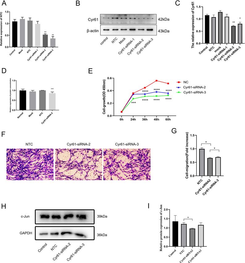
Cheng et al. BMC Molecular and Cell Biology       (2021) 22:21                                                                          Page 2 of 10

of axons which is important for subsequent nerve regen-                     secretion levels of Cyr61 in SC conditioned medium in-
eration. This is in part because of the ability of SCs to                   creased. This indicated that Cyr61 may participate in
support their survival through autocrine mechanisms                         SCs biology to facilitate nerve repair [17]. However, its
[6]. Factors that accelerate SCs proliferation during the                   role in SCs has not been fully elucidated.
early stages after peripheral nerve injury and/or promote                     In this study, we aimed to demonstrate the functional
SCs migration and myelination during the later stages                       effects of Cyr61 on SCs proliferation and migration. We
after nerve injury benefit nerve regeneration and func-                     found that Cyr61 affects proliferation and migration in
tional recovery [3]. However, the precise mechanisms for                    SCs through autocrine and paracrine mechanisms, and
this are unclear. Considering the critical role played by                   functions via αvβ3 integrin expressed on SCs and regu-
SCs, identifying factors that can accelerate the prolifera-                 lating c-Jun expression. Together, these findings suggest
tion and migration of SCs may help promote the repair                       that Cyr61 may contribute to peripheral nerve system
and regeneration of peripheral nerves after injury.                         (PNS) repair by supporting SC proliferation and migra-
   Cysteine-rich protein 61 (Cyr61, also known as CCN1)                     tion important for nerve regeneration.
is a member of the CCN family of matrix cell proteins.
Cyr61 is a secretory protein of the CCN family signal                       Results
protein related to ECM [7]. It can regulate a wide range                    In vivo and in vitro Cyr61 expression in SCs
of cell activities, including cell adhesion, migration, pro-                Western blot and immunocytofluorescence (ICF) assays
liferation, differentiation, apoptosis, and aging by inter-                 were used to determine the expression levels of Cyr61 in
acting with integrin receptors on the cell surface [8].                     cultured SCs. Primary cultured SCs are shown in Fig. 1a.
Previous studies have demonstrated that Cyr61 stimu-                        Under light microscopy, the cells were bright and ar-
lates the migration of smooth muscle cells [9], fibro-                      ranged in a regular pattern and depicted a typical SC
blasts [10], endothelial cells [11], and some cancer cells                  morphology. In cultured SCs, ICF assays using anti-
[12–14]. Cyr61 has also been observed in the nervous                        Cyr61 and the SC marker anti-S100β antibodies demon-
system. For example, Cyr61, as a dendrite growth regula-                    strated that Cyr61 expression overlapped with S100β
tor of hippocampal neurons, controls dendrite growth in                     (Fig. 1b). Western blot also demonstrated that Cyr61
an αβ1 integrin-dependent manner [15]. Cyr61 also plays                     protein (42KD) was expressed in cultured SCs (Fig. 1c).
a role in tissue repair. During the process of skin wound                   In transverse sections of normal rat sciatic nerves,
healing, Cyr61 can accelerate re-epithelialization by pro-                  immunohistofluorescence (IHF) assays using anti-Cyr61
moting the migration and proliferation of keratinocytes                     and anti-S100β antibodies demonstrated that Cyr61
[16]. A recent study found that SCs transformed into re-                    protein was colocalized with S100β protein expression
pair mediating SCs after FYT702P treatment and the                          (Fig. 1d).

 Fig. 1 In vivo and vitro expression of Cyr61. a Primary SC cultures under light microscopy; b SC primary cultures were immunostained using
 antibodies against Cyr61 (green color) and S100β (red color), with cell nuclei stained using Hoechst 33342 (blue color), Scale bar = 50 μm; c
 Western blots demonstrating Cyr61 protein expression in primary cultured SCs; d nerve transverse sections immunostained using antibodies
 against Cyr61 (green color) and S100β (red color), with cell nuclei stained using Hoechst 33342 (blue color), Scale bar = 50 μm
Cheng et al. BMC Molecular and Cell Biology        (2021) 22:21                                                                           Page 3 of 10

Inhibition of endogenous Cyr61 expression levels in SCs                      observed in cells transfected with non-targeting negative
attenuates cell proliferation and migration and                              control (NTC) siRNA compared to mock-transfected
downregulate c-Jun expression in SCs                                         and control cells (Fig. 2b). Compared to cells transfected
SCs were transfected with siRNA against Cyr61 to deter-                      with NTC siRNA, SCs transfected with siRNA-2 or
mine whether reduced Cyr61 expression levels affect cell                     siRNA-3 had reduced Cyr61 mRNA and protein expres-
proliferation and migration. Three siRNAs designated as                      sion levels (Fig. 2a-c). The secretion levels of Cyr61 have
Cyr61-siRNA-1, Cyr61-siRNA-2, Cyr61-siRNA-3 were                             no changes in SCs transfected with NTC siRNA com-
designed to reduce expression levels of Cyr61. The qPCR                      pared to mock-transfected SCs and the secretion levels
demonstrated that no changes in Cyr61 mRNA levels                            of Cyr61 in SCs after transfection with Cyr61-siRNA2
were observed in cells transfected with non-targeting                        were lower compared to SCs transfected with NTC, as
negative control (NTC) siRNA compared to mock-                               determined by ELISA (Fig. 2d). This indicated that
transfected cells (Fig. 2a). The western blot demon-                         Cyr61-siRNA transfection inhibited Cyr61 secretion in
strated that no changes in Cyr61 protein levels were                         SCs. Cell Counting Kit8 (CCK-8) assays demonstrated

 Fig. 2 Endogenous Cyr61 is required for the proliferation and migration of SCs and regulated c-Jun expression. a Histograms for three
 independent qPCR experiments of primary SCs transfected with Cyr61-specific siRNAs or with NTC siRNA; b Western blots demonstrating protein
 knockdown efficiency of three different Cyr61-targeting siRNAs; c Histograms for three independent western blot experiments of primary SCs
 were transfected with Cyr61-specific siRNAs or with NTC siRNA; d ELISA histograms of the results from three independent experiments for Cyr61
 secreted by SCs after transfection with Cyr61-specific siRNA or with NTC siRNA; e Histograms of three independent experiments of SCs cultured
 for 60 h after transfection with Cyr61-specific siRNAs or with NTC siRNA; f Representative images of SCs transfected with siRNAs that migrated to
 the underside of transwell membranes. g Histogram results from three independent experiments for cell migration. h Western blots
 demonstrating c-Jun protein expression in SCs and SCs transfected NTC siRNA or Cyr61-specific siRNAs. i Histograms for three independent
 western blot experiments of c-Jun expression in SCs and SCs transfected with Cyr61-specific siRNAs or with NTC siRNA. *, p < 0.05, **, p < 0.01, ***,
 p < 0.001, ****, p < 0.0001
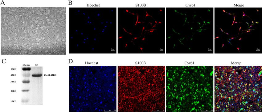
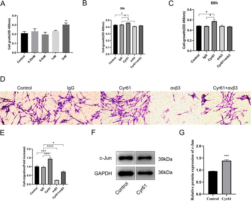
Cheng et al. BMC Molecular and Cell Biology       (2021) 22:21                                                                         Page 4 of 10

that proliferation rates of SCs transfected with Cyr61                     Cyr61 enhances SCs proliferation and migration and
siRNA-2 or Cyr61 siRNA-3 were also lower compared to                       enhanced c-Jun expression in SCs
cells transfected with NTC siRNA at different time points                  Cyr61 has been reported to stimulate proliferation and
from 0 to 60 h (Fig. 2e). Transwell-based migration assays                 migration of cancer cells, fibroblasts, and endothelial
and crystal violet staining were then used to determine                    cells [21], however, the effect of Cyr61 in SCs remained
the effects of Cyr61-siRNA-2 or Cyr61-siRNA-3 on SC                        to be deciphered. To determine whether Cyr61 influ-
migration. The number of cells that migrated through the                   ences the proliferation and migration of SCs, CCK-8 as-
transwell chamber, assessed using crystal violet staining,                 says, and transwell-based migration assays were
was substantially lower in SCs transfected with Cyr61-                     performed. To determine the effective concentration of
siRNA-2 or Cyr61-siRNA-3 compared to SCs transfected                       Cyr61 on SCs, different concentrations of Cyr61 on SC
with NTC siRNA (Fig. 2f and g). This indicated that                        proliferation was measured using CCK-8 assays. As
Cyr61 siRNA transfection suppressed cell migration. C-                     shown in Fig. 3a, when 2 nM exogenous Cyr61 was in-
Jun is essential for the normal activation of the SC repair                cluded in the culture media, the SC proliferation rate in-
programme and contributes to SC proliferation and mi-                      creased compared to control cells after 48 h. This result
gration [18–20]. Thun, western blot was used to detect c-                  indicated that the proliferation of cultured SCs could be
Jun expression. The results showed demonstrated that                       increased by exogenous Cyr61. Transwell assays were
compared to SCs transfected with NTC siRNA c-Jun pro-                      then performed to measure cell migration. Cells that
tein expression was lower in SCs transfected with Cyr61-                   were able to migrate to the lower chamber were quanti-
siRNA-2 while not significant in SCs transfected with                      tated to determine the effects of exogenous Cyr61. Re-
Cyr61-siRNA-3 (Fig. 2h and i).                                             sults of the migration assays demonstrated that 2 nM

 Fig. 3 Exogenous Cyr61 increases the proliferation and migration of SCs and upregulated c-Jun expression. a Histogram of SC proliferation
 cultured for 48 h after the addition of 0.25 nM, 0.5 nM, 1 nM, 2 nM of Cyr61 and control media measured using CCK-8 assays; b Histogram of SC
 proliferation cultured for 36 h after the addition of 2 nM of Cyr61, 0.5 μg neutralizing antibody αvβ3 and Mouse IgG1 Isotype Control (IgG), 2 nM
 Cyr61 + 0.5μg neutralizing antibody αvβ3 and control media measured using CCK-8 assays; c Histogram of SC proliferation cultured for 60 h after
 the addition of 2 nM of Cyr61, 0.5 μg neutralizing antibody αvβ3 and Mouse IgG1 Isotype Control (IgG), 2 nM Cyr61 + 0.5 μg neutralizing antibody
 αvβ3 and control media measured using CCK-8 assays; d Representative images of SCs treated with 2 nM of Cyr61, 0.5 μg neutralizing antibody
 αvβ3 and Mouse IgG1 Isotype Control (IgG), 2 nM Cyr61 + 0.5 μg neutralizing antibody αvβ3 and control media that migrated to the underside of
 the transwell membrane after 24 h. e Histograms from three independent cell migration experiments; f Western blots demonstrating c-Jun
 protein expression in SCs treated with 2 nM of Cyr61 or control medium; i Histograms for three independent western blot experiments of c-Jun
 expression in SCs treated with 2 nM of Cyr61 or control medium. *, p < 0.05, **, p < 0.01, ***, p < 0.001, ****, p < 0.0001
Cheng et al. BMC Molecular and Cell Biology (2021) 22:21 Page 5 of 10
Cyr61 added to the lower chamber of the transwell in- h (Fig. 3c). CCK-8 assays demonstrated that 2 nM of
creased the migration rate of SCs compared to media Cyr61 could increase the proliferation of SCs, but this
alone (Fig. 3d and e). Results of Western blot demon- effect could be inhibited by 0.5 μg αvβ3 neutralizing
strated that the protein expression of c-Jun in SCs was antibody after cultured for 36 h and 60 h (Fig. 3b and d).
increased after adding exogenous Cyr61(Fig. 3f and g). The number of cells that migrated through the transwell
This might suggest that Cyr61 could increase the prolif- chamber was assessed using crystal violet staining (Fig.
eration and migration of SCs and increased c-Jun pro- 3e). The addition of exogenous Cyr61 increased SC mi-
tein expression in SCs. gration and this effect could be inhibited by 0.5 μg αvβ3
neutralizing antibody. The addition of αvβ3 neutralizing
Cyr61 enhances SC proliferation and migration via antibody decreased SC migration rates (Fig. 3e). These
integrin αvβ3 results indicated that αvβ3 integrin was involved in SCs
As a secreted protein, Cyr61 binds to membrane recep- proliferation and migration and Cyr61 could increase
tors on the cell surface. Several studies have shown that the proliferation and migration of SCs via αvβ3.
αvβ3 integrin is a cell receptor for Cyr61. Cyr61 associ-
ates with αvβ3 integrin to promote endothelial cell adhe- Discussion
sion, migration, proliferation, survival, and tubular A better understanding of factors that facilitates nerve
formation [13]. Cyr61 modulates vascular formation by repair is essential for future improvement in regenerative
directly binding to αvβ3 to enhance endothelial cell ad- medicine [5]. Our findings indicate that SCs express
hesion, migration, and proliferation [19]. In addition, Cyr61 both in vivo and in vitro. We demonstrated that
Cyr61 has been shown to direct chondrosarcoma cell inhibition of Cyr61 expression in SCs can attenuate SC
migration through αvβ3 integrin [20]. However, the proliferation and migration ability. Cyr61 enhances SC
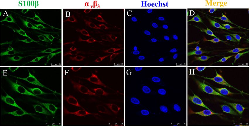
function of Cyr61 on SCs is unknown. Hence, immuno- proliferation and migration via αvβ3 integrin. Cyr61 en-
fluorescence assays were used to determine the expres- hances c-Jun expression in cultured SCs. Silencing
sion of αvβ3 integrin in SCs. The αvβ3 was expressed in Cyr61 in cultured SCs could decrease c-Jun expression.
S100β-positive cells, which indicated that αvβ3 was The unique regeneration ability of PNS is attributed to
expressed in SCs (Fig. 4). the function of SCs. Glial cells in the peripheral nerve
To determine whether Cyr61 influences the prolifera- have robust plasticity [22]. In addition, SCs secrete a var-
tion and migration of SCs via αvβ3 integrin, CCK-8, and iety of factors to create a microenvironment for cell re-
transwell-based migration assays were performed. As generation, including nerve growth factors [23].
shown in Fig. 3b-e, the proliferation and migration rate Neuroregulatory proteins secreted by SCs induce an
of SCs was inhibited by αvβ3 neutralizing antibody, and autocrine mechanism to promote SC proliferation [24].
the increased proliferation and migration rate of SCs SC survival after an injury is regulated by an autocrine
with addition of Cyr61 was blocked by αvβ3 neutralizing survival loop that includes the secretion of IGF-1,
antibody. The proliferation rate of SCs showed no differ- platelet-derived growth factor-BB, and NT-3 [25]. Un-
ence by αvβ3 neutralizing antibody after cultured for 60 derstanding how secreted proteins from SC influences
Fig. 4 αvβ3 expression levels in SCs determined using ICF. Primary SCs were immunostained using antibodies against S100β (green color) (a, e)
and αvβ3 (red color) (b, f), cell nuclei were stained using Hoechst 33342 (blue color) (c, f), a-d: Scale bar = 50 μm, e-h: Scale bar = 25 μmCheng et al. BMC Molecular and Cell Biology (2021) 22:21 Page 6 of 10 its function may contribute to the mechanistic under- receptors on the cell surface. Exogenous recombinant standing of nerve regeneration. Cyr61 has been reported to induce angiogenesis [30] and Previous studies have demonstrated increased Cyr61 promote cell proliferation, migration, adhesion, and dif- secretion by repair mediating SCs. The repair type SCs ferentiation [31]. At present, the known receptors for produce guidance tracks for regenerating axons called Cyr61 include integrin α6β11 [32, 33], αIIbβ3 [34], αmβ2 Büngner bands. Once SCs reprogrammed into repair [35], αvβ3 [36, 37], β1 [38], αDβ2 [39], and heparinase type cells, precursor/immature SC properties are re- (HSPGs) [9]. Cyr61 can associate with integrin αvβ3 on stored. Its proliferation and migration abilities increase. the surface of endothelial cells to promote endothelial However, the mechanism is yet to be deciphered. Repro- cell adhesion, migration, proliferation, survival, and graming SCs into repair phenotype is controlled tran- tubular formation [11]. By associating with integrin αvβ5 scriptionally by mechanisms involving the transcription and αvβ3 on bile duct cells, Cyr61 can induce the expan- factor c-Jun, which is rapidly upregulated in SCs after sion of the bile duct [40]. In addition, Cyr61 can syner- nerve injury [4, 26]. In this study, primary SCs were cul- gize with other mitogenic growth factors to enhance tured in vitro to determine the effects of Cyr61 on the growth factor-induced DNA synthesis in fibroblasts and proliferation and migration of SCs. Our results demon- endothelial cells through integrin αvβ3 [10, 41]. But the strated that compared to cells transfected with NTC function of Cyr61 with integrin αvβ3 on SCs is unclear. siRNA, Cyr61 secretion in cells transfected with cyr61 Using ICF, we demonstrated that SCs expressed the re- siRNA was reduced. In addition, the proliferation and ceptor αVβ3. A recent study showed that SPP1 might migration ability of SCs transfected with Cyr61 siRNA promotes SCs proliferation by binding receptors αvβ3 was reduced significantly. Silencing Cyr61 in cultured [42]. In our research, the ability of Cyr61 to promote the SCs also downregulated c-Jun expression. This suggested proliferation and migration of SCs was reduced by that lower Cyr61 secretion and expression in SCs leads blocking the αvβ3 receptor. This strongly suggested that to reduced proliferation and migration rates. And with Cyr61 played a significant role in SC proliferation and the decreasing of Cyr61 in cultured SCs, c-Jun expres- migration through the integrin receptor αvβ3, and we sion was inhibited. We next cultured SCs with exogen- hypothesize this was via an autocrine mechanism. ous Cyr61 to observe its biological effect. Our results indicated that exogenous Cyr61 could increase the pro- liferation and migration of SCs. In the meantime, c-Jun Conclusions expression was increased in cultured SCs with exogen- In summary, we demonstrated that Cyr61 promotes pro- ous Cyr61. These results suggested that Cyr61, through liferation and migration of SCs through an autocrine or an autocrine or paracrine mechanism, could significantly paracrine mechanism via αvβ3 integrin. Cyr61 might increase the proliferation and migration of SCs. Cyr61 modulate SC function by regulating c-Jun expression. could also influence the c-Jun expression in cultured Our results provide a functional mechanism of SC se- SCs. It has long been known that c-Jun is rapidly in- creted proteins to promote nerve regeneration which duced to high levels in the SCs of injured nerves [4]. C- may provide strategies for nerve repair. Jun promotes dedifferentiation of SCs and overexpres- sion of c-Jun alone might be sufficient to reprogram SCs of intact nerves into repair phenotype [27]. C-Jun signal- Methods ing involved in promoting many cell proliferation and Animals migration [20, 28, 29]. C-Jun-modified SCs showed en- Thirty neonatal 1 to 3 day old and three adult male hanced proliferation and migration abilities [20]. Here, Sprague-Dawley (SD) rats (180 g–220 g) were purchased we suppose that Cyr61 promotes SC proliferation and from the Experimental Animal Center at Nantong Uni- migration by regulating c-Jun expression. Since in- versity, China. The rats were specific pathogen free, ori- creased secretion of Cyr61 was found in repair mediat- ginally from Charles River Laboratories (Wilmington, ing SCs, Cyr61 might also could modulate SC phenotype MA) and bred in Laboratory Animal Research Center at by influence c-Jun protein expression. During the Nantong University. The animals were housed, in poly- process of peripheral nerve regeneration after injury, we carbonate cages with corn cob beddings, in a 12-h light/ hypothesized that SC repair type cells can survive in an dark schedule with ad libitum access to food and water environment that lacks the support of axons and other in a barrier unit. All animal experiments were performed cells. One of the reasons could be that this is through in accordance with the National Institutes of Health their ability to proliferate and migrate via the secretion (NIH) Guide for the Care and Use of Laboratory Ani- of Cyr61. mals and approved by the Administration Committee of Cyr61 has been previously demonstrated to regulate Experimental Animals of Nantong University, China cell proliferation and migration by binding to integrin (approval No. 20130410–006).
Cheng et al. BMC Molecular and Cell Biology   (2021) 22:21                                                   Page 7 of 10

SCs isolation and transfection                                protein levels of Cyr61 in the media were measured
Rat SCs were harvested as previously described [43] with      using a Cyr61 ELISA Kit (Cusbio, Wuhan, Hubei, China)
minor modifications. Briefly, the Sprague-Dawley rats (1      based on the manufacturer’s instructions. Measurement
to 3 d- old) were sanitized using 75% ethanol prior to        data were summarized from 3 independent experiments,
decapitation. Then sciatic nerves were harvested and en-      each run in triplicate.
zymatically dissociated by incubation at 37 °C sequen-
tially with 1% collagenase and 0.125% trypsin for 30 and      RNA extraction and quantitative real time RT-PCR (qPCR)
10 min, respectively. The mixture was triturated, centri-     Total RNA of each group was extracted using Trizol
fuged, and resuspended in 10% FBS in DMEM. The cell           (Invitrogen, Carlsbad, CA). Reverse transcription was
pellets were plated on poly-L-lysine precoated dishes         carried out with SuperScript First-Strand Synthesis Sys-
using the same media. The following day, 10 μM cyto-          tem (Invitrogen, Carlsbad, CA). Gene products were an-
sine arabinoside was added and incubated for an add-          alyzed using Fast EvaGreen qPCR Master Mix (Biotium,
itional 48 h to remove fibroblasts. The cell culture was      Hayward, CA) and specific primers in StepOne Real-
maintained in DMEM supplemented with 10% FBS,                 Time PCR System (Applied Biosystems). Reaction com-
2 μM forskolin (Sigma Aldrich, St. Louis, MO, USA),           ponents in each well were composed of 2× Fast Eva
and 2 ng/ml heregulin (HRG, R&D system, Minneapolis,          Green Master Mix, 10 μl; primers, 1 μl each; template,
MN, USA) to stimulate SC proliferation. For additional        1 μl; ROX, 2 μl; and H2O, 5 μl. Three step fast cycling
purification, the cell culture was gently trypsinized, pel-   protocol was performed. Relative gene expression levels
leted, and incubated with anti-Thy1.1 antibody (1:1000,       were calculated as ratios of the mRNA levels normalized
Sigma Aldrich, St. Louis, MO, USA; Cat# M7898, RRID:          against those of 18 s mRNA. All the results were
AB_477242; Clone number: TN26) on ice for 2 h,                expressed as the mean ± SD of three independent experi-
followed by incubation in complement (Sigma Aldrich,          ments. Primer sequences are provided in Additional file 1:
St. Louis, MO, USA) for an additional 2 h. All media and      Table 1.
supplements were purchased from Gibco-Invitrogen
(Carlsbad, CA, USA).                                          Western blot analysis
   For cell transfection, purified primary SCs were trans-    Total proteins from SCs were extracted using the M-
fected with Cyr61 siRNAs designated as ((Cyr61-siRNA-         PER cell protein extraction reagent (Pierce, Rockford, IL,
1 (sequence: GCAGACCCTGTGAATATAA), Cyr61-                     USA). Extracted proteins were quantified using the Fast
siRNA-2 (sequence: GGAATGGGTCTGTGATGAA),                      Silver Stain Kit (Beyotime, Haimen, Jiangsu Province,
Cyr61-siRNA-3 (sequence: GCTCCAGTGTGAAGAA                     China). Twenty microgram of total protein were loaded
ATA)) or NTC (Ribobio, Guangzhou, Guangdong,                  onto a 12% (w/v) SDS-PAGE, electrophoresed, and
China), using riboFECT CP Transfection kit (Ribobio,          transferred to a PVDF membrane (Millipore, Bedford,
Guangzhou, Guangdong, China) following the manufac-           MA). After blocking for 1 h with 5% (w/v) non-fat dry
turer’s instructions.                                         milk in TBS-T (0.05% (v/v) Tween 20 in Tris-buffered
                                                              saline), the membrane was incubated with specific pri-
Nerve tissue preparation                                      mary antibodies diluted in blocking buffer overnight at
The adult male SD rats were anesthetized intraperitone-       4 °C. The rabbit polyclonal antibody to Cyr61 (1:500,
ally using a mixture of 85 mg/kg trichloroacetaldehyde        Abcam, Cambridge, MA, USA; Cat# ab24448, RRID:
monohydrate (RichJoint, Shanghai, China), 42 mg/kg            AB_2088724) and rabbit monoclonal antibody to c-Jun
magnesium sulfate (Xilong Scientific, Guangzhou,              (1:2000, Abcam, Cambridge, MA, USA; Abcam Cat#
Guangdong, China), and 17 mg/kg sodium pentobarbital          ab40766, RRID: AB_731602, Clone number: EP693Y)
(Sigma Aldrich, St. Louis, MO, USA). After                    were used. Afterward, the membranes were washed with
anaesthetization, the rats were transcranial perfused se-     TBS-T and then incubated with HRP conjugated sec-
quentially with saline and 4% (v/v) paraformaldehyde in       ondary antibody diluted in blocking buffer (1:5000,
0.1 M PBS. Then, the sciatic nerve segments at 10 mm          Abcam, Cambridge, MA, USA) at RT for 2 h. Immuno-
above the bifurcation into the tibial and common fibular      reactive bands were visualized using enhanced chemilu-
nerves were harvested for frozen sections.                    minescence (Beyotime, Haimen, Jiangsu Province,
                                                              China). Densitometry analysis was performed using the
Enzyme-Linked Immunosorbent Assay (ELISA)                     Image J software (http://imagej.nih.gov/ij/).
Primary SCs were transfected with NTC or siRNA tar-
geting Cyr61. After transfection, the media was replaced      Immunofluorescent staining
with serum-free medium for an additional 12 h incuba-         Cells were plated on poly-L-lysine pre-coated coverslips
tion. The media was then harvested and filtered through       and cultured overnight. They were then fixed in 4%
a 0.22 μm filter (Millipore, Bedford, MA, USA). The           paraformaldehyde for 30 min at room temperature (RT).
Cheng et al. BMC Molecular and Cell Biology (2021) 22:21 Page 8 of 10
Sciatic nerve segments from adult rats were dissected, 37 °C in 5% CO2. Non-migrating cells were removed
fixed in 4% paraformaldehyde for 24 h, dehydrated in from the upper surface of the membrane using a cotton
30% sucrose at 4 °C, then cut and mounted onto micro- swab. Cells on the lower side of the membrane were
scope slides. Cells and sciatic nerve sections were stained with crystal violet, and migration was quantified
blocked for 2 h at 37 °C. Cells were incubated with the by counting cells from four microscope fields. Each
following antibodies overnight at 4 °C: mouse monoclo- treatment condition was run in triplicate.
nal or rabbit polyclonal antibody to S100β (1:100,
Abcam, Cambridge, MA, USA; Cat# ab14849, RRID: Statistical analysis
AB_301508, Clone number: 4B3; Cat# ab52642, RRID: Data were expressed as mean ± SEM, and statistical ana-
AB_882426), rabbit polyclonal antibody to Cyr61 (1:200, lysis was performed using GraphPad Prism Software
Abcam, Cambridge, MA, USA; Cat# ab24448, RRID: (GraphPad Software, LaJolla, CA). Comparisons between
AB_2088724) or mouse monoclonal antibody to αvβ3 (1: two groups were performed using the Student’s t-test.
200, R&D system, Minneapolis, MN, USA; Cat# Differences of p < 0.05 were considered statistically
MAB3050, RRID: AB_2128187; Clone number: #23C6). significant.
After washing, the cells and sciatic nerve sections were
incubated with FITC-conjugated rabbit anti-mouse IgG Abbreviations
Cyr61/CCN1: Cysteine-rich 61; DMEM: Dulbecco’s Modified Eagle Medium;
and Cy3-conjugated donkey anti-rabbit IgG (1:400, ELISA: Enzyme-linked immunosorbent assay; FBS: Fetal bovine serum;
Abcam, Cambridge, MA; Cat# ab6724, RRID: AB_ HRG: Heregulin; ICF: Immunocytofluorescence;
955315; Cat# ab97075, RRID: AB_10679955) for 2 h at IHF: Immunohistofluorescence; NTC: Non-targeting negative control;
OD: Optical density; PNS: Peripheral nerve system; qPCR: Quantitative Real
RT. Nuclei were counterstained with Hoechst 33342 dye Time RT-PCR; RT: Room temperature; SCs: Schwann cells; SD: Sprague-Dawley
(5 μg/mL, Sigma Aldrich, St. Louis, MO, USA). Fluores-
cence was visualized under a TCS SP5 confocal micro-
Supplementary Information
scope (Leica Microsystems, Wetzlar, Germany). The online version contains supplementary material available at https://doi.
org/10.1186/s12860-021-00360-y.
Cell proliferation analysis
SC proliferation was assessed after siRNA transfection, Additional file 1: Table 1. Primers used for qPCR analysis.
or exposure to recombinant Cyr61 with/without αvβ3 Additional file 2: Figure S1. Uncropped images of Western blots used
in figures.
neutralizing antibody (R&D system, Minneapolis, MN,
USA; Cat# MAB3050, RRID: AB_2128187; Clone num-
ber: #23C6) and mouse IgG1 isotype control (R & D, Acknowledgements
Not applicable.
Minneapolis, MN, USA; Cat# MAB002, RRID: AB_
357344; Clone number:11711) was used as a control for Authors’ contributions
neutralizing antibody. Cell Counting Kit8 (CCK-8) Author MS conceived and designed the work. ZHC and YWZ conducted
(Biyuntian Company, Jiangsu Province, China) was then experiments, YCT and YHC managed cell culture, YHJ and HW performed
data analysis, MS, FD and YHJ wrote the manuscript. All authors have read
used. Briefly, an equal number of cells were plated onto and approved the manuscript.
a 96-well plate. Cells in each treatment group were cul-
tured for 0-60 h. Then, 10 μL of CCK-8 solution was Funding
The work was supported by grants from the National Key R&D Program of
added to each well and incubated at 37 °C for an add-
China (No.2017YFA0104700), National Natural Science Foundation of China
itional 2 h. Optical density (OD) was determined at a (No. 31300942), Science and Technology Program of Nantong (JC2020034)
wavelength of 450 nm. and Jiangsu Provincial Key Medical Center. The funding bodies played no
role in the design of the study and collection, analysis, and interpretation of
data and in writing the manuscript.
Cell migration analysis
SC migration was monitored after siRNA transfection, Availability of data and materials
or exposure to recombinant Cyr61 with/without neutral- The complete blots of relevant figures are provided in Additional file 2.
Further datasets used and/or analysed during the current study available
izing antibody and mouse IgG1 isotype control was used from the corresponding author on reasonable request.
as a control for neutralizing antibody. The transwell mi-
gration assay was used as previously described [44]. Declarations
After transfection, 2 × 10⁴ cells in serum-free DMEM
Ethics approval and consent to participate
were plated onto the upper chamber of each transwell All animal experiments were performed in accordance with the National
with 8 μm pore size (Costar, Corning, Inc., NY). The Institutes of Health (NIH) Guide for the Care and Use of Laboratory Animals
lower chamber was supplemented with 800 μL of and approved by the Administration Committee of Experimental Animals of
Nantong University.
complete media (DMEM+ 10%FBS) or complete media
with Cyr61 or complete media with Cyr61 and αvβ3 Consent for publication
neutralizing antibodies. Cells were incubated for 24 h at Not applicable.Cheng et al. BMC Molecular and Cell Biology (2021) 22:21 Page 9 of 10
Competing interests 16. Du H, Zhou Y, Suo Y, Liang X, Chai B, Duan R, et al. CCN1 accelerates re-
The authors declare that they have no competing interests. epithelialization by promoting keratinocyte migration and proliferation
during cutaneous wound healing. Biochem Biophys Res Commun. 2018;
Author details 505(4):966–72. https://doi.org/10.1016/j.bbrc.2018.09.001.
1
Key Laboratory of Neuroregeneration of Jiangsu and Ministry of Education, 17. Schira J, Heinen A, Poschmann G, Ziegler B, Hartung HP, Stuhler K, et al.
Co-innovation Center of Neuroregeneration, NMPA Key Laboratory for Secretome analysis of nerve repair mediating Schwann cells reveals Smad-
Research and Evaluation of Tissue Engineering Technology Products, dependent trophism. FASEB J. 2019;33(4):4703–15. https://doi.org/10.1096/
Nantong University, Nantong 226001, People’s Republic of China. 2Jiangsu fj.201801799R.
Clinical Medicine Center of Tissue Engineering and Nerve Injury Repair, 18. Arthur-Farraj PJ, Latouche M, Wilton DK, Quintes S, Chabrol E, Banerjee A,
Nantong 226001, People’s Republic of China. 3Department of General et al. C-Jun reprograms Schwann cells of injured nerves to generate a repair
Surgery, Affiliated Hospital of Nantong University, Nantong, Jiangsu 226001, cell essential for regeneration. Neuron. 2012;75(4):633–47. https://doi.org/1
People’s Republic of China. 0.1016/j.neuron.2012.06.021.
19. Parkinson DB, Bhaskaran A, Droggiti A, Dickinson S, D'Antonio M, Mirsky R,
Received: 17 July 2020 Accepted: 29 March 2021 et al. Krox-20 inhibits Jun-NH2-terminal kinase/c-Jun to control Schwann
cell proliferation and death. J Cell Biol. 2004;164(3):385–94. https://doi.org/1
0.1083/jcb.200307132.
20. Huang L, Quan X, Liu Z, Ma T, Wu Y, Ge J, et al. C-Jun gene-modified
References Schwann cells: upregulating multiple neurotrophic factors and promoting
1. Wang H, Zhang P, Yu J, Zhang F, Dai W, Yi S. Matrix metalloproteinase 7 neurite outgrowth. Tissue Eng Part A. 2015;21(7–8):1409–21. https://doi.
promoted Schwann cell migration and myelination after rat sciatic nerve org/10.1089/ten.tea.2014.0416.
injury. Mol Brain. 2019;12(1):101. https://doi.org/10.1186/s13041-019-0516-6. 21. Chen MC, Tsai YC, Tseng JH, Liou JJ, Horng S, Wen HC, et al. Simvastatin
2. Assinck P, Duncan GJ, Hilton BJ, Plemel JR, Tetzlaff W. Cell transplantation Inhibits Cell Proliferation and Migration in Human Anaplastic Thyroid
therapy for spinal cord injury. Nat Neurosci. 2017;20(5):637–47. https://doi. Cancer. Int J Mol Sci. 2017;18(12):2690.
org/10.1038/nn.4541. 22. Hyung S, Im SK, Lee BY, Shin J, Park JC, Lee C, et al. Dedifferentiated
3. Yi S, Liu Q, Wang X, Qian T, Wang H, Zha G, et al. Tau modulates Schwann Schwann cells secrete progranulin that enhances the survival and axon
cell proliferation, migration and differentiation following peripheral nerve growth of motor neurons. Glia. 2019;67(2):360–75. https://doi.org/10.1002/
injury. J Cell Sci. 2019;132(6):jcs222059. glia.23547.
4. Jessen KR, Mirsky R. The repair Schwann cell and its function in 23. Heumann R, Korsching S, Bandtlow C, Thoenen H. Changes of nerve growth
regenerating nerves. J Physiol. 2016;594(13):3521–31. https://doi.org/1 factor synthesis in nonneuronal cells in response to sciatic nerve transection. J
0.1113/JP270874. Cell Biol. 1987;104(6):1623–31. https://doi.org/10.1083/jcb.104.6.1623.
5. Weiss T, Taschner-Mandl S, Bileck A, Slany A, Kromp F, Rifatbegovic F, et al. 24. Rosenbaum C, Karyala S, Marchionni MA, Kim HA, Krasnoselsky AL, Happel B,
Proteomics and transcriptomics of peripheral nerve tissue and cells unravel et al. Schwann cells express NDF and SMDF/n-ARIA mRNAs, secrete
new aspects of the human Schwann cell repair phenotype. Glia. 2016; neuregulin, and show constitutive activation of erbB3 receptors: evidence
64(12):2133–53. https://doi.org/10.1002/glia.23045. for a neuregulin autocrine loop. Exp Neurol. 1997;148(2):604–15. https://doi.
6. Jessen KR, Mirsky R. Why do Schwann cells survive in the absence of axons? org/10.1006/exnr.1997.6696.
Ann N Y Acad Sci. 1999;883(1):109–15. https://doi.org/10.1111/j.1749-6632.1 25. Meier C, Parmantier E, Brennan A, Mirsky R, Jessen KR. Developing Schwann
999.tb08573.x. cells acquire the ability to survive without axons by establishing an
7. Brigstock DR. The CCN family: a new stimulus package. J Endocrinol. 2003; autocrine circuit involving insulin-like growth factor, neurotrophin-3, and
178(2):169–75. https://doi.org/10.1677/joe.0.1780169. platelet-derived growth factor-BB. J Neurosci. 1999;19(10):3847–59. https://
8. Lau LF. CCN1/CYR61: the very model of a modern matricellular protein. Cell doi.org/10.1523/JNEUROSCI.19-10-03847.1999.
Mol Life Sci. 2011;68(19):3149–63. https://doi.org/10.1007/s00018-011-0778-3. 26. Arthur-Farraj PJ, Morgan CC, Adamowicz M, Gomez-Sanchez JA, Fazal SV,
9. Grzeszkiewicz TM, Lindner V, Chen N, Lam SC, Lau LF. The angiogenic factor Beucher A, et al. Changes in the coding and non-coding Transcriptome and
cysteine-rich 61 (CYR61, CCN1) supports vascular smooth muscle cell DNA Methylome that define the Schwann cell repair phenotype after nerve
adhesion and stimulates chemotaxis through integrin alpha (6) beta (1) and injury. Cell Rep. 2017;20(11):2719–34. https://doi.org/10.1016/j.celrep.2017.08.
cell surface heparan sulfate proteoglycans. Endocrinology. 2002;143(4):1441– 064.
50. https://doi.org/10.1210/endo.143.4.8731. 27. Figlia G. C-Jun in Schwann cells: stay away from extremes. J Neurosci. 2018;
10. Grzeszkiewicz TM, Kirschling DJ, Chen N, Lau LF. CYR61 stimulates human 38(14):3388–90. https://doi.org/10.1523/JNEUROSCI.0028-18.2018.
skin fibroblast migration through integrin alpha vbeta 5 and enhances 28. Ji R, Gu Y, Zhang J, Gao C, Gao W, Zang X, et al. TRIM7 promotes
mitogenesis through integrin alpha vbeta 3, independent of its carboxyl- proliferation and migration of vascular smooth muscle cells in
terminal domain. J Biol Chem. 2001;276(24):21943–50. https://doi.org/10.1 atherosclerosis through activating c-Jun/AP-1. IUBMB Life. 2020;72(2):247–58.
074/jbc.M100978200. https://doi.org/10.1002/iub.2181.
11. Leu SJ, Lam SC, Lau LF. Pro-angiogenic activities of CYR61 (CCN1) mediated 29. Luo Z, Han Z, Shou F, Li Y, Chen Y. LINC00958 accelerates cell proliferation
through integrins alphavbeta3 and alpha6beta1 in human umbilical vein and migration in non-small cell lung Cancer through JNK/c-JUN signaling.
endothelial cells. J Biol Chem. 2002;277(48):46248–55. https://doi.org/10.1 Hum Gene Ther Methods. 2019;30(6):226–34. https://doi.org/10.1089/hgtb.2
074/jbc.M209288200. 019.115.
12. Lin BR, Chang CC, Chen LR, Wu MH, Wang MY, Kuo IH, et al. Cysteine-rich 30. Babic AM, Kireeva ML, Kolesnikova TV, Lau LF. CYR61, a product of a growth
61 (CCN1) enhances chemotactic migration, transendothelial cell migration, factor-inducible immediate early gene, promotes angiogenesis and tumor
and intravasation by concomitantly up-regulating chemokine receptor 1 growth. Proc Natl Acad Sci U S A. 1998;95(11):6355–60. https://doi.org/10.1
and 2. Mol Cancer Res. 2007;5(11):1111–23. https://doi.org/10.1158/1541- 073/pnas.95.11.6355.
7786.MCR-06-0289. 31. Hsu PL, Chen JS, Wang CY, Wu HL, Mo FE. Shear-induced CCN1 promotes
13. Sun ZJ, Wang Y, Cai Z, Chen PP, Tong XJ, Xie D. Involvement of Cyr61 in Atheroprone endothelial phenotypes and atherosclerosis. Circulation. 2019;
growth, migration, and metastasis of prostate cancer cells. Br J Cancer. 2008; 139(25):2877–91. https://doi.org/10.1161/CIRCULATIONAHA.118.033895.
99(10):1656–67. https://doi.org/10.1038/sj.bjc.6604712. 32. Jun JI, Lau LF. The matricellular protein CCN1 induces fibroblast senescence
14. Kassis JN, Virador VM, Guancial EA, Kimm D, Ho AS, Mishra M, et al. and restricts fibrosis in cutaneous wound healing. Nat Cell Biol. 2010;12(7):
Genomic and phenotypic analysis reveals a key role for CCN1 (CYR61) in 676–85. https://doi.org/10.1038/ncb2070.
BAG3-modulated adhesion and invasion. J Pathol. 2009;218(4):495–504. 33. Chen N, Chen CC, Lau LF. Adhesion of human skin fibroblasts to Cyr61 is
https://doi.org/10.1002/path.2557. mediated through integrin alpha 6beta 1 and cell surface heparan sulfate
15. Malik AR, Urbanska M, Gozdz A, Swiech LJ, Nagalski A, Perycz M, et al. Cyr61, proteoglycans. J Biol Chem. 2000;275(32):24953–61. https://doi.org/10.1074/
a matricellular protein, is needed for dendritic arborization of hippocampal jbc.M003040200.
neurons. J Biol Chem. 2013;288(12):8544–59. https://doi.org/10.1074/jbc. 34. Jedsadayanmata A, Chen CC, Kireeva ML, Lau LF, Lam SC. Activation-
M112.411629. dependent adhesion of human platelets to Cyr61 and Fisp12/mouseCheng et al. BMC Molecular and Cell Biology (2021) 22:21 Page 10 of 10
connective tissue growth factor is mediated through integrin alpha (IIb)beta
(3). J Biol Chem. 1999;274(34):24321–7. https://doi.org/10.1074/jbc.2
74.34.24321.
35. Schober JM, Chen N, Grzeszkiewicz TM, Jovanovic I, Emeson EE, Ugarova TP,
et al. Identification of integrin alpha(M)beta (2) as an adhesion receptor on
peripheral blood monocytes for Cyr61 (CCN1) and connective tissue growth
factor (CCN2): immediate-early gene products expressed in atherosclerotic
lesions. Blood. 2002;99(12):4457–65. https://doi.org/10.1182/blood.V99.12.4457.
36. Lin MT, Chang CC, Chen ST, Chang HL, Su JL, Chau YP, et al. Cyr61
expression confers resistance to apoptosis in breast cancer MCF-7 cells by a
mechanism of NF-kappaB-dependent XIAP up-regulation. J Biol Chem. 2004;
279(23):24015–23. https://doi.org/10.1074/jbc.M402305200.
37. Menendez JA, Vellon L, Mehmi I, Teng PK, Griggs DW, Lupu R. A novel
CYR61-triggered 'CYR61-alphavbeta3 integrin loop' regulates breast cancer
cell survival and chemosensitivity through activation of ERK1/ERK2 MAPK
signaling pathway. Oncogene. 2005;24(5):761–79. https://doi.org/10.1038/sj.
onc.1208238.
38. Yoshida Y, Togi K, Matsumae H, Nakashima Y, Kojima Y, Yamamoto H, et al.
CCN1 protects cardiac myocytes from oxidative stress via beta1 integrin-Akt
pathway. Biochem Biophys Res Commun. 2007;355(3):611–8. https://doi.
org/10.1016/j.bbrc.2007.01.195.
39. Yakubenko VP, Yadav SP, Ugarova TP. Integrin alphaDbeta2, an adhesion
receptor up-regulated on macrophage foam cells, exhibits multiligand-
binding properties. Blood. 2006;107(4):1643–50. https://doi.org/10.1182/
blood-2005-06-2509.
40. Kim KH, Chen CC, Alpini G, Lau LF. CCN1 induces hepatic ductular reaction
through integrin alphavbeta (5)-mediated activation of NF-kappaB. J Clin
Invest. 2015;125(5):1886–900. https://doi.org/10.1172/JCI79327.
41. Kireeva ML, Mo FE, Yang GP, Lau LF. Cyr61, a product of a growth factor-
inducible immediate-early gene, promotes cell proliferation, migration, and
adhesion. Mol Cell Biol. 1996;16(4):1326–34. https://doi.org/10.1128/MCB.1
6.4.1326.
42. Wang JB, Zhang Z, Li JN, Yang T, Du S, Cao RJ, et al. SPP1 promotes
Schwann cell proliferation and survival through PKCalpha by binding with
CD44 and alphavbeta3 after peripheral nerve injury. Cell Biosci. 2020;10(1):
98. https://doi.org/10.1186/s13578-020-00458-4.
43. Shen M, Ji Y, Zhang S, Shi H, Chen G, Gu X, et al. A proteome map of
primary cultured rat Schwann cells. Proteome Sci. 2012;10(1):20. https://doi.
org/10.1186/1477-5956-10-20.
44. Yi S, Yuan Y, Chen Q, Wang X, Gong L, Liu J, et al. Regulation of Schwann
cell proliferation and migration by miR-1 targeting brain-derived
neurotrophic factor after peripheral nerve injury. Sci Rep. 2016;6(1):29121.
https://doi.org/10.1038/srep29121.
Publisher’s Note
Springer Nature remains neutral with regard to jurisdictional claims in
published maps and institutional affiliations.You can also read