Rapid identification of pathogens, antibiotic resistance genes and plasmids in blood cultures by nanopore sequencing - Nature
←
→
Page content transcription
If your browser does not render page correctly, please read the page content below
www.nature.com/scientificreports

            OPEN           Rapid identification of pathogens,
                           antibiotic resistance genes and
                           plasmids in blood cultures by
                           nanopore sequencing
                           Arne M. Taxt1,2, Ekaterina Avershina2, Stephan A. Frye1, Umaer Naseer3 & Rafi Ahmad2,4 ✉

                           Bloodstream infections (BSI) and sepsis are major causes of morbidity and mortality worldwide.
                           Blood culture-based diagnostics usually requires 1–2 days for identification of bacterial agent and
                           an additional 2–3 days for phenotypic determination of antibiotic susceptibility pattern. With the
                           escalating burden of antimicrobial resistance (AMR) rapid diagnostics becomes increasingly important
                           to secure adequate antibiotic therapy. Real-time whole genome sequencing represents a genotypic
                           diagnostic approach with the ability to rapidly identify pathogens and AMR-encoding genes. Here
                           we have used nanopore sequencing of bacterial DNA extracted from positive blood cultures for
                           identification of pathogens, detection of plasmids and AMR-encoding genes. To our knowledge, this is
                           the first study to gather the above-mentioned information from nanopore sequencing and conduct a
                           comprehensive analysis for diagnostic purposes in real-time. Identification of pathogens was possible
                           after 10 minutes of sequencing and all predefined AMR-encoding genes and plasmids from monoculture
                           experiments were detected within one hour using raw nanopore sequencing data. Furthermore, we
                           demonstrate the correct identification of plasmids and blaCTX-M subtypes using de novo assembled
                           nanopore contigs. Results from this study hold great promise for future applications in clinical
                           microbiology and for health care surveillance purposes.

                           Bloodstream infections (BSIs) and sepsis are major causes of morbidity and mortality worldwide. Epidemiological
                           data are scarce, but a recent estimate indicated that 31.5 million cases of sepsis and 5.3 million sepsis attributable
                           deaths occur annually1. This estimate is only based on data collected from high-income countries, and it therefore
                           likely underestimates the true burden of disease worldwide, especially in low-and-middle-income countries2.
                           Most studies on sepsis and BSIs report an increasing incidence over the last two decades3, particularly among
                           the immunocompromised, multimorbid, and elderly patients, or due to failure of empiric antibiotic regimens as
                           result of antimicrobial resistance (AMR)4.
                               With multi drug resistant pathogens spreading at an alarming rate, widely adopted empirical antibiotic treat-
                           ment regimens for sepsis based on penicillin (or aminopenicillin) in combination with gentamicin5 are being
                           challenged. In particular, the escalating burden of infections due to extended-spectrum β-lactamase (ESBL)
                           producing Gram negative bacteria represents a major health concern. These bacteria, mainly Escherichia coli
                           and Klebsiella pneumoniae, are not only resistant to all penicillins and third generation cephalosporins, but also
                           frequently express co-resistance to gentamicin. Consequently, treatment failure may occur, and clinicians increas-
                           ingly prescribe last-resort antibiotics such as carbapenems as initial antibiotic treatment of sepsis. This in turn
                           contributes to development and spread of AMR and to a further increase in the burden of infections caused by
                           resistant bacteria.
                               Current state-of-the art in diagnostics of BSIs is blood culture, which often takes 1–3 days to come out positive
                           and provide information on etiological agent. Time to positivity is influenced by a number of clinical and micro-
                           biological factors such as source of bacteraemia, level of bacteraemia, presence or absence of pre-administered

                           1
                            Department of Microbiology, Division of Laboratory Medicine, Oslo University Hospital, PB 4956, Nydalen, 0424,
                           Oslo, Norway. 2Department of Biotechnology, Inland Norway University of Applied Sciences, Holsetgata 22, 2317,
                           Hamar, Norway. 3Department of Zoonotic, Food- and Waterborne Infections, 0213 Oslo, Norwegian Institute of
                           Public Health, Oslo, Norway. 4Institute of Clinical Medicine, Faculty of Health Sciences, UiT - The Arctic University of
                           Norway, Hansine Hansens veg 18, 9019, Tromsø, Norway. ✉e-mail: rafi.ahmad@inn.no

Scientific Reports |   (2020) 10:7622 | https://doi.org/10.1038/s41598-020-64616-x                                                               1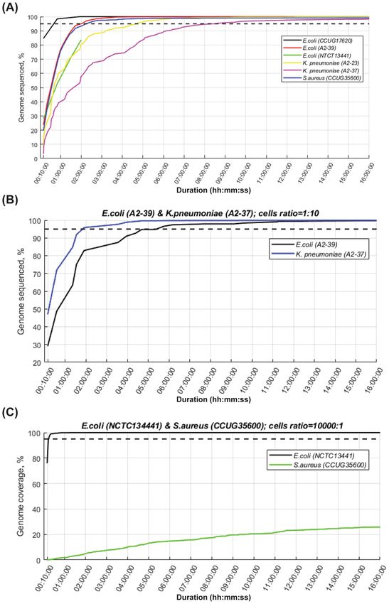
www.nature.com/scientificreports/                                                                  www.nature.com/scientificreports

                            Nr   Bacteria                AMR gene     CFU/ml         DNA [ng/µl]
                            1    E. coli (CCUG17620)     −            1,3 × 10⁹      60
                            2    E. coli (A2-39)         CTX-M-2      1,6 × 10⁹      57
                            3    E. coli (NCTC13441)     CTX-M-15     1,7 × 10⁹      49
                            4    K. pneumoniae(A2-23)    CTX-M-1      9,3 × 10⁸      54
                            5    K. pneumoniae(A2-37)    CTX-M-14     1,0 × 10⁹      56
                            6    S. aureus(CCUG35600)    mecA         2,6 × 10⁷      31
                                 E. coli (A2-239)        CTX-M-2      5,5 × 10⁷
                            7    +                                    +              45
                                 K. pneumoniae (A2-37)   CTX-M-14     4,8 × 10⁸
                                 E. coli (NCTC13441)     CTX-M-15     1,2 × 10⁹
                            8    +                                    +              54
                                 S. aureus (CCUG35600)   mecA         3,6 × 10⁵

                           Table 1. Overview of blood culture experiments.

                           antibiotics and the bacterial species 6. Use of matrix-assisted laser desorption/ionization time-of-flight
                           (MALDI-TOF) mass spectrometry is becoming widespread in clinical microbiology laboratories to identify
                           bacteria by analysis of pelleted blood-cultures when they are flagged as positive by the blood culture incuba-
                           tion system7. Phenotypic antimicrobial susceptibility testing (AST) however, requires subculture on solid media
                           overnight for colonies to form, and an additional 18+/−2 hours of subculture incubation with antibiotic discs
                           to obtain a result which can be interpreted according to official breakpoint guidelines and converted to sensitive
                           (S), intermediate (I) or resistant (R)8. Until then time-point choice of antibiotic treatment is based on clinical
                           assessment, empirical guidelines and local epidemiology on AMR. However, several studies have observed that
                           inappropriate antibiotic treatment is often initiated to patients with BSIs and this is associated with increased
                           mortality9,10.
                               Diagnosis of BSI and prescription of appropriate antimicrobial therapy is crucial for the reduction of mor-
                           bidity and mortality caused by BSI and WHO and Centers for Disease Control aim for a two hour turnaround
                           time11. For these reasons there is a growing interest and an urgent need for the development of molecular tech-
                           niques for rapid identification of pathogens and AMR from blood and blood cultures. The topic has been exten-
                           sively reviewed elsewhere12–14, and the currently available methods can be categorized in three groups; in situ
                           hybridization-based methods, DNA-microarray-based methods, nucleic acid amplification-based methods (such
                           as PCR and loop-mediated isothermal amplification (LAMP)), and combinations of these. Common to all these
                           techniques is that detection is limited to a predefined set of genetic targets, either specific for a particular path-
                           ogen or an AMR-encoding gene. Whole genome sequencing (WGS) on the other hand, provides comprehen-
                           sive genomic information and can potentially detect all AMR-encoding genes present in the bacterial genome.
                           Additionally, WGS-data provide vast opportunities for bacterial sequence typing, phylogenetics and virulence
                           analysis. The introduction of the real-time sequencing platform from Oxford nanopore technology (ONT) has
                           triggered studies to explore its application in blood culture diagnostics, either based on 16 s amplicon sequenc-
                           ing15, or by a whole-genome-sequencing approach16. This represents an unbiased approach to diagnostics with
                           the potential to identify any pathogen and AMR-encoding gene.
                               Here we present results from rapid blood culture diagnostics based on extraction of bacterial DNA from
                           positive blood cultures followed by nanopore sequencing and real-time data analysis for identification of
                           pathogens, detection of plasmids and AMR-encoding genes. The results have also been verified through WGS
                           using short-read Illumina sequencing and hybrid assembly using nanopore and Illumina sequences. This
                           proof-of-concept study represents a molecular-genetic approach to diagnosis of BSIs which can provide clinicians
                           with detailed information on etiologic agent and AMR within few hours of a blood culture becoming positive.

                           Results
                           Blood culture samples and nanopore sequencing.                    Seven blood cultures spiked with blaCTX-M posi-
                           tive E. coli and K. pneumoniae, mecA positive Staphylococcus aureus, or a combination of these were analysed.
                           In addition, one blood culture was spiked with E. coli reference strain CCUG17620 (non-ESBL) as control. All
                           cultures were incubated in a standard BD BACTEC FX blood culturing instrument with continuous monitoring
                           until flagged positive. Positive monocultures had bacterial concentrations ranging from 2.6 × 10⁷ to 1.6 × 10⁹
                           CFU/ml. When E. coli and S. aureus were co-cultured, we observed a 4-log difference in bacterial concentration
                           in favour of E. coli, while for the E. coli and K. pneumoniae co-culture the difference was less than one-log fold in
                           favour of K. pneumoniae (Table 1).
                               For extraction of bacterial DNA, the commercially available kits MolYsis Plus and BiOstic Bacteremia DNA
                           were both used initially. Subsequent experiments were conducted using the BiOstic Bacteremia DNA kit due to its
                           shorter protocol and higher DNA yield (Supplementary Fig. 1). Purified DNA was subject to nanopore sequenc-
                           ing on the MinION sequencing platform from ONT. The average amount of data generated was 334,113 ± 379,926
                           (mean ± standard deviation) sequencing reads per blood culture with an average read length of 3,529 ± 4,140 bp
                           (Supplementary Fig. 2). Only 15.4% ± 14.4% of the reads were shorter than 300 bp, which is the maximum
                           single-end read length generated by the latest version of the Illumina MiSeq system.

Scientific Reports |   (2020) 10:7622 | https://doi.org/10.1038/s41598-020-64616-x                                                             2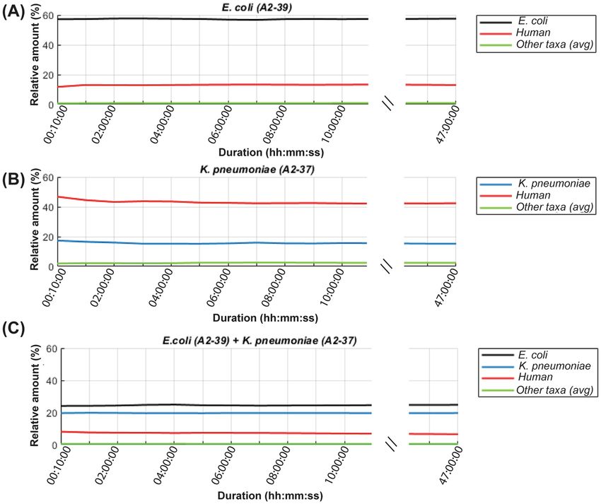
www.nature.com/scientificreports/ www.nature.com/scientificreports
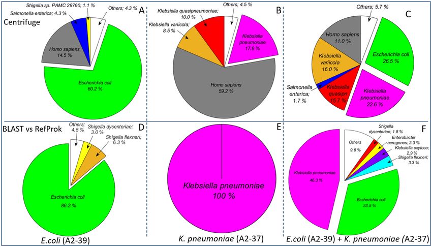
Figure 1. Relative distribution of reads in sequence data generated by nanopore sequencing of DNA purified
from the three selected blood cultures. The blood cultures were spiked with E. coli A2-39 (A and D), K.
pneumoniae A2-37 (B and E) and E. coli A2-39 + K. pneumoniae A2-37 (C and F). Upper panel (A–C) show
results obtained using Centrifuge and lower panel (D–F) show results based on BLAST search against the
RefProk database that contains prokaryotic sequence data only. The “Others” group represents taxa with relative
read counts below 1%. All results are based only on the first output file for each experiment from the MinION
sequencing platform, containing 4000 reads (available after approximately 10 minutes of sequencing).
Bacterial species identification using raw nanopore sequencing reads. Using default settings, the
Metrichor analysis platform from ONT performed real-time base calling generating 4,000 sequences per output
file. For taxonomy assignment, both Centrifuge17 classification (used by the ONT analysis tool What´s In My
Pot (WIMP)18) and BLAST search against the NCBI Prokaryotic RefSeq (RefProk) database took less than one
minute per sequencing file using four cores, which is the standard configuration on current laptops. We hereby
present detailed results for two monoculture experiments, E. coli A2-39 and K. pneumoniae A2-37, and for one
blood culture spiked with both of these isolates.
Based only on the first sequence file 80–100% of bacterial reads were classified correctly at the species level by
BLAST search against the RefProk database (Fig. 1D–F) and most of the ‘incorrect’ assignments, i.e. reads classi-
fied as non-target bacteria, comprised less than 1% per species (Fig. 2A–C). Also, relative amounts of the detected
species remained constant throughout the sequencing run (Fig. 2A–C).
Intriguingly, 4.3% of sequencing reads from E. coli A2-39 monoculture and 1.7% of reads from the E. coli
A2-39 + K. pneumoniae A2-37 mixed culture were classified as belonging to Salmonella enterica by Centrifuge,
but not by BLAST (Fig. 1A,C,D and F). Following a detailed analysis, the majority of these misclassified
reads were identified as E. coli specific sequences using BLAST against the RefProk and RefSeq databases
(Supplementary Fig. 3). The average nucleotide identity (ANI) values of these reads were 79.6% for Salmonella
enterica (AE014613.1) and 83.6% for E. coli (NC_000913.3). Around 9% of E. coli A2-39 culture reads were
classified as Shigella by BLAST search against RefProk database, with ANI values of 87.9% for Shigella flexneri
(AE014073.1) and 88.8% for E. coli (NC_000913.3).
In both analyses of the K. pneumoniae A2-37 isolate, in monoculture and in the E. coli A2-39 + K. pneumoniae
A2-37 mixed culture, K. pneumoniae A2-37 reads were evenly classified as K. pneumoniae, K. quasipneumoniae
or K. variicola by Centrifuge (Fig. 1B,C). However, BLAST search against RefProk classified all of these reads as
K. pneumoniae (Fig. 1E,F). ANI values for these reads were on average 85.2% for reads assigned to K. pneumoniae
(NC_016845.1), 84.2% for reads assigned to K. variicola (NZ_CP010523.2) and 87.0% for reads assigned to K.
quasipneumoniae (NZ_CP014696.2).
The other four monoculture experiments of E. coli, K. pneumoniae and S. aureus also showed very promising
results, with 94-100% of reads assigned correctly to target species using the first 4000 reads (Supplementary
Fig. 4). In the mixed culture experiment with E. coli and S. aureus however, 94.7% of sequences were assigned to
E. coli and only 0.05% of reads were assigned to S. aureus based on the first sequence file. Throughout the entire
sequencing run the relative number of reads classified as S. aureus remained below 1% and were thereby indistin-
guishable from other low-level misassignments. A probable explanation could be the 4-log difference in bacterial
concentration in favour of E. coli in the experiment (Table 1).
Scientific Reports | (2020) 10:7622 | https://doi.org/10.1038/s41598-020-64616-x 3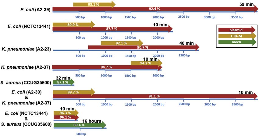
www.nature.com/scientificreports/                                                              www.nature.com/scientificreports

                           Figure 2. Centrifuge-based relative taxonomic assignments of sequence data from DNA purified from the
                           three selected blood cultures. Samples were spiked with - (A) E. coli A2-39, (B) K. pneumoniae A2-37 and (C)
                           E. coli A2-39 + K. pneumoniae A2-37. The “Others” group shows the average relative amount of all incorrectly
                           assigned (non-target) taxa. Blood cultures spiked with E. coli (A2-39) and with a combination of E. coli (A2-39)
                           and K. pneumoniae (A2-37) were sequenced for 68 and 64 hours respectively.

                           Detection of AMR-encoding genes and plasmids using raw nanopore sequencing reads. BLAST
                           search against the CARD and ResFinder databases showed similar AMR gene assignments and therefore these
                           results are presented together. In case of the reference strain (E.coli CCUG17620) spiked blood culture none of
                           the reads were identified as blaCTX-M positive. For all monocultures of blaCTX-M or mecA-positive bacteria, target
                           AMR genes were detected within the first hour of sequencing (Fig. 3). However, the unassembled sequencing data
                           did not allow correct identification of blaCTX-M gene variant (Supplementary Fig. 5). Sequencing reads containing
                           blaCTX-M genes were recognized as plasmid-borne when BLAST search of these reads against the plasmid database
                           from Brooks et al.19 yielded positive hits (Fig. 3). The mecA positive reads from the S. aureus spiked blood culture
                           were not detected in the plasmid database and therefore were labelled as chromosome borne. The first blaCTX-M
                           containing read from the E. coli A2-39 + K. pneumoniae A2-37 spiked blood culture was detected 10 minutes after
                           the start of sequencing. Ten minutes were also enough to capture the first blaCTX-M containing read from the E.
                           coli NCTC 13441 + S. aureus CCUG35600 spiked blood culture, but the first indication of the mecA gene came
                           only after 16 hours.

                           De novo assembled nanopore contigs enable identification of plasmids and bla CTX-M
                           gene-variants. Using de novo assembled contigs from E. coli A2-39 and K. pneumoniae A2-37 monoculture
                           experiments we searched for plasmids with the NCBI PlasmidFinder tool. E.coli A2-39 harboured IncHI2, IncI1
                           and p0111 plasmids, whereas K. pneumoniae A2-37 harboured IncFII and IncFI plasmids (Table 2). Assembled
                           contigs from sequencing of the blood culture spiked with both isolates also suggested presence of these plas-
                           mids. By performing additional BLAST searches with the plasmid-labelled contigs against the AMR databases the
                           blaCTX-M gene-variants could be identified (Table 2).
                              To assess whether the first raw sequencing reads that were recognized as containing bla CTX-M were
                           plasmid-borne, they were mapped to the de novo assembled contigs. The E. coli A2-39 blaCTX-M-containing
                           read detected after 59 minutes of sequencing mapped to the IncHI2-tagged contig with 92.1% identity
                           (length = 3,961 bp; query coverage = 100%; e-value = 0), whereas the K. pneumoniae A2-37 blaCTX-M con-
                           taining read detected after 10 minutes of sequencing mapped to the IncFII-tagged contig with 94% identity
                           (length = 2,055 bp; query coverage = 100%; e-value = 0). The first blaCTX-M containing read from the E. coli
                           A2-39 + K. pneumoniae A2-37 spiked blood culture that was detected after ten minutes of sequencing, mapped
                           only to the IncFII-tagged K. pneumoniae contig with 91.2% identity (length = 3 840 bp; query coverage = 100%;
                           e-value = 0). The first read that mapped to the IncHI2-tagged E. coli contig, was detected in the second output file
                           generated after 36 minutes of sequencing (length = 4 178 bp; query coverage = 99.9%; identity = 91.2%; e-value
                           = 0).

Scientific Reports |   (2020) 10:7622 | https://doi.org/10.1038/s41598-020-64616-x                                                           4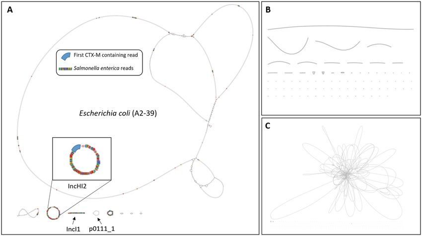
www.nature.com/scientificreports/                                                                   www.nature.com/scientificreports

                           Figure 3. Overview of the earliest generated reads from each sequencing experiment where blaCTX-M (yellow)
                           or mecA genes (green) and plasmid-derived sequences (red) were identified based on BLAST search. Blue
                           lines represent length of the nanopore reads, length of hits is illustrated by arrow-length and similarity to the
                           database entry is given on the arrow. The time of read generation is noted on top of each read.

                                                    PlasmidFinder                              Resistance genes database
                                                                    Query/HSP                  CTX-M
                            Bacterial culture       Plasmid type    length, bp   Identity, %   variant       Hit length, bp   Identity, % E-value
                                                    IncHI2          327/327      100
                            E. coli (A2-39)         IncI1           142/142      99.3          CTX-M-2       878              98.7       0
                                                    p0111           885/885      98.9
                                                    IncFII          231/230      98.3
                            K. pneumoniae (A2-37)   IncFIA          388/388      97.2          CTX-M-14      877              99.5       0
                                                    IncFIB          512/560      98.2
                                                    IncHI2          630/630      99.2
                                                                                               CTX-M-2       877              99.3       0
                                                    p0111           885/885      98.8
                            E. coli (A2-39) +K.
                                                    IncFIA          388/388      96.9
                            pneumoniae (A2-37)
                                                    IncFIB          560/560      98.7          CTX-M-14      876              100        0
                                                    IncFII          261/261      100

                           Table 2. BLAST search results of contigs harbouring plasmids against the AMR databases. HSP – high scoring
                           segment pair.

                           Identification of bacterial species, AMR-encoding genes and plasmids using raw Illumina
                           sequencing reads. To verify taxonomic classification, AMR-gene detection and plasmid identification, the
                           E. coli A2-39 and K. pneumoniae A2-37 isolates were sequenced on an Illumina MiSeq platform. Taxonomic
                           assignment of the unassembled Illumina reads by Centrifuge was similar to the results obtained with nanopore
                           data; where 4.7% of the E. coli A2-37 reads were assigned as S. enterica, and K. pneumoniae reads were assigned as
                           a combination of K. pneumoniae, K. quasipneumoniae and K. variicola group (Supplementary Fig. 6). For detec-
                           tion of AMR-encoding genes, CARD and ResFinder databases were searched using SPAdes assembled contigs
                           (see Supplementary Table 1 for details). This suggested that E. coli A2-39 harboured an IncHI2 plasmid encoding
                           the blaCTX-M-2 gene (hit length = 876 bp; identity = 100%; e-value = 0) and that K. pneumoniae A2-37 contained
                           an IncFII plasmid with the blaCTX-M-14 gene (hit length = 876 bp; identity = 100%; e-value = 0).

                           Hybrid de novo assembled data corroborate nanopore results.                     Hybrid de novo assembly using
                           Illumina reads and nanopore reads from blood cultures spiked with E. coli A2-39 or K. pneumoniae A2-37 was
                           performed using Unicycler (Fig. 4 and Supplementary Table 1). Generally, hybrid assembled data corroborated
                           results obtained using nanopore assembled data only, albeit with higher precision. The blaCTX-M-2 gene variant
                           was detected in E. coli A2-39 (identity = 100%; hit coverage = 100%; e-value = 0) and K. pneumoniae A2-37
                           was found to carry blaCTX-M-14 (identity = 100%; hit coverage = 100%; e-value = 0). Three contigs were tagged
                           as plasmid-borne by PlasmidFinder (Supplementary Table 2). Additional search with these contigs against the
                           PLSDB database revealed that one circular contig of the hybrid E. coli A2-39 assembly (length = 231,378 bp) had

Scientific Reports |   (2020) 10:7622 | https://doi.org/10.1038/s41598-020-64616-x                                                                  5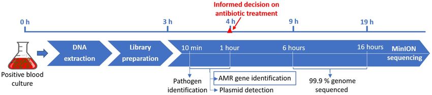
www.nature.com/scientificreports/                                                             www.nature.com/scientificreports

                           Figure 4. Assembly graphs for E. coli A2-39. (A) Hybrid assembly of nanopore and Illumina data; reads
                           tagged as S. enterica by Centrifuge and blaCTX-M are highlighted (S. enterica in multicolour, blaCTX-M in blue).
                           The blaCTX-M gene was located on a plasmid-derived read which mapped to the IncHI2 plasmid. (B) Unicycler
                           assembled nanopore generated data. (C) SPAdes assembled Illumina generated data.

                           99.8% identity to the IncHI2A E. coli RCS77_p plasmid (LT985297.1). The other circular contig (length = 95,977)
                           had 98.0% identity to the p0111_1 Enterobacteriaceae plasmid (NZ_CP033848.1) and a non-circular contig had
                           99.6% identity to the IncI1 E. coli pS51_1 plasmid (NZ_CP015996.1).
                               One circular contig of the K. pneumoniae A2-37 hybrid assembly (length = 84,474 bp; depth = 2.41×) labelled
                           as IncFII by PlasmidFinder had 99.8% identity to the IncFII E. coli pFAM22321 plasmid (KU288634.1). Two
                           non-circular contigs of the K. pneumoniae A2-37 hybrid assembly (length = 128,678 bp and length = 121,056 bp)
                           were recognized by PlasmidFinder as plasmids IncFIA(HI1) and IncFIB(K) (Supplementary Table 2). Additional
                           search of these contigs through the PLSDB database showed that they had 93.8% identity to K. pneumoniae pKp_
                           Goe_414-3 plasmid (NZ_CP018340.1) and 96.6% identity to K. pneumoniae strain AR_0049 unitig_2 plasmid
                           (NZ_CP018818.1) respectively.
                               The first blaCTX-M containing read from the E. coli A2-39 monoculture detected after one hour of sequencing
                           mapped to the IncHI2 plasmid contig with 92.4% identity (length = 4,100 bp; query coverage = 100%; e-value =
                           0). The first blaCTX-M containing read from the K. pneumoniae A2-37 monoculture detected after 10 minutes of
                           sequencing mapped to the IncFII contig with 94.8% identity (length = 2,109 bp; query coverage = 100%; e-value
                           = 0). Similar to the assembled nanopore data, the first blaCTX-M tagged read from the E. coli A2-39 + K. pneu-
                           moniae A2-37 spiked blood culture mapped to the K. pneumoniae A2-37 hybrid assembly with 91.2% identity
                           (length = 4,005 bp; query coverage = 100%; e-value = 0), but not to the E. coli A2-39 hybrid assembly. The first
                           blaCTX-M tagged read that mapped to the E. coli A2-39 hybrid assembly (length = 4,349 bp; query coverage =
                           99.9%; identity = 91.0%; e-value = 0), was found in the second output file generated 36 minutes after the sequenc-
                           ing start.

                           Plasmid sequence analysis suggests horizontal gene transfer between Salmonella species
                           and E. coli. Interestingly, 52.2% of the reads that were identified as S. enterica by Centrifuge in the E. coli
                           A2-39 experiment mapped to plasmid-tagged contigs. Two thirds of these reads (1117 out of 1917) mapped
                           to the blaCTX-M-2 harbouring IncHI2A plasmid contig (Fig. 4A–C). Moreover, this contig showed 99.7% iden-
                           tity to IncHI2 plasmids isolated from S. enterica subsp. enterica serovar Enteritidis strains (KM396300.1;
                           KM396299.1 and KM396298.1), suggesting horizontal gene transfer between Salmonella species and E. coli. Of
                           the Shigella-tagged reads from the same experiment the majority (81.6%) were evenly distributed across E. coli
                           A2-39 chromosomal contigs and 18.4% mapped to plasmid contigs, mainly to the IncHI2A plasmid contig (929
                           out of 950 reads) (Supplementary Fig. 7).

                           Eight hours of nanopore sequencing is sufficient for 95% genome coverage. For the majority of
                           monocultures, there was a steep increase to 95% genome coverage within the first two-four hours of sequencing
                           (Fig. 5). Then accumulation of new information slowed down and genome coverage reached 99.0–99.8% after
                           six hours of sequencing. In case of the K. pneumoniae A2-37 monoculture, which had more than 50% of human
                           reads, it took eight hours to reach 95% genome coverage, 20 hours to reach 98.5% and 44.5 hours to reach 99.0%
                           genome coverage (Fig. 5A). For the E. coli (A2-39) and K. pneumoniae (A2-37) spiked blood culture, it took only
                           2 hours to cover 95% of the K. pneumoniae (A2-37) genome as opposed to 5 hours to reach 95% coverage of E. coli
                           (A2-39) genome (Fig. 5B). For the E. coli (NCTC13441) and S. aureus (CCUG35600) spiked blood culture, where

Scientific Reports |   (2020) 10:7622 | https://doi.org/10.1038/s41598-020-64616-x                                                            6
www.nature.com/scientificreports/ www.nature.com/scientificreports
Figure 5. Genome coverage of target species over time. (A) Blood cultures spiked with monocultures.
Reference strain E. coli (NCTC13441) experiment was stopped after 2 hours; at that point it reached 84% of the
genome coverage. (B) Blood culture spiked with a combination of E. coli (A2-39) and K. pneumoniae (A2-23)
(C) blood culture spiked with a combination of E. coli (NCTC13441) and S. aureus (CCUG35600). S. aureus
reached 35% coverage by the end of the sequencing run of 33 hours. Dashed horizontal line denotes 95%
genome coverage.
the difference in bacterial concentration comprised 4-log, the E. coli (NCTC13441) genome was fully covered
within the first hour of sequencing, whereas at 16 hours, when the mecA gene was detected, sequencing reads
comprised information only on 25% of the S. aureus (CCUG35600) genome (Fig. 5C).
Discussion
In this proof-of-concept study we present a sequencing-based approach to blood stream infection diagnostics that
can identify pathogens and detect AMR-encoding genes within four hours from the time point when a blood cul-
ture is flagged as positive. By rapidly providing clinicians with bacterial pathogen identification and alerting them
to the presence of clinically relevant AMR-encoding genes, this can contribute to early optimization of antibiotic
therapy. Firstly, such an approach can save lives by triggering a change in antibiotic treatment in cases where inad-
equate therapy is given. Secondly, when established and validated further, this approach may contribute to early
de-escalation of broad-spectrum therapy and antibiotic stewardship when no clinically relevant AMR-encoding
genes are detected. Genotypic sequencing-based blood-culture diagnostics therefore represents a promising sup-
plement to conventional phenotypic antimicrobial susceptibility testing.
In this study, we have shown that by use of the first 4000 raw sequencing reads, which become available after
approximately 10 minutes of real-time sequencing, we can identify pathogens with a high degree of certainty.
However, results obtained with Centrifuge (which is also utilized by the ONT analysis software WIMP) and
search by BLAST differed substantially. Centrifuge is a very rapid tool for the classification of DNA sequences
from microbial samples, which uses an indexing scheme optimized for the metagenomic classification. Here we
have used the bacteria, archaea, virus and human indexing scheme for Centrifuge, which is derived from the
NCBI Reference sequences (RefSeq) database. For the BLAST searches we used the NCBI Reference Prokaryotic
Scientific Reports | (2020) 10:7622 | https://doi.org/10.1038/s41598-020-64616-x 7www.nature.com/scientificreports/ www.nature.com/scientificreports
(RefProk) database, which only contains prokaryotic genomic data. For this reason, human sequences are only
recognized with the former approach. Using Centrifuge 4.3% of E. coli A2-39 reads were classified as S. enterica,
and around half of these reads mapped to different plasmids (one third mapped to the IncHI2 plasmid), while the
rest mapped to the E. coli chromosome. Since these sequences were correctly assigned to E. coli by the RefProk
database search, possibly some of this E. coli genomic information is lacking in the indexed Centrifuge database,
or misclassification is caused by the Centrifuge algorithm. The misclassification of the plasmid-sequences by
Centrifuge highlights how mobile genetic elements may pose a challenge to sequence-based identification of bac-
teria. Bacterial identification should primarily be based on chromosomal gene content. Centrifuge also assigned
reads from the K. pneumoniae A2-37 experiment to three different subspecies of Klebsiella; K. pneumoniae, K.
quasipneumoniae and K. variicola, whereas BLAST search against RefProk classified 100% of reads as originating
from K. pneumoniae. Also, these reads were classified correctly as K. pneumoniae when using BLAST search
against the RefSeq database, indicating that the misclassification was potentially due to the Centrifuge search or
indexing algorithm and not the RefSeq database.
For our monoculture experiments, classification by BLAST against RefProk assigned 100% of reads correctly
for K. pneumoniae and S. aureus, whereas for the four E. coli experiments a slight proportion (from 3.1% to 9.3%)
where assigned to Shigella spp. The distinction between Shigella spp. and E. coli is a well-known challenge to
clinical microbiology since they share many biochemical, phenotypic and genetic properties. Shigella is widely
believed to have evolved from E. coli and the genus Shigella comprises several clusters interspersed in the E. coli
phylogeny20. Also, mapping of the Shigella tagged reads to E. coli A2-39 showed the reads to be distributed across
the chromosome (Supplementary Fig. 6). Moreover, only 9% of these Shigella-tagged reads had more than 80%
similarity to the essential E. coli genes downloaded from the database of essential genes21. The Shigella virulence
plasmid, which is the key molecular signature of Shigella spp., was not found. For the differentiation between E.
coli and Shigella, plasmid-encoded genetic information may be essential.
For this study we selected blaCTX-M carrying strains of E. coli and Klebsiella, and a mecA positive strain of S.
aureus for generation of mock blood-culture samples. These pathogens were selected because they are among
the most frequently isolated bacteria from BSIs. Furthermore, the ESBL phenotype of Enterobacteriaceae and
the MRSA phenotype of S. aureus are spreading globally at an alarming rate and are on the World Health
Organization’s list of priority pathogens for which new antibiotics is urgently needed. In our monoculture exper-
iments these target AMR-encoding genes were detected within the first hour (10–59 minutes) of sequencing. By
conventional microbiological methods the presence of ESBL or MRSA would not have been detected until 1–2
days later, a delay which in the setting of BSIs can be fatal. We have also shown that, for most of the samples one to
five hours of sequencing was enough to cover 95% of the target species genome. Also, even when more than half
of the generated reads were of human origin, eight hours of sequencing was enough.
For mixed cultures the sequencing-based approach performed well when bacteria were present in approximately
equal amounts, but for the E. coli and S. aureus experiment, where there was a 4-log difference in bacterial concentra-
tion in favour of E. coli, identification of both bacteria and AMR-encoding genes was challenging. After 10 minutes of
sequencing 95% of reads were classified as E. coli and only 0.05% as S. aureus. The blaCTX-M gene of E. coli was detected
in the first sequencing file, but the mecA gene of S. aureus was only detected after 16 hours of sequencing when ca.
25% of its genome was sequenced. Diagnosing mixed infections, however, is also problematic using conventional
microbiological methods. Analysis by MALDI-TOF of pelleted blood-cultures is often unsuccessful in these cases,
and overnight sub-cultures on solid culture-media are required for identification from bacterial colonies. Phenotypic
AST may take 3 days, since a monoculture is required before AST can be performed.
Raw nanopore reads allow for rapid detection of the blaCTX-M and mecA genes, but not further subtyping of
blaCTX-M gene-variants. However, by use of de novo assembled nanopore contigs we can correctly identify both
plasmids and blaCTX-M subtypes, thereby increasing the precision level substantially. This approach, possibly in
combination with rapid bacterial sequence typing22 harbours great potential for future health care surveillance
purposes. For Illumina the time taken from DNA extraction to normalized library was ~5.5 hours when using the
Nextera XT DNA library preparation kit as opposed to ca. 3 hours needed for Nanopore library preparation. The
additional time to be used for the sequencing by synthesis is very much dependent on the read length and in our
case with a maximum read length of 2 × 300 bp, a runtime lasted about 56 hours. Although we kept MinION runs
for up to 68 hours, the crucial difference between these two platforms is that in case of Nanopore sequencing, the
data become available in real-time, whereas in case of Illumina, one has to wait until the end of the sequencing
run. We are aware of very few studies which have applied nanopore whole genome sequencing for blood-culture
diagnostics15,16,23. Recently, Sakai et al.16 applied multiple samples on each flow cell by use of barcoding and col-
lected sequencing data for 30 minutes, thereby obtaining 100–3000 reads per sample only. Analysis was done
using the ONT analysis software WIMP, which utilizes Centrifuge. The authors report the top three bacteria with
the highest read count, and the species with the most assigned reads was used for diagnosis. This approach per-
formed well for Gram negative bacteria but performed poorly for Gram positive bacteria. The study supports the
feasibility of rapid nanopore-based identification of pathogens from blood cultures with a minimum of laboratory
requirements. Based on our data however, we believe this approach does not generate enough data per isolate for
reliable detection of all relevant AMR-encoding genes present in the sample.
It should be possible to develop a bioinformatic analysis pipeline that assembles nanopore data as they become
available, performs plasmids search and AMR search against a database of clinically important AMR-encoding
genes, and alerts the clinical microbiologist to important findings with very high precision. To implement such
an approach in a routine clinical microbiology laboratory the development of a graphic user interface is a neces-
sity, with the most basic information regarding bacterial identity and AMR presented in an easy-to-read fash-
ion13,14. More advanced analysis of virulence factors, sequence types, phylogeny and plasmids would require
bioinformatic skills. With the rapid development of sequencing-technology, computational power and bioin-
formatic tools, we believe that such an approach may be the future of routine analysis in clinical microbiology.
Scientific Reports | (2020) 10:7622 | https://doi.org/10.1038/s41598-020-64616-x 8www.nature.com/scientificreports/ www.nature.com/scientificreports
Figure 6. Timeline for the information gathered from nanopore sequencing of positive blood cultures.
Furthermore, costs of sequencing are decreasing steadily and flow cells suitable for on-demand sequencing of
bacterial genomes have reached the market. Currently, the price per Flongle flow cell, which according to the
manufacturer can deliver up to 1–2 gigabytes of data, is $90 (https://nanoporetech.com/). By use of barcoding
it should be possible to analyse several samples per flow cell, thereby reducing sequencing-costs to a level that is
comparable to other molecular tests which are available in many routine microbiology laboratories.
In conclusion, we have shown that with a sequencing-based approach to blood culture diagnostics it is possible
to identify pathogens and specific AMR-encoding genes using raw nanopore sequencing data, obtained within four
hours after a blood culture is flagged as positive by the incubation system. Identification of pathogens was possible
after 10 minutes of real-time sequencing, and all predefined AMR-encoding target genes and plasmids from the
monoculture experiments were detected within one hour (Fig. 6). Furthermore, we demonstrate correct identifica-
tion of plasmids and blaCTX-M subtypes using de novo assembled nanopore contigs. Results from this study hold great
promise for future applications in clinical microbiology and for health care surveillance purposes.
Methods
Bacterial strains. E. coli strain CCUG17620 and S. aureus strain CCUG35600 were obtained from Culture
Collection University of Gothenburg, Sweden. E. coli strain NCTC13441 produces CTX-M-15 ESBL and was
purchased from the culture collection at Public Health England and selected due to the availability of whole
genome sequence data. E. coli strain A2-39 (blaCTX-M-gr.2), Klebsiella pneumoniae strain A2-23 (blaCTX-M-gr.1), and
K. pneumoniae strain A2-37 (blaCTX-M-gr.9) were provided by the Norwegian National Advisory Unit on Detection
of Antimicrobial Resistance (K-Res), University Hospital of North Norway, Tromsø.
Inoculation and incubation of blood cultures. For incubation of blood cultures the BACTEC (BD)
system was used. Briefly, bacterial strains were grown overnight on agar plates and suspended into saline at a den-
sity of 0.5 McFarland units. The suspensions were diluted with saline to 10−6, and 500 µl of the bacterial dilution
together with 5 ml untreated human blood were added to one BD BACTEC Plus Aerobic medium flask. Human
blood was obtained from healthy anonymous donors via the blood bank at Oslo University Hospital. The flasks
were incubated in a BD BACTEC FX blood culture instrument overnight and growth was confirmed by the sys-
tem. Samples from the cultures were directly used for DNA extraction and dilutions were plated on agar plates for
CFU counting after overnight incubation.
Extraction of bacterial DNA from blood culture and nanopore sequencing. For extraction of
bacterial DNA to be used for nanopore sequencing two commercial systems were used, the QIAamp BiOstic
Bacteremia DNA Kit from Qiagen (Germany) and the MolYsis Plus kit from Molzym (Germany). DNA extrac-
tion was performed according to the manufacturer’s instructions. Purified DNA was then prepared for nanopore
sequencing using the Rapid Barcoding Sequencing kit SQK-RBK004 (Oxford Nanopore, UK) following the man-
ufacturer’s protocol. The optional purification and concentration step with the Agencourt AMPure XP system
(Beckman Coulter, USA) was included into the procedure. Sequencing was performed on MinION flow cells
(R9.4.1 FLO-MIN106, Oxford Nanopore) and data collected using the MinKNOW software v3.6.5 (https://nan-
oporetech.com/nanopore-sequencing-data-analysis). Basecalling was done online through the EPI2ME service
provided by Metrichor (UK).
Extraction of bacterial DNA and Illumina sequencing. DNA for Illumina sequencing was prepared
using the CTAB method described elsewhere24. DNA concentrations and purity were determined using the
NanoDrop One system and the Qubit 3.0 system with dsDNA HS assay kit (Thermo Fisher Scientific, USA).
Library preparation was performed using the KAPA HyperPlus Kit from Kapabiosystems (USA) and adjusted
to a final fragment length of about 600 bp. Adapters were NEXTflex DNA barcodes from Bioo Scientific (USA).
Sequencing was done for 601 cycles on a MiSeq system using the MiSeq reaction kit version 3 at the Norwegian
Sequencing Centre.
Nanopore sequencing data analysis. The MinKNOW platform generates sequencing data on the fly, out-
putting 4000 sequences per file using default settings. The first output file is produced approximately ten minutes
after the start of the sequencing run. For this work each output file was processed separately keeping track of the
time passed from the start of the sequencing. Combined sequencing reads from each complete run were de novo
assembled using the Unicycler25 assembly pipeline setting–min_fasta_length flag to 500 bp. Reads recognized as
generated from human DNA were omitted from further analysis and discarded.
Scientific Reports | (2020) 10:7622 | https://doi.org/10.1038/s41598-020-64616-x 9www.nature.com/scientificreports/ www.nature.com/scientificreports
Taxonomy classification. In addition to the online WIMP v3.2.118 ONT analysis tool, raw sequencing
reads (≥ 300 bp) were also taxonomically classified by Centrifuge 1.0.4 using default settings (minimum length
of partial hits min_hitlen = 22; at most k = 5 distinct assignments for each read; no preferred/excluded taxa) and
bacteria, archaea, virus and human indexing scheme (release 12.062016.)17. The output was summarized using the
centrifuge-kreport command. Additionally, we used the BLAST search algorithm with the NCBI RefSeq (release
93, 16.03.2019) and NCBI Prokaryotic RefSeq (RefProk, release 18.10.2018) databases. Only hits with ≥ 85%
similarity, E-value ≤ 10−6 and with ≥ 80% coverage were kept. The ANI was calculated using the orthoANIu
online calculator26.
Plasmid detection. Raw sequencing reads (≥ 300 bp) were searched against the comprehensive plasmid
sequences database from Brooks et al.19 using BLAST (downloaded March2019), Only hits with ≥ 80% similar-
ity, E-value ≤ 10−6 and with ≥ 80% coverage of the query were kept. Assembled contigs were initially searched
against the NCBI plasmid database using the PlasmidFinder v.2.0.227 and positive hits were additionally con-
firmed through the search against the PLSDB plasmid database v.2019_06_0328.
AMR genes search. Raw sequencing reads (≥ 300 bp) and assembled contigs tagged as plasmids, were
searched against the nucleotide-based CARD (v3.0.1, release February2019)29 and ResFinder (release February
2019)30 databases using BLAST. Only hits with ≥ 80% similarity, E-value ≤ 10-6 and with ≥ 50% coverage of the
database entry were kept.
Illumina sequencing data analysis. Raw sequencing reads were read error corrected and assembled
using SPAdes v.3.13.131. For taxonomy assignment, corrected unassembled reads were classified by Centrifuge
1.0.4 as described above. Assembled contigs were searched against the RefProk database using BLAST (con-
tigs ≥ 10000 bp; hit length ≥ 10000 bp; similarity ≥ 85%). For plasmid and AMR gene search assembled contigs
were analyzed using PlasmidFinder, CARD and ResFinder databases as described above.
Hybrid assembly, other software. Additionally, we used Unicycler for de novo hybrid assembly of com-
bined Illumina and nanopore reads using the “–min_fasta_length 500” flag and performed plasmid and AMR
gene search as described above.
Genome coverage analysis. Each output file from the nanopore sequencing was BLAST searched against
the reference genome sequence using 90% of the similarity threshold. The coverage was calculated as the propor-
tion of the genome that reads were mapping towards. BLAST results were summarized in additive fashion where
for each time point n all previous output files (n-1, n-2,…1) were also taken. Hybrid assembly was used as the
reference for E. coli A2-39, K. pneumoniae A2-37 and K. pneumoniae A2-23; nanopore assembly – for S. aureus
CCUG35600 as this strain was not sequenced on Illumina platform. Genome sequence of E. coli NCTC13441 was
downloaded from the ENA database (WGS project UFZF01) and of E. coli CCUG17620 (CP009072) - from the
NCBI database.
All results were summarized and visualized in Matlab R2018b (MathWorks Inc., MA, USA) unless stated
otherwise. Assembly graphs were visualized in Bandage32.
Data availability
The datasets used and analyzed during the current study are available from the corresponding author on request.
Received: 15 November 2019; Accepted: 20 April 2020;
Published: xx xx xxxx
References
1. Fleischmann, C. et al. Assessment of Global Incidence and Mortality of Hospital-treated Sepsis. Current Estimates and Limitations.
Am J Respir Crit Care Med 193, 259–272, https://doi.org/10.1164/rccm.201504-0781OC (2016).
2. Reinhart, K. et al. Recognizing Sepsis as a Global Health Priority — A WHO Resolution. New England Journal of Medicine 377,
414–417, https://doi.org/10.1056/NEJMp1707170 (2017).
3. Goto, M. & Al-Hasan, M. N. Overall burden of bloodstream infection and nosocomial bloodstream infection in North America and
Europe. Clin Microbiol Infect 19, 501–509, https://doi.org/10.1111/1469-0691.12195 (2013).
4. Cassini, A. et al. Attributable deaths and disability-adjusted life-years caused by infections with antibiotic-resistant bacteria in the
EU and the European Economic Area in 2015: a population-level modelling analysis. Lancet Infect Dis 19, 56–66, https://doi.
org/10.1016/S1473-3099(18)30605-4 (2019).
5. Fuchs, A., Bielicki, J., Mathur, S., Sharland, M. & Van Den Anker, J. N. Reviewing the WHO guidelines for antibiotic use for sepsis
in neonates and children. Paediatr. Int Child Health 38, S3–S15, https://doi.org/10.1080/20469047.2017.1408738 (2018).
6. Palmer, H. R., Palavecino, E. L., Johnson, J. W., Ohl, C. A. & Williamson, J. C. Clinical and microbiological implications of time-to-
positivity of blood cultures in patients with Gram-negative bacilli bacteremia. European Journal of Clinical Microbiology & Infectious
Diseases 32, 955–959, https://doi.org/10.1007/s10096-013-1833-9 (2013).
7. Faron, M. L., Buchan, B. W. & Ledeboer, N. A. Matrix-Assisted Laser Desorption Ionization–Time of Flight Mass Spectrometry for
Use with Positive Blood Cultures: Methodology, Performance, and Optimization. Journal of Clinical Microbiology 55, 3328, https://
doi.org/10.1128/JCM.00868-17 (2017).
8. EUCAST. EUCAST Disk Diffusion Test Methodology, http://www.eucast.org/ast_of_bacteria/disk_diffusion_methodology/ (2019).
9. Ibrahim, E. H., Sherman, G., Ward, S., Fraser, V. J. & Kollef, M. H. The influence of inadequate antimicrobial treatment of
bloodstream infections on patient outcomes in the ICU setting. Chest 118, 146–155, https://doi.org/10.1378/chest.118.1.146 (2000).
10. Kumar, A. et al. Initiation of inappropriate antimicrobial therapy results in a fivefold reduction of survival in human septic shock.
Chest 136, 1237–1248, https://doi.org/10.1378/chest.09-0087 (2009).
11. Buehler, S. S. et al. Effectiveness of Practices To Increase Timeliness of Providing Targeted Therapy for Inpatients with Bloodstream
Infections: a Laboratory Medicine Best Practices Systematic Review and Meta-analysis. Clin Microbiol Rev 29, 59–103, https://doi.
org/10.1128/CMR.00053-14 (2016).
Scientific Reports | (2020) 10:7622 | https://doi.org/10.1038/s41598-020-64616-x 10www.nature.com/scientificreports/ www.nature.com/scientificreports
12. Sinha, M. et al. Emerging Technologies for Molecular Diagnosis of Sepsis. Clin Microbiol Rev 31, https://doi.org/10.1128/
CMR.00089-17 (2018).
13. Li, Y., Yang, X. & Zhao, W. Emerging Microtechnologies and Automated Systems for Rapid Bacterial Identification and Antibiotic
Susceptibility Testing. SLAS Technol 22, 585–608, https://doi.org/10.1177/2472630317727519 (2017).
14. Molecular methods for antimicrobial resistance (AMR) diagnostics to enhance the Global Antimicrobial Resistance Surveillance
System. World Health Organisation. 64 (World Health Organization, Geneva, 2019).
15. Ashikawa, S. et al. Rapid identification of pathogens from positive blood culture bottles with the MinION nanopore sequencer. J
Med Microbiol 67, 1589–1595, https://doi.org/10.1099/jmm.0.000855 (2018).
16. Sakai, J. et al. An identification protocol for ESBL-producing Gram-negative bacteria bloodstream infections using a MinION
nanopore sequencer. J Med Microbiol 68, 1219–1226, https://doi.org/10.1099/jmm.0.001024 (2019).
17. Kim, D., Song, L., Breitwieser, F. P. & Salzberg, S. L. Centrifuge: rapid and sensitive classification of metagenomic sequences. Genome
Res 26, 1721–1729, https://doi.org/10.1101/gr.210641.116 (2016).
18. Juul, S. et al. What’s in my pot? Real-time species identification on the MinION. bioRxiv, 030742, https://doi.org/10.1101/030742
(2015).
19. Brooks, L., Kaze, M. & Sistrom, M. A Curated, Comprehensive Database of Plasmid Sequences. Microbiol Resour Announc 8, https://
doi.org/10.1128/MRA.01325-18 (2019).
20. The, H. C., Thanh, D. P., Holt, K. E., Thomson, N. R. & Baker, S. The genomic signatures of Shigella evolution, adaptation and
geographical spread. Nat Rev Microbiol 14, 235–250, https://doi.org/10.1038/nrmicro.2016.10 (2016).
21. Zhang, R., Ou, H. Y. & Zhang, C. T. DEG: a database of essential genes. Nucleic Acids Res 32, D271–272, https://doi.org/10.1093/nar/
gkh024 (2004).
22. Page, A. J. & Keane, J. A. Rapid multi-locus sequence typing direct from uncorrected long reads using Krocus. PeerJ 6, e5233, https://
doi.org/10.7717/peerj.5233 (2018).
23. Anson, L. W. et al. DNA extraction from primary liquid blood cultures for bloodstream infection diagnosis using whole genome
sequencing. J Med Microbiol 67, 347–357, https://doi.org/10.1099/jmm.0.000664 (2018).
24. Wilson, K. Preparation of Genomic DNA from Bacteria. 56, 2.4.1–2.4.5, https://doi.org/10.1002/0471142727.mb0204s56 (2001).
25. Wick, R. R., Judd, L. M., Gorrie, C. L. & Holt, K. E. Unicycler: Resolving bacterial genome assemblies from short and long
sequencing reads. PLoS Comput Biol 13, e1005595, https://doi.org/10.1371/journal.pcbi.1005595 (2017).
26. Yoon, S. H., Ha, S. M., Lim, J., Kwon, S. & Chun, J. A large-scale evaluation of algorithms to calculate average nucleotide identity.
Antonie Van Leeuwenhoek 110, 1281–1286, https://doi.org/10.1007/s10482-017-0844-4 (2017).
27. Carattoli, A. et al. In silico detection and typing of plasmids using PlasmidFinder and plasmid multilocus sequence typing.
Antimicrob Agents Chemother 58, 3895–3903, https://doi.org/10.1128/AAC.02412-14 (2014).
28. Galata, V., Fehlmann, T., Backes, C. & Keller, A. PLSDB: a resource of complete bacterial plasmids. Nucleic Acids Res 47, D195–D202,
https://doi.org/10.1093/nar/gky1050 (2019).
29. Jia, B. et al. CARD 2017: expansion and model-centric curation of the comprehensive antibiotic resistance database. Nucleic Acids
Res 45, D566–D573, https://doi.org/10.1093/nar/gkw1004 (2017).
30. Zankari, E. et al. Identification of acquired antimicrobial resistance genes. J Antimicrob Chemother 67, 2640–2644, https://doi.
org/10.1093/jac/dks261 (2012).
31. Bankevich, A. et al. SPAdes: a new genome assembly algorithm and its applications to single-cell sequencing. J Comput Biol 19,
455–477, https://doi.org/10.1089/cmb.2012.0021 (2012).
32. Wick, R. R., Schultz, M. B., Zobel, J. & Holt, K. E. Bandage: interactive visualization of de novo genome assemblies. Bioinformatics
(Oxford, England) 31, 3350–3352, https://doi.org/10.1093/bioinformatics/btv383 (2015).
Acknowledgements
This work was funded by the Research Council of Norway (grant nr. 273609 to AMR-Diag). The authors would
like to thank Ørjan Samuelsen for providing bacterial strains and reviewing the manuscript, and Harpreet Singh
and André Ingebretsen for fruitful scientific discussions.
Author contributions
A.M.T., S.F., E.A., R.A. designed experiments and wrote the manuscript; S.F. performed experimental work in
discussions with A.M.T.; E.A. analyzed the data in discussions with R.A.; U.N. edited the manuscript. All authors
read and edited the manuscript.
Competing interests
The authors declare no competing interests.
Additional information
Supplementary information is available for this paper at https://doi.org/10.1038/s41598-020-64616-x.
Correspondence and requests for materials should be addressed to R.A.
Reprints and permissions information is available at www.nature.com/reprints.
Publisher’s note Springer Nature remains neutral with regard to jurisdictional claims in published maps and
institutional affiliations.
Open Access This article is licensed under a Creative Commons Attribution 4.0 International
License, which permits use, sharing, adaptation, distribution and reproduction in any medium or
format, as long as you give appropriate credit to the original author(s) and the source, provide a link to the Cre-
ative Commons license, and indicate if changes were made. The images or other third party material in this
article are included in the article’s Creative Commons license, unless indicated otherwise in a credit line to the
material. If material is not included in the article’s Creative Commons license and your intended use is not per-
mitted by statutory regulation or exceeds the permitted use, you will need to obtain permission directly from the
copyright holder. To view a copy of this license, visit http://creativecommons.org/licenses/by/4.0/.
© The Author(s) 2020
Scientific Reports | (2020) 10:7622 | https://doi.org/10.1038/s41598-020-64616-x 11You can also read


